N
000-(Butan-2-ylidene)furan-2-carbo-hydrazide
Bu-wei Ma,aZhen-xin Zhaob* and He-ping Lic
aDepartment of Architectural Environment, and Energy Engineering, Henan University of Urban Construction, Pingdingshan 467044, People’s Republic of China,bDepartment of Chemistry and Chemical Engineering, Henan University of Urban Construction, Pingdingshan 467044, People’s Republic of China, andcSchool of Chemistry and Biological Engineering, Guilin University of Technology, People’s Republic of China
Correspondence e-mail: zhao_zhenxin@126.com
Received 19 September 2010; accepted 23 September 2010
Key indicators: single-crystal X-ray study;T= 293 K; mean(C–C) = 0.004 A˚; Rfactor = 0.044;wRfactor = 0.101; data-to-parameter ratio = 16.3.
The title Schiff base compound, C9H12N2O2, was obtained
from a condensation reaction of butan-2-one and furan-2-carbohydrazide. The furan ring and the hydrazide fragment are roughly planar, the largest deviation from the mean plane being 0.069 (2)A˚ , but the butanylidene group is twisted slightly with respect to this plane by a dihedral angle of 5.2 (3). In the crystal, intermolecular N—H O hydrogen bonds link pairs of inversion-related molecules, forming dimers ofR2
2
(8) graph-set motif.
Related literature
For general properties of Schiff bases, see: Kahwaet al.(1986); Santos et al. (2001). For related structures containing the furan-2-carbohydrazide fragment, see: Jing et al. (2007a,b); Yao & Jing (2007); Bakir & Gyles (2003); Taiet al.(2007a,b); Zhouet al.(2007); Butcheret al.(2007); Zhaoet al.(2007). For hydrogen-bond motifs, see: Bernsteinet al.(1995); Etteret al.
(1990).
Experimental
Crystal data
C9H12N2O2 Mr= 180.21
a= 8.2664 (15) A˚
b= 16.6687 (13) A˚
c= 7.5396 (11) A˚
= 113.171 (19)
V= 955.1 (2) A˚3
MoKradiation
= 0.09 mm1
T= 293 K
0.210.190.17 mm
Data collection
Bruker SMART CCD area-detector diffractometer
Absorption correction: multi-scan (SADABS; Bruker, 1998)
Tmin= 0.978,Tmax= 0.982
4182 measured reflections 1955 independent reflections 761 reflections withI> 2(I)
Rint= 0.040
Refinement
R[F2> 2(F2)] = 0.044
wR(F2) = 0.101
S= 0.74 1955 reflections
120 parameters
H-atom parameters constrained
max= 0.17 e A˚
3
min=0.17 e A˚
3
Table 1
Hydrogen-bond geometry (A˚ ,).
D—H A D—H H A D A D—H A
N1—H1 O2i 0.86 2.16 2.981 (2) 160
Symmetry code: (i)xþ1;yþ1;z.
Data collection:SMART(Bruker, 1998); cell refinement:SAINT
(Bruker, 1998); data reduction:SAINT; program(s) used to solve structure: SHELXTL (Sheldrick, 2008); program(s) used to refine structure:SHELXTL; molecular graphics:SHELXTL; software used to prepare material for publication:SHELXTL.
Supplementary data and figures for this paper are available from the IUCr electronic archives (Reference: DN2605).
References
Bakir, M. & Gyles, C. (2003).J. Mol. Struct.649, 133–135.
Bernstein, J., Davis, R. E., Shimoni, L. & Chang, N.-L. (1995).Angew. Chem. Int. Ed. Engl.34, 1555–1573.
Bruker (1998).SMART,SAINTandSADABS. Bruker AXS Inc., Madison, Wisconsin, USA.
Butcher, R. J., Jasinski, J. P., Kushawaha, S. K., Bharty, M. K. & Singh, N. K. (2007).Acta Cryst.E63, o4590–o4591.
Etter, M. C., MacDonald, J. C. & Bernstein, J. (1990).Acta Cryst.B46, 256–262. Jing, Z.-L., Yu, M. & Chen, X. (2007a).Acta Cryst.E63, o3899.
Jing, Z.-L., Yu, M. & Chen, X. (2007b).Acta Cryst.E63, o3992.
Kahwa, I. A., Selbin, I., Hsieh, T. C. Y. & Laine, R. A. (1986).Inorg. Chim. Acta,118, 179–185.
Santos, M. L. P., Bagatin, I. A., Pereira, E. M. & Ferreira, A. M. D. C. (2001).J. Chem. Soc. Dalton Trans.pp. 838–844.
Sheldrick, G. M. (2008).Acta Cryst.A64, 112–122.
Tai, X.-S., Hao, M.-Y. & Feng, Y.-M. (2007a).Acta Cryst.E63, o2267–o2268. Tai, X.-S., Yin, J., Hao, M.-Y. & Liang, Z.-P. (2007b).Acta Cryst.E63, o2144–
o2145.
Yao, X.-L. & Jing, Z.-L. (2007).Acta Cryst.E63, o3900.
Zhao, Y.-L., Zhang, Q.-Z., Chen, X. & Yu, M. (2007).Acta Cryst.E63, o2952– o2953.
Zhou, Q.-L., Wang, C.-L. & Jing, Z.-L. (2007).Acta Cryst.E63, o898–o899. Structure Reports
Online
supporting information
Acta Cryst. (2010). E66, o2657 [doi:10.1107/S1600536810038018]
N
′
-(Butan-2-ylidene)furan-2-carbohydrazide
Bu-wei Ma, Zhen-xin Zhao and He-ping Li
S1. Comment
The chemistry of Schiff base has attracted a great deal of interest in recent years. These compounds play an important
role in the development of various proteins and enzymes (Kahwa et al., 1986; Santos et al., 2001). In this paper, we
synthesized the title compound and reported its crystal structure of the title compound.
The molecular structure of (I) adopts an E conformation with respect to the C=N double bond (Fig.1). The furan ring
and the the C5/N1/N2/C6 group are roughly planar with the largest deviation from the mean plane being 0.069 (2)Å but
the butan C6/C7/C8/C9 group is slightly twisted with respect to this plane by a dihedral angle of 5.2 (3)°. Distances and
bond angles within the furan and the hydrazide moiety agree with related structures found in the literature (Jing et al.,
2007a,b; Yao & Jing, 2007; Bakir & Gyles, 2003; Tai et al., 2007a,b; Zhou et al., 2007; Butcher et al., 2007; Zhao et al.,
2007.
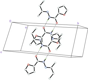
Intermolecular N—H···O hydrogen bonds link the molecules two by two around inversion centers to form dimers with a
R22(8) graph set motif (Etter et al., 1990; Bernstein et al., 1995) (Table 1, Fig. 2).
S2. Experimental
Furan-2-carbohydrazine (1 mmol, 0.126 g) was dissolved in anhydrous ethanol (10 ml), The mixture was stirred for
several minitutes at 351k, butan-2-one(1 mmol, 0.072 g) in ethanol (8 mm l) was added dropwise and the mixture was
stirred at refluxing temperature for 2 h. The product was isolated and recrystallized from methanol/dicholomethane(1:1),
colorless single crystals of (I) was obtained after 3 d.
S3. Refinement
All H atoms attached to C atoms and N atom were fixed geometrically and treated as riding with C—H = 0.96 Å
(methyl), 0.97 Å (methylene), 0.93Å (aromatic) and N—H = 0.86 Å with Uiso(H) = 1.2Ueq(C or N) or Uiso(H) =
Figure 1
Molecular view of the title compound with the atom labeling scheme. Displacement ellipsoids are drawn at the 30%
Figure 2
Partial packing view showing the formation of dimer through N-H···O hydogen bonds shown as dashed lines. H atoms
not involved in hydrogen bondings have been omitted for clarity.
N′-(Butan-2-ylidene)furan-2-carbohydrazide
Crystal data
C9H12N2O2
Mr = 180.21
Monoclinic, P21/c
Hall symbol: -P 2ybc
a = 8.2664 (15) Å
b = 16.6687 (13) Å
c = 7.5396 (11) Å
β = 113.171 (19)°
V = 955.1 (2) Å3
Z = 4
F(000) = 384
Dx = 1.253 Mg m−3
Mo Kα radiation, λ = 0.71073 Å Cell parameters from 1520 reflections
θ = 3.1–28.8°
µ = 0.09 mm−1
T = 293 K Block, colorless 0.21 × 0.19 × 0.17 mm
Data collection
Bruker SMART CCD area-detector diffractometer
Radiation source: fine-focus sealed tube Graphite monochromator
ω scans
Absorption correction: multi-scan (SADABS; Bruker, 1998)
Tmin = 0.978, Tmax = 0.982
θmax = 26.4°, θmin = 3.2°
h = −11→9
l = −6→9
Refinement
Refinement on F2
Least-squares matrix: full
R[F2 > 2σ(F2)] = 0.044
wR(F2) = 0.101
S = 0.74 1955 reflections 120 parameters 0 restraints
Primary atom site location: structure-invariant direct methods
Secondary atom site location: difference Fourier map
Hydrogen site location: inferred from neighbouring sites
H-atom parameters constrained
w = 1/[σ2(F
o2) + (0.0434P)2]
where P = (Fo2 + 2Fc2)/3
(Δ/σ)max = 0.003
Δρmax = 0.17 e Å−3
Δρmin = −0.17 e Å−3
Special details
Geometry. All esds (except the esd in the dihedral angle between two l.s. planes) are estimated using the full covariance matrix. The cell esds are taken into account individually in the estimation of esds in distances, angles and torsion angles; correlations between esds in cell parameters are only used when they are defined by crystal symmetry. An approximate (isotropic) treatment of cell esds is used for estimating esds involving l.s. planes.
Refinement. Refinement of F2 against ALL reflections. The weighted R-factor wR and goodness of fit S are based on F2,
conventional R-factors R are based on F, with F set to zero for negative F2. The threshold expression of F2 > σ(F2) is used
only for calculating R-factors(gt) etc. and is not relevant to the choice of reflections for refinement. R-factors based on F2
are statistically about twice as large as those based on F, and R- factors based on ALL data will be even larger.
Fractional atomic coordinates and isotropic or equivalent isotropic displacement parameters (Å2)
x y z Uiso*/Ueq
O1 0.4993 (2) 0.32297 (8) 0.4042 (2) 0.0598 (5) O2 0.4694 (2) 0.41298 (8) 0.1056 (2) 0.0670 (6) N1 0.6592 (3) 0.50672 (9) 0.2784 (3) 0.0511 (6)
H1 0.6497 0.5314 0.1746 0.061*
N2 0.7678 (3) 0.53804 (11) 0.4554 (3) 0.0505 (6) C1 0.5264 (4) 0.29006 (14) 0.5768 (4) 0.0586 (8)
H1B 0.4800 0.2411 0.5932 0.070*
C2 0.6274 (4) 0.33618 (14) 0.7195 (4) 0.0638 (8)
H2B 0.6641 0.3262 0.8509 0.077*
C3 0.6686 (3) 0.40406 (13) 0.6316 (4) 0.0582 (7)
H3A 0.7383 0.4473 0.6956 0.070*
C4 0.5899 (3) 0.39476 (11) 0.4416 (3) 0.0437 (6) C5 0.5674 (3) 0.43790 (12) 0.2653 (3) 0.0473 (7) C6 0.8540 (3) 0.60130 (13) 0.4535 (3) 0.0502 (7) C7 0.9668 (4) 0.63698 (13) 0.6436 (4) 0.0669 (8)
H7A 0.9302 0.6921 0.6457 0.080*
H7B 1.0872 0.6381 0.6529 0.080*
C8 0.9648 (4) 0.59519 (17) 0.8197 (4) 0.0933 (10)
H8A 1.0343 0.6251 0.9329 0.140*
H8B 1.0128 0.5422 0.8278 0.140*
H8C 0.8459 0.5916 0.8108 0.140*
H9A 0.8447 0.6059 0.1810 0.121*
H9B 0.9613 0.6740 0.3134 0.121*
H9C 0.7557 0.6803 0.2325 0.121*
Atomic displacement parameters (Å2)
U11 U22 U33 U12 U13 U23
O1 0.0962 (15) 0.0377 (8) 0.0454 (11) −0.0067 (9) 0.0276 (10) 0.0008 (7) O2 0.1069 (16) 0.0467 (9) 0.0373 (11) −0.0157 (9) 0.0174 (11) −0.0023 (8) N1 0.0748 (15) 0.0400 (10) 0.0386 (12) −0.0059 (11) 0.0224 (11) 0.0015 (9) N2 0.0599 (15) 0.0458 (11) 0.0440 (13) −0.0015 (10) 0.0186 (11) −0.0015 (9) C1 0.085 (2) 0.0456 (13) 0.0495 (18) −0.0007 (14) 0.0312 (16) 0.0107 (12) C2 0.077 (2) 0.0659 (16) 0.0435 (17) −0.0090 (15) 0.0182 (16) 0.0110 (13) C3 0.067 (2) 0.0543 (14) 0.0472 (17) −0.0152 (13) 0.0155 (15) 0.0028 (12) C4 0.0572 (18) 0.0318 (12) 0.0431 (15) 0.0011 (11) 0.0208 (13) 0.0017 (10) C5 0.0661 (19) 0.0369 (13) 0.0406 (16) 0.0038 (13) 0.0228 (15) −0.0010 (11) C6 0.0518 (18) 0.0415 (13) 0.0561 (17) 0.0021 (13) 0.0199 (14) 0.0008 (11) C7 0.060 (2) 0.0655 (16) 0.069 (2) −0.0095 (14) 0.0184 (17) −0.0050 (14) C8 0.092 (3) 0.123 (2) 0.059 (2) −0.0321 (19) 0.0227 (18) −0.0138 (18) C9 0.101 (3) 0.0641 (17) 0.075 (2) −0.0200 (16) 0.0328 (19) 0.0117 (14)
Geometric parameters (Å, º)
O1—C1 1.347 (2) C4—C5 1.457 (3)
O1—C4 1.381 (2) C6—C7 1.492 (3)
O2—C5 1.229 (2) C6—C9 1.493 (3)
N1—C5 1.357 (3) C7—C8 1.505 (3)
N1—N2 1.384 (2) C7—H7A 0.9700
N1—H1 0.8600 C7—H7B 0.9700
N2—C6 1.276 (3) C8—H8A 0.9600
C1—C2 1.319 (3) C8—H8B 0.9600
C1—H1B 0.9300 C8—H8C 0.9600
C2—C3 1.419 (3) C9—H9A 0.9600
C2—H2B 0.9300 C9—H9B 0.9600
C3—C4 1.329 (3) C9—H9C 0.9600
C3—H3A 0.9300
C1—O1—C4 106.56 (17) N2—C6—C9 126.8 (2)
C5—N1—N2 121.40 (19) C7—C6—C9 115.9 (2)
C5—N1—H1 119.3 C6—C7—C8 116.3 (2)
N2—N1—H1 119.3 C6—C7—H7A 108.2
C6—N2—N1 117.00 (19) C8—C7—H7A 108.2
C2—C1—O1 111.2 (2) C6—C7—H7B 108.2
C2—C1—H1B 124.4 C8—C7—H7B 108.2
O1—C1—H1B 124.4 H7A—C7—H7B 107.4
C1—C2—C3 106.0 (2) C7—C8—H8A 109.5
C1—C2—H2B 127.0 C7—C8—H8B 109.5
C4—C3—H3A 126.1 H8A—C8—H8C 109.5
C2—C3—H3A 126.1 H8B—C8—H8C 109.5
C3—C4—O1 108.49 (19) C6—C9—H9A 109.5
C3—C4—C5 139.3 (2) C6—C9—H9B 109.5
O1—C4—C5 112.16 (19) H9A—C9—H9B 109.5
O2—C5—N1 119.4 (2) C6—C9—H9C 109.5
O2—C5—C4 121.6 (2) H9A—C9—H9C 109.5
N1—C5—C4 118.9 (2) H9B—C9—H9C 109.5
N2—C6—C7 117.3 (2)
Hydrogen-bond geometry (Å, º)
D—H···A D—H H···A D···A D—H···A
N1—H1···O2i 0.86 2.16 2.981 (2) 160

![(E) 4 Methoxy N′ [(6 methyl 4 oxo 4H chromen 3 yl)methylidene]benzohydrazide monohydrate](data:image/gif;base64,R0lGODlhAQABAIAAAP///wAAACH5BAEAAAAALAAAAAABAAEAAAICRAEAOw==)