PTEN inhibits IL-2 receptor–mediated
expansion of CD4
+
CD25
+
Tregs
Patrick T. Walsh,1 Jodi L. Buckler,1 Jidong Zhang,1 Andrew E. Gelman,1 Nicole M. Dalton,1
Devon K. Taylor,1 Steven J. Bensinger,2 Wayne W. Hancock,3 and Laurence A. Turka1
1Department of Medicine, University of Pennsylvania, Philadelphia, Pennsylvania, USA. 2Cellular Immunology, La Jolla Institute for Allergy and Immunology,
San Diego, California, USA. 3Department of Pathology and Laboratory Medicine, Joseph Stokes Jr. Research Institute and Biesecker Pediatric Liver Center,
The Children’s Hospital of Philadelphia and University of Pennsylvania, Philadelphia, Pennsylvania, USA.
One of the greatest barriers against harnessing the potential of CD4
+CD25
+Tregs as a cellular immunotherapy
is their hypoproliferative phenotype. We have previously shown that the hypoproliferative response of Tregs to
IL-2 is associated with defective downstream PI3K signaling. Here, we demonstrate that targeted deletion of the
lipid phosphatase PTEN (phosphatase and tensin homolog deleted on chromosome 10) regulates the peripheral
homeostasis of Tregs in vivo and allows their expansion ex vivo
in response to IL-2 alone. PTEN deficiency does
not adversely affect either the thymic development or the function of Tregs, which retain their ability to
sup-press responder T cells in vitro
and prevent colitis in vivo. Conversely, reexpression of PTEN in PTEN-deficient
Tregs as well as in activated CD4
+T cells inhibits IL-2–dependent proliferation, confirming PTEN as a negative
regulator of IL-2 receptor signaling. These data demonstrate that PTEN regulates the “anergic” response of
Tregs to IL-2 in vitro and Treg homeostasis in vivo
and indicate that inhibition of PTEN activity may facilitate
the expansion of these cells for potential use in cellular immunotherapy.
Introduction
CD4+CD25+
Tregs are a naturally occurring population of T lym-phocytes with a key role in suppressing the response of self-reactive T cells that escape negative selection in the thymus (1). In addition to regulating responses against self antigens, it is also well estab-lished that Tregs can exert potent suppressive effects against most other types of T cell–mediated immune responses (1). These obser-vations highlight the potential of Treg subsets for use as cellular immunotherapy to inhibit the undesirable effects of certain class-es of immune response, such as those observed in autoimmune disease and transplant rejection (2–4). However, efforts to harness the therapeutic potential of these cells are significantly hindered by the relatively low number of Tregs present in the periphery of normal healthy individuals as well as their anergic phenotype ex vivo (5, 6). Accordingly a number of studies have reported strate-gies to overcome these obstacles, largely by using costimulatory antibodies against CD3 and CD28 in conjunction with extremely high doses of IL-2 (3, 7).
Tregs constitutively express all 3 chains (a, b, and g) of the high-affinity IL-2 receptor (IL-2R), and recent evidence indicates that a primary nonredundant function of IL-2 in vivois regulation of self tolerance through an essential role in the development and homeo-stasis of CD4+CD25+ Tregs (8, 9). Despite the widely recognized
importance of IL-2 in Treg homeostasis, very little is known about the intracellular mechanisms that regulate IL-2R signaling in these cells. Indeed, a defining characteristic of Tregs is their inability to expand in vitro upon stimulation with IL-2 alone despite expres-sion of all 3 chains of the high-affinity IL-2R (10). This observation
is in direct contrast with the established promitogenic effects of IL-2R signaling in activated IL-2R+ non-Tregs (11).
We have recently described a distinct IL-2R signaling pattern in Tregs, in which downstream mediators of PI3K are not activated while JAK/STAT-dependent signaling remains intact. This sig-naling pattern correlates with the hypoproliferative response of Tregs and is associated with expression of PTEN (phosphatase and tensin homolog deleted on chromosome 10) (12). PTEN, a phos-phoinositol 3,4,5-triphosphatase, catalyzes the reverse reaction of PI3K, thereby negatively regulating the activation of downstream signaling pathways (13). We have demonstrated that the expres-sion of PTEN is significantly downregulated after T cell activation yet remains relatively highly expressed in otherwise unmanipu-lated CD4+CD25+ Tregs (12). While a substantial body of evidence
exists indicating that PI3K-dependent signaling plays an essential role in driving IL-2–induced T cell proliferation (14, 15), a role for PTEN in regulating these events has not previously been defined.
In this report, we demonstrate that CD4+CD25+ Tregs develop
normally in mice with a specific deletion of PTEN in the T cell compartment (PTEN-DT). Furthermore, PTEN-DT Tregs prolifer-ate readily upon stimulation with IL-2 alone in vitroand exhibit enhanced peripheral turnover in vivo. PTEN-DT Tregs retain their ability to suppress effector T cell responses both in vitro and in vivo. Furthermore, ex vivo deletion of PTEN in Tregs, using an inducible Cre system, also facilitates IL-2–mediated expansion of these cells, demonstrating that this phenotype is distinct from T cell developmental defects that might occur in the absence of PTEN expression. Finally, enforced expression of PTEN in both PTEN-DT Tregs and recently activated CD4+ T cells inhibits their
ability to expand in response to IL-2, confirming the ability of this lipid phosphatase to negatively regulate IL-2–dependent prolifera-tion. These data identify PTEN as a negative regulator of IL-2R signaling in CD4+
T cells while defining the mechanism that regu-lates CD4+CD25+
Treg proliferation in response to IL-2R stimula-tion. These observations also indicate that targeting PTEN activity Nonstandard abbreviations used:
IL-2R, IL-2 receptor; MIGR1, MSCV-Ires-Gfp-EcoR1; NGFR, nerve growth factor receptor; 4-OHT, 4-OH tamoxifen; PTEN, phosphatase and tensin homolog deleted on chromosome 10; rIL-2, recombinant IL-2.
Conflict of interest: P.T. Walsh, S.J. Bensinger, and L.A. Turka own intellectual prop-erty related to the PTEN and regulatory T cells.
may facilitate expansion of CD4+CD25+ Tregs ex vivo or in vivo for
potential therapeutic use.
Results
CD4+CD25+ Tregs develop normally in the absence of PTEN. In normal
mice, PTEN is expressed at equivalent levels in Treg and CD4+CD25–
T cell subsets (Figure 1A). However, as PTEN deficiency in mice results in embryonic lethality, to examine the role of PTEN in CD4+CD25+ Tregs, we used mice with targeted deletion of PTEN
specific to the T cell compartment. Mice homozygous for expression of the Ptenflox allele were crossed with CD4–Cre transgenic mice. From
the resulting litters (with animals termed PTEN-DT mice), genomic DNA from purified CD4+
T cells was screened by PCR for expres-sion of the Cre transgene. Specific recombination at the Ptenflox locus
was detected using primers flanking the 5′ and 3′ loxP sites, which amplify an 849-bp product only after Cre-mediated deletion of
exons 4 and 5 (Figure 1B) (16). T cell–spe-cific deletion of PTEN was confirmed by Western blotting for total PTEN expres-sion in purified CD4+
T cells from 3-week-old homozygous mutant Ptenflox/floxCre+,
heterozygous mutant Ptenflox/+Cre+, or
wild-type Cre– mice (Figure 1C).
The precise phase of thymic develop-ment at which CD4+ T cells commit to
the Treg lineage is controversial, and although Cre recombinase becomes active during the double-positive phase of T cell development in the CD4–Cre
mouse (17), the in vivo half-life of PTEN protein expression may be sufficient that PTEN-DT Tregs develop in the presence of PTEN. Therefore, we examined levels of PTEN expression during different phases of T cell development in the thy- mus of 3-week-old PTEN-DT and wild-type mice. As shown in Figure 1D, PTEN protein levels were notably lower (com- pared with littermate controls) in dou-ble-positive thymocytes from PTEN-DT mice and were almost undetectable in CD4+ single-positive thymocytes.
To further determine whether PTEN deficiency affects Treg development we made bone marrow chimeric mice by reconstituting lethally irradiated congenic hosts with wild-type (Thy1.1) and/or PTEN-DT (Thy1.2) bone mar-row. Mice were reconstituted with either 100% wild-type, 100% PTEN-DT, or a 50% wild-type/50% PTEN-DT mixture of bone marrow. Ten weeks after reconstitution, thymic development appeared normal in all chimeras, with similar percent-ages of double-negative, double-posi-tive, and single-positive subsets (Figure 1E and data not shown). Furthermore, examination of Foxp3 expression among CD4 single-positive cells from all groups demonstrated equivalent levels of Treg development (Figure 1E). To analyze the relative proportion of wild-type and PTEN-DT–derived cells within the CD4+Foxp3+
Treg subsets of the mixed chimeras, we measured expression of congenic Thy1 markers. As shown in Figure 1E, although the pro-portion of wild-type–derived Tregs was slightly higher in the 50/50 chimeric mice (59.9% versus 39.9%), this was most likely due to residual host-derived T cell development, similar to what was seen in the 100% PTEN-DT chimeric mice, in which 92.8% of Tregs were PTEN deficient. Together, these data indicate that CD4+CD25+
Tregs develop normally in the absence of PTEN.
Intact suppression by PTEN-deficient CD4+CD25+ Tregs. Previous
reports have demonstrated that loss of PTEN in the T cell com-partment, achieved by crossing Ptenflox/– mice with lck-Cre
trans-genic mice, results in lethality by about 15 weeks of age due to the development of CD4+ T cell lymphomas (18). Our mice had
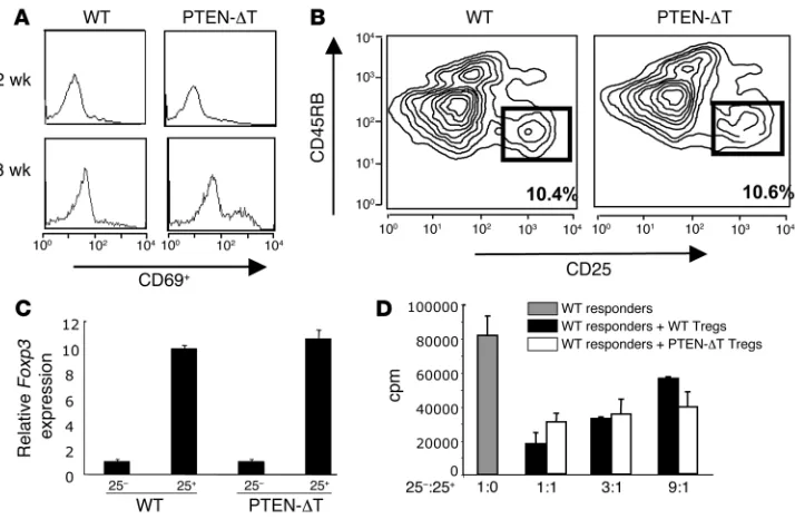
[image:2.585.44.377.82.403.2]a very similar phenotype, developing CD4+ T cell lymphomas by
Figure 1
10–12 weeks (data not shown). In agreement with these reports, lymphoma was preceded by an increased total number of CD4+
lymphocytes and an accumulation of activated CD4+ T cells in the
periphery, which became apparent after 6 weeks. This accumula-tion of activated CD4+ T cells in the periphery was detected as a
significant increase in the numbers of CD4+ cells expressing the
activation marker CD69 (Figure 2A). As previously reported, the expression levels of CD25 on CD4+
T cells did not vary signifi-cantly between PTEN-DT mice and wild-type littermate controls (data not shown).
We next sought to determine whether loss of PTEN in the T cell compartment affected the function of CD4+CD25+ Tregs. To
avoid contamination of putative Tregs with recently activated T cells, mice were sacrificed at 2–3 weeks of age, a time at which no increase in the number of CD69+CD4+ T cells was detectable
(Figure 2A). Phenotypic analysis of PTEN-DT mice demonstrated similar numbers of CD4+CD25+CD45RBlo cells when compared
with littermate controls (Figure 2B). After purification by FACS, real-time PCR demonstrated that cells from both PTEN-DT and control mice expressed comparable levels of Foxp3 mRNA (Figure 2C). Furthermore, PTEN-DT Tregs were able to suppress the pro-liferation of wild-type responder cells in vitro to the same extent as Tregs isolated from control mice (Figure 2D). Taken together, these data demonstrate that CD4+CD25+ Tregs develop normally
in the absence of PTEN.
PTEN-deficient CD4+CD25+ Tregs proliferate in response to IL-2. We
have previously demonstrated that the hypoproliferative response
of CD4+CD25+ Tregs to IL-2R
stimulation is associated with a distinct signaling pattern char- acterized by intact STAT5 phos-phorylation but an inability to activate pathways downstream of PI3K (12). In contrast, IL-2R stim-ulation of activated CD4+ T cells
readily activates PI3K-dependent signaling pathways and leads to a robust proliferative response. We further demonstrated that while CD4+CD25+ Tregs express high
levels of PTEN, a negative regula-tor of PI3K-dependent signaling, little or no PTEN is expressed in recently activated CD4+ T cells.
Therefore, we hypothesized that PTEN activity negatively regulates IL-2–induced expansion of Tregs. In order to test this hypothesis, we cultured CD4+CD25+CD45RBlo
cells from PTEN-DT mice in the presence of recombinant IL-2 (rIL-2) (100 U/ml) and assessed total cell numbers at various time points. As shown in Figure 3A, viable PTEN-DT Treg cell numbers increased an average of 15–25 fold over a 2-week period in culture while no accumulation of wild-type Tregs or CD4+CD25–
T cells from either wild-type or PTEN-DT mice was observed. CFSE dilution clearly illustrated the kinetics of cell division over the first 10 days in culture, and cell proliferation was also assessed by incorporation of tritiated thymidine after 48 hours in culture with rIL-2 (Figure 3, B and C). This expansion of PTEN-DT Tregs was due to the proliferative effects of IL-2R signaling, as no difference in the levels of cell sur- vival between wild-type and PTEN-DT Tregs was observed (Supple-mental Figure 1; supplemental material available online with this article; doi:10.1172/JCI28057DS1).
The proliferative response of PTEN-DT Tregs to IL-2 is dose dependent, with cell division evident at as low as 10 U/ml of rIL-2 (Figure 3D). This level of proliferation observed in PTEN-DT Tregs is less than that seen in activated CD4+ T cell blasts. In part, this
may be due to endogenous IL-2 secretion by the activated CD4+
blasts, which increases the effective dose of IL-2 in each well. How-ever, it also suggests that, in addition to PTEN, other parameters may also regulate IL-2 responsiveness of CD4+ T cells.
[image:3.585.46.403.81.314.2]One possible explanation for our observations of PTEN-DT Treg expansion in response to IL-2 alone is that these cells may have already encountered TCR stimulation in vivo before isola-tion, thus facilitating their IL-2 responsiveness. However, we were unable to detect any differences in phosphorylation of ZAP-70 or Erk between freshly isolated wild-type and PTEN-DT Tregs (data not shown), arguing against the possibility that PTEN-DT Tregs are intrinsically activated via TCR signaling pathways. Another consideration is that T cell developmental abnormalities that may occur in PTEN-DT mice (18, 19) could influence the behavior of
Figure 2
CD4+CD25+ Tregs. Although our mixed bone marrow chimera
data indicate that Tregs develop normally in the absence of PTEN, to definitively exclude the possibility that potential developmental or homeostatic defects that may arise in PTEN-DT mice may alter the response of PTEN-DT Tregs to IL-2, we crossed the Ptenflox/flox
mice with Cre-ER transgenic mice, in which Cre recombinase activ-ity occurs only in the presence of the estrogen homolog tamoxifen (20). Unlike in PTEN-DT mice, no defects in peripheral or central tolerance were observed in these mice, and the CD4+CD25+ Treg
compartments were identical to those of Cre-negative littermates (data not shown). Culture of purified CD4+ T cells from Cre-ER/
Ptenflox/flox mice in the presence of 1 nM 4-OH tamoxifen (4-OHT)
led to a significant decrease in the amount of PTEN expression
after 72 hours (Figure 3E). In addition, stimulation of Cre-ER/ Ptenflox/flox CD4+CD25+ Tregs with IL-2 in the presence of 1 nM
4-OHT led to a robust proliferative response after 7 days in cul-ture, as assessed by CFSE dilution (Figure 3F). Together, these data confirm that PTEN-deficient CD4+CD25+
Tregs can be read-ily expanded upon IL-2R stimulation and that this response is not due to developmental defects as a consequence of loss of PTEN.
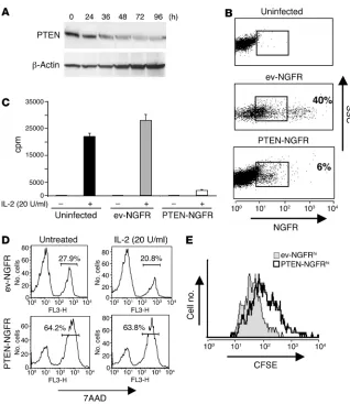
[image:4.585.67.518.78.410.2]Reexpression of PTEN blocks IL-2–mediated expansion of PTEN-DT Tregs . As loss of PTEN expression enabled IL-2–mediated prolif-eration of Tregs, we hypothesized that reexpression of the PTEN gene in PTEN-DT Tregs would restore the normal hypoprolifera-tive response to IL-2 that is observed in wild-type cells. We cloned the gene for human Pten into a bicistronic retroviral vector also
Figure 3
expressing the extracellular portion of human nerve growth factor receptor (NGFR)as a marker of cell transduction. Transduction of CFSE-labeled PTEN-DT Tregs with NGFR/MSCV-Ires-Gfp-EcoR1 (NGFR/MIGR1) empty vector typically resulted in 30–40% transduction efficiency, as determined by flow cytometric analysis of NGFR expression. In contrast, the Pten-containing retrovirus consistently resulted in 5–15% transduction efficiency (Figure 4). Since efficient retroviral transduction requires target cells to be progressing through the cell cycle, the significantly lower effi-ciency of transduction observed with the PTEN-containing virus is consistent with PTEN inhibiting cell cycle progression. After transduction, cells were cultured in the presence of rIL-2 (100 U/ml) for 4 days, and levels of CFSE dilution of NGFR+
cells were exam-ined. As shown in Figure 4, although transduction of PTEN-DT Tregs with empty vector did not affect IL-2–induced prolifera-tion, reexpression of PTEN resulted in complete inhibition of IL-2–mediated proliferation, restoring the hypoproliferative response observed in wild-type Tregs.
PTEN-deficient CD4+CD25+ Tregs do not proliferate in response to TCR
stimulation. In addition to their hypoproliferative response to IL-2R stimulation, it has also been established that CD4+CD25+ Tregs do
not divide after stimulation with anti-CD3 antibody alone. This is most likely related to the relative inability of Tregs to produce IL-2
[image:5.585.51.278.79.296.2](10). Thus, we also asked whether PTEN activity regulates the hypo- responsiveness of Tregs to TCR stimulation. Similar to their wild-type counterparts, stimulation of PTEN-DT Tregs with plate-bound anti-CD3 did not induce proliferation or any significant level of IL-2 production (Figure 5, A and B). It has also previously been demon-strated that the hypoproliferative response of Tregs can be broken by TCR stimulation in the presence of a relatively high dose of IL-2 (5, 10). Therefore, we examined the response of PTEN-DT Tregs upon stimulation with graded doses of anti-CD3 in the presence of rIL-2 (100 U/ml). PTEN-DT Tregs exhibited a more robust proliferative response in the presence of rIL-2 at all concentrations of anti-CD3 used in comparison with their wild-type counterparts (Figure 5C). Importantly, the differences in proliferation observed at each con-centration of anti-CD3 tested were equivalent to the difference seen in the presence of rIL-2 alone (i.e., the basal level of IL-2–induced pro-liferation in PTEN-DT Tregs). This suggests that while PTEN plays a significant role in regulating the IL-2 responsiveness of Tregs, the basal level of TCR responsiveness is unaltered in these cells.
Figure 4
[image:5.585.315.529.406.744.2]Reexpression of PTEN in PTEN-DT Tregs restores hypoproliferative response to IL-2. Purified PTEN-DT CD4+CD25+CD45RBlo cells were CFSE labeled and retrovirally transduced as described in Methods, with either MIGR1-NGFR empty vector (ev-NGFR) or PTEN-contain-ing virus (PTEN-NGFR). Cells were analyzed for expression of human NGFR 96 hours after infection and CFSE dilution of NGFR-positive cells analyzed by flow cytometry. Results are representative of 3 inde-pendent experiments. SSC, side scatter.
Figure 5
Taken together, these data confirm that Tregs can be expanded more readily in the absence of PTEN. However, they also indicate that the hypoproliferative responses of Tregs to IL-2R or TCR stimulation alone are mediated, at least in part, through distinct mechanisms.
PTEN-DT Tregs exhibit an enhanced rate of peripheral homeostatic turnover in vivo. Although CD4+CD25+ Tregs are characterized
by hypoproliferative responses in vitro, it is also clear that they readily undergo expansion in vivo (21, 22). Furthermore, in unmanipulated animals, Tregs in peripheral lymphoid organs exhibit higher levels of basal proliferation compared with their CD4+CD25– counterparts, and this response is known to depend,
at least in part, on IL-2 (21, 22). As we observed that PTEN-DT Tregs proliferate in response to IL-2 in vitro, we next examined whether the homeostasis of Tregs in vivowas altered in the absence of PTEN. PTEN-DT mice and littermate controls were injected with BrdU for 3 days, after which CD4+CD25+ and
CD4+CD25– T cells from both the thymi and spleens of these
mice were analyzed for BrdU incorporation. While similar levels of BrdU incorporation were seen in splenic CD4+CD25– cells from
control versus PTEN-DT mice, splenic PTEN-DT Tregs exhibited a significantly higher rate of BrdU staining compared with wild-type CD4+CD25+ T cells (Figure 6, B and C). Elevated BrdU uptake
in splenic Tregs was a result of increased peripheral turnover, as we found no difference in the levels of BrdU incorporation in either single-positive CD4+CD25– or CD4+CD25+ subsets from
PTEN-DT mice compared with wild-type littermates (Figure 6A). Together, these observations demonstrate that PTEN plays a role
in regulating the peripheral homeostasis of Tregs in vivo, without any significant effects on Treg development in the thymus.
Deletion of PTEN in Tregs facilitates downstream activation of PI3K-dependent signaling through the IL-2R. Previous studies on IL-2R signaling have shown that activation of both the JAK/STAT and PI3K/Akt pathways are critical for IL-2–induced proliferation (11). We have demonstrated that activation of signaling pathways downstream of PI3K does not occur in Tregs in response to IL-2R stimulation and have identified negative regulation of this signal- ing pathway by PTEN as a possible mechanism for this observa-tion. Therefore, we examined whether, in the absence of PTEN, IL-2R stimulation could activate signaling downstream of PI3K.
To this end, freshly isolated Tregs from wild-type mice or Tregs isolated from PTEN-DT mice and expanded for 8 days with IL-2 (100 U/ml) to obtain necessary cell numbers (see Methods) were rested overnight in complete media then stimulated for 30 min-utes with 100 U/ml rIL-2. Cell lysates were subsequently tested for activation of both JAK/STAT and PI3K-dependent signaling path-ways. As reported previously (12), stimulation of wild-type Tregs resulted in robust but isolated activation of JAK/STAT signaling, as shown by phosphorylation of STAT5, without any detectable activation of PI3K-dependent signaling through phosphorylation of p70 S6 kinase. In contrast, while activation of JAK/STAT signal-ing was also clearly detectable after IL-2R stimulation of PTEN-DT Tregs, we additionally observed phosphorylation of p70 S6 kinase, indicating that IL-2R stimulation activated PI3K-dependent sig-naling pathways in these cells (Figure 7).
Overexpression of PTEN inhibits IL-2–mediated expansion of activated CD4+ T cells
. The results shown above suggest that PTEN is neces-Figure 6
[image:6.585.70.254.79.418.2]PTEN-DT Tregs exhibit enhanced homeostatic expansion in the periph-ery. PTEN-DT mice and littermate controls were administered 1 mg BrdU every 12 hours for 3 days, at which time they were sacrificed and thymus and spleen cells were stained for BrdU. (A)Levels of BrdU incorpora-tion in CD4+CD8–CD25+ and CD4+CD8–CD25– thymocytes. (B)Levels of BrdU incorporation in CD4+CD25+ and CD4+CD25– splenocytes. (C) Percentage BrdU-positive CD4+CD25+ and CD4+CD25– splenocytes from PTEN-DT mice (n = 6) and littermate controls (n = 6).
Figure 7
[image:6.585.354.477.527.640.2]sary and sufficient for the hypoproliferative response of Tregs to IL-2. As IL-2 is known to enhance the proliferation of activated CD4+ T cells, we asked whether maintenance of its expression
would have a similar inhibitory effect on IL-2R–induced cell divi-sion in these cells.
Activation of nonregulatory CD4+ T cells both induces CD25
expression and downregulates PTEN (12) (Figure 8A). To examine
PTEN activity following T cell activation, we again used retrovi-ral transduction. Similar to our studies on PTEN-DT Tregs, infection of activated CD4+
T cells with NGFR/MIGR1 empty vector typ- ically resulted in 40–50% transduction effi-ciency while the Pten -containing virus result-ed in 5–10% transduction efficiency, again consistent with PTEN inhibiting cell cycle progression (Figure 8B). Purified NGFR-positive cells from both mock (ev-NGFR) and PTEN (PTEN-NGFR) transduced cells were subsequently cultured in the presence or absence of IL-2, and proliferation was assessed by thymidine incorporation. While cells expressing empty vector proliferated to an extent similar to that of nontransduced T cells, those expressing PTEN showed a dra- matically decreased level of thymidine incor-poration (Figure 8C).
Previous studies using ectopic expression of PTEN in T cell lines have supported a role for PTEN not only in inhibiting cell cycle pro-gression but also in inducing apoptotic cell death (23, 24). Therefore, we also examined the viability of purified NGFR-positive cells both in the presence and absence of rIL-2. Using 7-AAD staining as a measure of cell death, we found that PTEN overexpression leads to a significant increase in activated T cell death when compared with empty vec-tor–transduced cells (64% versus 28%). The addition of exogenous rIL-2 did not rescue this effect (Figure 8D).
To exclude the possibility that cells over-expressing PTEN failed to incorporate thymidine exclusively due to cell death, we measured CFSE dilution in live cells as an alternative means to assess proliferation after PTEN transduction. Freshly isolated CD4+
T cells were CFSE labeled prior to acti- vation with PMA and ionomycin and incu-bated for a period of 18 hours. These cells were then transduced with either empty vector or PTEN-expressing virus before sub-sequent culture with rIL-2 (10 U/ml) for a further 48 hours. Live cells were then identi- fied based on forward- and side-scatter pro- files, for NGFR expression, and CFSE dilu-tion was determined in live NGFR-positive subsets. For comparison, cells expressing identical levels of NGFR were analyzed. As shown in Figure 8E, proliferation of PTEN-overexpressing cells was significantly less than that observed in empty vector–transduced cells, confirming that PTEN acts to inhibit IL-2–induced proliferation of activated CD4+
T cells. As an appropriate comparison, cells expressing iden-tical levels of NGFR were analyzed (Figure 8B) although the results described were identical even when all NGFR-positive cells were included in the analysis (data not shown). While the inhibition of IL-2–induced proliferation by PTEN in recently activated CD4+
[image:7.585.42.360.78.445.2]cells was clear, it was far less significant than the almost complete
Figure 8
inhibition observed in retrovirally transduced PTEN-DT Tregs (Figure 4). This suggests that other factors in addition to PTEN may also regulate IL-2–mediated proliferation in activated non-regulatory CD4+ T cells.
Expanded PTEN-deficient CD4+CD25+ Tregs retain their suppressor
phe-notype. One of the largest drawbacks to exploiting the potential of Tregs in a therapeutic setting is the very low frequency with which these cells are found in normal, healthy individuals. Our results demonstrate that in the absence of PTEN activity, these cells can be readily expanded ex vivo using only rIL-2. These data highlight the potential for targeting this lipid phosphatase to facilitate ex vivo expansion of Tregs in response to cytokine stimulation alone. How-ever, such strategies could only be feasible if in vitro manipulation of these cells does not alter their regulatory potential. Therefore, to determine whether PTEN-deficient Tregs retain their regulatory phenotype after expansion with rIL-2, we examined their ability to
suppress the proliferation of wild-type CD4+ responder T cells in vitro as well
as their ability to suppress the develop-ment of inflammatory bowel disease in vivo. As shown in Figure 9, Tregs expanded for 8 days retained expression of Foxp3 mRNA and protein (Figure 9, A and B) and their ability to inhibit CD4+
effector T cell proliferation (Fig- ure 9, C and D). The level of suppres-sion was similar to that observed with freshly isolated wild-type Tregs.
Adoptive transfer of CD4+CD25–
CD45RBhi cells into immunodeficient
hosts has been demonstrated to result in the development of colitis, which can be prevented by cotransfer of Tregs (2, 25). To examine whether ex vivo– expanded, PTEN-deficient Tregs could prevent disease in vivo, we cotrans-ferred wild-type CD4+CD25–CD45RBhi
cells with either wild-type freshly iso-lated Tregs or PTEN-DT Tregs after expansion for 5 days in vitro with rIL-2. Similar to our in vitro observations, expanded PTEN-DT Tregs prevented the development of colitis in vivoto the same extent as freshly isolated wild-type Tregs (Figure 10).
A recent report has suggested that prevention of colitis in this model may be mediated by cell competition for an existing niche in the host rather than through the direct suppressive activity of Tregs (26). If this were the case in our system, one might also expect to see prevention of colitis if Tregs were replaced in this assay by PTEN-DT CD4+CD25–
T cells. However, coadop-tive transfer of PTEN-DT non-Tregs did not affect the development of colitis resulting from the transfer of wild-type CD4+CD25–
T cells (Supple-mental Figure 2).
Together, these data confirm that CD4+CD25+ Treg development
is intact in PTEN-DT mice and demonstrate that expansion of these cells with IL-2 in vitro does not alter their suppressor phenotype.
Discussion
Many studies have focused on the effects of IL-2 on CD4+CD25+
Tregs, with several recent reports providing substantial evidence of an essential role for IL-2 in Treg development and/or periph-eral homeostasis (27, 28). Despite these observations, very little is known about the intracellular pathways that regulate IL-2R sig-naling in CD4+CD25+
Tregs, and indeed, a defining characteris-tic of CD4+CD25+ Tregs is their relative inability to proliferate ex
[image:8.585.43.387.77.382.2]vivo in response to IL-2R stimulation. Recently, we characterized IL-2R signaling in Tregs and found that, unlike activated T cells, downstream mediators of the PI3K-dependent signaling pathway remain inactive following stimulation with IL-2. We associated
Figure 9
this distinct signaling pattern with relatively high levels of expres-sion of the lipid phosphatase PTEN (12). Therefore, here we tested the hypothesis that in the absence of PTEN, Tregs would respond to IL-2R stimulation by proliferating in a fashion similar to that of their activated CD4+ T cell counterparts.
In order to isolate Tregs deficient in PTEN expression, we used a Cre-LoxP system to generate mice with a T cell–specific deletion in PTEN. As described previously, these mice suffer a profound defect in T cell tolerance characterized by an accumulation of autoreactive T cells in the periphery, which precedes the development of a CD4+
T cell lymphoma (18). Given the importance of IL-2R signaling in Treg development and homeostasis, disrupted regulation of these signals, as may occur in the absence of PTEN, may be a potential contributing factor to the autoimmunity observed in these mice. Indeed, there have been several recent reports demonstrating that genetic alteration of downstream signaling pathways activated by IL-2 can have profound effects on Treg homeostasis. For example, mice expressing a constitutively active STAT5 transgene have an approximate 5-fold increase in the number of Tregs both in the thy- mus and periphery (29, 30). Our observations that Treg develop-ment and homeostasis appear normal in the thymus of PTEN-DT mice and that a functional Treg compartment can be isolated from these mice suggest that PTEN deficiency (and its effects on PI3K-dependent signaling) does not affect the development of these cells in vivo. In support of this observation, we have also found that mice expressing a constitutively active Akt transgene (mAkt mice)
have normal development of CD4+CD25+ Tregs (data not shown).
Together, these data suggest that JAK/STAT-dependent signaling plays a predominant role over PI3K-dependent pathways down-stream of the IL-2R in promoting Treg development.
These data also suggest that the breakdown in T cell tolerance observed in mice deficient in PTEN is the result of defects within non-Treg compartments. As described previously, PTEN-deficient T cells are hyperresponsive to activation stimuli and less suscepti-ble to activation-induced cell death when compared with wild-type controls (18, 31). As development of PTEN-DT Tregs appears nor- mal, it will be of interest to determine whether the altered respon-siveness of effector T cells lacking PTEN allows them to escape regulation by CD4+CD25+ T cells and contribute to disease.
Despite their apparently normal thymic development, PTEN-DT Tregs exhibited an increased rate of BrdU incorporation in the periphery (Figure 6), and could be expanded quite readily in response to IL-2 in vitro (Figure 3). Given these observations, and as IL-2 plays an important role in regulating the peripheral homeostasis of Tregs (22), one might expect to see an increased number of peripheral Tregs in PTEN-DT mice. However, the fre-quency of Tregs in PTEN-DT mice before the onset of disease was identical to that observed in littermate controls (Figure 1). This indicates that, in addition to exhibiting a higher level of peripheral homeostatic expansion, PTEN-DT Tregs also have a higher rate of turnover compared with their wild-type counterparts. Inter-estingly, however, a number of recent reports have demonstrated that peripheral CD4+CD25+ Tregs proliferate more readily than
CD4+CD25– effector T cells in the steady state in vivo, yet under
normal homeostatic conditions, the relative frequency of Treg ver-sus effector T cell subsets remains relatively stable over time (21, 22). These observations suggest that the higher rate of turnover of CD4+CD25+ Tregs (compared with CD4+CD25– T cells) may be
balanced by a higher rate of cell death, perhaps due to the lack of available “space” in the periphery (21). Interestingly, it has recent-ly been demonstrated that in vivo IL-2 therapy of lymphopenic patients results in a marked increase in the frequency of peripheral Tregs, suggesting that a preexisting niche must be present in the peripheral environment to facilitate an alteration in the relative frequency of Tregs versus effector T cells (32).
Arguably, the greatest single barrier to harnessing the therapeutic potential of naturally occurring CD4+CD25+
[image:9.585.63.267.81.440.2]Tregs for the treat-ment of immune disorders is their relative low frequency in normal, healthy individuals as well as their “anergic” phenotype ex vivo. Accordingly, several groups have reported a number of approaches aimed at overcoming these difficulties through the expansion of Tregs ex vivo for subsequent treatment in disease models (3, 7, 33, 34). Many of these strategies involve activating Tregs with a com- bination of stimulatory antibodies to the TCR and CD28 in con-junction with relatively high doses of rIL-2 (3, 7). Our data indicate that, by targeting PTEN activity, Tregs can be expanded in response
Figure 10
to rIL-2 alone without the need to stimulate the TCR and costim-ulatory receptors. Most importantly, we have found that ex vivo expansion of PTEN-deficient Tregs does not affect their regulatory phenotype, as illustrated by their ability to suppress the prolifera-tion of effector T cells in vitro as well as their ability to prevent the development of colitis in vivo (Figures 7 and 8).
Taken together, our data provide a mechanism that explains the hypoproliferative response of CD4+CD25+ Tregs to IL-2 in vitro
and also identify PTEN as a negative regulator of peripheral Treg homeostasis in vivo. These data indicate that negative regulation of PTEN activity in concert with IL-2R stimulation may facilitate expansion of CD4+CD25+
Tregs both ex vivo and in vivo for poten-tial therapeutic use.
Methods
Mice. Ptenflox/flox mice have been described previously and were a kind gift
from Tak Mak (University of Toronto, Toronto, Ontario, Canada) (18).
CD4–Cre
mice were a kind gift from Yongwon Choi (University of Penn-sylvania, Philadelphia, Pennsylvania, USA). Ptenflox/flox mice were crossed
with CD4–Cre transgenic mice to generate mice with a deletion of PTEN
specific to the T cell compartment. As T cell–specific deficiency of PTEN results in early lethality, Ptenflox/+ CD4–Cre+ mice were bred with Ptenflox/flox
mice to generate offspring. Mice were genotyped as described (18), and offspring that were found to express the Cre transgene and were homo-zygous for the Ptenflox allele were used for analysis as homozygous mutant
mice (PTEN-DT) while Cre-negative littermates were used as controls.
Cre-ER mice have been described previously (20); these were crossed with
Ptenflox/flox mice and genotyped as described above. Rag1–/– mice, C57BL/6
mice expressing the Thy1.1 congenic marker, and DO11.10 TCR trans-genic mice were purchased from Jackson Laboratory. All colonies were maintained under specific pathogen–free conditions at the animal facili-ties of the University of Pennsylvania. All experiments described in this manuscript were approved by the Institutional Animal Care and Use Committee at the University of Pennsylvania.
Media, reagents, antibodies, and flow cytometry. All cells were grown in RPMI 1640 (Mediatech Inc.) supplemented with 10% heat-inactivated FCS, 100 U/ml penicillin, 100 mg/ml streptomycin, 2 mM l-glutamine, 10 mM HEPES (all from Invitrogen), and 50 m M 2-mercaptoethanol (Sigma- Aldrich). Biotin–anti-CD25 (7D4), APC–anti-CD4 (RM4-5), FITC–anti-CD45RB, and streptavidin-PE were purchased from BD Biosciences — Pharmingen. Anti-Foxp3 staining kit was purchased from eBioscience. Murine recombinant IL-2 (IL-2) was purchased from R&D Systems. Cells were analyzed on a BD FACSCalibur using CellQuest 5.2 software (BD). Anti–phospho-STAT5, anti–phospho–p70 S6 kinase (all from Cell Signal- ing Technology), anti-PTEN (Cascade Bioscience), and anti-actin antibod-ies were used for Western blot analysis.
Bone marrow chimeras. C57BL/6 Thy1.1 mice were lethally irradiated (10
Gy) prior to reconstitution with 2 × 10⁶ total T cell–depleted bone marrow
cells at specified ratios from either wild-type (Thy1.1) or PTEN-DT (Thy1.2) mice. Recipient mice were treated with Neosporin (DSM Pharmaceuticals Inc.) for 1 week prior to and 2 weeks after reconstitution. Ten weeks after reconstitution, thymic subsets were analyzed by flow cytometry for expres-sion of Foxp3 and degree of chimerism.
CD4+CD25+ T cell isolation. Spleen and lymph node cells were isolated from
2- to 3-week-old Ptenflox/floxCre+ mice before the onset of disease and from
age-matched Cre-negative littermates. Cell preparations were stained with anti–CD4 APCs, anti–CD25 biotin, streptavidin-PE, and anti–CD45RB-FITC and subsequently purified into CD4+CD25+CD45RBlo cells by flow
cytometry on a FACSVantage Cell Sorter (BD Biosciences). Cell purity was routinely greater than 95%.
Proliferation assays. FACS-purified CD4+CD25+CD45RBlo cells were CFSE
labeled and cultured in complete medium supplemented with 100 U/ml rIL-2 in 96-well plates at a density of 105 cells/well. For TCR stimulation,
cells were cultured either with plate-bound anti-CD3 (2.5 m g/ml) and anti-CD28 (2.5 m g/ml) or in the presence of irradiated T cell–depleted spleno-cytes with varying doses of soluble anti-CD3. Activated CD4+ T cell blasts
were derived by stimulating purified CD4+ cells with PMA (0.2 mg/ml) and
ionomycin (1 mg/ml) for 24 hours and subsequent washing before culture in the presence of rIL-2. In the case of cells from Cre-ER/Ptenflox/flox mice,
1 nM 4-OHT was also added to the medium. Cell number was measured every 72 hours by Trypan blue exclusion, and cells were maintained at a density of 5 × 10⁵ cells/ml. To measure proliferation, CFSE dilution was
determined by FACS analysis at the indicated time points, or alternatively, after 48–72 hours, cells were pulsed with tritiated thymidine for a further 12 hours before harvest.
In vitro suppression assays. A total of 1.5 × 10⁵ Thy1.1+ CD4+CD25–
CD45RBhi T cells were CFSE labeled and cultured with 3 × 10⁵ irradiated
T cell–depleted splenocytes and anti-CD3 Abs (2C11; 0.5 mg/ml). The indicated ratios of CD4+CD25+CD45RBlo cells from either wild-type or
PTEN-DT mice (Thy1.2+
) were added to the cultures. To measure suppres-sion, CFSE dilution of Thy1.1-positive cells was assessed by flow cytometry after 72 hours. Alternatively, cells were pulsed with 0.5 m Ci of tritiated thy-midine for the final 12 hours before being harvested.
In vivo colitis model. Rag1–/– mice were injected i.v. with 6 × 10⁵ CD4+CD25–
CD45RBhi T cells either alone or with 3 × 10⁵ freshly isolated wild-type
CD4+CD25+CD45RBlo Tregs, PTEN-DT CD4+CD25–CD45RBhi T cells, or
PTEN-DT Tregs that had been expanded in vitro with rIL-2 (100 U/ml). Mice were weighed and examined every week for signs of disease and sacri- ficed for tissue harvest at 8 weeks. Tissues were fixed in 10% neutral buff-ered formalin (Fisher Scientific), cut into 5-mm sections, and stained with H&E. Severity of colitis was scored blindly as described previously (25).
Immunoblotting. As a result of the necessary breeding strategies employed,
the number of PTEN-DT mice generated per litter was typically 50% of the number of littermate controls. Therefore, in order to obtain sufficient cell numbers to analyze intracellular signaling pathways, it was neces-sary to expand isolated PTEN-DT Tregs in vitro in the presence of rIL-2 as described above while Tregs from littermate control mice were generated in numbers allowing analysis immediately subsequent to isolation and more-over were not expandable by IL-2 alone. Both cell types were subsequently rested overnight in complete medium before study. After stimulation, cells were lysed at 4°C in lysis buffer composed of 50 mM Tris-HCL (pH 6.8), 0.2% 2-mercaptoethanol, 20% glycerol, 4% SDS, and 0.001% bromophenol blue (all from Sigma-Aldrich). Cell lysates were clarified by centrifugation at 11,000 g for 10 minutes. Supernatants were boiled for 5 minutes, sepa-rated on a 10% SDS-PAGE at 1 × 10⁶ cell equivalents/well and blotted onto
Hybond ECL nitrocellulose membranes (Amersham Biosciences). Mem-branes were blocked for 1 hour in blocking reagent (Roche Diagnostics) at room temperature and probed with indicated antibodies at 1:1,000 dilution overnight at 4°C. Membranes were washed and probed with a horserad-ish peroxidase–conjugated anti-rabbit or anti-mouse antibody at 1:1,000 for 60 minutes at room temperature. Blots were visualized by enhanced chemiluminescence (Roche Diagnostics) according to the manufacturer’s protocol and on Hyperfilm ECL (Amersham Biosciences). Antibodies were subsequently stripped from membranes using Restore Western Blot Strip-ping Buffer (Pierce Biotechnology) and reprobed as above.
Retroviral transduction. The cDNA for human Pten, a kind gift from K.
labeled and stimulated for 18 hours with PMA (0.2 mg/ml) and ionomycin (1 m g/ml) for 18 hours. Cells were washed and transduced by spin infec-tion with retroviral supernatants containing 2 m g/ml polybrene (Sigma-Aldrich) at 500 g for 90 minutes. Cells were subsequently cultured for 48–96 hours in complete media supplemented with rIL-2. Cells were analyzed for transduction efficiency by measuring NGFR expression by flow cytometry.
Quantitative real-time PCR. Total RNA was extracted from FACS-purified
CD4+CD25+ and CD4+CD25– T cells with an RNeasy kit (QIAGEN) and
reverse transcription performed with the Superscript first-strand synthesis system (Invitrogen). Quantitative real-time PCR was performed with PRISM 7700 (Applied Biosystems) using primers, with an internal fluorescent probe specific for Foxp3and GAPDH obtained from Applied Biosystems.
In vivo BrdU labeling
. Mice were injected i.p. with 1 mg BrdU (BD Bio-sciences — Pharmingen) every 12 hours for 3 days (22). Mice were then sacrificed, and 2 × 10⁶ splenocytes or thymocytes were surface stained as
above for CD4, CD8, and CD25. BrdU staining with anti-BrdU FITC was performed using a BrdU labeling kit (BD Biosciences — Pharmingen) per the manufacturer’s instructions.
Fluorescence-linked immunosorbent assay. A total of 107 5-mm latex beads
(interfacial dynamics) were coated with anti–IL-2 capture Abs (10 mg/ml) (BD Biosciences — Pharmingen) in PBS for 90 minutes at 37°C. Then, 2 × 10⁵ beads were added to 100 ml of test supernatant or titrated amounts
of rIL-2 in 100 ml complete medium as standards. Bead-bound IL-2 was detected using PE-labeled anti–IL-2 (BD Biosciences — Pharmingen) and subsequent FACS analysis.
Statistics. As a measure of colitis progression, body weight was compared
using a 2-tailed Student’s t test, and differences were considered statisti-cally significant at P < 0.01.
Acknowledgments
We thank the University of Pennsylvania Cancer Center Flow Cytometry Core for expert assistance with FACS and the Mor-phology Core, Center for Molecular Studies in Digestive and Liver, for assistance with histology. We also thank Scott Adler, Somia Perdow-Hickman, Aaron Neumann, David Neujahr, Chuangqi Chen, and Margaret Lucitt for helpful discussions. This work was supported by NIH grant AI-43620 (to L.A. Turka). P.T. Walsh is supported by an American Society of Transplanta-tion basic science fellowship grant. Received for publication January 27, 2006, and accepted in revised form June 20, 2006. Address correspondence to: Laurence A. Turka, University of Penn- sylvania, 700 CRB, 415 Curie Boulevard, Philadelphia, Pennsylva-nia 19104-6144, USA. Phone: (215) 898-1018; Fax: (215) 573-2880; E-mail: turka@mail.med.upenn.edu. Steven J. Bensinger’s present address is: Department of Pathology and Laboratory Medicine, UCLA, Los Angeles, California, USA. 1. Sakaguchi, S. 2004. Naturally arising CD4+ regu-latory t cells for immunologic self-tolerance and negative control of immune responses. Annu. Rev. Immunol. 22:531–562.
2. Mottet, C., Uhlig, H.H., and Powrie, F. 2003. Cut-ting edge: cure of colitis by CD4+CD25+ regulatory T cells. J. Immunol. 170:3939–3943.
3. Taylor, P.A., Lees, C.J., and Blazar, B.R. 2002. The infusion of ex vivo activated and expanded CD4(+)CD25(+) immune regulatory cells inhib-its graft-versus-host disease lethality. Blood.
99:3493–3499.
4. Walsh, P.T., Taylor, D.K., and Turka, L.A. 2004. Tregs and transplantation tolerance. J. Clin. Invest.
114:1398–1403. doi:10.1172/JCI200423238.
5. Takahashi, T., et al. 1998. Immunologic self-toler-ance maintained by CD25+CD4+ naturally anergic and suppressive T cells: induction of autoimmune disease by breaking their anergic/suppressive state. Int. Immunol. 10:1969–1980.
6. Bluestone, J.A. 2005. Regulatory T-cell therapy: is it ready for the clinic? Nat. Rev. Immunol. 5:343–349.
7. Tang, Q., et al. 2004. In vitro-expanded antigen-specific regulatory T cells suppress autoimmune diabetes. J. Exp. Med. 199:1455–1465.
8. Malek, T.R., and Bayer, A.L. 2004. Tolerance, not immunity, crucially depends on IL-2. Nat. Rev. Immunol. 4:665–674.
9. Nelson, B.H. 2004. IL-2, regulatory T cells, and tol-erance. J. Immunol. 172:3983–3988.
10. Thornton, A.M., and Shevach, E.M. 1998. CD4+CD25+ immunoregulatory T cells suppress polyclonal T cell activation in vitro by inhibiting interleukin 2 production. J. Exp. Med. 188:287–296. 11. Nelson, B.H., and Willerford, D.M. 1998. Biology of
the interleukin-2 receptor. Adv. Immunol. 70:1–81. 12. Bensinger, S.J., et al. 2004. Distinct IL-2 receptor
signaling pattern in CD4+CD25+ regulatory T cells. J. Immunol. 172:5287–5296.
13. Leslie, N.R., and Downes, C.P. 2002. PTEN: the down side of PI 3-kinase signalling. Cell. Signal.
14:285–295.
14. Brennan, P., et al. 1997. Phosphatidylinositol
3-kinase couples the interleukin-2 receptor to the cell cycle regulator E2F. Immunity. 7:679–689. 15. Fruman, D.A., and Cantley, L.C. 2002.
Phos-phoinositide 3-kinase in immunological systems. Semin. Immunol. 14:7–18.
16. Backman, S.A., et al. 2001. Deletion of Pten in mouse brain causes seizures, ataxia and defects in soma size resembling Lhermitte-Duclos disease. Nat. Genet. 29:396–403.
17. Lee, P.P., et al. 2001. A critical role for Dnmt1 and DNA methylation in T cell development, function, and survival. Immunity. 15:763–774.
18. Suzuki, A., et al. 2001. T cell-specific loss of Pten leads to defects in central and peripheral tolerance. Immunity. 14:523–534.
19. Hagenbeek, T.J., et al. 2004. The loss of PTEN allows TCR alphabeta lineage thymocytes to bypass IL-7 and Pre-TCR-mediated signaling. J. Exp. Med.
200:883–894.
20. Hayashi, S., and McMahon, A.P. 2002. Efficient recombination in diverse tissues by a tamoxifen- inducible form of Cre: a tool for temporally regu-lated gene activation/inactivation in the mouse. Dev. Biol. 244:305–318.
21. Fisson, S., et al. 2003. Continuous activation of autoreactive CD4+ CD25+ regulatory T cells in the steady state. J. Exp. Med. 198:737–746.
22. Setoguchi, R., Hori, S., Takahashi, T., and Sakagu-chi, S. 2005. Homeostatic maintenance of natural Foxp3(+) CD25(+) CD4(+) regulatory T cells by inter-leukin (IL)-2 and induction of autoimmune disease by IL-2 neutralization. J. Exp. Med. 201:723–735. 23. Seminario, M.C., Precht, P., Wersto, R.P., Gorospe,
M., and Wange, R.L. 2003. PTEN expression in PTEN-null leukaemic T cell lines leads to reduced proliferation via slowed cell cycle progression. Oncogene. 22:8195–8204.
24. Xu, Z., Stokoe, D., Kane, L.P., and Weiss, A. 2002. The inducible expression of the tumor suppressor gene PTEN promotes apoptosis and decreases cell size by inhibiting the PI3K/Akt pathway in Jurkat T cells. Cell Growth Differ. 13:285–296.
25. Asseman, C., Mauze, S., Leach, M.W., Coffman,
R.L., and Powrie, F. 1999. An essential role for interleukin 10 in the function of regulatory T cells that inhibit intestinal inflammation. J. Exp. Med.
190:995–1004.
26. Barthlott, T., Kassiotis, G., and Stockinger, B. 2003. T cell regulation as a side effect of homeostasis and competition. J. Exp. Med. 197:451–460.
27. Murakami, M., Sakamoto, A., Bender, J., Kappler, J., and Marrack, P. 2002. CD25+CD4+ T cells contrib-ute to the control of memory CD8+ T cells. Proc. Natl. Acad. Sci. U. S. A. 99:8832–8837.
28. Malek, T.R., Yu, A., Vincek, V., Scibelli, P., and Kong, L. 2002. CD4 regulatory T cells prevent lethal autoimmunity in IL-2Rbeta-deficient mice. Implications for the nonredundant function of IL-2. Immunity. 17:167–178.
29. Burchill, M.A., et al. 2003. Distinct effects of STAT5 activation on CD4+ and CD8+ T cell homeostasis: development of CD4+CD25+ regu-latory T cells versus CD8+ memory T cells. J. Immu-nol. 171:5853–5864.
30. Antov, A., Yang, L., Vig, M., Baltimore, D., and Van Parijs, L. 2003. Essential role for STAT5 signal-ing in CD25+CD4+ regulatory T cell homeostasis and the maintenance of self-tolerance. J. Immunol.
171:3435–3441.
31. Di Cristofano, A., et al. 1999. Impaired Fas response and autoimmunity in Pten+/– mice. Science.
285:2122–2125.
32. Zhang, H., et al. 2005. Lymphopenia and interleu-kin-2 therapy alter homeostasis of CD4+CD25+ regulatory T cells. Nat. Med. 11:1238–1243. 33. Taylor, P.A., et al. 2004. L-Selectinhi but not the
L-selectinlo CD4+25+ T-regulatory cells are potent inhibitors of GVHD and BM graft rejection. Blood.
104:3804–3812.
34. Jiang, S., Camara, N., Lombardi, G., and Lechler, R.I. 2003. Induction of allopeptide-specific human CD4+CD25+ regulatory T cells ex vivo. Blood.
102:2180–2186.