Introduction
Ventilator-associated pneumonia (VAP) is defi ned as pneumonia that occurs 48–72 hours or thereafter follow-ing endotracheal intubation, characterized by the pre-sence of a new or progressive infi ltrate, signs of systemic infection (fever, altered white blood cell count), changes in sputum characteristics, and detection of a causative agent [1]. VAP contributes to approximately half of all cases of hospital-acquired pneumonia [1], [2]. VAP is estimated to occur in 9–27 % of all mechanically ventilated patients, with the highest risk being early in the course of hospitalization [1], [3]. It is the second most common nosocomial infection in the intensive care unit (ICU) and the most common in mechanically ventilated patients [4], [5]. VAP rates range from 1.2 to 8.5 per 1,000 ventilator days and are reliant on the defi nition used for diagnosis [6]. Risk for VAP is greatest during the fi rst 5 days of mechanical ventilation (3 %) with the mean duration between intubation and development of VAP being 3.3 days [1], [7]. Th is risk declines to 2 %/day between days 5 to 10 of ventilation, and 1 %/day thereafter [1], [8]. Earlier studies placed the attributable mortality for VAP at between 33–50 %, but this rate is variable and relies heavily on the underlying medical illness [1]. Over the years, the attributable risk of death has decreased and is more recently estimated at 9–13 % [9], [10], largely because of implementation of preventive strategies. Approximately 50 % of all antibiotics adminis-tered in ICUs are for treatment of VAP [2], [4]. Early onset VAP is defi ned as pneumonia that occurs within 4 days and this is usually attributed to antibiotic sensitive pathogens whereas late onset VAP is more likely caused by multidrug resistant (MDR) bacteria and emerges after 4 days of intubation [1], [4]. Th us, VAP poses grave implications in endotracheally intubated adult patients in
ICUs worldwide and leads to increased adverse outcomes and healthcare costs. Independent risk factors for develop ment of VAP are male sex, admission for trauma and intermediate underlying disease severity, with odds ratios (OR) of 1.58, 1.75 and 1.47–1.70, respectively [7].
Pathogenesis
Th e complex interplay between the endotracheal tube, presence of risk factors, virulence of the invading bacteria and host immunity largely determine the development of VAP. Th e presence of an endotracheal tube is by far the most important risk factor, resulting in a violation of natural defense mechanisms (the cough refl ex of glottis and larynx) against microaspiration around the cuff of the tube [4], [11]. Infectious bacteria obtain direct access to the lower respiratory tract via: (1) microaspiration, which can occur during intubation itself; (2) development of a biofi lm laden with bacteria (typically Gram-negative bacteria and fungal species) within the endotracheal tube; (3) pooling and trickling of secretions around the cuff ; and (4) impairment of mucociliary clearance of secretions with gravity dependence of mucus fl ow within the airways [11]–[13]. Pathogenic material can also collect in surrounding anatomic structures, such as the stomach, sinuses, nasopharynx and oropharynx, with replacement of normal fl ora by more virulent strains [11], [12], [14]. Th is bacterium-enriched material is also constantly thrust forward by the positive pressure exerted by the ventilator. Whereas reintubation following extu-bation increases VAP rates, the use of non-invasive positive pressure ventilation has been associated with signifi cantly lower VAP rates [4]. Host factors such as the severity of underlying disease, previous surgery and antibiotic exposure have all been implicated as risk factors for development of VAP [1].
In addition, it has recently been noted that critically ill patients may have impaired phagocytosis and behave as functionally immunosuppressed even prior to emergence of nosocomial infection [4], [15], [16]. Th is eff ect is attributed to the detrimental actions of the anaphylatoxin,
© 2010 BioMed Central Ltd
Ventilator-associated pneumonia in the ICU
Atul Ashok Kalanuria
1, Wendy Zai
2*, Marek Mirski
2This article is one of ten reviews selected from the Annual Update in Intensive Care and Emergency Medicine 2014 and co-published as a series in Critical Care. Other articles in the series can be found online at http://ccforum.com/series/annualupdate2014. Further information about the
Annual Update in Intensive Care and Emergency Medicine is available from http://www.springer.com/series/8901.
R E V I E W
*Correspondence: weziai@jhmi.edu
2Department of Anesthesiology/Critical Care Medicine, Johns Hopkins University School of Medicine, Baltimore, MD 21287, USA
Full list of author information is available at the end of the article
C5a, which impairs neutrophil phagocytic activity and impairs phagocytosis by neutrophils [15]. More recently, a combined dysfunction of T-cells, monocytes, and neu-tro phils has been noted to predict acquisition of noso-comial infection [16]. For example, elevation of regula-tory T-cells (Tregs), monocyte deactivation (measured by monocyte HLA-DR expression) and neutrophil dysfunc-tion (measured by CD88 expression), have cumulatively shown promise in predicting infection in the critically ill population, as compared to healthy controls [16].
Microbiology
Th e type of organism that causes VAP usually depends on the duration of mechanical ventilation. In general, early VAP is caused by pathogens that are sensitive to anti-biotics, whereas late onset VAP is caused by multi-drug resistant and more diffi cult to treat bacteria. However, this is by no means a rule and merely a guide to initiate antibiotic therapy until further clinical infor mation is available.
Typically, bacteria causing early-onset VAP include Streptococcus pneumoniae (as well as other streptococcus species), Hemophilus infl uenzae, methicillin-sensitive
Staphylo coccus aureus (MSSA), antibiotic-sensitive
enteric Gram-negative bacilli, Escherichia coli, Klebsiella pneumonia, Enterobacter species, Proteus species and Serratia marcescens. Culprits of late VAP are typically MDR bacteria, such as methicillin-resistant S. aureus (MRSA), Acinetobacter, Pseudomonas aeruginosa, and extended-spectrum beta-lactamase producing bacteria (ESBL) [4]. Th e exact prevalence of MDR organisms is variable between institutions and also within institutions [1]. Patients with a history of hospital admission for ≥ 2 days in the past 90 days, nursing home residents, patients receiving chemotherapy or antibiotics in the last 30 days and patients undergoing hemodialysis at out-patient centers are susceptible to drug resistant bacteria [1], [4]. Commonly found bacteria in the oropharynx can attain clinically signifi cant numbers in the lower airways. Th ese bacteria include Streptococcus viridans, Coryne-bacterium, coagulase-negative staphylococcus (CNS) and Neisseria species. Frequently, VAP is due to polymicrobial infection. VAP from fungal and viral causes has a very low incidence, especially in the immunocompetent host [1].
Pathogens causing VAP, their frequency (in paren-thesis) and their possible mode of multi-drug resistance, if any, are listed below [1]–[3]:
1. Pseudomonas (24.4 %): Upregulation of effl ux pumps, decreased expression of outer membrane porin channel, acquisition of plasmid-mediated metallo-beta-lactamases.
2. S. aureus (20.4 %, of which > 50 % MRSA): Production of a penicillin-binding protein (PBP) with reduced
affi nity for beta-lactam antibiotics. Encoded by the mecA gene.
3. Enterobacteriaceae (14.1 % – includes Klebsiella spp., E. coli, Proteus spp., Enterobacter spp., Serratia spp.,
Citro bacter spp.): Plasmid mediated production of
ESBLs, plasmid-mediated AmpC-type enzyme. 4. Streptococcus species (12.1 %).
5. Hemophilus species (9.8 %).
6. Acinetobacter species (7.9 %): Production of metallo-enzymes or carbapenemases.
7. Neisseria species (2.6 %).
8. Stenotrophomonas maltophilia (1.7 %). 9. Coagulase-negative staphylococcus (1.4 %).
10. Others (4.7 % – includes Corynebacterium, Moraxella, Enterococcus, fungi).
Diagnosis
At the present time, there is no universally accepted, gold standard diagnostic criterion for VAP. Several clinical methods have been recommended but none have the needed sensitivity or specifi city to accurately identify this disease [17]. Daily bedside evaluation in conjunction with chest radiography can only be suggestive of the presence or absence of VAP, but not defi ne it [18]. Clinical diagnosis of VAP can still miss about a third of VAPs in the ICU compared to autopsy fi ndings and can incorrectly diagnose more than half of patients, likely due to poor interobserver agreement between clinical criteria [8], [18], [19]. Postmortem studies comparing VAP diag-nosis with clinical criteria showed 69 % sensitivity and 75 % specifi city, in comparison to autopsy fi ndings [20].
Th e American Th oracic Society (ATS) and the
the criteria used to calculate the CPIS, only time-dependent changes in the PaO2/FiO2 ratio early in VAP may provide some predictive power for VAP outcomes in clinical trials, namely clinical failure and mortality [25]. However, a trial by Singh and colleagues [26] demonstrated that the CPIS is an eff ective clinical tool for determining whether to stop or continue antibiotics for longer than 3 days. In that study, antibiotics were discontinued at day 3 for patients who had been random-ized to receive ciprofl oxacin instead of standard of care, if their CPIS remained ≤ 6. Mortality and length of ICU stay did not diff er despite a shorter duration (p = 0.0001) and lower cost (p = 0.003) of antimicrobial therapy in the experimental as compared with the standard therapy arm, and the development of anti microbial resistance was lower among patients whose antibiotics were discontinued compared to those who received standard of care.
1. New or progressive radiographic consolidation or infi l-trate. In addition, at least 2 of the following:
2. Temperature > 38 °C
3. Leukocytosis (white blood cell count ≥ 12,000 cells/ mm3) or leukopenia (white blood cell count < 4,000
cells/mm3)
4. Presence of purulent secretions
Respiratory samples can be obtained using several techniques: Th e ATS/IDSA guidelines note that use of a bronchoscopic bacteriologic strategy has been shown to reduce 14-day mortality when compared with a clinical strategy (16.2 % vs. 25.8 %, p = 0.02) [1]. When samples are obtained by BAL techniques (BAL, mini-BAL or PSB), the diagnostic threshold is 103 colony forming units
(cfu)/ml for protected specimen brushing and 104 cfu/ml
for BAL. In one multicenter study, BAL- and PSB-based diagnosis was associated with signifi cantly more
antibiotic-free days (11.5 ± 9.0 vs. 7.5 ± 7.6, p < 0.001) compared to guideline-based clinical diagnosis alone [27]. Th is study also demonstrated short-term mortality benefi t in the BAL/PSB group. More recent evidence from the Canadian Clinical Trials study of 740 suspected VAP patients randomized to BAL or tracheal suctioning suggests that (excluding patients known to be colonized/ infected with pseudomonas species or MRSA) similar clinical outcomes and overall use of antibiotics is observed when either BAL with quantitative culture or endotracheal aspiration with non-quantitative culture is used for diagnosis [28]. Th is fi nding was confi rmed by a Cochrane meta-analysis of 1,367 patients which again found no diff erence in mortality in the invasive vs. non-invasive groups (26.6 % and 24.7 %, respectively), in quantitative versus qualitative cultures (relative risk 1.53, 95 % CI 0.54–4.39) or in antibiotic use [29].
1. Endotracheal aspirate: Easiest to obtain, does not require provider involvement.
2. Bronchoalveolar lavage (BAL): Requires bronchoscopic guidance.
3. Mini-bronchoalveolar lavage (mini-BAL): Performed ‘blind’, i. e., without bronchoscopic guidance.
4. Protected specimen brush (PSB): Utilizes a brush at the tip of the catheter which is rubbed against the bronchial wall.
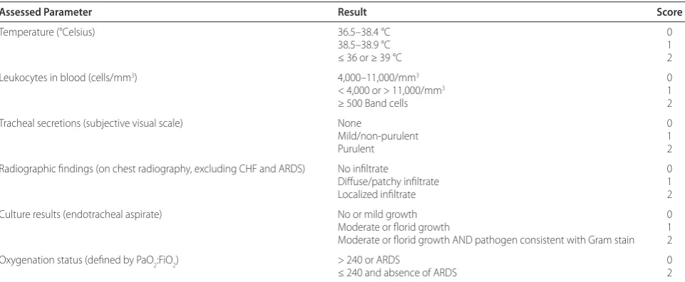
[image:3.612.66.547.99.297.2]Once specimens are obtained, the sample is sent for Gram stain, culture and sensitivity. Th e Gram stain can provide crucial initial clues to the type of organism(s) and whether or not the material is purulent (defi ned as ≥ 25 neutrophils and ≤ 10 squamous epithelial cells per low power fi eld) [1],[12]. Culture results can be reported as semi-quantitative and/or quantitative values. Semi-quantitative values obtained by endotracheal sampling are considered positive when the agar growth is moderate Table 1 The clinical pulmonary infection score (CPIS)
Assessed Parameter Result Score
Temperature (°Celsius) 36.5–38.4 °C 0
38.5–38.9 °C 1
≤ 36 or ≥ 39 °C 2
Leukocytes in blood (cells/mm3) 4,000–11,000/mm3 0
< 4,000 or > 11,000/mm3 1
≥ 500 Band cells 2
Tracheal secretions (subjective visual scale) None 0
Mild/non-purulent 1
Purulent 2
Radiographic fi ndings (on chest radiography, excluding CHF and ARDS) No infi ltrate 0
Diff use/patchy infi ltrate 1
Localized infi ltrate 2
Culture results (endotracheal aspirate) No or mild growth 0
Moderate or fl orid growth 1
Moderate or fl orid growth AND pathogen consistent with Gram stain 2
Oxygenation status (defi ned by PaO2:FiO2) > 240 or ARDS 0
≤ 240 and absence of ARDS 2
(+++) or heavy (++++), while quantitative positivity is defi ned as ≥ 105 cfu/ml. Exact speciation of pathogen
bacteria and their sensitivity to antibiotics can take a few days, but provides invaluable information.
Mechanically ventilated patients in the ICU receive frequent chest X-rays and presence of infi ltrate(s) and/or consolidation is considered part of diagnostic criteria and is widely used. However, there are several clinical conditions that have radiographic appearances similar to
VAP. Th ese conditions are commonly encountered in
mechanically ventilated patients and include aspiration and chemical pneumonitis, atelectasis, congestive heart failure, acute respiratory distress syndrome (ARDS), pleural eff usion and intra-alveolar hemorrhage to name a few. Hence, reliance on chest radiography for the diag-nosis of VAP is not advisable. Th ere is poor correlation between radiographic signs (alveolar infi ltrates, air broncho grams) and histopathological diagnosis of pneu-monia [12]. Th e sensitivity and specifi city of presence of infi ltrates on chest X-ray is also not encouraging [12]. On the fl ip-side, the negative predictive value of infi ltrates may have clinical utility. In a meta-analysis by Klompas, the presence or absence of fever, elevated white blood cell count, or purulent secretions did not substantively predict the probability of infection; however, the absence of a new infi ltrate on a plain radiograph lowered the likelihood of VAP [18].
VAP must be distinguished from tracheo-bronchitis. Clinical features of these diseases can overlap, but only VAP will demonstrate the presence of hypoxia and the presence of infi ltrate/consolidation on chest radiography [12].
Recently, the Centers for Disease Control and Prevention (CDC) rolled out new surveillance criteria for possible or probable VAP [17]. Th e goals were to capture other common complications of ventilator care, to improve objectivity of surveillance to allow comparability across centers for public reporting, and to minimize gaming [30]. Per these new criteria, a period of at least 2 days of stable or decreasing ventilator settings (daily minimum positive end-expiratory pressure [PEEP] or fraction of inspired oxygen [FiO2]) followed by consis-tently higher settings for at least 2 additional calendar days is required before a patient can be said to have a ventilator-associated condition (VAC). Most VACs are attributable to pneumonia, pulmonary edema, atelectasis, or ARDS, conditions which all have well researched prevention and management strategies [31]. Signs of infection/infl ammation (abnormal temperature or white-cell count and administration of one or more new antibiotics for at least 4 days) classify the patient as an “infection-related ventilator-associated complication,” or IVAC. Presence of purulent secretions (according to quantitative Gram staining criteria) and pathogenic
culture data will label the patient as possible or probable VAP. Patients with an IVAC and purulent secretions alone or pathogenic cultures alone have “possible pneu-monia”; those with both purulent secretions and positive quantitative or semiquantitative cultures have “probable pneumonia”. Probable pneumonia is also defi ned by suggestive histopathological features, positive pleural-fl uid cultures, or diagnostic tests for legionella and selected viruses. Chest radiograph fi ndings have been excluded in the new criteria because of their subjectivity without increased accuracy. Th is is not intended to reduce the role of radiography in clinical care. At the present time, the new CDC algorithm is for surveillance purposes only.
In the United States, VAP has been proposed as an indicator of quality of care in public reporting, and its prevention is a national patient safety goal. Th e threat of
non-reimbursement and fi nancial penalties for this
diagnosis has put pressure on hospitals to minimize VAP rates [13]. Th is has resulted in potential artifacts in surveil lance with more than 50 % of non-teaching medical ICUs in the United States reporting VAP rates close to zero [30], [32]. Th ese rates are an order of magnitude lower than those in European centers, which utilize similar preventive and treatment strategies suggesting that reductions in VAP rates may not refl ect improve-ments in prevention so much as subjective surveillance biases. It is anticipated that the new CDC surveillance paradigm for ventilator-associated events will help achieve a more realistic VAP rate.
Treatment
Selecting the appropriate antibiotic depends on the duration of mechanical ventilation. Late onset VAP (> 4 days) requires broad spectrum antibiotics whereas early onset (≤ 4 days) can be treated with limited spectrum antibiotics [1]. An updated local antibiogram for each hospital and each ICU based on local bacteriological patterns and susceptibilities is essential to guide optimally dosed initial empiric therapy [1]. With any empiric antibiotic regimen, de-escalation is the key to reduce emergence of resistance [33]. Delays in initiation of antibiotic treatment may add to the excess mortality risk with VAP [1]. Tables 2 and 3 highlight the recom-mended treatment regimens for VAP.
Owing to the high rate of resistance to monotherapy observed with P. aeruginosa, combination therapy is always recommended. Acinetobacter species respond best to carbapenems (also active against ESBL positive Enterobacteriaceae), colistin, polymyxin B and ampi-cillin/sulbactam [36], [37]. Although MDR organisms are usually associated with late-onset VAP, recent evidence suggests that they are increasingly associated with
antibiotics in the setting of failure of systemic antibiotics is unclear [1]. Th e usual duration of treatment for early-onset VAP is 8 days and longer in the case of late-early-onset VAP or if MDR organisms are suspected or identifi ed [39]–[41].
Despite therapy, if no response is observed, it may be prudent to reconsider the diagnosis, reassess the organism being treated or search for other reasons for signs and symptoms. Because of the challenges associated with diagnosing VAP, especially early in the course, the IDSA/ATS guidelines highlight the importance of re-assessing patients at 48–72 hours once pertinent data are available to determine whether the patient should con-tinue antibiotic therapy for VAP or whether an alternative diagnosis should be pursued. In one study, Swoboda et al. [42] found that half of the empiric antibiotic use for VAP in two surgical ICUs was prescribed for patients without pneumonia.
Prevention
[image:5.612.69.548.108.465.2]Th ere are multiple recommended measures for preven-tion of VAP. Th ese measures are summarized in Table 4 [43]–[46]. Institutions or ICUs may observe a reduction in VAP rates by utilizing a ‘VAP-bundle’ approach [44], [47] using elements depicted in Table 4. Th e 5-element Institute of Healthcare Improvement (IHI) VAP bundle [47] includes: Head of bed elevation, oral care with chlorhexidine, stress ulcer prophylaxis, deep venous thrombosis prophylaxis, and daily sedation assessment and spontaneous breathing trials. Each of these elements has been shown to reduce the incidence of VAP although the quality of evidence supporting the eff ectiveness and importance of each intervention has been questioned. Even studies using VAP bundles have been criticized as failing to demonstrate clinical and cost eff ectiveness [48]. A before-after study which systematically implemented a VAP prevention bundle using IHI methodology showed a Table 2 Comparison of recommended initial empiric therapy for ventilator-associated pneumonia (VAP) according to time of onset [1], [34], [41]
Early-onset VAP Late-onset VAP
Second or third generation cephalosporin: e. g., ceftriaxone: 2 g daily;
cefuroxime: 1.5 g every 8 hours;
cefotaxime: 2 g every 8 hours
OR
Fluoroquinolones
e. g., levofl oxacin: 750 mg daily;
moxifl oxacin: 400 mg daily
OR
Aminopenicillin + beta-lactamase inhibitor e. g., ampicillin + sulbactam: 3 g every 8 hours
OR
Ertapenem
1 g daily
Cephalosporin
e. g., cefepime: 1–2 g every 8 hours;
ceftazidime 2 g every 8 hours
OR
Carbepenem
e. g., imipenem + cilastin: 500 mg every 6 hours or 1 g every 8 hours;
meropenem: 1 g every 8 hours
OR
Beta-lactam/beta-lactamase inhibitor
e. g., piperacillin + tazobactam: 4.5 g every 6 hours
PLUS
Aminoglycoside
e. g., amikacin: 20 mg/kg/day;
gentamicin: 7 mg/kg/day;
tobramycin: 7 mg/kg/day
OR
Antipseudomonal fl uoroquinolone
e. g., ciprofl oxacin 400 mg every 8 hours;
levofl oxacin 750 mg daily
PLUS
Coverage for MRSA
e. g., vancomycin: 15 mg/kg every 12 hours
OR
linezolid: 600 mg every 12 hours
signifi cant reduction in VAP rates, antibiotic use and MRSA acquisition [43]. Th ere was no reduction, however, in duration of mechanical ventilation or ICU admission. Th e IHI emphasizes the need for high (95 %) overall com-pliance rates with VAP bundles although this particular study reported overall bundle compliance rates of 70 %.
[image:6.612.64.549.108.430.2]Issues with completeness of documentation may under-estimate compliance, which remains an important feature of VAP bundle prevention strategies. Another important contribution towards VAP prevention and shortening periods of antibiotic exposure was a recent prospective study (n = 129), which concluded that a single-dose of Table 3 Recommended therapy for suspected or confi rmed multidrug resistant organisms and fungal VAP [1], [34], [35], [41]
Pathogen Treatment
Methicillin-resistant Staphylococcus aureus (MRSA) See Table 2
Pseudomonas aeruginosa Double coverage recommended. See Table 2
Acinetobacter species Carbapenem
e. g., imipenem + cilastin; 1 g every 8 hours;
meropenem 1 g every 8 hours
OR
Beta-Lactam/beta-lactamase inhibitor
e. g., ampicillin + sulbactam: 3 g every 8
hours
OR
Tigecycline: 100 mg loading dose, then 50 mg every 12 hours
Extended-spectrum beta-lactamase (ESBL) positive enterobacteriaceae Carbepenem
e. g., imipenem + cilastin: 1 g every 8 hours;
meropenem: 1 g every 8 hours
Fungi Fluconazole: 800 mg every 12 hours;
caspofungin: 70 mg loading dose, then 50 mg daily;
voriconazole (for aspergillus species): 4 mg/kg every 12 hours
Legionella Macrolides (e. g., azithromycin)
OR
Fluoroquinolones (e. g., levofl oxacin)
Table 4 Suggested measures for prevention of ventilator-associated pneumonia (VAP) [41], [42], [49]
ICU focused measures Institution focused measures
Alcohol-based hand washing policy Surveillance program for pathogen profi ling and creation of “antibiogram”
Early discontinuation of invasive devices Frequent educational programs to reduce unnecessary antibiotic prescription
Reduce reintubation rates Propagate use of non-invasive positive pressure ventilation (NIPPV)
Use of oropharyngeal vs. nasopharyngeal feeding tubes Endotracheal tubes (ETTs) with potential benefi t:
Polyurethane-cuff ed ETT
Silver/antibiotic coated ETT
Aspiration of subglottic secretions (HI-LO ETT)
Semi-recumbent patient positioning (30–45°) Maintain policy for oral decontamination Selective digestive decontamination (SDD)
Endotracheal tube cuff pressure ~ 20 cm H2O Early weaning and extubation
Early tracheostomy Daily sedation holds
Small bowel feeding instead of gastric feeding Preference on using heat-moisture exchangers over heater humidifi ers
[image:6.612.70.533.458.657.2]antibiotics within 4 h of intubation may be eff ective in preventing early onset VAP in a cohort of comatose patients [49]. A randomized clinical trial is needed to address this question.
Conclusion
VAP occurs frequently and is associated with signifi cant morbidity in critically ill patients. Th e primary obstacle in diagnosing VAP is the absence of gold standard criteria and, therefore, VAP continues to be an inconspicuous clinical syndrome. Th ere is enough evidence to indicate that VAP is preventable and that hospitals can decrease VAP rates, a factor that the new CDC VAP defi nitions are poised to demonstrate more objectively. Th e diagnostic challenge of VAP has multiple implications for therapy. Although a CPIS score > 6 may correlate with VAP, the sensitivity, specifi city and inter-rater agreement of this criterion alone are not encouraging. Microbiological data should be used for tailoring antibiotic therapy and not be restricted only to diagnosis. Th e pitfall in using empiric antibiotics for suspicion of VAP is the potential for antibiotic overuse, emergence of resistance, unnecessary adverse eff ects and potential toxicity. Th e major goals of VAP management are early, appropriate antibiotics in adequate doses followed by de-escalation based on microbiological culture results and the clinical response of the patient. Antimicrobial stewardship programs involving pharmacists, physicians and other healthcare providers optimize antibiotic selection, dose, and
duration to increase effi cacy in targeting causative
pathogens and allow the best clinical outcome.
List of abbreviations used
ARDS: acute respiratory distress syndrome; ATS: American Thoracic Society; BAL: Bronchoalveolar lavage; CDC: Centers for Disease Control and Prevention; CHF: congestive heart failure; CI: confi dence interval; CNSL coagulase-negative staphylococcus; CPIS: clinical pulmonary infection score; ESBL: extended-spectrum beta-lactamase producing bacteria (ESBL); FiO2: fraction of inspired oxygen; ICU: intensive care unit; IVAC: infection-related ventilator-associated complication; IDSA: Infectious Diseases Society of America; MDR: multi drug resistant; Mini-BAL – mini-bronchoalvelolar lavage; MRSA: methicillin-resistant
Staphylococcus aureus; MSSA: methicillin-senstive Staphylococcus aureus; OR: odds ratios; PBP: penicillin-binding protein; PEEP: positive end-expiratory pressure; PSB: protected specimen brush; Tregs: regulatory T-cells; VAP: ventilator-associated pneumonia.
Competing interests
The authors declare that they have no competing interests.
Declarations
Publication costs for this article were funded by the authors’ institutions.
Author details
1Department of Neurology, University of Maryland School of Medicine, Baltimore, MD 21201, USA. 2Department of Anesthesiology/Critical Care Medicine, Johns Hopkins University School of Medicine, Baltimore, MD 21287, USA.
Published: 18 March 2014
References
1. A merican Thoracic Society, Infectious Diseases Society of America: Guidelines for the management of adults with hospital-acquired,
ventilator-associated, and healthcare-associated pneumonia. Am J Respir
Crit Care Med 2005, 171:388–416.
2. V incent JL, Bihari DJ, Suter PM, Bruining HA, White J, Nicolas-Chanoin MH, Wolff M, Spencer RC, Hemmer M: The prevalence of nosocomial infection in
intensive care units in Europe. JAMA 1995, 274:639–644.
3. C hastre J, Fagon JY: State of the art: ventilator-associated pneumonia.Am J Respir Crit Care Med 2002, 165:867–903.
4. H unter JD: Ventilator associated pneumonia. BMJ 2012, 344(e3325):e3325. 5. A fshari A, Pagani L, Harbarth S: Year in review 2011: Critical care – infection.
Crit Care 2012, 16:242–247.
6. S krupky LP, McConnell K, Dallas J, Kollef MH: A comparison of ventilator-associated pneumonia rates as identifi ed according to the National Healthcare Safety Network and American College of Chest Physicians
Criteria. Crit Care Med 2012, 40:281–284.
7. R ello J, Ollendorf D, Oster G, Vera-Llonch M, Bellm L, Redman R, Kollef MH, VAP Outcomes Scientifi c Advisory Group: Epidemiology and outcomes of
ventilator-associated pneumonia in a large US database. Chest 2002,
122:2115–2121.
8. C ook DJ, Walter SD, Cook RJ, Griffi th LE, Guyatt GH, Leasa D, Jaeschke RZ, Brun-Buisson C: Incidence of and risk factors for ventilator-associated
pneumonia in critically ill patients.Ann Int Med 1998, 129:433–440.
9. M elsen WG, Rovers MM, Koeman M, Bonten MJM: Estimating the attributable mortality of ventilator-associated pneumonia from
randomized prevention studies. Crit Care Med 2011, 39:2736–2742.
10. M elsen WG, Rovers MM, Groenwold RH, Bergmans DC, Camus C, Bauer TT, Hanisch EW, Klarin B, Koeman M, Krueger WA, Lacherade JC, Lorente L, Memish ZA, Morrow LE, Nardi G, van Nieuwenhoven CA, O’Keefe GE, Nakos G, Scannapieco FA, Sequin P, Staudinger T, Topeli A, Ferrer M, Bonten MJ: Attributable mortality of ventilator-associated pneumonia: a
meta-analysis of individual patient data from randomised prevention studies.
Lancet Infect Dis 2013, 13:665–671.
11. Z olfaghari PS, Wyncoll DL: The tracheal tube: gateway to
ventilator-associated pneumonia.Crit Care 2011, 15:310–317.
12. G rgurich PE, Hudcova J, Lei Y, Sarwar A, Craven DE: Diagnosis of ventilator-associated pneumonia: controversies and working toward a gold
standard.Curr Opin Infect Dis 2013, 26:140–150.
13. M ietto C, Pinciroli R, Patel N, Berra L: Ventilator associated pneumonia:
evolving defi nitions and preventive strategies. Respir Care 2013,
58:990–1007.
14. R ocha LA, Marques Ribas R, da Costa Darini AL, Gontijo Filho PP: Relationship between nasal colonization and ventilator-associated pneumonia and the role of the environment in transmission of Staphylococcus aureus in
intensive care units. Am J Infect Control 2013, 41:1236–1240.
15. M orris AC, Brittan M, Wilkinson TS, McAuley DF, Antonelli J, McCulloch C, Barr LC, McDonald NA, Dhaliwal K, Jones RO, Mackellar A, Haslett C, Hay AW, Swann DG, Anderson N, Laurenson IF, Davidson DJ, Rossi AG, Walsh TS, Simpson AJ: C5a-mediated neutrophil dysfunction is RhoA-dependent
and predicts infection in critically ill patients. Blood 2011, 117:5178–5188.
16. C onway Morris A, Anderson N, Brittan M, Wilkinson TS, McAuley DF, Antonelli J, McCulloch C, Barr LC, Dhaliwal K, Jones RO, Haslett C, Hay AW, Swann DG, Laurenson IF, Davidson DJ, Rossi AG, Walsh TS, Simpson AJ: Combined dysfunctions of immune cells predict nosocomial infection in critically ill
patients. Br J Anaesth 2013, 3:1–10.
17. N ational Healthcare Safety Network (NHSN) July 2013 CDC/NHSN Protocol
Clarifi cations 2013, Available at: http://www.cdc.gov/nhsn/PDFs/
pscManual/10-VAE_FINAL.pdf Accessed Oct 2013
18. K lompas M: Clinician’s Corner: Does this patient have ventilator-associated
pneumonia?JAMA 2013, 297:1583–1593.
19. P etersen IS, Aru A, Skødt V, Behrendt N, Bols B, Kiss K, Simonsen K: Evaluation
of pneumonia diagnosis in intensive care patients. Scand J Infect Dis 1999,
31:299–303.
20. Fà bregas N, Ewig S, Torres A, Al-Abiary M, Ramirez J, de La Bellacasa JP, Bauer T, Cabello H: Clinical diagnosis of ventilator associated pneumonia revisited: comparative validation using immediate post-mortem lung
biopsies. Thorax 1999, 54:867–873
21. Joh anson WG, Pierce AK, Sanford JP, Thomas GD: Nosocomial respiratory infections with gram-negative bacilli. The signifi cance of colonization of
22. Pug in J, Auckenthaler R, Mili N, Janssens JP, Lew PD, Suter PM: Diagnosis of ventilator-associated pneumonia by bacteriologic analysis of bronchoscopic and nonbronchoscopic “blind” bronchoalveolar lavage
fl uid.Am Rev Respir Dis 1991, 143:1121–1129.
23. Sha n J, Chen HL, Zhu JH: Diagnostic accuracy of clinical pulmonary infection score for ventilator-associated pneumonia: a meta-analysis. Respir Care 2011, 56:1087–1094.
24. Zil berberg MD, Shorr AF: Ventilator-associated pneumonia: the clinical pulmonary infection score as a surrogate for diagnostics and outcome. Clin Infect Dis 2010, 1:S131–S135.
25. Sho rr AF, Cook D, Jiang X, Muscedere J, Heyland D: Correlates of clinical failure in ventilator-associated pneumonia: insights from a large,
randomized trial.J Crit Care 2008, 23:64–73.
26. Sin gh N, Rogers P, Atwood CW, Wagener MM, Yu VL: Short-course empiric antibiotic therapy for patients with pulmonary infi ltrates in the intensive care unit. A proposed solution for indiscriminate antibiotic prescription. Am J Respir Crit Care Med 2000, 162:505–511.
27. Fag on JY, Chastre J, Wolff M, Gervais C, Parer-Aubas S, Stéphan F, Similowski T, Mercat A, Diehl JL, Sollet JP, Tenaillon A: Invasive and noninvasive strategies for management of suspected ventilator-associated pneumonia.
A randomized trial.Ann Intern Med 2000, 132:621–630.
28. Cana dian Critical Care Trials Group: A randomized trial of diagnostic
techniques for ventilator-associated pneumonia.N Engl J Med 2013,
355:2619–2630.
29. Bert on DC, Kalil AC, Cavalcanti M, Teixeira PJ (2012) Quantitative versus qualitative cultures of respiratory secretions for clinical outcomes in patients with ventilator-associated pneumonia Chocrane Database Syst Rev CD006482
30. Klom pas M: Complications of mechanical ventilation – the CDC’s new
surveillance paradigm. N Engl J Med 2013, 368:1472–1475.
31. Haya shi Y, Morisawa K, Klompas M, Jones M, Bandeshe H, Boots R, Lipman J, Paterson DL: Toward improved surveillance: the impact of ventilator-associated complications on length of stay and antibiotic use in patients
in intensive care units. Clin Infect Dis 2013, 56:471–477.
32. Dude ck MA, Horan TC, Peterson KD, Allen-Bridson K, Morrell G, Pollock DA, Edwards JR:National Healthcare Safety Network (NHSN) Report, data
summary for 2010, device-associated module. Am J Infect Control 2011,
39:798–816.
33. Mast erton RG: Antibiotic de-escalation. Crit Care Clin 2011, 27:149–162. 34. Torr es A, Ewig S, Lode H, Carlet J: Defi ning, treating and preventing hospital
acquired pneumonia: European perspective. Intensive Care Med 2009,
35:9–29.
35. Walk ey AJ, O’Donnell MR, Wiener RS: Linezolid vs glycopeptide antibiotics for the treatment of suspected methicillin-resistant Staphylococcus aureus nosocomial pneumonia: a meta-analysis of randomized controlled
trials.Chest 2011, 139:1148–1155.
36. Muno z-Price LS, Weinstein RA: Acinetobacter Infection. N Engl J Med 2008, 358:1271–1281.
37. Mart in-Loeches I, Deja M, Koulenti D, Dimopoulos G, Marsh B, Torres A, Niderman MS, Rello J, EU-VAP Study Investigators: Potentially resistant microorganisms in intubated patients with hospital-acquired pneumonia:
the interaction of ecology, shock and risk factors.Intensive Care Med 2013,
39:672–681.
38. Pasq uale TR, Jabrocki B, Salstrom SJ, Wiemken TL, Peyrani P, Hague NZ, Scerpella EG, Ford KD, Zervos MJ, Ramirez JA, File TM Jr, IMPACT-HAP Study Group: Emergence of methicillin-resistant Staphylococcus aureus USA300 genotype as a major cause of late-onset nosocomial pneumonia in
intensive care patients in the USA. Int J Infect Dis 2013, 17:e398–e403.
39. Cape llier G, Mockly H, Charpentier C, Annane D, Blasco G, Desmettre T, Roch A, Faisy C, Cousson J, Limat S, Mercier M, Papazian L: Early-onset ventilator-associated pneumonia in adults randomized clinical trial: comparison of 8
versus 15 days of antibiotic treatment.PloS one 2012, 7:e41290.
40. Chas tre J, Wolff M, Fagon J-Y, Chevret S, Thomas F, Wermert D, Clementi E, Gonzalez J, Jusserand D, Asfar P, Perrin D, Fieux F, Aubas S, PneumA Trial Group: Comparison of 8 vs 15 days of antibiotic therapy for
ventilator-associated pneumonia in adults: a randomized trial.JAMA 2003,
290:2588–2598.
41. Dimo poulos G, Poulakou G, Pneumatikos IA, Armaganidis A, Kollef MH, Matthaiou DK: Short- versus long-duration antibiotic regimens for ventilator-associated pneumonia: a systematic review and meta-analysis. Chest 2013, 144:1759–1767.
42. Swob oda SM, Dixon T, Lipsett PA: Can the clinical pulmonary infection score
impact ICU antibiotic days?Surg Infect (Larchmt) 2006, 7:331–339.
43. Morr is AC, Hay AW, Swann DG, Everingham K, McCulloch C, McNulty J, Brooks O, Laurenson IF, Cook B, Walsh TS: Reducing ventilator-associated
pneumonia in intensive care: impact of implementing a care bundle.Crit
Care Med 2011, 39:2218–2224.
44. Alha zzani W, Almasoud A, Jaeschke R, Lo BW, Sindi A, Altayyar S, Fox-Robichaud A: Small bowel feeding and risk of pneumonia in adult critically ill patients: a systematic review and meta-analysis of randomized trials. Crit Care 2013, 17:R127.
45. Musc edere J, Rewa O, McKechnie K, Jiang X, Laporta D, Heyland DK: Subglottic secretion drainage for the prevention of ventilator-associated
pneumonia: a systematic review and meta-analysis.Crit Care Med 2011,
39:1985–1991.
46. Morr ow LE, Kollef MH: Recognition and prevention of nosocomial pneumonia in the intensive care unit and infection control in mechanical
ventilation.Crit Care Med 2010, 38:S352–S362.
47. Youn gquist P, Carroll M, Farber M, Macy D, Madrid P, Ronning J, Susag A:
Implementing a ventilator bundle in a community hospital.Jt Comm J
Qual Patient Saf 2007, 33:219–225.
48. Zilb erberg MD, Shorr AF, Kollef MH: Implementing quality improvements in
the intensive care unit: Ventilator bundle as an example. Crit Care Med 2009,
37:305–309.
49. Vall és J, Peredo R, Burgueño MJ, Rodrigues de Freitas AP, Millán S, Espasa M, Martín-Loeches I, Ferrer R, Suarez D, Artigas A: Effi cacy of single-dose antibiotic against early-onset pneumonia in comatose patients who are
ventilated. Chest 2013, 143:1219–1225.
doi:10.1186/cc13775
Cite this article as: Kalanuria AA, et al.: Ventilator-associated pneumonia in
![Table 2 Comparison of recommended initial empiric therapy for ventilator-associated pneumonia (VAP) according to time of onset [1], [34], [41]](https://thumb-us.123doks.com/thumbv2/123dok_us/1528539.696008/5.612.69.548.108.465/comparison-recommended-initial-empiric-ventilator-associated-pneumonia-according.webp)
![Table 4 Suggested measures for prevention of ventilator-associated pneumonia (VAP) [41], [42], [49]](https://thumb-us.123doks.com/thumbv2/123dok_us/1528539.696008/6.612.64.549.108.430/table-suggested-measures-prevention-ventilator-associated-pneumonia-vap.webp)
