Original Article
Overexpression of CAMK2N1 indicates
good prognosis for glioma and regulates androgen
receptor-associated cell proliferation and apoptosis
Dejun Bao1,2,3, Wanxiang Niu1,2,3, Chenxu Zhou1,2,3, Chuandong Cheng1,2,3, Yang Yang1,2,3, Chaoshi Niu1,2,3
1Department of Neurosurgery, Anhui Provincial Hospital Affiliated to Anhui Medical University, China; 2Anhui
Provincial Stereotactic Neurosurgical Institute, China; 3Anhui Province Key Laboratory of Brain Function & Brain
Disease, Hefei 230001, Anhui, China
Received July 22, 2018; Accepted October 10, 2018; Epub January 15, 2019; Published January 30, 2019
Abstract: Glioma is the most common form of primary central nervous system tumors. The present study aimed to identify the prognostic roles of Calcium/calmodulin-dependent protein kinase II inhibitor 1 (CAMK2N1) and its regulation of glioma cell proliferation and apoptosis. Quantitative reverse transcription polymerase chain reaction (qRT-PCR) and immunohistochemistry (IHC) were used to detect expression of CAMK2N1 in glioma. Kaplan-Meier survival analysis was used to analyze the association of CAMK2N1 with overall survival (OS) of glioma patients.
Cell growth was detected by MTT assay and cell apoptosis was tested by flow cytometry and Western blot analysis. Results revealed that expression of CAMK2N1 was significantly lower in high-grade glioma tissue compared with
low-grade glioma. High expression of CAMK2N1 indicates better outcomes for glioma patients. Univariate and mul-tivariate analysis suggested that CAMK2N1 expression was an independent risk factor for OS. Overexpression of CAMK2N1 may inhibit proliferation and promote apoptosis in glioma cell U87 by activating apoptosis regulatory kinases BAX, Bcl-2, Cleaved-caspase3, and Cleaved-PARP. This could be reversed by synthetic androgen (R1881),
a standard androgen receptor (AR) signaling agonist. These findings characterize CAMK2N1 as a tumor suppressor
gene that regulates glioma cell proliferation and apoptosis, most likely through AR signaling pathways.
Keywords: CAMK2N1, glioma, androgen receptor, proliferation, apoptosis
Introduction
Glioma accounts for almost half of primary cen-tral nervous system tumors and is the most prevalent malignancy of brain tumors [1]. The
World Health Organization (WHO) has classified
glioma into grades I-IV, according to their cellu-lar origin and degree of malignancy [2]. Current therapeutic approaches include surgery, che-motherapy, radiotherapy, and molecular target-ed therapy. However, the most malignant glio-mas, grade IV glioglio-mas, also called glioblastomas (GBMs), only have a 5-year survival rate of 9.8% at best [3]. Genetic and epigenetic alterations are major culprits of cellular transformation and therapy resistance [4]. Recent genomic studies have unveiled the complexity of tumor heterogeneity in glioblastomas, providing new insight into the genomic landscape of tumor cells that survive and initiate tumor recurrence
[5]. Therefore, it is critical to develop novel and effective molecular makers to assist early diag-nosis and accurate prediction of progdiag-nosis in patients suffering from glioma.
Ca2+-calmodulin stimulated protein kinase II (CaMKII) is a multifunctional Ser/Thr protein kinase which is enriched in the brain [6]. Its bio-logical function is cell type and cell contest dependent [7]. It plays a central role in synapse formation, neurotransmitter synthesis and se- cretion, receptor and ion channel function, syn-aptic plasticity, and memory [8]. However, in cancer cells, it participates in the activation of ERK pathways by oncogenic Ras and RET rear-rangements (RET/PTC), thus modulating tumor cell proliferation [9].
protein kinase II), has recently been shown to affect tumorigenesis and tumor growth [10-12]. A previous study suggested that it inhibited human colon adenocarcinoma cell growth and was negatively correlated with severity of hu- man colon adenocarcinoma, indicating a pivot-al role of CAMK2N1 in the development and progression of carcinomas [12]. An integrated analysis of genome-wide DNA methylation and
RNA expression profiles using cervical cancer tissues identified 19 novel cervical
cancer-related genes, including CAMK2N1, which sup-pressed mRNA expression regulated by DNA methylation [13]. Genome-wide miRNA
expres-sion profiling also revealed that CAMK2N1 may
regulate cell apoptosis and the cell-cycle pro-cess in osteosarcoma [14]. Recently, CAMK2N1 was found to inhibit prostate cancer progres-sion through androgen receptor (AR)-dependent signaling in vitro and in vivo, indicating cross-regulation of AR and CAMK2N1 in cancers [11]. Data suggests that CAMK2N1 may play an important role in cancer progression. However, the molecular mechanisms and functional link between CAMK2N1 and glioma remain un- known.
The present study aimed to detect expression levels of CAMK2N1 in glioma and to identify its correlation with clinicopathologic features in gl- ioma patients. Moreover, this study investigat-ed its regulatory role in proliferation and apop-tosis of glioma cells and explored its relation-ship with AR signaling.
Methods
Patients and samples
For reverse transcription-quantitative polyme- rase chain reaction (RT-qPCR) assay, eight fresh glioma tissues with low grade, eight fresh glioma tissues with high grade, and eight nor-mal non-cancerous tissue samples were ob- tained from patients with glioma, undergoing surgical resection at Anhui Provincial Hospital
Affiliated to Anhui Medical University, between
January 2016 to January 2017. Human glioma tissue microarray (n=180) was purchased from Outdo Biotech (Shanghai, China) for Kaplan-Meier survival analysis. The operation time of patients was from February 2008 to October 2011, with follow-ups ending in July 2017. No patients received chemotherapy or
radiothera-py prior to surgery. Patient clinical features were obtained from medical records. The medi-an age of this cohort was 23 years (rmedi-ange, 3-80 years). Written informed consent was obtained from each patient for the use of tissue samples for research purposes. This study was carried out with the approval of the Ethics Committee
of Anhui Provincial Hospital Affiliated to Anhui
Medical University. Reagents
Human anti-CAMK2N1 antibody and anti-GAP-DH antibody were purchased from Santa Cruz Biotechnology (Santa Cruz, CA, USA). Human BAX, Bcl-2, caspase3, and anti-PARP antibodies were obtained from Cell Sig- naling Technology (Beverly, MA, USA). R1881, also known as methyltrienolone, was purcha- sed from SIGMA.
Cell culture
Human glioma U87 cells were obtained from ATCC and were maintained in Roswell Park Memorial Institute (RPMI)-1640 medium sup-plemented with 10% fetal bovine serum (Gibco,
Grand Island, NY) at 37°C in a humidified atmo
-sphere of 95% air and 5% CO2.
Lentiviral construction and transfections
Human CAMK2N1 genes were polymerase
chain reaction (PCR) amplified from normal
genomic DNA and cloned into lentiviral ve- ctor GV166 (Ubi-MCS-3FLAG-IRES-puromycin; Shanghai Genechem, Shanghai, China) for ectopic expression of CAMK2N1. Expression of
CAMK2N1 was confirmed by Western blot
analysis.
Quantitative real-time PCR (qRT-PCR)
Total RNA was isolated and reversely tran-scribed to cDNA using TRIzol Reagent (In- vitrogen) and the PrimeScript RT Reagent kit (RR037A; Takara), respectively, according to manufacturer instructions. qRT-PCR was car-ried out via Bio-Rad CFX96 Real-Time PCR Detection System with SYBR Green Supermix (Bio-Rad). Relative gene expression was
nor-malized and calculated by the 2-ΔΔCt method.
Immunohistochemistry (IHC) staining and
Western blot analysis
Immunohistochemical analysis of human glio-ma tissues was conducted using huglio-man priglio-ma- prima-ry antibody anti-CAMK2N1. Human glioma tis-sue microarray was purchased from Outdo Biotech (Shanghai, China). Western blot was performed on U87 cell lines, as previously indi-cated. Cells were pelleted and lysed in buffer supplemented with a protease inhibitor cock-tail (Roche Diagnostics, Mannheim, Germany). Protein samples were separated by sodium dodecyl sulfatepolyacrylamide gel
electropho-resis and transferred to polyvinylidene fluoride
membranes (Millipore, Billerica, MA). Immun- oblots were performed by incubating
polyvinyli-dene fluoride membranes with 5% non-fat milk
in TRIS-buffered saline and 2.5% Tween-20 for 1 hour at room temperature. Each membrane was then incubated with primary antibodies at 4°C overnight and incubated with secondary antibodies at room temperature for 1.5 hours. Each blot was stripped and re-probed with a GAPDH antibody as an internal control. Images were captured with SynGene G: Box Chemi XRQ (Alpha Metrix Biotech, Germany). Experiments were performed in triplicate and repeated at least 3 times.
Cell proliferation and apoptosis analysis
First, 2 × 10³ stable cells were seeded in 96-well plates in normal growth medium. Cell growth was measured daily by MTT assays using 3-(4, 5-dimethylthiazol-2-yl)-2, 5-diphen-yltetrazolium bromide. The experiment was per-formed at least 3 times. PE-Annexin-V Apoptosis Detection Kit (BD Biosciences) was used to
detect apoptosis by flow cytometry, according
to manufacturer instructions. Each sample was
analyzed by flow cytometry with a FACScan
Fl-ow Cytometer (Becton-Dickinson Biosciences,
are presented as means of at least 3 indepen-dent experiments. Statistical comparisons we- re performed using Student’s t-test, with P
val-ues <0.05 considered statistically significant.
Kaplan-Meier survival analysis was used to illustrate the prognostic relevance of CA- MK2N1 in glioma patients. P values <0.05 are
considered statistically significant.
Results
Expression of CAMK2N1 in glioma of different grades
CAMK2N1, characterized as an inhibitor of Ca- MKII, plays a critical role in tumorigenesis in dif-ferent cancers. CAMK2N1 mRNA expression of para-cancerous normal tissue and glioma sp- ecimens was measured by performing quanti-tative RT-PCR (qRT-PCR). As shown in Figure
1A, mRNA levels of CAMK2N1 were
signifi-cantly reduced as tumor malignancy degree increased. Moreover, protein expression of
CAMK2N1 was significantly decreased in tumor
tissues with high grade compared to those with low grade. Next, CAMK2N1 protein expression in human glioma specimens was analyzed by performing immunohistochemical (IHC) stain-ing usstain-ing tissue microarray. CAMK2N1 showed higher IHC scores in low grade (Figure 1B) than in high grade, consistent with mRNA expression in gliomas. Figure 1C shows representative IHC pictures from the tissue microarray. CAMK2N1 showed whole cell distribution, with stronger staining in the nucleus, in low grade when com-pared with high grade.
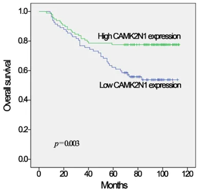
Relationship between CAMK2N1 expression and overall survival in glioma
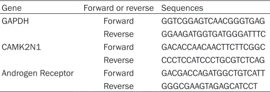
[image:3.612.91.362.96.189.2]Kaplan-Meier survival analysis showed that patients with glioma expressing high expres-sion of CAMK2N1 had good overall survival (P=0.003, Figure 2). Univariate analyses were Table 1. Primer sequences used for quantitative reverse
transcription-polymerase chain reaction
Gene Forward or reverse Sequences
GAPDH Forward GGTCGGAGTCAACGGGTGAG Reverse GGAAGATGGTGATGGGATTTC CAMK2N1 Forward GACACCAACAACTTCTTCGGC Reverse CCCTCCATCCCTGCGTCTCAG Androgen Receptor Forward GACGACCAGATGGCTGTCATT Reverse GGGCGAAGTAGAGCATCCT
Mansfield, MA) using a 488 nm
laser. A minimum of 10,000 ev- ents were collected to maximize the statistical validity of compart-mental analysis. The experiment was performed at least 3 times. Statistical analysis
blot analysis of CAMK2N1 expression demon-strated the upregulation of CAMK2N1 in U87-CAMK2N1 (Figure 3A). As shown in Figure 3B, MTT assay showed that CAMK2N1 inhibited proliferation in U87-CAMK2N1, compared to U87-vector. Moreover, it was found that CA- MK2N1 could upregulate the proportion of apoptosis of glioma cells, as shown in Figure 3C. After upregulating the expression of CA- MK2N1, the proportion of apoptotic cells was
significantly increased, compared with Mock cells. Differences were statistically significant
[image:4.612.92.372.73.372.2](P<0.05). As shown in Figure 3D, 3E, lentiviral-mediated transfection of CAMK2N1 overexpr- ession induced visual increases of protein ex- pression of BAX (P<0.01), Bcl-2 (P<0.001), Cleaved-Caspase3 (P<0.01), and Cleaved-PA- RP (P<0.05), representing cell apoptosis in the U87 cell line. Results suggest that upregulating CAMK2N1 could inhibit the proliferation of glio-ma through promoting apoptosis in glioglio-ma cells.
Figure 1. Expression of CAMK2N1 in glioma of different grades. A. Reverse transcription-polymerase chain reaction detection of CAMK2N1 mRNA in normal tissues (n=8), low grade glioma tissues (n=8), and high-grade glio-ma tissues (n=8). B. CAMK2N1 IHC score in the glioglio-ma tissue microarray (N=180). C. Representative examples of immunohistochemical staining for CAMK2N1 in low and high grade of glioma tissue as indicated. Values are expressed as mean ± SEM (*P<0.05, **P<0.01***P<0.001).
Figure 2. Significant positive correlation between
CAMK2N1 expression and overall survival in glioma. In Kaplan-Meier survival analysis, patients with gli-oma expressing high expression of CAMK2N1 had good overall survival (P=0.003).
performed using age, gender, WHO grade, and CAMK2N1 expression as possible
vari-ables. Results identified age
(P<0.01), WHO grade (P<0.01), and CAMK2N1 expression
(P<0.01) as significant predic -tors of overall survival (OS).
Multivariate analysis
confirm-ed that WHO grade (HR=3.3- 64; 95% CI=2.1464.844; P< 0.01) and CAMK2N1 expre- ssion (HR=1.175; 95% CI=0.3- 71-2.597; P<0.01) were in- dependent risk factors for OS (Table 2). Results revealed th- at high expression of CAMK- 2N1 is associated with go- od prognosis for glioma pa- tients.
CAMK2N1 inhibits cell prolifer
-ation and promotes apoptosis
[image:4.612.91.286.475.661.2]Table 2. Univariate and multivariate analyses were performed to analyze clinical characteristics and overall survival in glioma
Variable Univariate Cox Multivariate Cox HR (95% CI) p HR (95% CI) p
Age
Increasing years 1.146 (1.078-1.253) <0.01 1.023 (0.964-1.137) 0.221 Gender
Female vs Male 0.945 (0.809-1.146) 0.355 WHO Grade
High vs Low 4.681 (3.021-6.181) <0.01 3.364 (2.146-4.844) <0.01 CAMK2N1
Low vs High 2.446 (1.268-4.353) <0.01 1.175 (0.371-2.597) <0.01
inhibition of CaMKII in medullary thyroid carci-noma (MTC) cells induced a reduction of Raf-1, MEK, and ERK phosphorylation, cyclin D expression, and cell proliferation. Moreover, mRNA expression of CAMK2N1 inversely corre-lates with severity of MTC [16].
The present study provides evidence that, in human glioma cancer tissue, endogenous CA- MK2N1 expression was positively correlated with prognosis of glioma. It was found that CAMK2N1 showed higher IHC scores in low grade than in high grade, consistent with mR- NA expression in glioma. Patients with high CAMK2N1 levels had longer overall survival. Univariate analyses and multivariate analysis
were conducted, finding that CAMK2N1
ex-pression was an independent risk factor for OS. Results were consistent with those in other
cancers. At the cellular level, it was confirmed
that CAMK2N1 inhibited cell proliferation and promoted apoptosis through activating BAX, Bcl-2, caspase3, and PARP proteins.
Recent studies have shown that CAMK2N1 and AR signaling formed an auto-regulatory nega-tive feedback loop, where CAMK2N1 was down regulated by AR activation in response to andro-gen, while CAMK2N1 inhibited AR expression and activity through CAMKII pathways [11].
Researchers have also found significantly
upregulated AR expression in GBM tissue, com-pared to the periphery normal brain tissue, in patients. Further in vitro experiments have sug-gested that AR signaling might promote tumori-genesis of GBM in adult men by inhibiting TGF-beta receptor signaling [17]. To study the relationship between CAMK2N1 and AR in glio-ma, the present study detected the expression
has been reported to suppress prostate canc- er progression depend-ing on androgen receptor (AR)-associated signal-ing [11]. In ovarian can-cer, hypermethylation of CAMK2N1 resulting in loss of CAMK2N1 is as- sociated with poor clini-cal outcomes in Type II EOC, also after macro-scopic complete resec-tion [15]. Previous re- search has revealed that
Effects of CAK2N1 on cell proliferation and apoptosis could be reversed by androgen re
-ceptor-associated signaling (AR) signaling ac -tivation
To explore the association of CAMK2N1 with AR signaling in glioma, U87 cells were treated with 10 nm R1881 (AR signaling activator) for 0-48 hours. Expression of CAMK2N1 decre- ased with time after R1881 was treated (Figure 4A). Next, expression of AR in U87-CAMK2N1 and U87-vector was detected. It was found that AR was down-expressed in U87-CAMK2N1, compared with U87-vector (Figure 4B, P< 0.01). As shown in Figure 4C, proliferation of U87-CAMK2N1 was rescued when treated with R1881. Flow cytometry demonstrated that R1- 881 could inhibit apoptosis of CAMK2N1 over-expressed glioma U87 cells (Figure 4D and 4E, P<0.01). As shown in Figure 4F, 4G, R1881 induced a visual decrease on protein ex- pression of BAX, Cleaved-Caspase3 (P<0.05, respectively), Bcl-2, and Cleaved-PARP (P< 0.01, respectively). Results suggest that prolif-eration inhibition and apoptosis promotion effects of CAMK2N1 could be reversed by acti-vation of androgen receptor-associated sig- naling in glioma.
Discussion
of AR in U87-CAMK2N1 and U87-vector, finding
[image:6.612.96.518.74.618.2]that AR was down regulated in CAMK2N1- overexpressed glioma cells. Next, this study used R1881, known as a synthetic androgen, Figure 3. CAMK2N1 inhibits cell proliferation and promotes apoptosis. A. CAMK2N1 expression was detected in U87, U87-vector, and U87-CAMK2N1cells by Western blots. B. Cell proliferation activity of U87, U87-vector, and U87-CAMK2N1 were measured daily by MTT assays for three days. C. U87-vector and U87-CAMK2N1 were then
analyzed for cell apoptosis by flow cytometry. D, E. Western blot analysis of apoptosis related protein expression
to activate AR, finding that activation of AR res -cued CAMK2N1 related cell proliferation inhibi-tion and apoptosis increased as expected. This could prove that CAMK2N1 interacts with AR in glioma cells.
[image:7.612.95.517.72.554.2]AR plays a critical role in the development of prostate cancer [18, 19]. In prostate cancer cells, AR promotes proliferation and invasion to promote progression of prostate cancer th- Figure 4. Effects of CAMK2N1 on cell proliferation and apoptosis could be reversed by androgen receptor-associated signaling (AR) signaling activation. A. CAMK2N1 gene expression was detected by quantitative PCR in U87 treated with 10 nm R1881 for 0-48 hours. B. Androgen receptor expression was measured by quantitative PCR in U87-vector and U87-CAMK2N1 cell line. C. Cell proliferation activity of U87-U87-vector and U87-CAMK2N1 treated with or without R1881was measured daily by MTT assays for three days. Experiments were performed at least 3 times. D, E.
U87-CAMK2N1 treated with or without R1881 for 48 hours were then analyzed for cell apoptosis by flow cytometry.
ry kinases [20], including BAX/Bcl-2, Bad, cas-pase4, caspase7, and activating PI3K/AKT and MEK/ERK signaling pathways [14]. In this study, it was observed that CAMK2N1 activated apop-tosis regulatory kinases BAX, Bcl-2, caspase3, and PARP protein expression through blocking AR signaling pathways in glioma. Collectively, results revealed the tumor suppressive role of CAMK2N1 in glioma.
In conclusion, the presents study identified a
possible functional link between CAMK2N1 and androgen receptor signaling in glioma. Ov- erexpression of CAMK2N1 countered glioma growth, indicating good prognostic outcomes in glioma patients. Present results may provide a novel and convincing method of treating glioma by targeting the functional interaction between CAMK2N1 and AR associated signaling. Acknowledgements
This study was support by the Science and Technology Project grant from Anhui Province (No: 140802285MKL69, No: 1606c08235, No: 1604a0802069).
Disclosure of conflict of interest
None.
Address correspondence to: Dr. Chaoshi Niu, De- partment of Neurosurgery, Anhui Provincial Hos-
pital Affiliated to Anhui Medical University, China;
Anhui Provincial Stereotactic Neurosurgical Institute, China; Anhui Province Key Laboratory of Brain Function & Brain Disease, No. 17 Lujiang Road, Hefei 230001, Anhui, China. Tel: 0551-62284149; Fax: 0551-62283292; E-mail: niuchaoshi@163.com
References
[1] Siegel RL, Miller KD and Jemal A. Cancer sta-tistics, 2017. CA Cancer J Clin 2017; 67: 7-30. [2] Van Meir EG, Hadjipanayis CG, Norden AD, Shu
HK, Wen PY and Olson JJ. Exciting new advanc-es in neuro-oncology: the avenue to a cure for malignant glioma. CA Cancer J Clin 2010; 60: 166-93.
[3] Wang Y and Jiang T. Understanding high gra- de glioma: molecular mechanism, therapy and comprehensive management. Cancer Lett 2013; 331: 139-46.
[4] Domingo-Fernandez R, Watters K, Piskareva O, Stallings RL and Bray I. The role of genetic and epigenetic alterations in neuroblastoma dis-ease pathogenesis. Pediatr Surg Int 2013; 29: 101-19.
[5] Kalpathy-Cramer J, Gerstner ER, Emblem KE, Andronesi O and Rosen B. Advanced magnetic resonance imaging of the physical processes in human glioblastoma. Cancer Res 2014; 74: 4622-37.
[6] Ling KH, Hewitt CA, Beissbarth T, Hyde L, Che-ah PS, Smyth GK, Tan SS, HChe-ahn CN, Thomas T, Thomas PQ and Scott HS. Spatiotemporal reg-ulation of multiple overlapping sense and nov-el natural antisense transcripts at the Nrgn and Camk2n1 gene loci during mouse cere-bral corticogenesis. Cereb Cortex 2011; 21: 683-97.
[7] Wu P, Zuo X, Deng H, Liu X, Liu L and Ji A. Roles of long noncoding RNAs in brain development,
functional diversification and neurodegenera -tive diseases. Brain Res Bull 2013; 97: 69-80. [8] Wayman GA, Lee YS, Tokumitsu H, Silva AJ and
Soderling TR. Calmodulin-kinases: modulators of neuronal development and plasticity. Neu-ron 2008; 59: 914-31.
[9] Rusciano MR, Salzano M, Monaco S, Sapio MR, Illario M, De Falco V, Santoro M, Campiglia P, Pastore L, Fenzi G, Rossi G and Vitale M. The Ca2+-calmodulin-dependent kinase II is acti-vated in papillary thyroid carcinoma (PTC) and mediates cell proliferation stimulated by RET/ PTC. Endocr Relat Cancer 2010; 17: 113-23. [10] Wang C, Li N, Liu X, Zheng Y and Cao X. A novel
endogenous human CaMKII inhibitory protein suppresses tumor growth by inducing cell cycle arrest via p27 stabilization. J Biol Chem 2008; 283: 11565-74.
[11] Wang T, Guo S, Liu Z, Wu L, Li M, Yang J, Chen R, Liu X, Xu H, Cai S, Chen H, Li W, Xu S, Wang L, Hu Z, Zhuang Q, Wang L, Wu K, Liu J, Ye Z, Ji JY, Wang C and Chen K. CAMK2N1 inhibits prostate cancer progression through androgen receptor-dependent signaling. Oncotarget 20- 14; 5: 10293-306.
[12] Won HH, Lee J, Park JO, Park YS, Lim HY, Kang WK, Kim JW, Lee SY and Park SH. Polymorphic markers associated with severe oxaliplatin-in-duced, chronic peripheral neuropathy in colon cancer patients. Cancer 2012; 118: 2828-36. [13] Lee HS, Yun JH, Jung J, Yang Y, Kim BJ, Lee SJ,
Yoon JH, Moon Y, Kim JM and Kwon YI. Identi-
fication of differentially-expressed genes by
DNA methylation in cervical cancer. Oncol Lett 2015; 9: 1691-8.
[14] Baumhoer D, Zillmer S, Unger K, Rosemann M, Atkinson MJ, Irmler M, Beckers J, Siggelkow H, von Luettichau I, Jundt G, Smida J and
Nath-rath M. MicroRNA profiling with correlation to
gene expression revealed the oncogenic miR-17-92 cluster to be up-regulated in osteosar-coma. Cancer Genet 2012; 205: 212-9. [15] Hafner N, Steinbach D, Jansen L, Diebolder H,
CAM-K2N1 hypermethylation as prognostic marker for epithelial ovarian cancer. Int J Cancer 2016; 138: 217-28.
[16] Russo E, Salzano M, De Falco V, Mian C, Barol-lo S, Secondo A, Bifulco M and Vitale M. Calci-um/Calmodulin-dependent protein kinase II and its endogenous inhibitor alpha in medul-lary thyroid cancer. Clin Cancer Res 2014; 20: 1513-20.
[17] Yu X, Jiang Y, Wei W, Cong P, Ding Y, Xiang L and Wu K. Androgen receptor signaling regu-lates growth of glioblastoma multiforme in men. Tumour Biol 2015; 36: 967-72.
[18] Wu W, Karelia D, Pramanik K, Amin SG, Shar-ma AK, Jiang C and Lu J. Phenylbutyl isoseleno-cyanate induces reactive oxygen species to - inhibit androgen receptor and to initiate p53-mediated apoptosis in LNCaP prostate cancer cells. Mol Carcinog 2018; 57: 1055-66.
[19] Liao Y, Xia X, Liu N, Cai J, Guo Z, Li Y, Jiang L, Dou QP, Tang D, Huang H and Liu J. Growth ar-rest and apoptosis induction in androgen re-ceptor-positive human breast cancer cells by inhibition of USP14-mediated androgen recep-tor deubiquitination. Oncogene 2018; 37: 18- 96-1910.