The avermectins are a group of macrocyclic lactones with exceptional insecticidal and anthelmintic activity (Campbell and Benz, 1984). They are generally believed to exert a paralysing effect on the muscle of target organisms through the opening of Cl−channels (Martin and Pennington, 1989; Arena et al. 1991). Injection of C. elegans mRNA into Xenopus laevis oocytes results in the expression of a glutamate-gated Cl− (Glu-Cl) channel that is potentiated by avermectin (Arena et al. 1992). The relative efficacy of different avermectin derivatives has been shown to correlate with their potency at this channel (Arena et al. 1995), supporting the claim that this represents the major target for avermectins in nematodes. Glutamate-gated Cl− channels have also been proposed as a target for avermectins in insects and crustaceans (Duce and Scott, 1985; Zufall et al. 1989).
Cully et al. (1994) have expression-cloned an avermectin receptor from C. elegans. Two subunits were isolated, a glutamate-sensitive β-subunit and an avermectin-sensitive α-subunit which, when co-expressed in Xenopus oocytes, formed avermectin-sensitive glutamate-gated Cl− channels. In sequence comparisons, both subunits show highest sequence identity to previously cloned mammalian and invertebrate
GABAAand glycine receptors. In vertebrates, γ-aminobutyric acid (GABA) and glycine are the major inhibitory neurotransmitters, mediating their effects via Cl− channels. GABA is also a major inhibitory neurotransmitter in invertebrates, and GABA receptor subunits have been isolated from Drosophila melanogaster (ffrench-Constant et al. 1991; Henderson et al. 1993; Harvey et al. 1994), Aedes aegypti (Thompson et al. 1993) and the freshwater snail Lymnaea stagnalis (Harvey et al. 1991; Hutton et al. 1993). On the basis of their primary sequence, these receptors have the same general structure as the vertebrate subunits. In nematodes, GABA is the major neuromuscular inhibitory transmitter (Del Castillo et al. 1964a,b), and the pharmacology of the receptor resembles that of vertebrate GABAAreceptors, although there are important differences, especially regarding the antagonist profile and modulatory binding sites (Holden-Dye et al. 1989; Duittoz and Martin, 1991). A number of previous studies have shown modulatory effects on nematode muscle GABA receptors by avermectin (Holden-Dye et al. 1988; Holden-Dye and Walker, 1990). More recently, however, the focus of attention has turned to the related class of avermectin-sensitive glutamate receptors discovered in C. elegans.
JEB0703
Gene promoter/LacZ reporter constructs were made in order to analyse the expression of the ββ-subunit of the
Caenorhabditis elegans glutamate-gated Cl−−channel (Glu-Cl) receptor. Southern blot analysis of the C. elegans cosmid C35E8 identified a 4 kbp EcoRI fragment which contained the 5′′portion of the Glu-Cl ββcoding sequence together with 5′′ flanking sequences. This was subcloned and used as the template for polymerase chain reaction (PCR) amplification of a DNA fragment encoding the first 24 amino acid residues of Glu-Cl ββtogether with 1.4 kbp of 5′′genomic sequence. The fragment was subcloned into the LacZ expression vector pPD22.11 to form a translational reporter fusion. After injection of the construct into worms, six stably transformed lines were established and assayed
for ββ-galactosidase activity. Stained nuclei were observed in the pharyngeal metacorpus in adults and in all larval stages, and stained nuclei were seen in many embryos undergoing morphogenesis. Additional stained nuclei towards the terminal bulb of the pharynx were observed in larval stages. These results provide further evidence that the Glu-Cl receptor mediates the glutamatergic inhibition of pharyngeal muscle via the M3 motor neurone and point to inhibition of pharyngeal pumping as a major mode of action for avermectins.
Key words: nematode, Caenorhabditis elegans, glutamate-gated Cl− channel, anthelmintic, reporter gene, avermectin receptor.
Summary
Introduction
REPORTER GENE CONSTRUCTS SUGGEST THAT THE CAENORHABDITIS
ELEGANS AVERMECTIN RECEPTOR ββ
-SUBUNIT IS EXPRESSED SOLELY IN
THE PHARYNX
DAVID L. LAUGHTON*, GEORGE G. LUNT ANDADRIAN J. WOLSTENHOLME† School of Biology and Biochemistry, University of Bath, Claverton Down, Bath BA2 7AY, UK
Accepted 11 March 1997
Although a putative avermectin receptor has been cloned from C. elegans, the mode of action of avermectins is still unknown. Pharmacologically relevant concentrations of avermectin have no visible effect on motility in susceptible nematode species, suggesting that muscle GABA receptors are not the target for avermectin action (Geary et al. 1992). However, pharyngeal pumping in C. elegans is extremely sensitive to avermectin (Avery and Horvitz, 1990), and avermectin concentrations between 0.1 and 1 nmol l−1paralyse
the pharynx of Haemonchus contortus (Geary et al. 1993) and reduce feeding in Trichostrongylus colubriformis (Bottjer and Bone, 1985). Gill et al. (1995) have looked at the effects of avermectins on the inhibition of larval development in H.
contortus. Concentrations of avermectin greater than
30 nmol l−1 were required for the effective paralysis of L1
larvae soon after hatching, but much lower concentrations (approximately 1 nmol l−1) were sufficient to inhibit larval
development to the L3 stage, most probably through the inhibition of pharyngeal pumping and feeding. These results clearly demonstrate that pharyngeal muscle is much more sensitive to the effects of avermectin than is body muscle.
We have employed the DNA transformation methods developed by Fire (1986) and Mello et al. (1991) to analyse the expression pattern of the C. elegans Glu-Cl β-subunit. These methods involve the construction of reporter fusions where the promoter elements for the gene of interest are linked to a reporter gene such as the Escherichia coli lacZ gene, encoding β-galactosidase (Fire et al. 1990). Transgenic nematodes are generated by micro-injection of the reporter construct into the adult gonad followed by the selection of transformed F1 progeny and the establishment of transgenic
lines. Animals are fixed and incubated with the chromogenic substrate 5-bromo-4-chloro-3-indolyl-β-D-galactosidase (X-gal) to localise β-galactosidase activity. These methods allow for the temporal and spatial characterisation of gene expression patterns in C. elegans.
Materials and methods
The N2 strain of Caenorhabditis elegans was used throughout the work (Brenner, 1974). Nematodes were routinely cultured on NGM agar plates seeded with OP50, a uracil-requiring mutant of Escherichia coli (Sulston and Brenner, 1974). Plates were incubated at 20 °C and required subculturing approximately every 7 days.
Southern analysis
Restriction digests of cosmid DNA were electrophoresed through a 1 % agarose gel and transferred onto Hybond-N nylon membrane (Amersham). Radiolabelled cDNA probes were prepared by random-primed labelling using random hexameric oligonucleotides and [32P]dCTP in the presence of
T7 DNA polymerase. Filters were hybridised in Rapid-hyb
buffer (Amersham) at 68 °C for 3 h. Filters were washed once in 50 ml of 2×SSC (0.15 mol l−1 NaCl, 0.015 mol l−1 sodium
citrate), 0.1 % SDS for 20 min at room temperature and twice
in 0.1×SSC, 0.1 % SDS for 15 min at 65 °C, air-dried and exposed to X-ray film overnight.
C. elegans micro-injection
Micro-injections of adult hermaphrodites were performed as described by Mello and Fire (1995) using a Zeiss Axiovert 10 microscope and a Narashige model 202 micromanipulator. Reporter constructs together with the plasmid pRF4 (100–200µg ml−1) were injected into the cytoplasmic
syncytium of the gonad. pRF4 carries the dominant mutation
su1006 in the rol 6 collagen gene, which causes animals to roll
and move in circles, acting as a marker for transformation. Rolling F1 progeny were picked and the subsequent F2
generations surveyed for transformants. Progeny showing a high frequency of transmission (10–50 %) were chosen for the establishment of transgenic lines.
β-Galactosidase assay
Animals were fixed and assayed for activity using a modification of the protocol described by Fire (1992). Briefly, nematodes were washed off agar plates using water, placed onto a slide and overlaid with a coverslip. The slide was transferred to a metal plate on dry ice for 5 min, after which the coverslip was levered off and the slide plunged into methanol at −20 °C for 5 min. The slide was transferred to acetone at −20 °C for a further 5 min. Slides were air-dried, 25µl of staining mix (0.024 % X-gal in 0.2 mol l−1 sodium phosphate, 1 mmol l−1 magnesium chloride, 5 mmol l−1 potassium ferricyanide and 5 mmol l−1potassium ferrocyanide, 0.04 % SDS, 0.075 mg ml−1 kanamycin sulphate) was added and a coverslip overlaid. Slides were incubated at 37 °C until staining was clearly visible (3–24 h).
Synchronous C. elegans cultures
Synchronous cultures were established as described by Sulston and Hodgkin (1988). Eggs were obtained by digesting populations containing many gravid worms with alkaline hypochlorite (1 % sodium hypochlorite, 0.25 mol l−1 KOH). After centrifugation, the pelleted eggs were transferred to S medium (0.1 mol l−1NaCl, 0.05 mol l−1potassium phosphate, pH 6.0, 5 mg l−1 cholesterol, 0.01 mmol l−1 potassium citrate, pH 6.0, 0.03 mmol l−1 CaCl2, 0.03 mmol l−1 MgSO4, 50µmol l−1Na2EDTA, 250µmol l−1FeSO4.7H2O, 10µmol l−1 MnCl2.4H2O, 10µmol l−1 ZnSO4.7H2O, 1µmol l−1 CuSO4.5H2O) without bacteria and allowed to hatch overnight at 20 °C. The subsequent L1 larvae were then transferred to NGM plates with bacteria, allowing the development of a synchronised culture.
Results
Reporter constructs
membranes (Amersham). Each filter was then hybridised with a cDNA probe corresponding to either the N-terminal 154 amino acid residues or the C-terminal 185 amino acid residues of the Glu-Cl β-subunit. Hybridising bands in each filter were compared and a 4 kbp EcoRI fragment was identified which contained the N-terminal but not the C-terminal region of the Glu-Cl β-subunit gene. This EcoRI fragment was subcloned into pBluescript SK+ for DNA
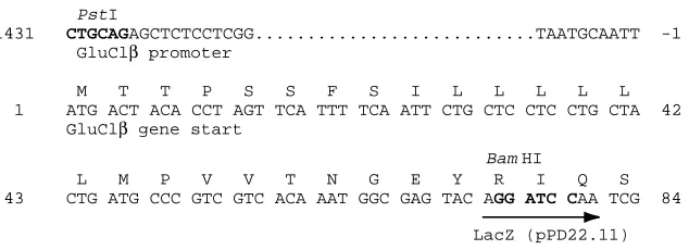
sequencing, and the construct was used as a template for PCR. A forward primer incorporating a PstI site (5′-TGTGTTCTGCAGAGCTCTCCTCGGAGCCCA-3′) was used with a reverse primer incorporating a BamHI site (5′-CCGATTGGATCCTGTACTCGCCAT-3′) to amplify a product consisting of 1.4 kb of 5′ sequence together with sequence encoding the first 24 amino acid residues of the Glu-Cl β-subunit. The PCR product was subcloned into the expression vector pPD22.11 (Fire et al. 1990) to form a translational fusion with the lacZ gene (Fig. 1).
Analysis of Glu-Cl β-subunit expression in transgenic
C. elegans
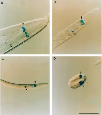
Following micro-injections of the Glu-Cl β reporter construct, six heritably transformed C. elegans lines were established and assayed for β-galactosidase activity. The pPD22.11 fusion vector contains the nuclear localisation signal from the SV40 T-antigen, which targets β-galactosidase to the nucleus of cells expressing this construct. Adults from all six transformed lines showed consistent staining of nuclei within the pharynx (Fig. 2A), and no staining was observed in other organs. From their position and the shape of the corresponding cells, six of these stained nuclei were identified as those of the m4 pharyngeal muscle cells in the metacorpus (Fig. 3). In adults, no nuclei outside the metacorpus consistently stained for X-gal, although very faint staining of nuclei in the terminal bulb was sometimes seen (indicated by the small arrowhead in Fig. 2A).
Developmental expression of Glu-Cl β-subunit
Synchronous cultures were set up for two of the transformed lines to analyse the developmental expression of the Glu-Cl β-subunit. All the larval stages showed staining of pharyngeal nuclei (Fig. 2B,C), as seen in the adults, and stained nuclei were also observed in many eggs undergoing morphogenesis (Fig. 2D). In addition to the metacorpus staining, larvae (L1–L3) also exhibited staining of nuclei towards the terminal
bulb of the pharynx (see Fig. 2B–D). Exact identification of these nuclei in the terminal bulb proved difficult owing to the diffuse nature of the staining observed at the earlier stages of development. This staining was most pronounced in eggs undergoing the later stages of morphogenesis and generally decreased throughout development until it was completely absent or barely visible in adults (Fig. 2A).
Discussion
These LacZ reporter experiments have shown that the Glu-Cl β-subunit promoter is active in pharyngeal cells of C.
elegans. Adults and all larval stages appear to express this
subunit in the three m4 pharyngeal muscle cells, which comprise the metacorpus, and these are the only nuclei that consistently stained in adults. These muscles are required for pharyngeal pumping/feeding, and their relaxation is triggered
via the fast inhibitory motor neurone M3 (Raizen and Avery,
1994). Although reporter gene fusions do not always faithfully reflect the expression patterns of the intact gene, perhaps because of the absence of enhancer elements located downstream or within introns, there are additional physiological data to support the conclusion that this subunit is expressed on pharyngeal muscle cells. Raizen and Avery (1994) have shown that the inhibitory M3 motor neurone is unlikely to be GABAergic and have suggested more recently that it is glutamatergic (L. Avery, M. W. Davis, W. Denk, J. Dent and G. Hess, personal communication) and, given that pharyngeal pumping is extremely sensitive to the avermectins (Avery and Horvitz, 1990), it seems likely that transmission is mediated via the glutamate-gated avermectin receptor cloned by Cully et al. (1994). Martin (1996) has made two-microelectrode current-clamp recordings from pharyngeal muscle preparations of Ascaris suum and has demonstrated the presence of glutamate-gated Cl− channels on the pharyngeal muscle of this important parasitic nematode. In addition, he demonstrated a dose-dependent potentiation of the glutamate response by the avermectin analogue milbemycin D.
The data reported here thus provide further evidence that an inhibitory glutamate-gated Cl− channel is expressed on nematode pharyngeal muscle. This channel, the ‘avermectin’ receptor, may be the primary target of the avermectins, and the effects of the drug may be due to the paralysis of pharyngeal muscle. How does this account for the anthelmintic activity? Paralysis of the pharynx will prevent nematodes from feeding,
PstI
-1431 CTGCAGAGCTCTCCTCGG...TAATGCAATT -1 GluClβ promoter
M T T P S S F S I L L L L L 1 ATG ACT ACA CCT AGT TCA TTT TCA ATT CTG CTC CTC CTG CTA 42 GluClβ gene start
Bam HI
L M P V V T N G E Y R I Q S 43 CTG ATG CCC GTC GTC ACA AAT GGC GAG TAC AGG ATC CAA TCG 84
[image:3.609.252.569.70.185.2]LacZ (pPD22.11)
leading to arrested development, starvation and eventual death. The inhibition of larval development has been demonstrated in
vitro for Haemonchus contortus, where concentrations of
avermectin as low as 1 nmol l−1 were sufficient to prevent
development of larvae to the L3 stage (Gill et al. 1995). Nevertheless, it remains unclear how this could account for the rapid expulsion of adult parasites from the treated host. Furthermore, there is evidence that many gastrointestinal nematodes are able to absorb nutrients through the cuticle as well as by ingestion (Ho et al. 1990; Geary et al. 1993). It is possible that avermectins kill the parasite through the interruption of other vital functions served by the pharynx, for example the regulation of turgor pressure. Adult filarial nematodes depend on the transcuticular absorption of nutrients (Howells, 1980). Here, the lack of avermectin anthelmintic activity on macrofilariae fits with the proposed site of action and the interruption of oral ingestion. The localisation of avermectin receptors in parasitic nematode species will be necessary to verify the results obtained from C. elegans.
Avermectins may act at additional sites in nematodes. Our observations on the developmental expression of the Glu-Cl β-subunit suggest that there may be some developmentally regulated expression of this subunit within the pharynx. One
of these sites is probably the m5 muscle cell which forms the isthmus of the pharynx, nuclei of which are occasionally weakly stained in adults. Interestingly, Albertson and Thomson (1976) noted that the glutamatergic M3 motor neurone which innervates m4 pharyngeal muscle cells also sends an occasional synapse to m5 muscle cells. Although the pattern of expression in adults is largely limited to the m4 cells, it is clear the reporter gene is active in additional pharyngeal nuclei at earlier stages of development, especially in the terminal bulb, and that the pattern of staining becomes simpler through development. The diffuse nature of this larval staining makes it difficult to identify the specific nuclei concerned unequivocally, but it is likely that additional pharyngeal cells are expressing the subunit at earlier stages of development and possible that the effects of avermectins on larval development may be related to this expression.
Although the glutamate-sensitive β-subunit of the avermectin receptor appears to be expressed exclusively in the pharynx, we have not localised the avermectin-sensitive α-subunit. This subunit may co-assemble with other subunits to form additional avermectin receptors that are expressed at other sites. For example, the α6-subunit of the mammalian
[image:4.609.228.560.75.450.2]brain GABAAreceptor exists as distinct α6γand α6δreceptor
populations in the cerebellum (Quirk et al. 1994). A further complication is the growing evidence that there may be multiple inhibitory glutamate receptors composed of a heterogeneous family of subunits. We have cloned two additional subunits from C. elegans (Cegbr2 and Cegbr3) which show a high level of sequence identity to the avermectin receptor subunits (D. L. Laughton, G. G. Lunt and A. J. Wolstenholme, manuscript in preparation) and J. Dent and L. Avery (personal communication) have recently cloned a second α-subunit from C. elegans (Glu-Cl α2) which has 85 % identity at the amino-acid level to Glu-Cl α. Similar LacZ reporter studies for the α-subunits and the use of subunit-specific antibodies may help to elucidate further the mode of action of avermectins.
Taken together, these data provide strong evidence that the pharynx is a major target for avermectins. However, the exact nature and composition of the avermectin receptor(s) remains to be determined.
We thank Ian Hope (University of Leeds) for his help with
the construction of transgenic worms, Donna Albertson (University of California, San Francisco) for helping us in the identification of stained nuclei and for allowing us to adapt Fig. 3 from her original observations, and Leon Avery and co-workers (University of Texas Southwestern Medical Centre) for the use of personal communications. This work was supported by a grant from the Wellcome Trust (award no. 039228).
References
ALBERTSON, D. G. AND THOMSON, J. N. (1976). The pharynx of Caenorhabditis elegans. Trans. R. Soc. Lond. B 275, 299–325. ARENA, J. P., LIU, K. K., PARESS, P. S. ANDCULLY, D. F. (1991).
Avermectin-sensitive chloride currents induced by Caenorhabditis elegans RNA in Xenopus oocytes. Molec. Pharmac. 40, 368–374. ARENA, J. P., LIU, K. K., PARESS, P. S., FRAZIER, E. G. ANDCULLY, D. F. (1995). The mechanism of action of avermectins in Caenorhabditis elegans: correlation between activation of glutamate sensitive chloride current, membrane binding and biological activity. J. Parasitol. 81, 286–294.
ARENA, J. P., LIU, K. K., PARESS, P. S., SCHAEFFER, J. M. ANDCULLY, D. F. (1992). Expression of a glutamate-activated chloride current in Xenopus oocytes injected with Caenorhabditis elegans RNA: evidence for modulation by avermectin. Molec. Brain Res. 15, 339–348.
AVERY, L. AND HORVITZ, H. R. (1990). Effects of starvation and neuroactive drugs on feeding in Caenorhabditis elegans. J. exp. Zool. 253, 263–270.
BOTTJER, K. P. AND BONE, L. W. (1985). Trichostrongylus colubriformis: Effect of anthelmintics on ingestion and oviposition. Int. J. Parasitol. 15, 501–503.
BRENNER, S. (1974). The genetics of Caenorhabditis elegans.
Genetics 77, 71–94.
CAMPBELL, W. C. ANDBENZ, G. W. (1984). Ivermectin: a review of efficacy and safety. J. vet. pharm. Ther. 7, 1–16.
CULLY, D. F., VASSILATIS, D. K., LIU, K. K., PARESS, P. S., VAN DER PLOEG, L. H. T., SCHAEFFER, J. M. ANDARENA, J. P. (1994). Cloning of an avermectin-sensitive glutamate-gated chloride channel from Caenorhabditis elegans. Nature 371, 707–711.
DEL CASTILLO, J., DE MELLO, W. C. AND MORALES, T. (1964a). Inhibitory action of γ-aminobutyric acid (GABA) on Ascaris muscle. Experientia 20, 141–143.
DEL CASTILLO, J., DE MELLO, W. C. AND MORALES, T. (1964b). Mechanism of the paralysing action of piperazine on Ascaris muscle. Br. J. Pharmac. 22, 463–477.
DUCE, I. R. ANDSCOTT, R. H. (1985). Actions of avermectin B1a on insect muscle. Br. J. Pharmac. 85, 395–401.
DUITTOZ, A. H. AND MARTIN, R. J. (1991). Effects of the arylaminopyridazine-GABA derivatives, SR95103 and SR95531, on the Ascaris muscle GABA receptor: the relative potency of the antagonists in Ascaris is different to that at vertebrate GABAA receptors. Comp. Biochem. Physiol. 98, 417–422.
FFRENCH-CONSTANT, R. H., MORTLOCK, D. P., SHAFFER, C. D., MACINTYRE, R. J. ANDROUSH, R. T. (1991). Molecular cloning and transformation of cyclodiene resistance in Drosophila: An invertebrate γ-aminobutyric acid subtype A receptor locus. Genetics 88, 7209–7213.
FIRE, A. (1986). Integrative transformation of C. elegans. EMBO J. 5, 2673–2680.
a b
c
d
e
f
g
h
[image:5.609.120.242.68.358.2]FIRE, A. (1992). Histochemical techniques for locating Escherichia coliβ-galactosidase activity in transgenic organisms. Genet. analyt. Tech. Appl. 9, 152–160.
FIRE, A., HARRISON, S. ANDDIXON, D. (1990). A modular set of Lac
Z fusion vectors for studying gene expression in Caenorhabditis elegans. Gene 93, 189–198.
GEARY, T. G., KLEIN, R. D., VANOVER, L., BOWMAN, J. W. AND THOMPSON, D. P. (1992). The nervous systems of helminths as targets for drugs. J. Parasitol. 78, 215–230.
GEARY, T. G., SIMS, S. M., THOMAS, E. M., VANOVER, L., DAVIS, J. P., WINTERROWD, C. A., KLEIN, R., HO, N. F. H. ANDTHOMPSON, D. P. (1993). Haemonchus contortus ivermectin-induced paralysis of the pharynx. Exp. Parasitol. 77, 88–96.
GILL, J. H., REDWIN, J. M., VANWYK, J. A. ANDLACEY, E. (1995). Avermectin inhibition of larval development in Haemonchus contortus – Effects of ivermectin resistance. Int. J. Parasitol. 25, 463–470.
HARVEY, R. J., SCHMITT, B., HERMANS-BORGMEYER, I., GUNDELFINGER, E. D., BETZ, H. AND DARLISON, M. G. (1994). Sequence of a Drosophila ligand-gated ion-channel polypeptide with an unusual amino-terminal extracellular domain. J. Neurochem. 62, 2480–2483.
HARVEY, R. J., VREUGDENHIL, E., ZAMAN, S. H., BHANDAL, N. S., USHERWOOD, P. N. R., BARNARD, E. A. AND DARLISON, M. G. (1991). Sequence of a functional invertebrate GABAA receptor subunit which can form a chimeric receptor with a vertebrate α subunit. EMBO J. 10, 3239–3245.
HENDERSON, J. E., SODERLUND, D. M. ANDKNIPPLE, D. C. (1993). Characterisation of a putative γ-aminobutyric acid (GABA) receptor βsubunit gene from Drosophila melanogaster. Biochem. biophys. Res. Commun. 193, 474–482.
HO, N. F. H., GEARY, T. G., RAUB, T. J., BARSUHN, C. L. AND THOMPSON, D. P. (1990). Biophysical transport properties of the cuticle of Ascaris suum. Molec. Biochem. Parasitol. 41, 153–166. HOLDEN-DYE, L., HEWITT, G. M., WANN, K. T., KROSGSGAARD -LARSEN, P. AND WALKER, R. J. (1988). Studies involving avermectin and the 4-aminobutyric acid (GABA) receptor of Ascaris suum muscle. Pestic. Sci. 24, 231–245.
HOLDEN-DYE, L., KROGSGAARD-LARSEN, P., NIELSEN, L. AND WALKER, R. J. (1989). GABA receptors on the somatic muscle cells of the parasitic nematode, Ascaris suum: stereoselectivity indicates similarity to a GABAA type agonist recognition site. Br. J.
Pharmac. 98, 841–850.
HOLDEN-DYE, L. AND WALKER, R. J. (1990). Avermectin and avermectin derivatives are antagonists at the 4-aminobutyric acid
(GABA) receptor on the somatic muscle cells of Ascaris; is this the site of anthelmintic action? Parasitology 101, 265–271.
HOWELLS, R. E. (1980). Filariae: Dynamics of the surface. In The Host–Invader Interplay (ed. H. Vanden-Bossche), pp. 69–84. Amsterdam: Elsevier/North-Holland Biomedical Press.
HUTTON, M. L., HARVEY, R. J., EARLEY, F. G. P., BARNARD, E. A. AND DARLISON, M. G. (1993). A novel invertebrate GABAA receptor-like polypeptide. FEBS Lett. 326, 112–116.
LAUGHTON, D. L., WHEELER, S. V., LUNT, G. G. ANDWOLSTENHOLME, A. J. (1995). The β-subunit of Caenorhabditis elegans avermectin receptor responds to glycine and is encoded by chromosome I. J. Neurochem. 64, 2354–2357.
MARTIN, R. J. (1996). An electrophysiological preparation of Ascaris suum pharyngeal muscle reveals a glutamate-gated chloride channel sensitive to the avermectin analogue, milbemycin D. Parasitology 112, 247–252.
MARTIN, R. J. ANDPENNINGTON, A. J. (1989). A patch-clamp study of effects of dihydroavermectin on Ascaris muscle. Br. J. Pharmac. 98, 747–756.
MELLO, C. ANDFIRE, A. (1995). DNA transformation. In Methods in
Cell Biology, vol. 48 (ed. H. F. Epstein and D. C. Shakes), pp. 451–482. San Diego, London: Academic Press.
MELLO, C. C., KRAMER, J. M., STINCHCOMB, D. AND AMBROS, V. (1991). Efficient gene transfer in C. elegans: Extrachromosomal maintenance and integration of transforming sequences. EMBO J. 10, 3959–3970.
QUIRK, K., GILLARD, N. P., RAGAN, C. I., WHITING, P. J. ANDMCKERNAN, R. M. (1994). Model of subunit composition of γ-aminobutyric acid A receptor subtypes expressed in rat cerebellum with respect to their
αand γ/δsubunits. J. biol. Chem. 269, 16020–16028.
RAIZEN, D. M. AND AVERY, L. (1994). Electrical activity and behaviour in the pharynx of Caenorhabditis elegans. Neuron 12, 483–495.
SULSTON, J. E. ANDBRENNER, S. (1974). The DNA of Caenorhabditis
elegans. Genetics 77, 95–104.
SULSTON, J. E. ANDHODGKIN, J. (1988). Methods. In The Nematode Caenorhabditis elegans (ed. W. B. Wood), pp. 587–606. Cold Spring Harbor, New York: Cold Spring Harbor Laboratory Press. THOMPSON, M., SHOTKOSKI, F. AND FFRENCH-CONSTANT, R. (1993).
Cloning and sequencing of the cyclodiene insecticide resistance gene from the yellow fever mosquito Aedes aegypti. FEBS Lett. 325, 187–190.