Although it is well known that fish increase cardiac output (Q) during exercise to meet increased metabolic demands, there is a controversy concerning the basic hemodynamic mechanisms underlying this increase. One view is that stroke volume (Vs) and, to a lesser extent, heart rate (fH) are
responsible for the rise in Qin swimming fish, especially in salmonids (Dunmall and Schreer, 2003; Farrell, 1991; Farrell and Jones, 1992; Jones and Randall, 1978; Kiceniuk and Jones, 1977). This view has been questioned by others, who suggest that increased fHis the primary means of increasing Qduring swimming (Altimiras and Larsen, 2000; Axelsson et al., 1994; Cooke et al., 2003; Korsmeyer et al., 1997; Lefrancois et al., 1998; Priede, 1974). While this difference in opinion may exist because different species do indeed have different strategies for increasing Q, we know little of one of the primary determinants of Vs in fish, i.e., cardiac preload (= central
venous pressure, PCV). Ventricular end-systolic volume, at least in rainbow trout, is close to zero and hence cannot be
reduced much further during exercise (Forster and Farrell, 1994; Franklin and Davie, 1992). Thus, any increase in Vshas
to come about by increasing end-diastolic volume, which must come about through increased PCVand a concurrent increase in the myocardial force of contraction due to the Frank-Starling relationship (Farrell, 1991; Farrell and Jones, 1992). Consequently, without information on the changes in PCV during swimming it is difficult to evaluate fully the role of increasing Vs.
This line of logic ignores the fact that under steady-state conditions, Qequals venous return and the heart can pump only what it gets back from the venous circulation. Moreover, any increase in PCV during exercise will reduce the pressure gradient for venous return (flow) to the heart from the venous periphery, if end-capillary blood pressure and venous resistance remain unaltered. Thus, in addition to increasing cardiac filling pressure, a proportional increase in peripheral venous pressure is expected to ensure that venous return can
doi:10.1242/jeb.01606
Cardiac preload (central venous pressure,PCV), mean circulatory filling pressure (MCFP), dorsal aortic blood pressure (PDA) and relative cardiac output (Q) were measured in sea bass (Dicentrarchus labrax) at rest and while swimming at 1 and 2·BL·s–1. MCFP, an index of venous capacitance and the upstream venous pressure driving the return of venous blood to the heart, was measured as the plateau in PCV during ventral aortic occlusion. Compared with resting values, swimming at 1 and 2·BL·s–1 increased Q (by 15±1.5 and 38±6.5%, respectively), PCV (from 0.11±0.01·kPa to 0.12±0.01 and 0.16±0.02·kPa, respectively), MCFP (from 0.27±0.02·kPa to 0.31±0.02 and 0.40±0.04·kPa, respectively) and the calculated pressure gradient for venous return (∆PV, from 0.16±0.01·kPa to 0.18±0.02 and 0.24±0.02·kPa, respectively), but not PDA. In spite of an increased preload, the increase in Qwas exclusively mediated by an
increased heart rate (fH, from 80±4·beats·min–1 to 88±4 and 103±3·beats·min–1, respectively), and stroke volume (Vs) remained unchanged. Prazosin treatment (1·mg·kg–1
Mb) abolished pressure and flow changes during swimming at 1·BL·s–1, but not 2·BL·s–1, indicating that other control systems besides an α-adrenoceptor control are involved. This study is the first to address the control of venous capacitance in swimming fish. It questions the generality that increased Qduring swimming is regulated primarily through Vsand shows that an increased cardiac filling pressure does not necessarily lead to an increased Vs in fish, but may instead compensate for a reduced cardiac filling time.
Key words: cardiac preload, cardiac output, exercise, heart rate, mean circulatory filling pressure, prazosin, sea bass, stroke volume, teleost, venous capacitance, venous return.
Summary
Introduction
Cardiac preload and venous return in swimming sea bass
(
Dicentrarchus labrax
L.)
Erik Sandblom
1,*, Anthony P. Farrell
2, Jordi Altimiras
3, Michael Axelsson
1and Guy Claireaux
41Department of Zoology, Göteborg University, Box 463, S-405 30 Göteborg, Sweden,2Faculty of Agricultural
Sciences and Department of Zoology, University of British Columbia, Vancouver, BC, V6T 1Z4, Canada,
3Department of Biology, Institute of Physics and Measurement Technology, Linköpings Universitet,
S-58183 Linköping, Swedenand 4Centre de Recherche sur les Ecosystèmes Marins et Aquacoles (CNRS-IFREMER),
Place du Séminaire, F-17137, L’Houmeau, France *Author for correspondence (e-mail: erik.sandblom@zool.gu.se)
match the increase in Q. Venous return can be estimated from measurements of PCVand venular blood pressures.
The mean circulatory filling pressure (MCFP) is an index of venous capacitance. It is measured as the venous pressure after a short (5–10·s) cardiac arrest and also represents the upstream venous (venular) pressure that drives venous return (Pang, 2001; Rothe, 1986, 1993; Sandblom and Axelsson, 2005). MCFP can increase due to an increased smooth muscle tone and/or a decreased compliance in venous capacitance vessels (Conklin et al., 1997; Hoagland et al., 2000; Olson et al., 1997; Pang, 2001; Rothe, 1986, 1993; Zhang et al., 1998). While comprehensive studies have examined the nervous and humoral control of venous capacitance in unaesthetized fish under resting conditions (Conklin et al., 1997; Hoagland et al., 2000; Olson et al., 1997; Sandblom and Axelsson, 2005; Zhang et al., 1998), none have considered the changes in venous capacitance that are likely to occur during exercise. Furthermore, basic information on PCV during exercise is limited to a few studies and what data exist are compromised by noisy signals and/or experiments on a small number of animals (Jones and Randall, 1978; Kiceniuk and Jones, 1977; Stevens and Randall, 1967).
The primary objective of this study was therefore to measure changes in PCV and MCFP in a fast swimming teleost, the European sea bass (Dicentrarchus labrax L.). By combining PCVand MCFP measurements, it was also possible to assess the degree to which the pressure gradient for venous return, venous capacitance and cardiac preload change during the periods of increased Qassociated with exercise. Also, the role of α-adrenoceptor control of these responses was examined, since Zhang et al. (1998) identified that venous capacitance in resting rainbow trout (Oncorhynchus mykiss) can be altered by α-adrenergic mechanisms.
Materials and methods
Animals
Results from 13 sea bass (279–648·g and 31–38·cm) are presented in this study. The fish were obtained from the Ferme Marine des Baleines (Ile de Ré, France) and maintained at CREMA under a natural photoperiod in indoor 400·l fibre glass tanks supplied with recirculating, biofiltered seawater at ambient temperature (21–24°C). The fish were fed commercial dry pellets on a regular basis.
Surgical procedures
Prior to surgery the fish were anaesthetized in seawater containing MS-222 (approx 100·mg·l–1) and placed on water-soaked foam on a surgery table. During surgery, the fish was covered with wet tissue paper and the gills were continuously irrigated with aerated, chilled (~11–16°C) seawater containing MS-222 (~50·mg·l–1). The ventral aorta was exposed with an incision on the right side of the isthmus and dissected free. A Perspex cuff-type 20 MHz Doppler flow probe (Iowa Doppler products; Iowa City, IA, USA), with an inner diameter of 1.8–2.0·mm, was positioned around the aorta proximal to the
bulbus to measure relative changes in Q(Fig.·1). Also, a cuff-type vascular occluder (i.d. 2.5–4.3·mm) was positioned posterior to the flow probe to obtain zero-flow during measurement of MCFP. The occluder was constructed from Perspex, a heat-flared water-filled PE-50 catheter and a piece of latex rubber (model Thin, Dental Dam, Coltène/Whaledent Inc, USA and Canada). The rubber was tied with a 4–0 suture around the flared end of the PE tubing (Fig.·1). The sinus venosus was non-occlusively cannulated to measure PCV (Altimiras and Axelsson, 2004). Briefly, the operculum was retracted and the left lateral part of the ductus of Cuvier was carefully exposed and dissected free with an incision between the cleithrum and the fifth branchial arch. A small portion of the vessel wall was lifted with forceps and secured with a 4–0 suture, allowing the vessel to be gently lifted during the cannulation procedure. This procedure prevents blood loss, and is an improvement on the method used by Farrell and Clutterham (2003). A PE-50 catheter, with 2–3 side-holes to keep it patent and a bubble 1·cm from the tip, was inserted into the ductus of Cuvier through a small incision made close to the suture holding the vessel. A 4–0 suture was used to close the vessel wall around the catheter, leaving the bubble located on the luminal side of the vessel wall. The catheter was secured with silk sutures to the skin. The third efferent branchial artery on the left side was occlusively cannulated to measure dorsal aortic pressure (PDA). To do this, the first two gill arches were retracted to expose the third gill arch, which was gently
PDA
PCV
Q
a
b VO
a
b
retracted. The branchial artery was dissected free close to the angle of the branchial arch, occluded upstream with a 4–0 silk suture and cannulated downstream with a tapered PE-50 catheter (Fig.·1). The catheter was secured with 3–0 silk sutures around the gill arch and secured to the skin with 3–0 silk sutures. The catheters and the lead from the flow probe were collectively secured with a 3–0 suture to the back of the fish. Both blood pressure catheters were filled with physiological saline (0.9% NaCl). Following surgery, the fish were revived in fresh seawater, placed in plastic floating tubes in the holding tanks and allowed a 24·h recovery before experiments commenced.
Experimental protocol
A stainless steel, Brett-type swimtunnel respirometer was used in the present study. This tunnel had been designed to exercise individual fish in a non-turbulent water flow with a uniform cross-sectional water velocity. The total water volume was 48·l and the swim chamber had a square cross-sectional area of 290·cm2. A propeller downstream of the swim chamber
generated water flow. The flow in the swimtunnel was calibrated (Marsh-McBirney 200 flow meter; Frederick, MD, USA) in cm·s–1, which was converted to swimming speeds in
body·lengths·s–1(BL·s–1). The respirometer was thermostatted
by immersion in a large outer stainless steel tank that received a flow of aerated water. Since venous pressure is low readings can be affected by any changes in the pressure head on the propeller in the swim-channel when water velocity is changed.
To minimise this problem the lid to the swim chamber was removed. Water pressure was measured with a saline-filled catheter immersed in the channel, and no pressure fluctuations were observed at the swimming speeds used (up to 2·BL·s–1).
The swim channel was covered with an opaque black plastic sheet to avoid visual disturbance of the fish.
All experiments were conducted at a temperature of 22°C. The experimental protocol started with a 2·min recording at rest, i.e., the fish oriented into a low water velocity and maintaining position without swimming. MCFP was measured at rest by occluding the ventral aorta for 8·s. Water velocity was then gradually increased over a 5·min period until the fish reached a swimming speed of 1·BL·s–1, which was maintained
for 15·min. MCFP was remeasured at the end of this period. The same procedure was used for a swimming speed of 2·BL·s–1. Water velocity was then reduced to the resting
condition over a 2·min period and an α-adrenoceptor antagonist (prazosin, 1·mg·kg–1M
b; Pfizer, Sandwich, UK) was
administeredvia the venous catheter. The entire protocol was repeated 1.5–2.0·h later. Preliminary experiments had revealed that an exercise period of 15·min was well beyond the time necessary to establish stable cardiovascular measurements in untreated fish.
Successful ventral aortic occlusion always resulted in a rapid fall in PDA, a rise in PCVand a complete cessation of ventral aortic flow (Fig.·2). Although the ventral aorta was easily accessible, the vessel proved to be relatively fragile as compared with rainbow trout and several sea bass died due to
A
B
C
D
0 0.1 0.2 0.3 0.4 0.50 20 40 60 80 100 120 140
P
CV
(kP
a)
Q
(relati
v
e)
f
H
(beats min
–1
)
P
DA
(kP
a)
Untreated Prazosin
8 s ventral aortic occlusion 8 s ventral aortic occlusion 0
0 1 2 3 4 5
fatal rupture of the ventral aorta during occlusion. This generally occurred after the first occlusion and might have been due to mechanical abrasion from the vascular occluder during recovery. Another unusual finding was the low occurrence of blood clotting in the catheters during routine surgery and this was the reason why heparin was omitted from the saline.
Data acquisition, calculations and statistical analysis Both blood pressure catheters were connected to pressure transducers (model DPT-6100, pvb Medizintechnik, Kirchseeon, Germany), calibrated against a static water column with the water surface of the swim channel serving as the zero reference pressure. The signals from the pressure transducers were amplified with a 4ChAmp amplifier (Somedic, Hörby, Sweden). Relative cardiac output (Q) was recorded with a directional-pulsed Doppler flow meter (model 545C-4, University of Iowa, USA). The digital signals were fed into a portable computer running a custom made program,
General Acquisition (Labview version 6.01, National Instruments, Austin, TX, USA).
Heart rate (fH) was obtained from either pulsatile pressure or flow records. Relative changes in cardiac stroke volume (Vs)
were calculated as Q/fH. Total systemic resistance (Rsys) was
calculated from the pressure drop in the circulation divided by cardiac output Rsys=(PDA–PCV)/Q. For the Vs, Q and Rsys
calculations raw data was used and the initial untreated resting value for each fish was arbitrarily set to 100%. The plateau pressure in PCVduring ventral aortic occlusion was assumed to equal mean circulatory filling pressure (MCFP), and the average pressure between the 5th and 7th seconds of ventral aortic occlusion is reported here. This time interval is sufficient to obtain reasonably steady plateau values, while minimising the compromising effects of barostatic reflexes (Sandblom and Axelsson, 2005). A similar method has been used previously to measure MCFP in the trout (Hoagland et al., 2000; Zhang et al., 1998). No correction for inequalities in blood remaining in the Rest 1 BL s–1 2 BL s–1
1 2 3 4 5
0
0 0.1 0.2 0.3
80 100 120 140 160
Q PCV
PDA
Rsys
VS fH
kP
a
kP
a
%
†
†
Rest 1 BL s–1 2 BL s–1 †
*
*
*
*
*
*
*
*
*
*
*
A
B
C
D
E
F
†† †
60 80 100 120
%
† 80
100 120 140
%
60 80 100 120
beats min
–1
Fig.·3. Mean cardiovascular variables at rest and while swimming at 1 and 2·BL·s–1 in the sea bass, Dicentrarchus labrax. Solid lines represent untreated fish (N=8–9) and hatched lines (N=7) represent fish after treatment with prazosin (1·mg·kg–1 M
arterial circulation after occlusion was performed, because the difference was assumed to be negligible due to the large differences in compliances and volumes of the arterial and the venous compartments (Rothe, 1993; Zhang et al., 1998). The pressure gradient (∆PV) for venous return was calculated as
∆PV= MCFP–PCV. (1)
Resistance to venous return (Rv) was calculated as
Rv= (MCFP–PCV)/Q, (2)
assuming that Qequals venous return.
Data (mean values ±S.E.M.) are presented only for fish that performed steady state swimming at both water velocities. Partial data from fish that exhibited either erratic swimming behaviours, burst swimming, died after prazosin treatment, lost a catheter, or the ventral aorta ruptured were discarded. Cardiovascular data were taken from the last 90·s of each exercise period. For the calculations of venous variables, i.e. Fig.·4, PCVwas taken as the average of a 30·s period before ventral aortic occlusion. Wilcoxon matched pairs signed-ranks test, with a fiduciary level of P0.05 was used to evaluate statistically significant differences in cardiovascular variables at different swimming speeds and between treatments. To compensate for multiple two-group comparisons, a modified Bonferroni-test was applied (Holm, 1979).
Results
Cardiovascular responses to swimming in untreated fish Fig.·2 shows representative resting cardiovascular recordings for an individual fish during ventral aortic occlusion. Fig.·3 summarises the mean data for fish at rest and
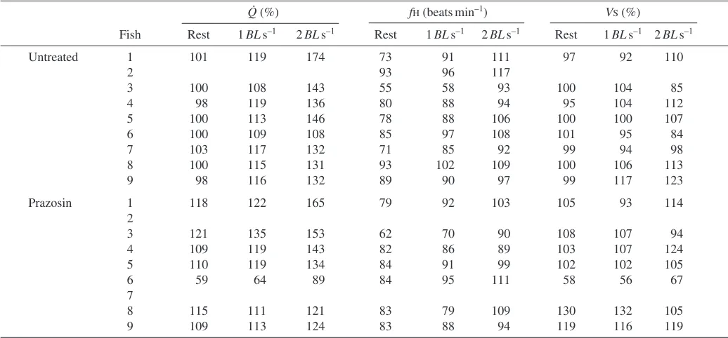
during steady state swimming at 1 and 2·BL·s–1, while Table·1 shows changes in Q, Vs and fH for individual fish. Exercise
increased Q by 15±1.5% and 38±6.5% of the resting value, respectively, at 1 and 2·BL·s–1. These increases in Q were mediated by significant increases in fHfrom 80±4·beats·min–1 at rest to 88±4·beats·min–1 at 1·BL·s–1 and 103±3·beats·min–1 at 2·BL·s–1. By contrast, Vsremained unaltered during exercise.
Nevertheless, the increases in Q were associated with significant rises in PCV from 0.11±0.01·kPa at rest to 0.12±0.01·kPa at 1·BL·s–1 and 0.16±0.02·kPa at 2·BL·s–1. However, PDAand Rsyswere unchanged during exercise.
Fig.·4 shows the rise in PCVduring exercise and illustrates changes in venous variables during exercise. In addition to an increase in PCV with increased swimming speed, MCFP increased significantly (0.27±0.02·kPa at rest to 0.31±0.02·kPa and 0.40±0.04·kPa, respectively). Because the rise in MCFP was proportionally larger than that in PCV, ∆PV increased significantly from 0.16±0.01·kPa at rest to 0.18±0.02·kPa at 1·BL·s–1 and 0.24±0.02·kPa at 2·BL·s–1, whereas Rv remained
unchanged.
Effects of α-adrenoceptor blockade on cardiovascular performance during swimming
Blockade of α-adrenergic receptors with prazosin showed that the cardiovascular system is at least partially controlled via α-adrenoceptors, both at rest as well as during exercise (Figs·3 and 4). At rest, prazosin treatment significantly decreased Rsys, producing a significant arterial hypotension
while PCVincreased significantly.
[image:5.612.48.566.97.338.2]During swimming after prazosin treatment, the increases in PCVand fHwere absent at 1·BL·s–1, but not at 2·BL·s–1. In fact, the increase in PCVwas accentuated significantly at 2·BL·s–1 Table 1. Changes in cardiac output, heart rate and stroke volume from nine individual sea bass, Dicentrarchus labrax, at rest
and while swimming at 1 and 2BLs–1
Q(%) fH(beats min–1) VS(%)
Fish Rest 1BLs–1 2BLs–1 Rest 1BLs–1 2BLs–1 Rest 1BLs–1 2BLs–1
Untreated 1 101 119 174 73 91 111 97 92 110
2 93 96 117
3 100 108 143 55 58 93 100 104 85
4 98 119 136 80 88 94 95 104 112
5 100 113 146 78 88 106 100 100 107
6 100 109 108 85 97 108 101 95 84
7 103 117 132 71 85 92 99 94 98
8 100 115 131 93 102 109 100 106 113
9 98 116 132 89 90 97 99 117 123
Prazosin 1 118 122 165 79 92 103 105 93 114
2
3 121 135 153 62 70 90 108 107 94
4 109 119 143 82 86 89 103 107 124
5 110 119 134 84 91 99 102 102 105
6 59 64 89 84 95 111 58 56 67
7
8 115 111 121 83 79 109 130 132 105
after prazosin. Prazosin also significantly accentuated the decrease in Rsysat 2·BL·s–1(from the resting value of 85.7±3.2
to 73.5±3.6%) and the corresponding hypotension (PDAfrom 2.9±0.1 to 2.5±0.1·kPa). Again, Vs remained unchanged
throughout the swim challenges after prazosin treatment despite the increase in PCV(Fig.·3). After prazosin, MCFP also remained unaltered at 1·BL·s–1, but increased significantly from 0.28±0.02·kPa at rest to 0.40±0.05·kPa during swimming at 2·BL·s–1, but no more so than before prazosin treatment (Fig.·4). As a result ∆PVincreased significantly compared with the resting value at 2·BL·s–1, although this response was not as pronounced as pre-prazosin treatment. Rvremained unaltered
throughout the experiment (Fig.·4).
Discussion
In its natural pelagic habitat, sea bass are subjected to strong
water currents requiring long periods of sustained exercise. This ecological fact makes sea bass an excellent experimental animal for exercise studies. However, existing cardiovascular data of sea bass is sparse. Axelsson et al. (2002) reported a resting fH of 51·beats·min–1 at 16°C. In the present study at 22°C, fHwas 80·beats·min–1, a difference that would represent a Q10 of 2.1 between the two studies and suggests that the
difference was simply a temperature effect. Furthermore, judging from the variable heart rate in resting fish (Fig.·2), it is likely that the fish had a functional cholinergic tone, which is indicative of an acceptable decay in post-surgical stress (Campbell et al., 2004).
Hemodynamics of venous return and cardiac filling pressure in sea bass
The present study is the first to demonstrate an active control of the venous vasculature during exercise in any species of fish.
A
B
D
C
*
*
*
*
*
*
*
*
*
Rest 1 BL s–1 2 BL s–1
0 0.1 0.2 0.3
0 40 80 120 160
PCV
MCFP
kP
a
%
†
Rest 1 BL s–1 2 BL s–1 †
0 0.1 0.2 0.3
kP
a
0 0.1 0.2 0.3 0.4 0.5
kP
a
RV ∆PV †
† 0
0.2 0.4 0.6 0.8
kP
a
Fig.·4. Upper trace shows a representative recording of venous pressure during a full swim protocol (~30·min) in an untreated sea bass, Dicentrarchus labrax. Ventral aortic occlusions for MCFP measurements at the end of each swim speed (rest, 1·BL·s–1and 2·BL·s–1, respectively) are marked with vertical arrows. Lower panels (A–D) show mean values (+ S.E.M.) of measured and calculated venous variables at rest and while swimming at 1 and 2·BL·s–1. Filled bars represent untreated fish (N=8–9) and open bars (N=7) represent fish after treatment with prazosin (1·mg·kg–1M
Very few studies have successfully measured cardiac filling pressure during swimming in teleost fish. Kiceniuk and Jones (1977) found that PCVin the common cardinal vein of four rainbow trout increased significantly during swimming only when the fish swam at critical swimming speed (Ucrit), and not
at intermediate swimming speeds. This finding is surprising because Vs increased significantly at intermediate swimming
speeds, suggesting that these increases in Vswere not a result
of increased filling pressure. It is likely that increased adrenergic stimulation of the heart, which is known to both increase during swimming (Axelsson, 1988) and increase the sensitivity of the heart to filling pressure (Farrell et al., 1986) permitted these increases in Vswithout a concomittent increase
in PCV. Stevens and Randall (1967) measured venous pressure and flow in the subintestinal vein (= hepatic portal vein), and found that venous pressure increased whereas flow decreased. Since the hepatic portal vein drains the gastrointestinal tract and is located upstream of the liver, and arterial gut blood flow decreases during exercise (Axelsson et al., 1989; Axelsson and Fritsche, 1991; Farrell et al., 2001; Thorarensen et al., 1993), it is uncertain to what extent these changes directly affected cardiac performance (for further discussion, see Jones and Randall, 1978).
In the present study, Qand fHincreased during swimming. The increase in fHwould have reduced cardiac filling time, but it is clear that the observed increase in preload would have compensated for this, leaving Vsunchanged. As pointed out in
the introduction, the increase in PCVin itself could decrease the pressure gradient driving venous return from the periphery to the heart. However, a proportionally larger increase in MCFP ensured that the pressure gradient for venous return actually increased and since Rvwas unchanged, venous return
would be increased to support the increase in Q(Fig.·4). The increase in MCFP could be attributed to either an increased venous tone, a decreased venous compliance or a combination of both (Conklin et al., 1997; Hoagland et al., 2000; Olson et al., 1997; Pang, 2001; Rothe, 1986, 1993; Zhang et al., 1998). In rainbow trout, adrenaline increases venous tone through an α-adrenergic control and decreases venous compliance (Sandblom and Axelsson, 2005; Zhang et al., 1998). Since vascular capacitance curves could not be constructed in the present study, we do not know the exact mechanism for the increase in MCFP. Nevertheless, the observation that the increases in MCFP, PCV, Q, ∆PVand fH during swimming at 1·BL·s–1 were abolished after α -adrenoceptor blockade (Figs·3 and 4), suggests an important α-adrenergic control mechanism for the venous vasculature in sea bass during exercise, which can mobilize venous blood towards the heart and increase cardiac preload. This control mechanism was evident in resting fish as well. About 2·h after prazosin treatment, resting cardiovascular variables in the sea bass were significantly altered (Figs·3 and 4); PCVincreased and both Rsysand PDAdecreased. It is unlikely that this increase
in PCVwas mediated by either an increased transcapillary fluid uptake, thus increasing blood volume, or an up-regulation of some compensatory vasoactive system since this would have
affected MCFP as well. The importance of an α-adrenergic tone on the arterial side of the circulation has been previously demonstated at rest and during swimming in other fish species (Axelsson and Fritsche, 1991; Axelsson and Nilsson, 1986; Smith, 1978), and was confirmed here because after prazosin treatment sea bass could not maintain Rsys at 2·BL·s–1 and
suffered a major systemic hypotension. In view of this, it could be argued that the increase in PCVwas only a consequence of the reduction in Rsys, but then MCFP would not have increased.
Instead, the accentuated reduction in arterial pressure at 2·BL·s–1 possibly triggered the activation of some unknown vasoactive system, acting primarily on the venous vasculature. One concern with the present study is that an increased adrenergic tone on resistance vessels may have counteracted a further decrease in Rsys during exercise and resulted in an
unaltered resistance in untreated fish. It is unknown whether the adrenergic control of MCFP is mediated by adrenergic nerves and/or circulating catecholamines. In cod (Axelsson and Nilsson, 1986; Butler et al., 1989; Smith et al., 1985) and trout (Smith, 1978) it has been demonstrated that the increase in arterial blood pressure observed during moderate exercise is exclusively mediated by adrenergic nerves. Whether the same is true for the venous circulation in fish is not yet known.
A possible consequence of a decreased venous capacitance, manifested as the increase in MCFP, is that blood volume from the venous compartment is redistributed to other parts of the circulation, such as muscle capillary beds, respiratory organs and central veins (Pang, 2001). It is possible that much of the blood redistributed from the venous system in the sea bass during exercise, in addition to increasing cardiac preload, served to fill gill vasculature and muscle capillary beds. In mammals the splanchnic circulation has a high capacitance and is the primary reservoir for blood volume mobilization during exercise (Pang, 2001; Rothe, 1986). To what extent splanchnic venous blood volume is mobilized during exercise in fish is unclear, even though Stevens and Randall (1967) demonstrated that blood flow in the subintestinal vein (e.g. portal vein) decreased and venous pressure increased in rainbow trout. Albeit somewhat meager evidence, the observations are consistent with blood volume mobilization from the splanchnic venous compartment during exercise in fish. As judged from the drop in PDA and Rsys during swimming at 2·BL·s–1after
prazosin, it is possible that the gut circulation continued to be perfused (unlike the normal decrease with exercise) as perfusion of locomotory muscles increased (Axelsson and Fritsche, 1991; Axelsson et al., 2000; Farrell et al., 2001; Thorarensen et al., 1993). Further research in this area is clearly needed.
Heart rate versusstroke volume regulation during exercise Increased Qobserved after force feeding in sea bass was due primarily to tachycardia (Axelsson et al., 2002). Similarly, in the present experiments, sea bass increased Q through tachycardia with no significant change in Vs, despite the fact
necessarily lead to an increased Vs in fish, but may instead
compensate for a reduced cardiac filling time associated with an increase in fH. Thus, within the scope of the present exercise challenge, Qin sea bass was frequency regulated.
Other studies on fish with various life-strategies also suggest that control of Q by fH during exercise, might be more important than previously thought (Farrell, 1991; Farrell and Jones, 1992; Jones and Randall, 1978). Korsmeyer et al. (1997) found in the highly active yellowfin tuna (Thunnus albacares) that Qincreased by 13.6% during exercise at 24°C exclusively through tachycardia and, in fact, Vs decreased. Similarly,
during forced swimming at 0°C the Antarctic borch (Pagothenia borchgrevinki) increased Qby 75% by doubling fH(Axelsson et al., 1992). Furthermore, Cooke et al. (2003) investigated the relative contribution of Vsand fHto maximum
cardiac output at 3°C in three North American species with various degrees of winter quiescence. Largemouth bass (Micropterus salmoides), a winter inactive species, increased Qby means of a 124% increase in fHwith a 24% reduction in Vs. Similarly, the intermediately active black crappie (Pomoxis
nigromaculatus) increased Qby a 156% increase in fHwith a 56% reduction in Vs. By contrast, in the winter active white
bass (Morone chrysops) maximum Qwas attained by a 45% increase in fHand a 55% increase in Vs. Within a species,
temperature may modulate the relative contributions of fHand Vsduring exercise. For example, for maximum Qat 5°C and
10°C in largescale suckers (Catostomus macrocheilus), increased Vscontributed 58% and 62%, respectively (Kolok et
al., 1993), whereas at 16°C fH contributed 70% of maximum Q during exercise. Altogether these results indicate the importance of frequency regulation of Qin various fish species under various conditions but, of course, contrasts with several studies (mainly on salmonids) where increased Vs was the
major means of increasing Q during exercise (Dunmall and Schreer, 2003; Farrell, 1991; Farrell and Jones, 1992; Jones and Randall, 1978; Kiceniuk and Jones, 1977).
Under the present experimental conditions, exercising sea bass used only tachycardia to increase Q, but to what degree Vsmight be modulated in sea bass at higher swimming speeds
and different temperatures awaits further study. By performing the study at 22°C, which is in the upper range of the temperature preferendum for sea bass (Claireaux and Lagardere, 1999), it is possible that at lower water temperatures Vs could increase during swimming. Another
concern is that fish only swam to 2·BL·s–1and this resulted in a relatively small increase in Q(38%). Therefore, it is possible that at higher swimming speeds further increases in Q could occur through increased Vs.
In conclusion, this is the first study to measure variables related to venous return and cardiac filling pressure during exercise and to provide evidence for an active involvement of the venous vasculature during exercise in any species of fish. An α-adrenoceptor mediated control system was partially responsible for a decrease in venous capacitance, as reflected as an increase in MCFP in the swimming sea bass. This control would serve to: (1) maintain or increase the pressure gradient
for venous return, thus matching venous return and cardiac output; (2) Increase central venous blood volume, and consequently cardiac preload; and (3) Possibly redistribute venous blood to other parts of the circulation, such as the gills and muscle capillaries. These results highlight the fact that an increased cardiac preload does not necessarily result in an increased Vs, but can instead compensate for the reduced filling
time when fH is increased, thereby offsetting potential decreases in Vs. In fact, under the present conditions, the
exercise-induced increase in Q in sea bass was exclusively mediated by tachycardia.
Symbols
fH heart rate
Mb body mass
MCFP mean circulatory filling pressure
PCV central venous pressure
PDA dorsal aortic blood pressure ∆PV pressure gradient for venous return
Q relative cardiac output
Rsys total systemic resistance
Rv resistance to venous return
Ucrit critical swimming speed
Vs stroke volume
We are delighted to acknowledge Kenneth R. Olson for valuable comments on the manuscript and David J. McKenzie who kindly lent us the swim tunnel used in these experiments. E.S. and M.A. were supported by the Swedish research council (VR). A.P.F. was supported by NSERC Canada. J.A. was supported by a travel grant from the Swedish research council (VR). Financial help by IFREMER (G.C.) is also acknowledged.
References
Altimiras, J. and Axelsson, M. (2004). Intrinsic autoregulation of cardiac output in rainbow trout (Oncorhynchus mykiss) at different heart rates. J. Exp. Biol.207, 195-201.
Altimiras, J. and Larsen, E. (2000). Non-invasive recording of heart rate and ventilation rate in rainbow trout during rest and swimming. Fish go wireless. J. Fish. Biol.57, 197-209.
Axelsson, M. (1988). The importance of nervous and humoral mechanisms in the control of cardiac performance in the Atlantic cod Gadus morhua at rest and during non-exhaustive exercise. J. Exp. Biol.137, 287-301.
Axelsson, M., Altimiras, J. and Claireaux, G. (2002). Post-prandial blood flow to the gastrointestinal tract is not compromised during hypoxia in the sea bass Dicentrarchus labrax. J. Exp. Biol.205, 2891-2896.
Axelsson, M., Davison, B., Forster, M. and Nilsson, S. (1994). Blood pressure control in the antarctic fish Pagothenia borchgrevinki. J. Exp. Biol. 190, 265-279.
Axelsson, M., Davison, W., Forster, M. E. and Farrell, A. P. (1992). Cardiovascular responses of the red-blooded antarctic fishesPagothenia bernacchiiand P. borchgrevinki. J. Exp. Biol.167, 179-201.
Axelsson, M., Driedzic, W. R., Farrell, A. P. and Nilsson, S. (1989). Regulation of cardiac output and gut blood flow in the searaven, Hemitripterus americanus. Fish Physiol. Biochem.6, 315-326.
Axelsson, M. and Fritsche, R. (1991). Effects of exercise, hypoxia and feeding on the gastrointestinal blood flow in the Atlantic cod Gadus morhua. J. Exp. Biol.158, 181-198.
Axelsson, M., Thorarensen, H., Nilsson, S. and Farrell, A. P. (2000). Gastrointestinal blood flow in the red Irish lord, Hemilepidotus hemilepidotus: Long-term effects of feeding and adrenergic control. J. Comp. Physiol.170, 145-152.
Butler, P. J., Axelsson, M., Ehrenstrom, F., Metcalfe, J. D. and Nilsson, S. (1989). Circulating catecholamines and swimming performance in the Atlantic cod Gadus morhua. J. Exp. Biol.141, 377-388.
Campbell, H. A., Taylor, E. W. and Egginton, S. (2004). The use of power spectral analysis to determine cardiorespiratory control in the short-horned sculpin Myoxocephalus scorpius. J. Exp. Biol.207, 1969-1976.
Claireaux, G. and Lagardere, J. P. (1999). Influence of temperature, oxygen and salinity on the metabolism of the European sea bass. J. Sea Res.42, 157-168.
Conklin, D., Chavas, A., Duff, D., Weaver, L., Zhang, Y. and Olson, K. R. (1997). Cardiovascular effects of arginine vasotocin in the rainbow trout Oncorhynchus mykiss. J. Exp. Biol.200, 2821-2832.
Cooke, S. J., Grant, E. C., Schreer, J. F., Philipp, D. P. and DeVries, A. L. (2003). Low temperature cardiac response to exhaustive exercise in fish with different levels of winter quiescence. Comp. Biochem. Physiol. A 134, 157-165.
Dunmall, K. M. and Schreer, J. F. (2003). A comparison of the swimming and cardiac performance of farmed and wild Atlantic salmon, Salmo salar, before and after gamete stripping. Aquaculture220, 869-882.
Farrell, A. P. (1991). From hagfish to tuna: a perspective on cardiac function in fish. Physiol. Zool.64, 1137-1164.
Farrell, A. P. and Clutterham, S. M. (2003). On-line venous oxygen tensions in rainbow trout during graded exercise at two acclimation temperatures. J. Exp. Biol.206, 487-496.
Farrell, A. P. and Jones, D. R. (1992). The heart. In Fish Physiology, The
Cardiovascular System., vol. XII (ed. W. S. Hoar, D. J., Randall and A. P. Farrell), pp. 1-88. New York: Academic Press Inc.
Farrell, A. P., MacLeod, K. R. and Chancey, B. (1986). Intrinsic mechanical properties of the perfused rainbow trout heart and the effects of catecholamines and extracellular calcium under control and acidotic conditions. J. Exp. Biol.125, 319-345.
Farrell, A. P., Thorarensen, H., Axelsson, M., Crocker, C. E., Gamperl, A. K. and Cech, J. J., Jr (2001). Gut blood flow in fish during exercise and severe hypercapnia. Comp. Biochem. Physiol.128A, 551-563. Forster, M. E. and Farrell, A. P. (1994). The volumes of the chambers of
the trout heart. Comp. Biochem. Physiol. A 109, 127-132.
Franklin, C. E. and Davie, P. S. (1992). Dimensional analysis of the ventricle of an in situperfused trout heart using echocardiography. J. Exp. Biol.166, 47-60.
Hoagland, T. M., Weaver, L., Jr, Conlon, J. M., Wang, Y. and Olson, K. R. (2000). Effects of endothelin-1 and homologous trout endothelin on cardiovascular function in rainbow trout. Am. J. Physiol.278, R460-R468.
Holm, S. (1979). A simple sequentially rejective multiple test procedure. Scand. J. Stat.6, 65-70.
Jones, D. R. and Randall, D. J. (1978). The respiratory and circulatory systems during exercise. In Fish Physiology (ed. W. S. Hoar and D. J. Randall), pp. 425-492. New York, London: Academic Press.
Kiceniuk, J. W. and Jones, D. R. (1977). The oxygen transport system in trout (Salmo gairdneri) during sustained exercise. J. Exp. Biol.69, 247-260. Kolok, A. S., Spooner, R. M. and Farrell, A. P. (1993). The effect of exercise on the cardiac output and blood flow distribution of the largescale sucker Catostomus macrocheilus. J. Exp. Biol.183, 301-321.
Korsmeyer, K. E., Lai, N. C., Shadwick, R. E. and Graham, J. B. (1997). Heart rate and stroke volume contributions to cardiac output in swimming yellowfin tuna: Response to exercise and temperature. J. Exp. Biol. 20, 1975-1986.
Lefrancois, C., Claireaux, G. and Lagardere, J. P. (1998). Heart rate telemetry to study environmental influences on fish metabolic expenditure. Hydrobiologia371/372, 215-224.
Olson, K. R., Conklin, D. J., Farrell, A. P., Keen, J. E., Takei, Y., Weaver, L., Jr, Smith, M. P. and Zhang, Y. (1997). Effects of natriuretic peptides and nitroprusside on venous function in trout. Am. J. Physiol.273, R527-R539.
Pang, C. C. (2001). Autonomic control of the venous system in health and disease: effects of drugs. Pharmacol. Ther.90, 179-230.
Priede, I. G. (1974). The effect of swimming activity and section of the vagus nerves on heart rate in rainbow trout. J. Exp. Biol.60, 305-319.
Rothe, C. F. (1986). Physiology of venous return. An unappreciated boost to the heart. Arch. Intern. Med.146, 977-982.
Rothe, C. F. (1993). Mean circulatory filling pressure: its meaning and measurement. J. Appl. Physiol.74, 499-509.
Sandblom, E. and Axelsson, M. (2005). Baroreflex mediated control of heart rate and vascular capacitance in trout. J. Exp. Biol.208, 821-829. Smith, D. G. (1978). Neural regulation of blood pressure in rainbow trout
(Salmo gairdneri). Can. J. Zool.56, 1678-1683.
Smith, D. G., Nilsson, S., Wahlqvist, I. and Eriksson, B. M. (1985). Nervous control of the blood pressure in the Atlantic cod, Gadus morhua. J. Exp. Biol.117, 335-437.
Stevens, E. D. and Randall, D. J. (1967). Changes in blood pressure, heart rate and breathing rate during moderate swimming activity in rainbow trout. J. Exp. Biol.46, 307-315.
Thorarensen, H., Gallaugher, P. E., Kiessling, A. K. and Farrell, A. P. (1993). Intestinal blood flow in swimming chinook salmon Oncorhynchus tshawytschaand the effects of haematocrit on blood flow distribution. J. Exp. Biol.179, 115-129.