Fulminant Multiple Sclerosis
Gwendolyn Niebler,1'4 Todd Harris,3 Tom Davis,2 and Karen Roos1
Summary: The authors describe the MR imaging characteristics
and clinical course of a 22-year-old man with acute disseminated demyelinating disease, either Marburg multiple sclerosis or
recurrent (relapsing) acute disseminated perivenous
encephalo-myelitis.
Index terms: Sclerosis, multiple; Demyelinating disease
Multiple sclerosis (MS) is an autoimmune
de-myelinating disease that typically has a
relapsing-remitting
course
.
A rare fulminant form of MS
(acute fulminant MS of the Marburg type) is
associated with high morbidity and mortality. We
present radiographic and pathologic findings of a
case
of fulminant MS that resolved with
aggres-sive immunosuppresaggres-sive therapy.
Case
Report
A 22-year-old man developed a flu-like illness with severe headache, low-grade fever, vomiting, and diarrhea. Five days into the illness, he became increasingly lethargic and was admitted to the hospital. Computed tomography (CT) and magnetic resonance (MR) imaging studies were obtained {Figs. 1 A-1 D).
The patient was treated for 1 0 days with intravenous acyclovir for presumed herpes encephalitis. He became increasingly obtunded and was transferred to University Hospital 1 month after the onset of symptoms.
Epidemiologic history was unrevealing. The patient was a printer who worked at the same plant for 3 years. He lived in a trailer park across from a landfill. There was no history of recent spider, tick, or mosquito bites, travel outside of Indiana, blood transfusions, intravenous drug use, or time spent in wooded areas. The patient has a single sibling, a brother, who suffered from a recurrent illness characterized by fever to 1 02°F (38.9°C), night sweats, and lethargy. The etiology of the patient's brother's illness is unknown. The patient's past medical history was unremarkable except for varicella (chickenpox) 3 years prior to the onset of this illness.
On admission, the patient was afebrile, stuporous, and disoriented. There was evidence of a right relative afferent
pupillary defect, bilateral papilledema, and nuchal rigidity. He had a right hemiparesis with bilateral Babinski signs.
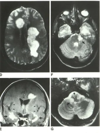
MR images obtained at the time of admission to Univer -sity Hospital showed white matter lesions in the frontal and temporal lobes, the left parietal lobe, and the genu of the corpus callosum (Figs. 2A-2E). Additional lesions were present in the left internal capsule, left thalamus, left pons,
and right medulla (Figs. 2F and 2G). In the right parietal lobe, there were small lesions that involved the subcortical white matter, with possible gray matter involvement. All lesions were of high signal intensity on T2-weighted images (T2WI). The large lesions were visible on T1-weighted images (T1 WI) as low-signal foci with very low-signal cen -ters. Virtually all lesions showed central enhancement with Gd-DPT A. Compared to the imaging studies done at the onset of the patient's illness, all the lesions had increased in size and degree of enhancement, but the pattern and number of lesions were unchanged.
A brain biopsy was obtained through a right frontal burr hole. Histologic sections stained with hematoxylineosin showed large plaques representing demyelination. These were diffuse and not perivenous in nature. Special stains for acid-fast bacteria and fungi were negative. Immunohis -tochemical stains for cytomegalovirus, herpes simplex vi-rus, papovavirus, and Toxoplasma were negative. DNA probes for herpes simplex virus, cytomegalovirus, and JC virus were negative. Multiple sections of brain tissue were examined by electron microscopy, but no viral inclusions were identified.
Cerebrospinal fluid (CSF) obtained at the time of brain biopsy revealed a protein level of 186 mg/dl , four white blood cells per cubic millimeter and 2045 red blood cells per cubic millimeter. CSF T cells were normal except for a mild decrease in T suppressor cells. There were no oli go-clonal bands. CSF lgG was 8.69 mg% (normal = <7 mg%). Serum electrophoresis was normal except for a borderline increase in lgM = 304 mg% (normal< 194
±
82 mg%). Serum toxoplasmosis titer was 1 :512. A test for antibodies to human immunodeficiency virus was negative. The a n-tinuclear antibody test was negative and the Westergren sedimentation rate was 11. Serum Epstein-Barr virus titer was 1 :40. There was no increase in serum acute and convalescent titers to respiratory syncytial virus, influenza A and B, adenovirus, echovirus, Coxsackie B, ChlamydiaReceived October I, 1991; revision requested November 6; final revision received February 2. 1992 and accepted February I 0.
'Address reprint requests to Gwendolyn Niebler. DO, Department of Neurology, Indiana University. Emerson Hall Room 125, Indianapolis. IN 46202. 1
Departments of 'Neurology, 2Pathology, and 3Radiology, Indiana University, Indianapolis, IN. AJNR 13:1547-1551, Nov/Dec 1992 0195-6108/92/1306-1547 (t) American Society of Neuroradiology
1548 NIEBLER
trachomatis, Epstein Barr virus, parainfluenza virus, and Toxoplasma gondii. Cold agglutinins for /VIycoplasma pneu-moniae were negative. Viral culture of the CSF for herpes simplex virus was negative. A Lyme disease titer was not obtained.
The patient was treated with intravenous methy lpred-nisolone 1000 mg/day for 5 days followed by oral predni-sone 80 mg/day. Seven days after the initiation of trea
t-ment, he was alert and following commands. He was able
to ambulate with assistance 1 week later. A repeat post-contrast MR scan obtained 11/2 weeks after initiation of
AJNR: 13, November/December 1992
treatment revealed a decrease in the size of most of the lesions with a markedly reduced degree of enhancement (Figs. 3A and 3B).
The patient was discharged on 80 mg of prednisone per day to a rehabilitation facility 3 weeks after admission. This daily dose was tapered by 10 mg per week. The patient developed optic neuritis in the left eye while on 50 mg of prednisone per day. The dose of prednisone was then increased to 100 mg per day and the daily dosage was tapered again at a rate of 10 mg every 14 days. The patient's visual acuity and pupillary examination returned
Fig. 1. A, A noncontrasted CT obtained at the onset of the patient's illness (at outside
hospital) demonstrates periventricular white matter (PVWM) low-density lesions, espe
-cially in the right frontal region (arrow).
8, Axial 500/13/2(TR/TE/excitations). Unenhanced T1 WI image obtained at the
same stage in the patient's illness as the CT in A. In addition to the right frontal lesion, the left parieto-occipital PVWM lesion is more noticeable (arrow).
C, Axial 500/13. Gd-DTPA-enhanced T1WI image obtained at the same time as A
and 8 shows the enhancement of both the right frontal and left parieto-occipital lesions.
D, Axial 2000/1 00. T2WI image obtained at the same time as A-C shows extensive
abnormal increased signal in the periventricular and deep white matter.
[image:2.612.53.561.237.737.2]AJNR: 13, November /December 1992 FULMINANT MULTIPLE SCLEROSIS 1549
A
B
c
Fig. 2. A, Axial 800/20/1. Unenhanced T1 WI image obtained at University Hospital 1 month after onset of symptoms shows an increase in low-signal abnormalities in frontal and parieto-occipital periventricular white matter (PVWM) (arrows) compared to Figure lB.
B, Axial 800/20. Gd-DTPA-enhanced Tl WI image obtained at the same time as A shows marked PVWM enhancement with
prominent callosal involvement. Note the increase in degree of enhancement and size of the lesions compared to Figure 1 C.
C, Axial 2500/80. T2WI image obtained at the same time as A and B reveals an increase in the abnormal signal in the PVWM, the genu of the corpus callosum, and left internal capsule compared to Figure 1 D. The arrow indicates lack of insular involvement.
to normal. There was mild pallor of the right optic disc,
but the left eye was normal. Nine months after discharge,
the patient developed an acute bout of optic neuritis in the
right eye and was again treated with pulse methyl
predniso-lone therapy. He is presently maintained on 15 mg of
prednisone per day.
Discussion
The MR scan demonstra
t
ed a d
i
sease process
almost exclusively confine
d
to white matter with
both supra- and infratentorial involvement. There
was
marked breakd
o
wn of the blood
-
brain barrier
as ev
i
denced by intense contrast enhancement.
Herpes simplex type I typically involves the tem
-poral lobes, causing both gray and white matter
disease, and has a predilection for insular cortex
(1)
.
In this case, the majority of the disease was
located outside of the insula. Lyme disease, a
spirochetal
infection that m9y involve the
central
nervous system (CNS), might have imaging
find-ings
similar to our case. MR of CNS Lyme disease
has revealed multifocal white matter disease
in-volving
the cerebrum and brain stem (2)
.
Multiple
ring-enhancing
lesions have been described in a
patient
with Lyme disease. However
,
our patient
's
history, physical
examination,
and disease
pro-gression were not consistent with this diagnosis
.
Neoplastic considerations
for the radiographic
disease pattern included lymphoma
.
Pr
i
mary
in-tracranial lymphoma often presents as
hyper-dense or isohyper-dense lesion on noncontrasted
CT.
Enhanced CT or MR
shows
homogeneous,
or less
commonly
,
ring-enhancing lesions (3)
.
The brain
biopsy ruled out
t
his diagnosis.
The clinical
course, radiographic findings, and
microscopic
examination
of brain t
iss
ue were
suggestive of a fulminant demyelinating process.
Adrenoleukodyst
r
ophy
,
which causes
widespread
demyelination
,
was considered and excluded
based on the patient
's
age
,
history
(acute
onset
and intact adrenals)
,
and MR. Progressive
multi-focal
leukoencephalopathy,
a
viral demyelinating
disease associated
with immunosuppression, was
also
considered.
However
,
our
pati
e
nt had
no
clinical or laboratory
evidence of
immuno-suppression
and the electron microscopic
exam-ination of
the
brain biops
y
excluded
thi
s
diagno-sis
.
[image:3.612.53.551.82.337.2]1550 NIEBLER
Fig. 2. -Continued. 0, Axial 2500/ 80. T2WJ image above the lateral ventri -cles obtained at the same time as Figures 2A-2C indicates large confluent left p
a-rietal and right frontal plaques.
£, Coronal 800/20.
Gd-DTPA-en-hanced Tl WI image obtained at the same
time as Figures 2A-20 shows white mat
-ter disease of the temporal lobes (large arrows) and right parietal operculum. There is also a left PVWM lesion. Arrow-heads indicate minimal insular involv e-ment.
Fand G, Axial2500/80. T2WI images of posterior fossa obtained at the same
time as Figures 2A-2£ reveal lesions in the left pons and right medulla resp ec-tively (arrows).
D
E
and fu
l
minant MS based on clinical
,
neuroimaging
and pathologic findings
.
ADEM is a demyelinating
disease that deve
l
ops subsequent to a viral
infec-tion or following vaccinainfec-tion against viral diseases
and may represent CNS damage caused either
by direct infection by the virus
,
damage by a viral
toxin or, most likely
,
an autoimmune reaction
.
MS is an acquired inflammatory demyelination of
the CNS
.
ADEM and MS may represent different
manifestations of the same pathologic process.
ADEM is usually a monophasic acute disease
,
whereas MS is usually a polyphasic chronic
dis-ease.
It is unusual for MS to present in a highly
malignant form; however
,
when it does
,
diffuse
CNS symptoms can evolve over a few weeks.
Coma and death generally occur in a few weeks
AJNR: 13, November /December 1992
F
G
to months
,
often w
i
thout a period of remission
(4-7).
Unfortunately
,
a sing
l
e MR examination usually
does not allow one to distinguish between MS
and ADEM and follow-up studies are required
(8-9). ADEM and MS may have mu
l
t
i
focal white
matter
l
esions represented by areas of increased
signal
on T2WI
i
mages. The lesions of both
dis-eases
may enhance with gadolinium and this has
been correlated with clinical disease activity in
MS
.
The diagnosis of acute MS in this patient is
suggested
by the MR
evidence
of multiple
irreg-ularly
enhancing
lesions in
the
periventricular and
subcortical
white matter areas
, the
clinical cours
e
[image:4.614.225.564.74.524.2]-AJNR: 13, November /December 1992
A
8
ritis
and the pattern of diffuse, not perivenous,
demyelination on microscopic examination of the
brain
biopsy. In ADEM, the areas of
demyelina-tion
are predominantly perivenous in distribution.
The
microscopic differences between the
fulmi-nant
and the typical form of MS are that in the
f
ulminant form of MS, the plaques are all of the
sa
me age, and there is a tendency toward a
merging
of the areas of demyelination resulting
in
large plaque formation (10)
.
We followed the recent recommendations of
the
Mayo Clinic for the therapy of acute
exacer-ba
tions of MS, with a slight modification, in
managing
the patient
.
For the treatment of
mod-era
te to severe relapsing-remitting MS,
intrave-nous
methylprednisolone 1000 mg/ day for 5
days
followed by prednisone 60 mg/ day on
a
ta
pering schedule over
18
days, is recommended
(11).
We
used a longer course of daily prednisone
t
herapy
because our patient developed an
addi-t
ional acute
exacerbation
of the disease,
charac-ter
ized by optic neuritis, during the initial attempt
t
o taper
the daily prednisone dose. The patient
s
urvived with minimal neurologic
deficit.
FULMINANT MULTIPLE SCLEROSIS 1551
References
Fig. 3. A, Axial 2500/80. T2WI im
-age obtained 1112 weeks after initiation of
treatment with high-dose methylpredni
s-olone reveals decreased size of periven
-tricular lesions.
B, Axial 800/20. Gd-DTPA-enhanced
Tl WI image obtained at the same time
as A shows marked decrease in enhan
ce-ment of periventricular lesions.
I. Schroth G, Gawehn J, Thron A, Vallbracht A, Voigt K. Early diagnosis
of herpes simplex encephalitis by MRI. Neurolgy 1987;37: 179-183 2. Fernandez RE, Rothberg M, Ferencz G, Wujack D. Lyme disease of
the CNS: MR imaging findings in 14 cases. AJNR 1990; I I :479-481
3. Lee Y, Bruner JM, Van Tassel P, Libshitz HI. Primary central nervous system lymphoma: CT and pathologic correlation. AJR 1986; 147:
747-752
4. Marburg 0. Die sogenante "akute multiple sklerosis" (Encephalom
ye-litis periacialis scleroticans). J Psychialr Neurol 1906;27:211-312
5. Johnson MD, Lavin P, Whetsell WO Jr. Fulminant monophasic multiple sclerosis, Marburg's type. J Neural Neurosurg Psychiatry
1990;53:918-921
6. Mendez MF, Pogacar S. Malignant monophasic multiple sclerosis or "Marburg's disease." Neurology 1988;38: 1153-1155
7. Adams RD, Victor M. Principles of neurology. 4th ed. New York: McGraw-Hill, 1989:762
8. Kresselring J, Miller DH, Robb SA, et al. Acute disseminated
enceph-alomyelitis: MRI findings and the distinction from multiple sclerosis.
Brain 1990; 113:291-302
9. Atlas SW. Grossman Rl, Goldberg HI, Hackney DB, Bilaniuk L T,
Zimmerman RA. MR diagnosis of acute disseminated encephal
omye-litis. J Compul Assisl Tomogr 1986; 10:798-801
I 0. Burger PC. Surgical pathological considerations in inflammatory and
transmissible diseases of the central nervous system. Am J Surg Pal hoi 1987; 11 (Suppl 1 ):38-46
11. Carter J, Rodriguez M. Immunosuppression in multiple sclerosis.
[image:5.612.55.394.82.323.2]