. / . exp. Biol. 151, 371-385 (1990) 3 7 1 f Printed in Great Britain © The Company of Biologists Limited 1990
AN ANALYTICAL ELECTRON MICROSCOPY STUDY OF
IRON-RICH TEETH FROM THE BUTTERFLYFISH
(CHAETODON ORNATISSIMUS)
BY N. H. C. SPARKS1, P. J. MOTTA2, R. P. SHELLIS3, V. J. WADE1 AND S. MANN1
^School of Chemistry, University of Bath, Claverton Down, Bath BA2 7AY, UK, 2Department of Biology, University of South Florida, Tampa, FL 33620-5150,
USA and 3MRC Dental Unit, The Dental School, Lower Maudlin Street, Bristol BS1 2LY, UK
Accepted 8 February 1990
Summary
Iron is known to be a common constituent of vertebrate teeth, yet little is known about the form it takes or the extent to which it modifies the structural properties of the tooth. Analytical electron microscopy was used to study the iron-rich tooth cap of the butterflyfish Chaetodon ornatissimus Cuvier, 1831. Images of thin sections of the tooth cap showed three distinct layers of hydroxyapatite crystals; a densely packed cuticle, a crystallographically organized superficial enameloid and a randomly oriented inner enameloid. Energy dispersive X-ray analysis (edXa) showed that iron was concentrated in the cuticular layer of the tooth cap, the concentration increasing to a maximum at the tip. Elemental analysis showed the level of calcium to be inversely related to iron concentration. An identical relationship was found for synthetic iron-doped hydroxyapatite. The implications of these results for the functional role of iron in the tooth caps are discussed.
Introduction
Iron has long been recognized as a constituent of the tooth surface of many invertebrates and vertebrates (Schmidt, 1958, 1959; Schmidt and Keil, 1971; Preuschoft et al. WIA; Shellis and Berkovitz, 1976; Motta, 1985), although its function, if any, is uncertain. Some authors have suggested that iron serves to harden the tooth, making it more resistant to abrasion and cracking (Preuschoft
et al. 1974; Shellis and Berkovitz, 1976). Indeed, Motta (1987) suggested that the
level of iron in the tooth cap of chaetodontid butterflyfishes was related to the hardness of the coralline prey.
alogy of the iron in vertebrate teeth is not certain. Kerr (1960) believed that the iron in axolotl teeth was present as haematite (a>-Fe203) and observations by Selvig and Halse (1975) indicate that in rat incisor enamel the iron is not present as a separate phase but is bound to the surface of the hydroxyapatite crystals.
The aim of this study was twofold. First, to determine the spatial localization of iron within the cap of the butterflyfish jaw tooth, using energy dispersive X-ray analysis, and to identify its structural form. Second, to examine the potential of iron-doped synthetic hydroxyapatite as a model system for these biogenic materials.
Materials and methods
Fish teeth Specimen preparation
Jaw teeth from the butterflyfish Chaetodon ornatissimus, cleaned of cellular material, were obtained according to previously described methods (Motta, 1984). The teeth selected for examination were those with the darkest coloration, as iron content and coloration have been positively correlated (Motta, 1987).
Scanning electron microscopy (SEM)
Sections comprising four teeth (cap, shaft and the underlying bone) were dissected from the jaw, serially dehydrated in acetone and critical-point dried. The sections were mounted on aluminium stubs using colloidal carbon and sputter-coated with gold/palladium. The specimens were examined with a Jeol 35C electron microscope operated at 20 keV.
Transmission electron microscopy (TEM)
Tooth caps were separated from the lower jaw by cutting through the tooth shaft. The caps were dehydrated in an acetone series and embedded in Taab resin. The teeth were oriented so that longitudinal sections could be cut. Ultra-thin sections were made using a diamond knife fitted to a Reichert OMLJ3 ultramicro-tome and placed on nitrocellulose-covered, carbon-coated copper mesh grids. The sections were examined using a Jeol 2000FX electron microscope operated at 200 keV with an objective aperture of 80 ^m. This instrument was capable of a point-to-point resolution of 0.28nm. Electron diffraction patterns were recorded in the selected area mode.
Energy dispersive X-ray analysis (edXa)
Energy dispersive X-ray analysis was carried out in both scanning and transmission modes. The SEM was fitted with an inclined, and the TEM with a horizontal, ultra-thin-window, Li-drifted detector (Link AN10000 and associated software).
Butterflyfish teeth 373
[image:3.451.120.333.52.177.2]25/im
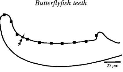
Fig. 1. Schematic diagram of a thin section of a tooth cap showing the approximate position of the sites examined by energy dispersive X-ray analysis. Lingual edge ( • ) ; normal to the tooth-cap surface (A).
approximately 20//m in diameter, for 100 s) at 30 sites. The shaft was analysed at five sites. The smaller probe diameter of the TEM enabled areas approximately
2 pan in diameter to be analysed (for 100 s) at approximately 15 ^m intervals along
an ultra-thin longitudinal section of the lingual edge of the cap (Fig. 1). Analyses were also carried out at intervals of approximately 2 pan along a line perpendicular to the tooth surface which bisected the tooth long axis at approximately one-third of the distance from the tip (Fig. 1).
Counts due to background interference were deducted (Link AN 10000 soft-ware) and the K<*and K/S peaks for calcium, phosphorus and iron were expressed as a percentage of the total number of counts.
Synthetic hydroxyapatite
Two samples of hydroxyapatite [Ca5(PO4)3OH], one doped with iron (iron hydroxyapatite), were synthesised at pH7.4 and 37°C, by slow addition of Ca(CH3COO)2 ( O . l m o i r1) and (NH^PCX, (0.06 molT1) solutions to CH3COONH4 ( O m o i r1) solution. FeCl3 was added to one Ca(CH3COO)2 solution at the atomic ratio 1:10 (Fe: Ca) (Okazaki et al. 1985). The slurries were incubated for 2 h at 37°C followed by 2 days at 25°C prior to filtration, exhaustive washing and drying at 25°C over P2O5.
Transmission electron microscopy - synthetic hydroxy apatites
The hydroxyapatite samples were prepared for TEM by suspending powders in chloroform (Analar grade), briefly sonicating (3 s), and then loading on nitrocellu-lose-covered, carbon-coated copper grids. The grids were viewed, and electron diffraction patterns recorded from single crystals. These were indexed on the basis of a hexagonal unit cell (space group P ^ / M ) with unit cell dimensions
a = b = 0.9418 nm, c = 0.6884nm.
X-ray diffraction
and a xenon proportional counter (PW1711/10). As a sample size of approxi-mately 1 g was required to fill the specimen holder, only synthetic hydroxyapatite/ iron-doped hydroxyapatite samples were analysed.
Atomic absorption
The synthetic samples were dissolved in hydrochloric acid and then diluted to approximately 5 p.p.m. Ca to bring them within the range of the standards. The solutions were run on a Varian AA-275 atomic absorption spectrophotometer.
Results
Butterflyfish teeth
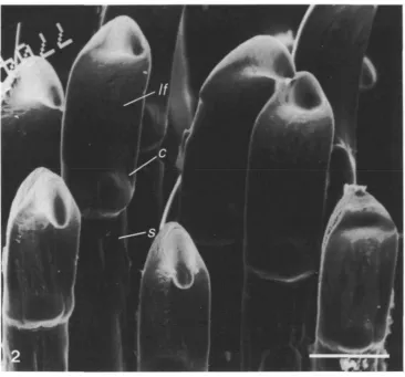
Butterflyfish jaw teeth lie on the premaxilla and dentary bones, forming rows of varying number. Inner teeth are usually smaller and more villiform, and outer or more labial teeth are more spatulate. The teeth are composed of a cap and a shaft (Fig. 2) which is firmly ankylosed to the bony pedestals. The cuticle, which is deposited during tooth development, is restricted to the tooth cap. Occasionally, abrasion of the pointed tooth cap results in a more rounded profile.
Perl's reaction was used to confirm the presence of iron in a sample of teeth from the butterflyfish (C. ornatissimus). This test uses 2moll"1 hydrochloric acid to dissolve the ferric iron which is then reacted with potassium ferrocyanide to form a blue precipitate of ferric ferrocyanide. The tooth caps, but not the shafts, were positive for iron, indicating that the iron in the cap must be in an ionizable state or relatively loosely bound since no reaction occurs if the iron is firmly bound in a complex molecule (Miles, 1963).
Elemental analysis (edXa) using a scanning electron microscope (SEM) showed calcium and phosphorus to be the main constituents of the tooth-cap surface with only low levels of iron present (data not shown). The low level of iron recorded was not consistent with Perl's reaction which showed the tooth cap to be coated with iron. The failure of the edXa system, when used under SEM conditions, to detect comparable quantities of iron may be due in part to substantial absorption of the emitted iron K a X-rays by the curved surface of the cap (Fig. 2).
In thin sections, the tooth cap was composed of three crystal layers; a cuticular and a superficial enameloid layer (Fig. 3) and, towards the centre of the tooth, an inner enameloid layer. Electron diffraction powder patterns gave d-spacings for each layer corresponding to standard hydroxyapatite d-spacings (data not shown). Thus, the differences in the layers result from a change in the orientation and morphology of the constituent hydroxyapatite crystals. There was no electron diffraction evidence for the presence of crystalline iron minerals.
Butterfiyfish teeth
375Fig. 2. Scanning electron micrograph of tooth caps (c) from a butterfiyfish showing the connection with the shaft (s). Lingual face, //. Scale bar, 50 jan.
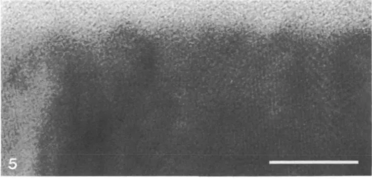
(0002) and (1010) lattice planes were parallel and perpendicular, respectively, to the cuticle surface (Fig. 5). This indicates that the crystal c-axis in these crystals was preferentially aligned perpendicular to the cuticle surface.
Fig. 3. Transmission electron micrograph of the cuticular layer (c) and underlying superficial enameloid (se). The gap between the two layers is an artefact of specimen preparation. Scale bar, 500 nm.
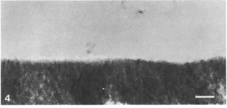
Fig. 4. Micrograph of the electron-dense cuticular layer. Note the lack of fissuring across the layer. Scale bar, 100nm.
[image:6.451.45.412.327.501.2]Butterflyfish teeth 377
Fig. 5. High-resolution transmission^ micrograph of the cuticular layer showing the smooth outer surface, (0002) and (1010) lattice planes running parallel and perpendicu-lar, respectively, to the surface. Scale bar, 20 nm.
(linear correlation coefficient of r = -0.97, P<0.001, Fig. 12). Iron was confined to the cuticular layer of the cap, the counts falling below detectable levels 2/zm from the cap surface (Fig. 13).
Synthetic iron-doped hydroxyapatite
The atomic absorption spectra for a sample of synthetic iron-doped hydroxy-apatite (4.2% iron w/w) showed iron to be present at an atomic ratio of 1:6.8 (Fe: Ca). The iron-doped sample showed an additional infrared peak at 1565 cm"1 (Fig. 14) due to N - H vibrations from occluded ammonium ions. Carbonate vibrational modes were observed in infrared spectra of both doped and non-doped samples, consistent with the preparation having been made in air.
The X-ray diffraction patterns for the non-doped and iron-doped hydroxyapa-tites were identical, each showing the major d-spacings for hydroxyapatite. Thus, there was no apparent change in the cell dimensions for the doped sample under these levels of iron incorporation.
The particles of synthetic hydroxyapatite were plate-like with an irregular outline (Fig. 15). There was no visible difference between the iron-doped and the non-doped samples. Lattice fringes (data not shown) were a feature of all the particles, indicating that they were single crystals with a high degree of structural perfection. Selected area electron diffraction (SAD) patterns (Fig. 16) were characteristic of single crystals and indicated that the top of the plates corre-sponded to the prismatic (1100) face of hydroxyapatite. There was no difference between the SAD patterns of the doped and the non-doped samples (data not shown).
Butterflyfish teeth
379
Fig. 6. Elongated hydroxyapatite crystals in the superficial enameloid layer arranged in 'bundles' (b) running parallel to the long axis of the tooth cap. The small crystals separating the bundles are probably elongated crystals viewed in cross-section. Scale bar, 500 nm.
[image:9.451.52.402.192.414.2]Fig. 7. Detail from Fig. 6 showing the oriented bundles (b) of hydroxyapatite crystals. Scale bar, 100 nm.
Fig. 8. TEM of the inner enameloid layer showing loosely packed, randomly oriented hydroxyapatite crystals. Scale bar, 500nm.
Fig. 9. Detail from Fig. 8. Scale bar, 50 nm.
Ca
Fe
Cu
KeV
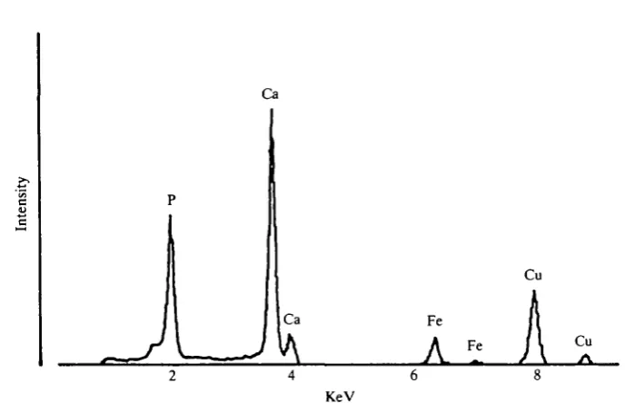
Fig. 10. Energy dispersive X-ray analysis spectrum from the cuticular layer (TEM mode).
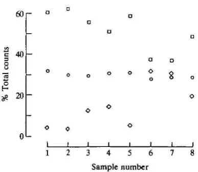
crystals (Fig. 17), the inverse relationship (r= -0.99, P<0.001) between the iron and calcium counts was consistent for all crystals examined (Fig. 18). Phosphorus levels remained stable in the crystals, being independent of changes in the iron/calcium ratio (Fig. 17). No iron was detected in the non-doped sample.
Discussion
The tooth cap of the butterflyfish is composed of three mineralized (hydroxy-apatite) layers; an iron-rich cuticle, the superficial enameloid and the inner enameloid. Each layer is formed from morphologically distinct arrangements of hydroxyapatite crystals.
60
-40
20
O • D o a D D
o o o o o o o o
O . O O O
O o o
I I I I I I I I I I I " 0 2 4 6 8 10 Tip Base
[image:10.451.164.308.75.362.2]Site analysed
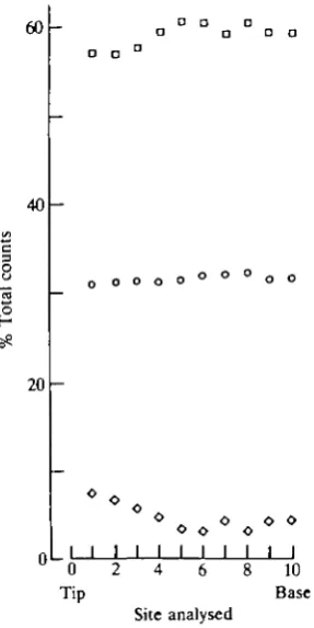
Fig. 11. Comparison of calcium ( • ) , phosphorus (O) and iron (O) counts (edXa) from the cuticular layer (lingual edge) of the tooth cap. The sites were approximately 15 jzm apart.
10
3?
i i i
55 56 57 58 59 % Total counts Ca
60
[image:10.451.118.353.435.606.2]Butterflyfish teeth
381
60
8 40
3? 20
o1-L o I
o I
o
I
0 1 2 3 Site analysed
Fig. 13. Comparison of calcium (D), phosphorus (O) and iron « » counts (edXa) along a line perpendicular to the cuticular layer (site 0) through the superficial enameloid. The sites were approximately 2 /an apart.
Iron apatite
Control apatite
2000 1800 1600 1400 1200 Wavenumber (cm"1)
1000 800 600
Fig. 14. Infrared spectra of non-doped and iron-doped synthetic hydroxyapatites, A = v(H2O), B = v(NH4+), C = v(CO32"), D = v(PO43").
piranha (Shellis and Berkovitz, 1976) it is a mixed inorganic/organic phase. It has been postulated (Preuschoft et al. 1974) that the cuticle inhibits the initiation of cracks in the underlying enameloid. Structurally the butterflyfish tooth-cap cuticle would appear to be well suited to this task, being formed from closely packed oriented hydroxyapatite crystals which result in a smooth, fissure-free surface.
15
Fig. 15. Image showing plate-like morphology of single crystals of synthetic hydroxy-apatite (iron-doped). Scale bar, 100nm.
Fig. 16. Single crystal electron diffraction pattern of iron-doped synthetic hydroxy-apatite. The pattern corresponds to the [1120] zone of hydroxyapatite, A = (0002), B = (1102), C = (2202). Camera length 166cm.
60
I 40
20
I I I I I I I
1 2 3 4 5 6 7
Sample number
[image:12.451.135.329.403.571.2]Butterflyfish teeth
38340
r-30
3 20
10
o o
I
36 40 44 48 52 % Total counts Ca
[image:13.451.75.377.58.270.2]56 60
Fig. 18. Linear correlation of iron and calcium counts (edXa) from the cuticular layer (lingual edge) of the tooth cap ( • ) and the iron-doped synthetic hydroxyapatite (O); r= _ 0 . 9 9 , P<0.001.
analyses showed the levels of iron and calcium within the cuticle to be inversely related to each other, indicating that the iron was closely associated with the hydroxyapatite phase. Similarly, Meridith-Smith (1967) found that the iron-pigmented enamel of urodele amphibians gave electron diffraction patterns consistent with hydroxyapatite, even though the ferritin granules in the adjacent enamel epithelium gave patterns for crystalline Fe2O3.nH2O.
Our results indicate that phosphorus levels were not affected by iron incorpor-ation. Phosphorus levels were the same in the cuticle as in the underlying enameloid. They were also constant among the synthetic iron hydroxyapatite crystals, even though the maximum iron level (30.8%) greatly exceeded iron levels observed in the cuticle (7.5 % ) . These results agree with findings by Okazaki
et al. (1985) on a series of apatites with increasing iron content. Moreover,
phosphorus levels in the cuticle were not significantly different from those in the synthetic crystals.
The stability of the phosphorus levels provides a good internal control for the efficiency of the edXa apparatus. In addition, in both biological and synthetic systems iron and calcium counts were inversely related ( r = -0.99, P<0.001) and not statistically different, with regression lines of similar (P<0.05) slope and displacement (intercepts). From this we infer that both systems use essentially the same mechanism to incorporate iron into the hydroxyapatite lattice.
smaller radius (0.064 nm) than calcium (0.099 nm). Since direct substitution results in loss of electroneutrality, it is possible that two calcium ions are replaced for a combination of Fe3 + and NH4+. This would explain the presence of the unique N - H vibration in the infrared spectra of the iron-doped sample. This is consistent with the findings of Okazaki et al. (1985). The ability of iron and calcium to substitute reversibly for each other within the hydroxyapatite lattice may explain the observation that a marked decrease in the iron pigmentation of rat enamel was associated with a diet high in calcium (Brudevold and Soremark, 1967).
The wide range of iron counts obtained from individual single crystals suggests that iron was distributed heterogeneously throughout the synthetic sample. This was consistent with the broad lines observed in the Mossbauer spectra of this material (D. P. E. Dickson, unpublished data). It is notable that the synthetic hydroxyapatite crystal lattice was able to accommodate iron at levels of about 4 % (w/w) without significant lattice distortion.-Assuming the same mechanism to be responsible for iron incorporation in both the biogenic and synthetic samples, the cuticular layer is far from being saturated with iron and could have a wide range of iron levels, depending on the iron loading within the system.
The butterflyfish tooth is structurally adapted for feeding from hard surfaces. However, although this study has shown iron to be concentrated at the point of maximal loading, this does not necessarily represent a genetic adaptation leading to increased durability of the cuticle. The small size of the individual tooth caps precluded the use of hardness tests (i.e. diamond indentation) to assess the effect on cuticle strength of increasing iron levels. Motta (1987) assessed iron levels (degree of tooth-cap pigmentation) and reported that species of butterflyfish feeding on the harder coral polyps had raised levels of iron in their teeth. Similarly, Selvig and Halse (1975) reported that the iron-pigmented layer of rat incisor enamel confers increased hardness and increased resistance to 5 % formic acid. In contrast, Suga (1984) contended that the incorporation of iron into the cuticular layer of fish teeth was not related to the hardness of the prey or feeding habits. Rather, he suggested that the enameloid provided a storage and detoxifica-tion site for excess amounts of iron and fluoride present in the body fluids. This supposition is supported by the fact that teeth are continually being replaced and offer a means of removing these elements into the environment in a stable form. The incorporation of iron into the synthetic hydroxyapatite lattice decreased the sensitivity of the crystals to damage by the electron beam. The ability of iron to stabilize synthetic hydroxyapatite crystals has also been reported by Okazaki et al. (1985). They noted an increase in acid resistance (decrease in crystal solubility) with increasing iron content, which may be of functional importance.
Butterflyfish teeth. 385
we suggest that synthetic iron-doped hydroxyapatite systems are useful models for studying iron incorporation into iron-rich calcined tissues.
References
BRUDEVOLD, F. AND SOREMARK, R. (1967). Chemistry of the mineral phase of enamel. In
Structural and Chemical Organization of Teeth, vol. 2 (ed. A. E. W. Miles), pp. 247-290. London: Academic Press.
GARANT, P. R. (1970). Observations on the ultrastructure of the ectodermal components during odontogenesis in Helostoma temmincki. Anat. Rec. 166, 167-188.
HALSE, A. (1973). Effect of dietary iron deficiency on the pigmentation and iron content of rat incisor enamel. Scand. J. dent. Res. 81, 319-334.
KERR, T. (1960). Development and structure of some actinopterygian and urodele teeth. Proc. tool. Soc. Lond. 133, 401-422.
LOWENSTAM, H. A. (1967). Lepidocrocite, an apatite mineral, and magnetite in teeth of chitons (Polyplacophora). Science 136, 1373-1375.
MERiDiTH-SMrrH, M. (1967). Studies on the structure and development of urodele teeth. PhD thesis, University of London.
MILES, A. E. W. (1963). Pigmented enamel. Proc. R. Soc. Med. 56, 918-920.
MOTTA, P. J. (1984). Tooth attachment, replacement, and growth in the butterflyfish, Chaetodon miliaris (Chaetodontidae, Perciformes). Can. J. Zool. 62, 183-189.
MOTTA, P. J. (1985). Functional morphology of the head of Hawaiian and Mid-Pacific butterflyfishes (Chaetodontidae, Perciformes). Environ. Biol. Fish. 13, 253-276.
MOTTA, P. J. (1987). A quantitative analysis of ferric iron in butterflyfish teeth (Chaetodontidae, Perciformes) and the relationship to feeding ecology. Can. J. Zool. 65, 106-112.
OKAZAKI, M., TAKAHASHI, J. AND KIMURA, H. (1985). Iron uptake of hydroxyapatite. J. Osaka Univ. Dent. Sch. 25, 17-24.
PREUSCHOFT, H., REIF, W.-E. AND MOLLER, W. H. (1974). Funktionsanpassungen in Form und Struktur an Haifischzahnen. Z. Anat. EntwGesch. 143, 315-344.
RANDALL, M. (1966). Electron microscopical demonstration of ferritin in the dental epithelial cells of urodeles. Nature, Lond. 210, 1325-1362.
REIF, W.-E. (1973). Morphologie und Ultrastruktur des Hai "Schmelzes". Zoologica Scripta 2, 231-250.
SCHMIDT, W. J. (1958). Zur histologie und Farbung der Zahne des japanischen Riesensalamanders. Z. Zellforsch. mikrosk. Anat. 49, 47-57.
SCHMIDT, W. J. (1959). Durodentin bei einem Devonischen Fisch (Laccognathus panderi). Z. Zellforsch. mikrosk. Anat. 49, 493-514.
SCHMIDT, W. J. AND KEIL, A. (1971). Polarizing Microscopy of Dental Tissues (translated by D. F. G. Poole and A. I. Darling). Oxford: Pergamon Press.
SELVIG, K. A. AND HALSE, A. (1975). The ultrastructural localization of iron in rat incisor enamel. Scand. J. dent. Res. 83, 88-95.
SHELLIS, R. P. AND BERKOVTTZ, K. B. (1976). Observations on the dental anatomy of piranhas (Characidae) with special reference to tooth structure. J. Zool., Lond. 180, 69-84.