Randall T. Higashida1 Van V. Halbach1 Stanley L. Barnwell1 Christopher Dowd1 Bill Dormandy2 Julie Bell2 Grant B. Hieshima 1
Received October 30, 1989; revision requested January 3, 1990; revision received February 9, 1990; accepted February 22, 1990.
' Departments of Radiology and Neurological Surgery, lnterventional Neuroradiology Section, L-352, 505 Parnassus Ave., San Francisco, CA 94143-0628. Address repritn requests to R. T. Higashida.
2 lnterventional Therapeutics Corporation, 385 Oyster Point Blvd., Suite 6, South San Francisco, CA 94080.
0195-6108/90/1104-0633
© American Society of Neuroradiology
Treatment of Intracranial
Aneurysms with Preservation of
the Parent Vessel:
Results of
Percutaneous Balloon Embolization in 84
Patients
633
Treatment of intracranial arterial aneurysms by interventional neurovascular
tech-niques is now being performed in selected cases. From a transfemoral approach, under
local anesthesia, a detachable silicone microballoon can be guided through the
intracra-nial circulation, directed into the aneurysm, inflated with a polymerizing agent for
solidification, and detached. The goal is to exclude the aneurysm from the circulation
and preserve flow through the parent artery. Since 1984, 84 patients have been treated
by this technique. The patients ranged in age from 15 to 83 years (mean age, 48) and
included 63 females and 21 males. The distribution of aneurysms included 59 in the
anterior circulation and 25 in the posterior circulation. The presenting symptom or cause
was mass effect in 45 patients (53.6%), subarachnoid hemorrhage in 31 patients (36.9%),
carotid-cavernous sinus fistula resulting from rupture of an intracavernous aneurysm in
six cases (7.1%), trauma in one case, and transient cerebral ischemia due to emboli in
one case. Permanent complications directly related to therapy included 15 deaths and
nine cases of stroke. Clinical and radiologic follow-ups were performed 1, 3, and 12
months after treatment; duration of follow-up ranged from 3 to 68 months (mean, 35.5
months). In 65 cases (77.4%) there was evidence of complete aneurysmal occlusion,
and in 19 cases 922.6%) there was subtotal occlusion greater than 85%.
lnterventional techniques for treatment of intracranial aneurysms may be useful as a
therapeutic alternative in those patients not amenable to standard surgical therapy.
AJNR 11:633-640, July I August 1990
Surgical exploration and clipping remains the treatment of choice for the majority of patients diagnosed with an intracranial aneurysm. However, in certain types of cases, aneurysms may not be accessible by surgery owing to size or anatomic location, or patients may be too unstable medically to undergo general anesthesia.
In these instances, treatment with interventional neurovascular techniques has emerged as a therapeutic alternative.
lnterventional techniques using detachable balloons were first described by Serbinenko [1] in 197 4. Since then, recent advances in high-resolution digital subtraction angiography, road mapping technique, microballoon technology, and solidifying materials have allowed the treatment of selected patients with intracranial aneurysms from an endovascular approach. Under direct fluoroscopic imaging, the balloon can be guided directly into the aneurysm, filled with a polymerizing sub-stance, and detached, thus excluding the aneurysm from the circulation and preserving blood flow through the parent artery [2, 3]. For large and giant aneu
634
HIGASHIDA ET AL. AJNR:11, July/August 1990Materials and Methods
Patient Population
Since 1984, 84 patients, 63 females and 21 males, have been treated by detachable balloon occlusion of intracranial aneurysms with preservation of the parent artery. The patients ranged in age from 15 to 83 years (mean age, 49 years). Table 1 lists the locations
of the various types of aneurysms treated. Fifty-nine patients were treated for an aneurysm in the anterior circulation and 25 were treated
for an aneurysm in the posterior circulation. Nineteen patients (22.6%) were treated for a giant aneurysm greater than 2.5 em in diameter. In 45 cases (53.6%) the presenting symptom was mass effect
accom-panied by pain, ophthalmoplegia, headaches, and compression of adjacent brain structures causing hydrocephalus, visual loss, or
pro-gressive neurologic deficit depending on aneurysm location. in 31 patients (36.9%), subarachnoid hemorrhage (SAH) due to aneurysm
rupture was the presenting symptom. Six patients (7.1 %) had
evi-dence of a direct carotid-cavernous sinus fistula, which included
symptoms of retroorbital bruit, chemosis, ophthalmoplegia, andjor
visual decline due to rupture of a preexisting intracavernous aneu-rysm. One patient presented with a pseudoaneurysm following
trauma and one patient presented with symptoms of transient
cere-bral ischemia from thromboembolic disease presumably originating
from the aneurysm.
Indications for Treatment
Our current indications for treatment by detachable balloon em-bolization therapy include prior surgical exploration of an aneurysm with inability to clip the neck, aneurysms in surgically difficult anatomic regions such as the cavernous segment of the internal carotid artery and midbasilar artery, fusiform aneurysms without a well-defined
neck, inability to tolerate general anesthesia owing to an underlying medical condition, and aneurysms in which the risks of surgical clipping are judged to be excessively high owing to th-eir size or location. Therefore, this series favored a very high-risk group of
patients in whom standard neurosurgical treatment failed or who were poor candidates for surgery. All patients gave complete
in-formed consent prior to therapy.
Technique
All procedures are performed in the interventional neuroa
ngiogra-phy suite with the use of high-resolution digital subtraction
angiog-TABLE 1: Locations of Aneurysms Treated by Detachable Balloon Therapy
Location No.
Anterior circulation
High cervicaljpetrous internal carotid artery 3 Cavernous internal carotid artery 24 Supraclinoid internal carotid artery 24
Middle cerebral artery 6
Anterior cerebral artery 2
Total 59
Posterior circulation
Distal vertebral/posterior inferior cerebellar artery 2
Mid-basilar artery 3
Distal basilar artery 19
Posterior cerebral artery 1
T~~ ~
raphy, road-mapping technique, and rapid-sequence filming. From a transfemoral approach, using 1% xylocaine for local anesthesia and IV diazepam and morphine sulfate for mild sedation, a 7.5-French
sheath is placed into the femoral artery and an activated clotting time
(ACT) is measured. A complete four-vessel cerebral arteriogram is
obtained to assess aneurysm size, shape, axis, relationship of the neck to the parent vessel, evidence of intraluminal thrombus, and
collateral blood flow. Correlation with prior MR and CT scans is made to determine if recent thrombus is present within the aneurysm [4]. If fresh thrombus of less than 6-weeks duration is evident and the patient is clinically stable, we prefer to postpone therapy because of the increased risk of dislodging clot during balloon manipulation within
the aneurysm. Following the diagnostic evaluation, 5000 units of heparin (for a 70-kg patient) are given IV for systemic anticoagulation to prevent thrombus formation on the balloon and microcatheters. A repeat ACT measurement is obtained to ensure adequate anticoag-ulation.
Balloon selection is determined by aneurysm size and shape. Two different types of balloons, a standard balloon and a collarless balloon, each in several different sizes and three release ranges, are currently
available (Table 2). The balloon that we have been using for endo-vascular aneurysm therapy (Fig. 1) is manufactured as an
investiga-tional device by lnterventional Therapeutics Corp. (South San F ran-cisco, CA) and is currently undergoing Food and Drug Administration clinical trials [5]. The balloon is attached to a 2.0-French polyethylene
catheter or a 2.0-French Tracker catheter (Target Therapeutics Corp.,
San Jose, CA).
For aneurysms in unfavorable locations relative to the parent
vessel, the polyethylene catheter can be steam-formed into different shapes or the Tracker catheter can be used in conjunction with a 0.014-in. (0.035-cm) steerable guidewire to aid in placing the balloon
directly into the aneurysm. If the small or medium-sized balloon is used, a 7.3-French nontapered catheter is placed through the femoral artery sheath into the proximal internal carotid or vertebral artery, depending on aneurysm location. For the large balloon, an 8.0-French nontapered catheter is used. Through this guiding catheter, the balloon and microcatheter are then advanced, with continuous
per-fusion of heparinized saline between the two catheters to avoid thrombus formation. The balloon is filled with metrizamide, at a
concentration of 200 mg % iodine, for opacification.
A "road map," which is the subtracted neurovascular architecture
superimposed on the real-time fluoroscopic image, is then obtained
of the aneurysm and proximal artery. The balloon is flow-directed through the intracranial circulation and carefully guided into the aneu-rysm. Once within the aneurysm, the balloon is inflated so as to
TABLE 2: Characteristics of Silicone Detachable Balloons
Dimension, Volume, Dimension, Detachment
Size Uninflated Unrestricted Maximum Inflated Strengths,
(mm)
(ml) (mm) All Sizes (g)
Small 0.85 X 4.10 0.10 3.80 X 9.00 20-30 0.85 X 5.10 0.20 4.20 X 12.0 30-40 0.85x 7.10 0.40 4.50 X 16.0 40-55
Medium 1.50 X 4.10 0.20 5.00 X 6.00 20-30 1.50 X 4.60 0.30 6.50 X 9.50 30-40
1.50 X 5.10 0.50 7.50 X 13.50 40-55 1.50 X 7 30 0.90 8.50 X 21.00
[image:2.612.313.556.586.740.2] [image:2.612.53.300.586.738.2]AJNR:11, July/August 1990 BALLOON THERAPY OF INTRACRANIAL ANEURYSMS
635
Fig. 1.-A. Silicone detachable balloon used for intravascular treatment of intracranial aneu-rysms. Balloons shown are uninflated, partially inflated, and fully inflated.
B, Newer type of silicone balloon currently available shows that self-sealing miter valve is internalized and does not protrude beyond valve base.
A
produce complete occlusion of the aneurysm dome, body, and base.
The volume required to produce complete occlusion is measured and
then the contrast material is aspirated from the balloon and refilled
with 2-hydroxyethyl methacrylate (HEMA). HEMA is a liquid
mon-omer, which can be cross-linked and catalyzed to produce
polymeri-zation that yields a nonbiodegradable solid substance. This is used
as a fill material within the silicone balloon to ensure that a permanent
embolic material remains within the aneurysm, should the balloon
shell deteriorate or the self-sealing miter valve fail over time (2, 3].
To overcome the dead space of our catheter system, several
ex-changes with HEMA or the use of a 0.006-inc. (0.015-cm) vent tube
are undertaken during this stage of the procedure. Once the HEMA
is solidified, confirmed by comparing the HEMA within the balloon with an outside control, the balloon is detached by gentle traction on
the 2.0-French catheter. If the neck of the aneurysm is broad-based,
a second nondetachable balloon can be temporarily placed across
the neck to aid in detachment, and then removed. For giant
aneu-rysms, more than one balloon may be required to completely fill the
aneurysm. After the embolization procedure, protamine sulfate is
given IV to reverse systemic heparinization (1 0 mg of protamine will
reverse 1000 units of heparin). A final arteriogram is obtained to
check for aneurysm occlusion and patency of the parent vessel.
Neurologic assessment is also made of the patient's condition.
All patients are observed in the neurosurgical observation unit for
2-4 days after treatment and if stable are discharged. Clinical
follow-up is performed in all patients at 1, 3, and 12 months after treatment.
Postembolization arteriograms are obtained at 1-3 months and at
3-12 months after treatment. In addition, plain skull radiographs along
with CT andfor MR head scans are obtained during these intervals
to check balloon placement and aneurysm thrombosis, particularly
for large and giant aneurysms.
Representative Case Reports
Supraclinoid Aneurysm with Hemorrhage
A 56-year-old woman presented with a large SAH primarily
involv-ing the right side. Cerebral angiography demonstrated a small right
middle cerebral artery aneurysm as well as a large supraclinoid carotid
artery aneurysm measuring 13 x 16 x 23 mm. In addition, the right
anterior cerebral artery was hypoplastic (Figs. 2A and 2B). Surgical
exploration of both right-sided aneurysms was performed. The right
middle cerebral artery aneurysm was successfully clipped; however,
exploration of the right supraclinoid aneurysm showed the neck
extending below the clinoid process into the cavernous sinus.
Be-cause this patient was still at risk for rehemorrhage she was referred
8
for balloon embolization treatment. Test occlusion of the cervical right
internal carotid artery was performed under local anesthesia. The
mean arterial pressure of the carotid artery was 93 mm Hg; however,
on temporary balloon occlusion the mean arterial pressure dropped
to 19 mm Hg, and at 2 min the patient became lethargic, unresponsive
to commands, and dysarthric; she also developed a left-sided
hemi-paresis. The balloon was immediately deflated, and after 15 min the
neurologic deficits resolved.
Because the patient did not tolerate temporary occlusion of the
carotid artery, it was important to preserve flow of the distal intracra
-nial vessels. Under direct fluoroscopic visualization, two detachable
balloons were flow-directed through the intracranial carotid circulation
and directed into the large supraclinoid aneurysm. An exchange of
HEMA was made into each balloon, allowed to solidify, and then
detached. The postembolization angiogram demonstrated obli
tera-tion of the aneurysm with preservation of the parent artery. The
patient was discharged several days later in stable neurologic
condi-tion without any major deficits. A 1-year follow-up angiogram
contin-ued to show obliteration of both aneurysms with normal filling of the
intracranial circulation (Figs. 2C and 20); clinically she continued to remain neurologically intact.
Large lntracavernous Aneurysm
A 47-year-old woman presented with retroorbital headaches, nau
-sea, and visual difficulties. A CT head scan followed by cerebral
angiography demonstrated a large right-sided intracavernous
aneu-rysm projecting medially and measuring 12 x 15 x 20 mm (Figs. 3A
and 3B). Test occlusion of the internal carotid artery demonstrated
an arterial back pressure of 18 mm Hg with temporary balloon
occlusion. Within 30 sec left-sided hemiparesis and aphasia
devel-oped. Thus, the patient was unable to tolerate carotid artery occlusion
owing to poor collateral circulation around the circle of Willis and via
the external carotid artery.
From a transfemoral approach, a 1.5-mm medium detachable
balloon was guided through the carotid artery and directed into the
aneurysm. The balloon was filled with 0.38 ml of HEMA, allowed to
solidify, and detached. Her immediate, 4-week, and 4-month
follow-up postembolization cerebral arteriograms demonstrated obliteration
of the intracavernous aneurysm (Figs. 3C-3E).
Large Basilar Artery Aneurysm
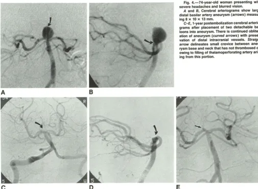
A 74-year-old woman presented with severe throbbing headaches
and blurred vision. A CT head scan and cerebral angiogram
[image:3.614.222.556.78.227.2]636 HIGASHIDA ET AL. AJNR:11, July/August 1990
mm (Figs. 4A and 48). Two silicone detachable balloons were
flow-directed up the posterior circulation, through the basilar artery, and into the aneurysm. After placing HEMA into the balloons for solidifi-cation, the balloons were detached, with obliteration of the aneurysm and preservation of the distal posterior cerebral arteries. After the
procedure the patient developed a mild visual field deficit, most likely
from a small embolus to the posterior cerebral artery from the
microcatheter during the procedure. She was discharged several
days later in stable neurologic condition without further difficulties. A follow-up arteriogram at 1 year demonstrated continued occlu -sion of the aneurysm with normal filling of the distal intracranial vessels (Figs. 4C-4E). At 2 years of clinical follow-up she is still
neurologically intact except for a mild visual field deficit.
Results
Since 1984, we have guided a balloon directly into an aneurysm and preserved the parent artery in 84 patients [6, 7]. In 19 cases (22.6%), the patients presented with a giant aneurysm, and often more than one balloon was required to occlude the dome and body of the aneurysm and still preserve the parent vessel.
Complications associated directly with aneurysm treatment included 15 deaths (17 .9%). In 1 0 of these, rupture of the aneurysm occurred as a result of incomplete aneurysm occlu-sion, resulting in an SAH. Five of these patients had presented
~
A
8
c
D
previously with an SAH and five with symptoms of mass effect. Six of the 1 0 patients died from aneurysm rupture within 5 days after treatment; in the other four, rupture was delayed. One death was due to rupture of the balloon and a second death was from valve leakage; both occurred during polymerization of HEMA, resulting in occlusion of the distal intracranial vessels. Two other deaths in this series occurred in patients who developed myocardial infarcts 24 hr after the procedure; a third patient died from a pulmonary embolus 6 days after treatment.
Three other deaths (3.6%) occurred in this series, although not because of the endovascular procedure for aneurysm occlusion. These were caused, respectively, by an intracranial hematoma from a prior surgical craniotomy, severe vaso-spasm from prior SAH, and hemorrhage from an associated arteriovenous malformation 16 months after successful bal-loon embolization therapy of a basilar artery aneurysm.
Nine patients (1 0.7%) developed a stroke as a result of therapy. In five of these patients, the stroke was due to thrombus dislodging either within the aneurysm or on the balloon and catheters during the procedure, despite systemic anticoagulation. in two patients, the balloon narrowed the parent vessel after it was detached, causing occlusion and stroke. In one patient, a stroke developed owing to balloon rupture during HEMA polymerization; in another patient,
pre-Fig. 2.--,-Patient presenting with large sub-arachnoid hemorrhage from aneurysm rupture.
A and 8, Frontal (A) and lateral (8) views of right internal carotid arteriogram show small middle cerebral artery aneurysm (curved arrows) and large supraclinoid internal carotid artery aneurysm (straight arrows). Note that anterior cerebral artery is hypoplastic.
[image:4.612.54.390.372.736.2]AJNR:11, July/August 1990 BALLOON THERAPY OF INTRACRANIAL ANEURYSMS 637
Fig. 3.-A and 8, Selective right internal ca-rotid arteriograms, frontal (A) and lateral (8)
views, show large intracavernous aneurysm
measuring 12 x 15 x 20 mm (straight arrows). Aneurysm projects medially; neck is seen well angiographically (curved arrow).
C-E, After placement of a single detachable balloon into aneurysm (arrows), 4-month
follow-up arteriogram shows obliteration of aneurysm
with preservation of intracranial vessels. This is demonstrated best on left anterior oblique pro-jection (C), but is seen also on frontal (D) and
lateral (E) views.
c
A
D
mature balloon detachment resulted in an embolus to the middle cerebral artery. Long-term follow-up demonstrated improvement in neurologic function in seven of these nine patients: three had only mild to moderate expressive aphasia, two had a visual field deficit, one was recovering from a frontal lobe stroke, and one had a mild hemiparesis. One patient required surgical repair of the right and left femoral artery due to pseudoaneurysms that developed after treatment from the femoral artery puncture for therapy of intracranial aneurysms; this patient had no other difficulty.
Follow-up has ranged from 3 to 68 months (mean, 35.5 months). Radiologic follow-up has demonstrated complete aneurysm occlusion in 65 cases (77.4%), with good clinical
recovery of the presenting symptoms. In the other 19 patients
(22.6%), subtotal occlusion of the aneurysm was observed with greater than 85% occlusion of the dome and body. Two patients had evidence of the balloon shifting and occluding the parent vessel on follow-up studies. Ten patients (11.8%) required more than one balloon embolization treatment owing to shift of the balloon(s) or enlargement of the aneurysm on follow-up studies. These patients with subtotal occlusion tended to have large and giant aneurysms, often requiring placement of multiple balloons, and evidence of intraluminal thrombus within the aneurysm prior to therapy. These pa-tients continue to have follow-up clinical and radiologic studies
B
E
to ensure that no further change occurs in the size of the residual neck.
Discussion
Intravascular detachable balloon embolization therapy for treatment of intracranial aneurysms has been reported from several different centers. The earliest report was by Serbi-nenko (1] in 197 4, who described successful occlusion of aneurysms involving the internal carotid artery using latex balloons. In 1981, Debrun et al. (8] and in 1984, Berenstein et al. (9,1 0] described a small number of patients who underwent carotid occlusion for treatment of giant unclippable aneurysms in the anterior circulation with only one permanent complication. In 1987, Fox et al. (11] described the use of detachable balloons for treating 68 patients with aneurysms by parent vessel occlusion therapy. They reported a 13.8% complication rate, consisting of delayed cerebral events and no permanent morbidity. These researchers have demon-strated that proximal occlusion of the parent artery can be performed with acceptable morbidity and mortality to alleviate symptoms from an intracranial aneurysm.
[image:5.613.57.560.76.446.2]lnsti-638 HIGASHIDA ET AL. AJNR:11, July/August 1990
c
D
tute of Neurosurgery. In 1982, they reported 119 cases, including 118 cases involving the anterior circulation and one
patient treated for a posterior inferior cerebellar artery
aneu-rysm. In 93 cases (78.2%), they were able to occlude the
aneurysm and preserve patency of the parent artery. Fiften patients (12.6%) required parent artery vessel occlusion and in 11 cases (9.2%) the aneurysm could not be occluded. In 89 (96.7%) of 92 patients treated with preservation of the parent artery, Romodoanov and Shcheglov reported good to
excellent results with complete aneurysm occlusion. In 1989,
Shcheglov updated the series and described 617 patients
treated by intravascular detachable balloon therapy.
(Shcheg-lov VI. Endovascular occlusion of saccular intracranial
aneu-rysms: results in 617 patients. Presented at the annual meet-ing of the American Society of Neuroradiology, Orlando, FL,
March 1989.) The aneurysm was successfully occluded and
the parent artery preserved in 91% of cases. The reported
mortality rate was 1.7% in 338 patients who were in fair
condition when treated and 22% in 71 patients who were in
poor condition. Currently, Shcheglov proposes that patients
with an intracranial aneurysm first undergo detachable balloon
embolization therapy, and only if that technique fails should they be treated by surgical clipping.
The experience of our group differs from that of Shcheglov in several important aspects. Since we consider this technique
Fig. 4.-74-year-old woman presenting with severe headaches and blurred vision.
A and B, Cerebral arteriograms show large distal basilar artery aneurysm (arrows) measur-ing 8 x 10 x 13 mm.
C-E, 1-year postembolization cerebral arteria-grams after placement of two detachable bal-loons into aneurysm. There is continued obliter-ation of aneurysm (curved arrows) with preser-vation of distal intracranial vessels. Straight
arrow delineates small crevice between aneu-rysm base and neck that has not thrombosed off owing to filling of thalamoperforating artery aris-ing from this portion.
E
to be somewhat developmental, our criteria for patient selec-tion are more limited. Patients are included only if standard surgical exploration for clipping aneurysms in surgically diffi-cult anatomic locations has failed or if patients cannot tolerate general anesthesia [5]. We also tend to treat patients who have an acute SAH shortly after presentation, because of the increased risk of recurrent hemorrhage within the first 4-6 weeks [13-15]. Shcheglov prefers waiting at least 2-4 weeks after the initial hemorrhage; we believe that during this time patients may rehemorrhage or develop severe vasospasm.
Our patient population therefore favors a higher-risk group of patients to undergo this form of therapy. Our complication rate compares favorably with neurosurgical results for large and giant aneurysms in difficult anatomic locations. The over-all reported operative morbidity and mortality statistics range
from 16% to over 50% for complex aneurysms, such as those involving the posterior circulation, from several large neuro-surgical centers [16-23].
We have also developed a different detachable balloon catheter system for aneurysm therapy. Our group has devel-oped a silicone, rather than latex, balloon system for several different reasons. Silicone is much more stable than latex, is biocompatible, and will not degrade within the intravascular
system [24, 25]. In order to ensure that the aneurysm remains
[image:6.620.56.561.78.448.2]AJNR:11, July/August 1990 BALLOON THERAPY OF INTRACRANIAL ANEURYSMS 639
silicone balloon, which may not be compatible with latex
balloons [5]. Shcheglov and his group fill their latex balloons with silicone; however, this is a very viscous material and is not water soluble. Therefore, the dead space of the attached catheter must be overcome to ensure the balloon is com-pletely filled. For aneurysms involving the anterior circulation, the Kiev group uses direct puncture of the cervical carotid artery. For posterior circulation aneurysms, other approaches are required. We perform all our procedures from a transfem-oral arterial approach, thus permitting access to both the anterior and posterior circulation.
Complications associated with endovascular therapy are different from those of conventional surgery. Since balloons and catheters are being manipulated within the blood vessel lumen, thrombus may form on these materials. To avoid this, we place our patients on systemic anticoagulation by admin-istering IV heparin prior to the balloon procedure and recheck-ing an activated clottrecheck-ing time. Despite this precaution, embolic episodes related to the procedure still occurred in five of our patients. Also in patients with a recently ruptured aneurysm, systemic anticoagulation may increase the prevalence of an-other severe hemorrhage during the procedure. Dislodgment of thrombus within an aneurysm by the balloon is also possible during the procedure, and we suspect that in two of our pateints this occurred, resulting in transient cerebral ischemia. The most critical phase with the use of HEMA is during the polymerization phase, lasting 20-60 min after mixing with the catalyst. If the balloon were to rupture or the valve leak during this interval, HEMA could leak out into the distal intracranial vasculature as a semisolid embolic agent, resulting in a stroke. Other complications include premature detachment of the balloon, resulting in parent vessel occlusion or distal emboli-zation, aneurysm rupture during balloon inflation, and delayed thromboembolic events in an incompletely treated aneurysm. The majority of deaths in our series were due to incomplete aneurysm occlusion with recurrent hemorrhage, occurring in 1 0 of 12 patients. Unless the aneurysm is completely occluded at the base by the balloon, there is the potential for aneurysm regrowth at its base or shift of the balloon within a thrombus already present within the aneurysm. This may result in persistent filling of the aneurysm and the potential for further hemorrhage or increase in mass effect. This is particularly important in giant aneurysms, in which more than one balloon may be required for complete occlusion. In 22.6% of our patients, only subtotal occlusion was apparent on follow-up studies; these patients are being closely followed for any change in residual size at the aneurysm base. Ten patients in our series required more than one procedure for aneurysm treatment owing to balloon shift within thrombus or regrowth of the aneurysm on follow-up studies.
The advantage of this procedure is that it is performed under local anesthesia from a transfemoral arterial approach; therefore, neurologic monitoring of the patient can be contin-uous. The suppleness of the catheter and balloon system aids in making placement of the balloon directly into the aneurysm possible for almost all types of aneurysms around the circle of Willis. For smaller aneurysms, a vent tube can be used during the exchange for HEMA to allow an adequate volume for permanent solidification of the entire contents of
the balloon. Some of the disadvantages associated with this
procedure include the need for a high degree of experience
with detachable balloon systems, polymerizing materials, and
high-resolution fluoroscopic equipment with road-mapping
ca-pability.
Long-term follow-up is still required to assess the clinical outcome and efficacy in patients with subtotal occlusion. In our experience, after an aneurysm is occluded by detachable
balloons, any remaining small crevice still filling between mul
-tiple balloons placed within the aneurysm and at the aneurysm base will thrombose in the majority of patients. However, in those patients in whom persistent filling is seen on studies
after 1-3 months, we have observed late hemorrhage and
regrowth of the aneurysm. As with incomplete surgical clip
-ping, these patients need to be assessed continuously to check further change in residual filling and to see if retreat -ment is necessary [26]. Preliminary animal studies of experi
-mentally created aneurysms treated by endovascular plac
e-ment of detachable silicone balloons into the aneurysm have
indicated that thrombosis with reendothelialization does occur at the aneurysm base, with patency of the parent vessel. (Negoro M. Endovascular treatment of cerebral aneurysm,
clinical and experimental aspects. Presented at the Inte rna-tional Workshop on Intracranial Aneurysms, Nagoya, Japan, April 1989.) Further studies need to be carried out to assess
changes with subtotal occlusion and hemodynamic changes
that occur within the aneurysm lumen, particularly with giant aneurysms that require multiple balloons.
For now it is believed that intravascular detachable balloon embolization therapy is a viable alternative for treating pa
-tients with intracranial aneurysms. This technique allows oc -clusion of the aneurysm with preservation of the parent vessel, which may be the only alternative in patients in whom surgical clipping has failed and who cannot tolerate parent
vessel occlusion therapy. As continued improvements in this
technique evolve, the indications for therapy may broaden
also.
REFERENCES
1. Serbinenko FA. Balloon catheterization and occlusion of major cerebral
vessels. J Neurosurg 1974;41 :125-145
2. Goto K, Halbach VV, Hardin CW, Higashida RT, Hieshima GB. Permanent inflation of detachable balloons with a low viscosity hydrophilic pol
ymeriz-ing system. Radiology 1988;169:787-790
3. Taki W, Handa H, Yamagata S, et al. Radio-opaque solidifying liquids for
releasable balloon technique: a technical note. Surg Neural 1980;
13:140-142
4. Tsuruda JS, Halbach VV, Higashida RT, Mark AS, Hieshima GB, Norman
D. MR evaluation of large and giant aneurysms using cine low flip angle gradient-refocusing imaging. AJNR 1988;9:415-424
5. Higashida RT, Halbach VV, Dormandy B, Bell J, Hieshima GB. Endovas-cular treatment of intracranial aneurysms using a new silicone microballoon device: technical considerations and indications for therapy. Radiology
1990;174:687-691
6. Halbach VV, Higashida RT, Hieshima GB. Treatment of intracranial aneu
-rysms by balloon embolization therapy. Semin lnterven Radio/
1987;4(4):261-268
7. Higashida RT, Halbach VV, Cahan LD, Hieshima GB, Konishi Y. Detachable balloon embolization therapy of posterior circulation intracranial aneu
-rysms. J Neurosurg 1989;71 :512-519
640
HIGASHIDA ET AL. AJNR:11, July/August 19901981:2:167-173
9. Kupersmith MJ, Berenstein A, Choi IS, Ransohoff J, Flamm ES. Percuta-neous trans vascular treatment of giant carotid aneurysms: neuro-ophthal-mologic findings. Neurology 1984;34:328-335
10. Berenstein A, Ransohoff J, Kupersmith M, Flamm E, Graeb D. Transvas-cular treatment of giant aneurysms of the cavernous carotid and vertebral arteries: functional investigation and embolization. Surg Neural 1984; 21:3-12
11. Fox A, Vinuela F, Pelz DM, et al. Use of detachable balloon for proximal artery occlusion in the treatment of unclippable cerebral aneurysms. J Neurosurg 1987;66:40-46
12. Romodanov A, Shcheglov VI. Intravascular occlusion of saccular aneu-rysms of the cerebral arteries by means of a detachable balloon catheter. In: Krayenbuhl H, ed. Advances and technical standards in neurosurgery, vol. 9. Zurich: Springer-Verlag, 1982:25-48
13. Sahs AL, Perret GE, Locksley HB, et al. Intracranial aneurysms and subarachnoid hemorrhage. A cooperative study. Philadelphia. Lippincott, 1969
14. Jane JA, Winn HR. Richardson AE. The natural history of intracranial aneurysms: rebleeding rates during the acute and long-term period and implication for surgical management. C/in Neurosurg 1976;24:176-184 15. Wilkins RH. Update-subarachnoid hemorrhage and saccular intracranial
aneurysm. Surg Neural 1981;15:92-1 01
16. Jamieson KG. Aneurysms of the vertebrobasilar system. Surgical
interven-tion in 19 cases. J Neurosurg 1964;21 :781-797
17. Logue V. Posterior fossa aneurysms. Clin Neurosurg 1963;11 :183-219 18. Logue V. The surgical treatment of aneurysms in the posterior fossa. J
Neural Neurosurg Psychiatry 1958;21 :66-67
19. Drake CG. Cerebral aneurysm surgery: an update. In Scheinberg P, ed. Cerebral vascular diseases. 10th Princeton Conference. New York: Raven, 1976:289-31 0
20. Peerless SJ, Drake CG. Management of aneurysms of the posterior circulation, In: Youmans JR, ed. Neurological surgery, 2nd ed. Philadelphia: Saunders, 1982:1715-1763
21. McMurtry JG, Housepian EM, Bowman FO, et al. Surgical treatment of basilar artery aneurysms. Elective circulatory arrest with thoracotomy in 12 cases. J Neurosurg 1974;40:486-494
22. Wilson CB, Hoi Sang U. Surgical treatment for aneurysms of the upper
basilar artery. J Neurosurg 1976;44:537-543
23. Yasargil MG, Antic J, Laciga R, et al. Microsurgical pterional approach to
aneurysms of the basilar bifurcation. Surg Neurol1976;6:83-91
24. Arkles B. New generation silicones for medical devices. Medical Device and Diagnostics Industry 1981;3(4):31-35
25. Miller F. Elastomers in medicine. Elastomerics 1985;117(7):15-20 26. Drake CG, Vanderlinden RG. The late consequences of incomplete surgical
treatment of cerebral aneurysms. J Neurosurg 1967;27:226-238


![rac Bis[μ N salicylidene N' (α phenylsulfanylmethyl 2 oxidobenzyl)propylenediamine N,N′,O,O,O′]nickel(II) dichloromethane solvate](data:image/gif;base64,R0lGODlhAQABAIAAAP///wAAACH5BAEAAAAALAAAAAABAAEAAAICRAEAOw==)