ACID-BASE REGULATION AND BLOOD GAS TRANSPORT
FOLLOWING EXHAUSTIVE EXERCISE IN AN AGNATHAN,
THE SEA LAMPREY PETROMYZON MARINUS
B Y B . L .
TUFTS
Department of Biology, Queen's University, Kingston, Ontario, Canada K7L 3N6
Accepted 19 April 1991
Summary
Exhaustive exercise in cannulated sea lampreys, Petromyzon marinus, resulted in a marked extracellular acidosis in the arterial blood which had both a respiratory and a metabolic component. Blood CO2 tension (Pco2) returned to
control levels within an hour after exercise, but the metabolic acidosis had a somewhat longer time course and the extracellular pH (pHe) did not fully recover until the 4h recovery sample. The magnitude and duration of the changes in both the plasma lactate concentration and the concentration of metabolic protons were very similar and the maximal proton deficit after exercise was, therefore, only 1.5mequivl~\ In contrast to the changes in pHe, there were no significant changes in the erythrocyte pH (pHi) following the exercise period. The regulation of pHi was apparently not adrenergically mediated, however, since addition of catecholamines to lamprey blood in vitro had no significant effect on pHi. In addition, the period of exhaustive exercise in vivo was not associated with any significant changes in the mean cellular hemoglobin concentration. The total carbon dioxide concentration in the arterial whole blood and true plasma were both significantly reduced after exercise, but the total carbon dioxide concen-tration within the erythrocytes was transiently increased. Finally, there was a marked decrease in the arterial PO j immediately after exercise, which was
associated with a significant reduction in the amount of oxygen bound to hemoglobin; however, within 30min, these values had both returned to normal. The maintenance of pHi presumably contributes to the regulation of oxygen transport in lampreys and it may be particularly important during the brief period immediately after exercise when oxygen transport is clearly compromised. Although several studies have provided evidence that chloride/bicarbonate exchange limitations may exist in agnathan blood in vitro, the present results demonstrate that the characteristics of carbon dioxide transport and acid-base regulation after exercise in P. marinus are not markedly different from those in other lower vertebrates.
Introduction
The sea lamprey Petromyzon marinus usually spends the parasitic phase of its
life in the ocean, but spawns in freshwater rivers and streams. Landlocked forms of this lamprey also now exist. The parasitic phase of landlocked lampreys develops in large bodies of fresh water, but then migrates into rivers and streams in the early summer months prior to spawning. In many cases, the lampreys' spawning streams are the fast-flowing type cohabited by anadromous salmonids such as Salmo salar. Thus, the spawning migration in sea lampreys may also involve periods of burst activity to scale obstacles and ascend through the turbulent flow to the actual spawning site.
The physiological characteristics of exhaustive exercise in salmonids have been extensively studied. In contrast, there is a paucity of information on the physiology of exhaustive exercise in relatively primitive vertebrates such as the agnathans. Moreover, in vitro studies on agnathan blood have described several unique characteristics of their erythrocytes which may influence gas transport and acid-base regulation in these animals after exercise in vivo. The erythrocytes of both Lampetra fluviatilis and P. marinus regulate pH in vitro via a sodium/proton exchange mechanism on the erythrocyte membrane (Nikinmaa, 1986; Nikinmaa
et al. 1986; B. L. Tufts, unpublished results). In L. fluviatilis, the erythrocyte pH is
also regulated in vivo during chronic acid exposure (Mattsoff and Nikinmaa, 1988). In salmonids, regulation of erythrocyte pH contributes to the regulation of oxygen transport after exhaustive exercise (Primmett et al. 1986). Clearly, erythrocyte pH regulation may also have important consequences for the regulation of oxygen transport in lampreys after exercise, but this has not been investigated. Recent in vitro investigations have also demonstrated that chloride/ bicarbonate exchange activity across the erythrocyte membrane may be absent or very limited in agnathan blood (Nikinmaa and Railo, 1987; Ellory et al. 1987; Tufts and Boutilier, 1989,1990). This exchange mechanism is an integral part of the CO2
transport system in most vertebrates (Roughton, 1964; Randall and Daxboeck, 1984; Cameron, 1978; Perry, 1986). Rapid chloride/bicarbonate exchange also provides a link between carbon dioxide and oxygen transport via the Bohr effect and enables hemoglobin to buffer metabolic acid rapidly during acid-base disturbances in the true plasma. Thus, any chloride/bicarbonate exchange limitations in agnathan erythrocytes might be expected to have profound consequences on gas transport and acid-base regulation in these animals after exercise. The purpose of the present investigation was, therefore, to exercise cannulated sea lampreys, P. marinus, exhaustively, and determine whether the unique features that have been previously described in agnathan erythrocytes in
vitro impact significantly on the characteristics of gas transport and acid-base
regulation during recovery from exercise in vivo.
Materials and methods
Animals
^Ontario. The animals were transported to the Biology Department at Queen's University where they were held in freshwater tanks at 8-10°C for at least 2 weeks prior to the experiments. The composition of the water used to hold the animals and in the experiments was as follows: [Na+]=2.1; [K+]=0.05, [Ca2+]=2.2,
] = 1.5mequivr1, pH7.4.
Surgery
The lampreys were anesthetized in an aerated and pH-balanced solution of tricaine methane sulfonate (66.7mgI"1 MS-222 and 133.3mg I"1 NaHCO3). The
animals were then transferred to a surgical table and a mid-ventral incision (3-4 cm) was made approximately half-way down the body to allow access to the dorsal aorta. A cannula of PE50 tubing was implanted in the dorsal aorta and secured to the body wall. The cannula extended through the incision, which was then closed with sutures. During the surgical procedure, the lamprey's head and gills were kept moist by intermittent immersion in the anesthetic solution and the body was wrapped in a damp cloth. Following the 5-10 min of surgery, the lampreys recovered in a lightproof Perspex box with aerated flowing fresh water at 10°C for 24-48 h prior to the experiment.
In vivo experiments
Following recovery, a 1.0 ml sample of blood was taken into a Hamilton gas-tight syringe. Whole-blood carbon dioxide content (Ccoji oxygen content (COz)
and oxygen tension (PoJ were measured immediately using about 400 pA of the sample. Triplicate hematocrit measurements were made with approximately 200 /zl of blood and the remaining blood was dispensed into a 0.5 ml Eppendorf tube and centrifuged at 10 000 g for 4 min at 10°C. The CCo2 ° ft r u e plasma was determined
on a 100/xl sample of plasma taken from the hematocrit tubes using a 100 ^il gas-tight Hamilton syringe. Following centrifugation, the plasma pH (pHe) was measured immediately from the supernatant of the Eppendorf tube. A further 200 /A of the plasma supernatant was added to 200 /A of chilled 8 % perchloric acid (PCA) and then frozen in liquid nitrogen for the subsequent determination of plasma lactate concentrations. The remaining plasma was removed from the red cell pellet and the pellet was immediately frozen in liquid nitrogen prior to the determination of erythrocyte pH (pHi).
In vitro experiments
In a second series of experiments, 4 ml of blood was removed from a cannulated lamprey and equilibrated with a humidified 1 % CO2:air mixture in paired
intermittently rotating tonometers at 10°C with 2 ml of blood per tonometer. Following a 60min equilibration period, a 0.5 ml blood sample was removed from each tonometer and analyzed forpHe andpHi. At this point, 100 (A of either saline (sham) or saline plus catecholamines (final concentrations 10~5 mol I"1 adrenaline and 10~5molP1 noradrenaline) was added to each blood pool. The blood was then equilibrated for a further 15min, at which time a final 0.5 ml of blood was removed from each tonometer for another determination of pHe and pHi.
Analyses
Plasma (pHe) and erythrocyte (pHi) pH were determined with a PHM73pH meter and associated micro-pH unit (Radiometer, Copenhagen, Denmark) thermostatted at 10°C. Erythrocyte pellets were frozen and thawed twice in liquid nitrogen prior to the determination of pHi according to the method of Zeidler and Kim (1977). The whole-blood POl was measured with an E5046 oxygen electrode
(Radiometer, Copenhagen, Denmark), also thermostatted at 10°C, and an associated oxygen meter (Cameron Instrument Co., Texas, USA). Another E5046 oxygen electrode was used to determine the total oxygen content (Co,) of whole-blood samples using the Tucker method (Tucker, 1967). Total CO2 contents
(Cco2) °f whole blood and plasma were measured with a Corning model 965 CO2
analyzer (Ciba Corning Canada Inc.). Analysis of hemoglobin concentration was performed by Drabkin's method (Drabkin and Austin, 1935) with Sigma reagents. The concentration of plasma lactate was measured using the L-lactate dehydrogen-ase method (Loomis, 1961), also using Sigma reagents.
Measured values of true plasma total CO2 and pHe were used to determine
Pco-and true plasma bicarbonate concentration ([HCO3~]t.pi) via a rearrangement of
the Henderson-Hasselbalch equation with the values for pK' determined accord-ing to Boutilier et al. (1984) and a CO2 solubility coefficient (aCO2) of
8.29xlO"3mmoir1kPa"1 (Tufts and Boutilier, 1990). The concentration of metabolic protons added to the plasma (A[H+]m) over any given time period (e.g.
time 1 to time 2) was calculated according to McDonald et al. (1980) using the following equation:
A[H+]m = [HCO3-]t pU - [HCO3-]t.p,,2 - jS(pHe, - PH e2) ,
where /S is the nonbicarbonate buffer value of true plasma (—3.1mequivl-1pH unit"1; Tufts and Boutilier, 1990). Finally, the erythrocyte Cc o, (Cco^ryth) was
determined from the Cco2 of whole blood (w.bl) and true plasma (t.pl.) and the
hematocrit (Hct) according to the following equation:
CcO2eryth = [Cco2w.bl ~ CCo2t.pl X (1-Hct)]/Hct .
hematocrit (Hct) and the blood hemoglobin concentration (Hb) using the
following equation:
MCHC = Hb/(Hct/100).
Statistics
All values are presented as means ± standard error (in vivo, control, N=6, exercise, N=8; in vitro, N=6). A repeated-measures analysis of variance (ANOVA) was used to assess the significance of observed differences in both the
in vivo and the in vitro experiments. If the ANOVA indicated significance
(P<0.05), a Dunnett's multiple comparisons test was then used to determine significant differences (P<0.05) between resting values and post-exercise values in the in vivo experiment. An unpaired Student's Mest was also used to compare the exercise values to the control values in Fig. 3C.
Results
Exhaustive exercise in Petromyzon marinus resulted in an immediate drop in the extracellular pH (pHe) of 0.350 units (Fig. 1A). Thereafter, pHe began to recover and the extracellular acidosis only persisted until the l h sample. The acidosis was not apparent, however, at the level of the erythrocyte and throughout the recovery period the erythrocyte pH (pHi) was not significantly different from the pre-exercise resting value of 7.502±0.030 (Fig. IB). The changes in the pH gradient (ApH=pHe—pHi) across the erythrocyte membrane reflect the consider-able difference between extracellular and erythrocyte pH regulation after exhaus-tive exercise in the lamprey (Fig. 1C). There is a reduction of 0.322 units in this gradient immediately after exercise as the values for pHe and pFIi become almost identical. The pH gradient steadily increases during the recovery period; however, owing to the recovery of pHe by the 1 h sample, the pH gradient is no longer significantly different from the resting value. Further evidence of the consistent regulation of erythrocyte pH in P. marinus is obtained when the individual data points for the relationship between pHe and pHi are plotted (Fig. 2). The slope of the regression line through these points is only 0.12; again demonstrating that pHi is hardly influenced by changes in pHe in vivo.
The regulation of erythrocyte pH in vivo did not appear to be adrenergically mediated. No significant change in erythrocyte pH was observed after catechol-amines had been added to lamprey blood in vitro (Table 1). It should also be noted that there were no significant changes in the mean cellular hemoglobin concen-tration of lamprey erythrocytes after exhaustive exercise (Table 2). Thus, the in
vivo regulation of erythrocyte pH in P. marinus does not appear to be associated
with any significant changes in erythrocyte water content.
7.4
7.6 X
D. 7.4
7.2
R 0 1 7.8, B
I *
R 0 1 2 3 4
0 1 2 3 4 Time (h)
Fig. 1. (A) Extracellular pH (pHe), (B) erythrocyte pH (pHi) and (C) the pH gradient (ApH=pHe—pHi) across the erythrocyte membrane at rest (R) and 0, 0.5,1, 4 and 8h following exhaustive exercise in Petromyzon marinus. The dashed line represents the 10-15 min period of exhaustive exercise. Values are means ± standard error (control,
N=6; exercise, N=8). Asterisks denote a significant difference from the resting value.
variables in the control animals, however, and the control hematocrit values were not significantly different (unpaired f-test; P<0.05) from the exercise hematocrit values at any of the sample times. Moreover, all the measured variables had returned to resting levels well before the largest reductions in hematocrit were observed. Thus, it is unlikely that the reductions in hematocrit had a significant impact on the observed differences in the exercised animals.
The extracellular acidosis observed in the lampreys after exercise was due to in-creases in both the arterial PCCh and the concentration of metabolic protons ([H
+
]m)
in the plasma (Fig. 3). PCOl increased to a maximal value of 0.46±0.02kPa
7.6
= 7.4
7.2
7.0
7.4 76 7.8
pHe
8.0
Fig. 2. Extracellular pH (pHe) versus erythrocyte pH (pHi) for blood from Petromy-zon marinus in vivo. The regression equation for this relationship is pHi=0.117pHe+6.575, ^=0.126.
Table 1. Effect of catecholamines on extracellular pH (pHe), erythrocyte pH (pHi)
and the pH gradient (ApH) across the membrane of Petromyzon marinus erythrocytes
Control Treatment Sham
pHe pHi ApH
Catecholamines pHe
pHi ApH
7.631+0.046 7.374±0.016 0.258±0.044 7.652±0.049 7.407±0.032 0.246+0.045
7.606±0.034 7.399±0.019 0.207±0.039 7.632+0.048 7.403±0.029 0.229±0.051 Values are means ± standard error (N=6).
No treatment values were significantly (P<0.05) different from the control values.
should be taken into consideration. Certainly, any relative differences in arterial
PCo2 wiH t>e reflected in these values, but it is important to note that the absolute
values may be somewhat different if, for any reason, the CO2 reactions in the
plasma have not reached equilibrium. The increase in the plasma lactate concentration, [lactate], and the associated increase in [H+]m occurred somewhat
more slowly than the rise in Pco2- These values did not peak until 0.5 h into the
recovery period and had only fully recovered at the 4h sample time. The maximal change in plasma [lactate] was 4.4mequivP1, whereas that for [H+]m was only
2.9mequivl~1. These values can be used to calculate the proton deficit, which is the difference between the change in [lactate] and the change in [H+]m at any
Table 2. Effect of exhaustive exercise on the hematocrit and mean cellular hemoglo
concentration (MCHC) in Petromyzon marinus
Time
Rest Oh 0.5 h l h 4h 8h Hematocrit (%)
Control 24.6±3.3 22.5±3.5* 21.8±3.6* 20.0±3.1* 18.3±2.7* 17.7±3.0* Exercise 28.4±1.5 26.7±1.6 25.6±1.6* 23.8±1.3* 20.6±1.2* 16.8±1.3* MCHC
(g Hb d l " ' ery throcytes)
Control 26.3±0.5 26.6±0.9 27.4±0.7 27.4±0.9 26.7±1.0 27.8±1.0 Exercise 25.2±1.2 27.8±0.3 27.4±0.5 25.6±1.6 24.7±1.7 24.9±2.1 Values are means ± standard error (control, N=6; exercise, N=8).
Asterisks denote values that are significantly (P<0.05) different from the resting value.
The total CO2 concentration in the plasma (CCo2tpi) fell significantly after
exercise, but had recovered by l h (Fig. 4A). The lowest value for CCo2t.Pi was
observed 0.5 h into the recovery period and amounted to a l.Smmoll"1 change from the resting value. In contrast, erythrocyte total CO2 concentration (Ccozeryth) w a s significantly elevated for the first 30min after exercise (Fig. 4C). In this case, the maximal change (2.4mmolP1) was observed at Oh. Immediately following the exercise period, the changes in the plasma and the erythrocytes appeared to offset each other and there was no significant change in the whole-blood total CO2 concentrations (CCo2w.bi) (Fig- 4B). Thereafter, the Cco2w.t>i
decreased, but the magnitude of this decrease was smaller than that observed in the plasma.
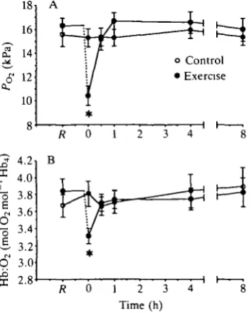
Exhaustive exercise in P. marinus was also associated with significant changes in the oxygen transport characteristics of the arterial blood The partial pressure of oxygen (Pod had fallen by 36% immediately after exercise (Fig. 5A). POl quickly
recovered to values that were not significantly different from the resting value, but the large initial decrease was also associated with a significant decline in the amount of oxygen bound to hemoglobin (Hb:O2; Fig. 5B). Hb:O2 had also
returned to normal within 30min.
Discussion
0.6
<£ 0.4
0.2
o Control • Exercise
R 0 81 B
2 2
1 2 3 Time (h) 0 1 2 3 4
iv
l
a" 4
3
2
1
0
- 1 C
* *
\ *
H
Fig. 3. (A) Arterial CO2 tension (Pco2)> (B) plasma lactate concentration ([lactate])
and (C) plasma metabolic proton load (A[H+]m) at rest (R) and 0, 0.5, 1, 4 and 8h
following exhaustive exercise in Petromyzon marinus. The dashed line represents the 10-15 min period of exhaustive exercise. Values are means ± standard error (control,
N=6; exercise, N=8). Asterisks denote significant differences from resting values in A
and B and a significant difference from the control values in C.
immediately after exhaustive exercise (Wood and Perry, 1985). The 78% increase observed in lampreys in the present study is comparable with the lower portion of this range. The magnitude of the increase in blood [lactate] following exercise also covers a broad range in fish and is largely dependent on species. Maximal values range from less than 2mmoll~1 in benthic species like the flounder Platichthys
stellatus and sole Hippoglossoides elassodon to more than 25mmoll~1 in very active species like the salmon Salmo salar (Turner et al. 1983; Milligan and Wood, 1987; Black, 1957; Tufts et al. 1991). The peak plasma [lactate] after exercise in
P. marinus also falls within the documented range for fish, but [lactate] only
reaches 5.6±0.9mmoll~1 (Fig. 3B). It is noteworthy that the maximal [lactate] in
[image:9.451.137.316.78.403.2]- 6 o
£
o Control • Exercise
R 0 1 2 3 4
0 1 2 3 4
K 0 1 2 3 4 Time (h) Fig. 4. Total carbon dioxide concentration CQO2 >
n
(A) whole blood (w.bl), (B) true plasma (t.pl) and (C) erythrocytes (eryth) at rest (/?) and 0, 0.5, 1, 4 and 8h following exhaustive exercise in Petromyzon marinus. The dashed line represents the 10-15 min period of exhaustive exercise. Values are means ± standard error (control, N=6; exercise, N=8). Asterisks denote a significant difference from the resting value.
species of fish, peak lactate concentrations are not observed until about 2h (Heisler, 1986; Wood and Perry, 1985). The relationship between [H+]m and
[lactate] in the plasma of P. marinus is intermediate between the two general patterns described by Wood and Perry (1985) for benthic versus active fishes. In the present study, the calculated plasma A[lactate] marginally exceeds A[H+]m
during the entire recovery period, but the dynamics of these two variables are quite similar and the maximal proton deficit calculated for the plasma only reaches l.Smequivl"1 (Fig. 3B,C). Finally, although there is a substantial drop in pHe after exercise in P. marinus, there are relatively few metabolic protons added to the plasma (Fig. 3C). Since the increase in Pco2 >
s a
18 16
cu 14
10
4.2 4.0 3.8 3.6 3.4 3.2 3.0 2.8
o Control • Exercise
R 0 1
0 1 2 3 4 Time (h)
Fig. 5. (A) Arterial oxygen tension (Po2) and (B) hemoglobin:oxygen carriage
(Hb:O2) at rest (R) and 0, 0.5,1, 4 and 8h following exhaustive exercise in Petromyzon
marinus. The dashed line represents the 10-15 min period of exhaustive exercise.
Values are means ± standard error (control, iV=6; exercise, N=8). Asterisks denote a significant difference from resting values.
relatively low nonbicarbonate buffer value of the true plasma in P. marinus (Tufts and Boutilier, 1989, 1990).
[image:11.451.140.317.75.295.2](Tufts etal. 1987a,b; Tufts and Randall, 1989). In both L.fluviatilis and P.
mari-nus, pHi is regulated in vitro, however, by a sodium/proton exchange mechanism
that is not dependent upon catecholamine levels, but which appears to be stimulated by changes in pHe (Nikinmaa, 1986; Nikinmaa et al. 1986; B. L. Tufts, unpublished observations). The present results suggest that this mechanism is probably also operating in P. marinus after exercise in vivo (Figs IB, 2). Moreover, this study demonstrates that the mechanism of pHi regulation in the sea lamprey, although independent of catecholamines, is clearly as effective, if not more effective, at regulating pHi in vivo than the adrenergic systems that have been described in many species offish. Indeed, the slope of the in vivo relationship between pHe and pHi is only 0.12 in the present experiments, whereas it is 0.20 in a similar series of experiments carried out on Oncorhynchus mykiss, a species with a relatively large adrenergic response (Fig. 2; Milligan and Wood, 1987; Salama and Nikinmaa, 1989).
In most vertebrates, CO2 transport is largely dependent on erythrocyte
chloride/bicarbonate exchange (Roughton, 1964; Randall and Daxboeck, 1984; Cameron, 1978; Perry, 1986). In agnathans, however, it has been suggested this anion exchange protein may be absent or only present in very limited quantities within the erythrocyte membrane (Ellory et al. 1987; Nikinmaa and Railo, 1987). The in vitro CO2 transport properties and ion distributions in P. marinus blood
also provide evidence to support this view (Tufts and Boutilier, 1989, 1990). In fish, it has been suggested that temporary inhibition of bicarbonate flux through the erythrocytes may contribute to the PQO2 rise in the arterial blood immediately
after exercise (Wood and Perry, 1985). In a similar manner, the observed increase in arterial Pco2 after exercise in the present study could, therefore, reflect a
chloride/bicarbonate exchange limitation in P. marinus erythrocytes (Fig. 3A). In both fish and lampreys, however, titration of plasma bicarbonate by metabolic protons from muscle and by protons extruded from the erythrocyte via sodium/ proton exchange would also be expected to contribute to the temporary rise in arterial PQO2- Since the relative importance of these potential sources of change in
Pcoi cannot be differentiated in these experiments, it is impossible to determine
whether the PQO2 increase does, in fact, reflect an in vivo anion exchange
limitation in P. marinus blood. Further study into the nature of the CO2 reactions
in agnathan blood in vivo is required before any conclusions can be made on this point.
The observed changes in CQO2
m
arterial blood after exercise do not provide any indication that CO2 transport is adversely effected in vivo by possible
chloride/bi-carbonate exchange limitations (Fig. 4). Indeed, the only significant change observed in the CCo2w.t>i was a decrease between 0.5 and 1 h of the recovery period
(Fig. 4A). This was apparently due to an even larger reduction in the Ccchi.pi in
P. marinus blood after exercise (Fig. 4B). Again, as in fish, the reduction in
Cc02t.pi can be explained as the titration of plasma HCO3~ by protons arising from
Significant quantities of carbonic anhydrase (CA) have been found within the erythrocytes of agnathans (Nikinmaa et al. 1986). Thus, the increase in Cco^ryth in the arterial blood of P. marinus after exercise probably does not reflect any inhibition of CO2 transport at the level of the erythrocyte. During passage of blood
through the gills, any bicarbonate within the erythrocyte would already have had access to CA and could have been dehydrated to CO2 for excretion. Rather, the
increase in Cco^ryth would be expected if erythrocyte sodium/proton exchange extruded protons from the erythrocyte after exercise at the same time that arterial
Pco2 was transiently elevated (Fig. 3A). Together, these two factors would
increase the apparent bicarbonate concentration of the erythrocyte and result in the observed increase in CCo2
eryth-In contrast to CO2 transport, it is clear that O2 transport is adversely affected by
exhaustive exercise in P. marinus. In exhausted lampreys, there is a substantial drop in the arterial POz immediately after the exercise period (Fig. 5A). Transient
decreases in arterial POl have also been observed immediately after exercise in fish
and have been attributed to a reduction in ventilation frequency in particularly exhausted animals (Primmett et al. 1986; Milligan and Wood, 1987). The present reduction in PO2 probably also reflects a brief reduction in ventilatory frequency in
exhausted lampreys. The drop in PO2 was sufficient to have a significant effect on
arterial Hb:O2 carriage (Fig. 5B). Concurrent with the 36% fall in P^, there was
a 14 % reduction in the amount of O2 bound to hemoglobin. The hemoglobin of P.
marinus has a significant Bohr effect (Manwell, 1963). Thus, the maintenance of
pHi in the sea lamprey probably enhances oxygen uptake at the gills after exercise and the decline in Hb:O2 carriage would probably have been even greater in these
animals had the pHi not been so well regulated.
In summary, exhaustive exercise in the agnathan P. marinus results in a significant extracellular acidosis, which is very similar to that observed in other lower vertebrates. In addition, despite reports that chloride/bicarbonate exchange limitations exist in agnathan erythrocytes in vitro, the present results provide no evidence that acid-base regulation or carbon dioxide transport is adversely affected after exercise in vivo. Finally, the extracellular acidosis is not transferred to the erythrocyte and the regulation of pFIi may have an important role in the transport of oxygen immediately after exercise in the sea lamprey.
This study was supported by an NSERC Operating Grant to B.L.T. The author would also like to thank the Lamprey Control Center (Department of Fisheries and Oceans) in Sault Ste Marie, Ontario, for their assistance in obtaining the lampreys. Finally, technical assistance by B. Cameron was greatly appreciated.
References
BLACK, E. C. (1957). Alterations in the blood level of lactic acid in certain salmonid fishes following muscular activity. III. Sockeye salmon, Oncorhynchus nerka. J. Fish. Res. Bd Can. 14, 807-814.
use in fish respiratory physiology. In Fish Physiology, vol. XA (ed. W. S. Hoar and D. J. Randall), pp. 401-430. New York: Academic Press.
CAMERON, J. N. (1978). Chloride shift in fish blood. J. exp. Biol. 206, 289-295.
COSSINS, A. (1989). Intracellular pH regulation by fish red cells. Nature 340, 20-21.
DRABKIN, D. L. AND AUSTIN, J. H. (1935). Spectrophotometric studies. II. Preparations from washed blood cells; nitric oxide hemoglobin and sulfhemoglobin. J. biol. Chem. 112, 51-65.
ELLORY, J. C , WOLOWYK, M. W. AND YOUNG, J. D. (1987). Hagfish {Eptatretus stouti) erythrocytes show minimal chloride transport activity. /. exp. Biol. 129, 377-383.
HEISLER, N. (1986). Acid-base regulation in fishes. In Acid-Base Regulation in Animals (ed. N. Heisler), pp. 309-356. Amsterdam: Elsevier.
HOFFMAN, E. K. AND SIMONSEN, O. (1989). Membrane mechanisms in volume and pH regulation in vertebrate cells. Physiol. Rev. 69, 315-382.
LOOMIS, M. E. (1961). An enzymatic fluorometric method for determination of lactic acid in serum. J. Lab. din. Med. 57, 966-972.
MANWELL, C. (1963). The blood proteins of cyclostomes. A study of phylogenetic and ontogenetic biochemistry. In The Biology of Myxine (ed. A. Brodal and R. Fange), pp. 372-455. Oslo: Universitetsforlaget.
MATSOFF, L. AND NIKINMAA, M. (1988). Effects of external acidification on the blood acid-base status and ion concentrations of lamprey. /. exp. Biol. 136, 351-361.
MCDONALD, D. G., BOUTILIER, R. G. AND TOEWS, D. P. (1980). The effects of enforced activity on ventilation, circulation and blood acid-base status in the semi-terrestrial anuran, Bufo
marinus. J. exp. Biol. 84, 273-287.
MILUGAN, C. L. AND WOOD, C. M. (1987). Regulation of blood oxygen transport and red cell pHi after exhaustive activity in rainbow trout (Salmo gairdneri) and starry flounder
{Platichthysstellatus). J. exp. Biol. 133, 263-282.
NIKINMAA, M. (1986). Red cell pH of lamprey {Lampetra fluviatilis) is actively regulated.
J. comp. Physiol. B 156, 747-750.
NIKINMAA, M., CECH, J. J., JR AND MCENROE, M. (1984). Blood oxygen transport in stressed striped bass {Morone saxitilis): role of beta-adrenergic responses. J. comp. Physiol. B 154, 365-369.
NIKINMAA, M., KUNNAMO-OJALA, T. AND RAILO, E. (1986). Mechanisms of pH regulation in lamprey {Lampetra fluviatilis) red blood cells. J. exp. Biol. 122, 355-367.
NIKINMAA, M. AND RAILO, E. (1987). Anion movements across lamprey {Lampetra fluviatilis) red cell membrane. Biochim. biophys. Ada 899, 134-136.
NIKINMAA, M. AND TUFTS, B. L. (1989). Regulation of acid and ion transfer across the membrane of nucleated erythrocytes. Can. J. Zool. 67, 3039-3045.
PERRY, S. F. (1986). Carbon dioxide excretion in fishes. Can. J. Zool. 64, 565-572.
PRIMMETT, D. R. N., RANDALL, D. J., MAZEAUD, M. AND BOUTILIER, R. G. (1986). The role of catecholamines in erythrocyte pH regulation and oxygen transport in rainbow trout {Salmo
gairdneri) during exercise. J. exp. Biol. 122, 139-148.
RANDALL, D. J. AND DAXBOECK, C. (1984). Oxygen and carbon dioxide transfer across fish gills. In Fish Physiology, vol. XA (ed. W. S. Hoar and D. J. Randall), pp. 263-314. New York: Academic Press.
ROUGHTON, F. J. W. (1964). Transport of oxygen and carbon dioxide. In Handbook of
Physiology, vol. 1 (ed. W. O. Fenn and H. Rahn), pp. 767-825. Washington, DC: American
Physiological Society.
SALAMA, A. AND NIKINMAA, M. (1989). Species differences in the adrenergic responses of fish red cells: studies on whitefish, pikeperch, trout and carp. Fish Physiol. Biochem. 6, 167-173.
TUCKER, V. A. (1967). Method for oxygen content and dissociation curves on microliter blood samples. /. appl. Physiol. 23, 410-414.
TUFTS, B. L. AND BOUTILIER, R. G. (1989). The absence of rapid chloride/bicarbonate exchange in lamprey erythrocytes: implications for CO2 transport and ion distributions between plasma and erythrocytes in the blood of Petromyzon marinus. J. exp. Biol. 144, 565-576.
TUFTS, B. L. AND BOUTILIER, R. G. (1990). CO2 transport in agnathan blood: evidence of CT/HC03~ exchange limitations. Respir. Physiol. 80, 335-348.
circulating catecholamines and pH and water content of erythrocytes in the toad. J. exp. Biol. 128,411-418.
TUFTS, B. L., NIKINMAA, M., STEFFENSEN, J. F. AND RANDALL, D. J. (1987i>). Ion exchange mechanisms on the erythrocyte membrane of the aquatic salamander, Amphiuma
tridactylum. J. exp. Biol. 133, 329-338.
TUFTS, B. L. AND RANDALL, D. J. (1989). The functional significance of adrenergic pH regulation in fish erythrocytes. Can. J. Zool. 67, 235-238.
TUFTS, B. L., TANG, Y., TUFTS, K. AND BOUTTUER, R. G. (1991). Exhaustive exercise in 'wild' Atlantic salmon (Salmo salar): Acid-base regulation and blood-gas transport. Can. J. Fish.
aquat. Sci. (in press).
TURNER, J. D., WOOD, C. M. AND HOBE, H. (1983). Physiological consequences of severe exercise in the benthic flathead sole {Hippoglossoides elassodon): a comparison with active pelagic trout (Salmo gairdneri). J. exp. Biol. 104, -288.
WOOD, C. M. AND PERRY, S. F. (1985). Respiratory, circulatory, and metabolic adjustments to exercise in fish. In Circulation, Respiration and Metabolism (ed. R. Gilles), pp. 1-22. Berlin: Springer-Verlag.
![Fig. 3. (A) Arterial CO2 tension (Pco2)> (B) plasma lactate concentration ([lactate])and (C) plasma metabolic proton load (A[H+]m) at rest (R) and 0, 0.5, 1, 4 and 8hfollowing exhaustive exercise in Petromyzon marinus](https://thumb-us.123doks.com/thumbv2/123dok_us/1168864.638871/9.451.137.316.78.403/arterial-concentration-metabolic-hfollowing-exhaustive-exercise-petromyzon-marinus.webp)