SLOW ACTIVE POTENTIALS IN WALKING-LEG MOTOR
NEURONES TRIGGERED BY NON-SPIKING
PROPRIOCEPTIVE AFFERENTS IN THE CRAYFISH
BY KEITH T. SILLAR* AND ROBERT C. ELSON
School of Veterinary Science, Department of Physiology, University of Bristol, Park Row, Bristol BS1 5LS, UK
Accepted 29 July 1986
SUMMARY
Intracellular recordings have been made from walking-leg motor neurones of the crayfish, Pacifastacus leniusculus, in isolated preparations of the thoracic ganglia. Some motor neurones display slow depolarizations that can drive bursts of spikes and resemble 'plateau' potentials described in other invertebrate and vertebrate neurones. Evidence is presented which suggests that the potentials are regenerative and endogenous to the motor neurones, and are not the result of feedback from a neural network.
These potentials can be induced by synaptic inputs from the non-spiking afferent neurones of the thoracic-coxal muscle receptor organ, a basal limb proprioceptor. Reflex input from this receptor is augmented during the active depolarization of the motor neurone. The results are discussed in terms of the control of rhythmic motor output and the central modulation of reflexes in the crayfish's thoracic nervous system.
INTRODUCTION
The basic motor patterns underlying cyclical behavioural activities, such as respiration, mastication and locomotion, are thought to result from the synaptic in-teractions of networks of neurones in the central nervous system (CNS) (Delcomyn, 1980). However, the cellular properties of individual neurones that could participate in producing patterned activity are also important. One such mechanism involves voltage-dependent inward currents that give rise to prolonged, regenerative depolar-izations which drive bursts of spikes. Such 'plateau' or 'driver' potentials, found in invertebrate endogenous burster neurones as well as vertebrate heart muscle, have recently been discovered in neurones of the crustacean cardiac and stomatogastric ganglia, where they underlie burst production (Russell & Hartline, 1978, 1982; Benson, 1980; Tazaki & Cooke, 1983; reviewed in Benson & Cooke, 1984). The neuronal membrane potential can adopt two quasi-stable values: a resting potential
•Present address: Department of Zoology, University of Bristol, Woodland Road, Bristol, BS8 1UG, UK.
and a second, more depolarized plateau. The transitions between the two states are rapid, regenerative, threshold processes (Russell & Hartline, 1982). Such neurones may also have mechanisms for pacemaking and endogenous repolarization, leading to repetitive bursting (Miller & Selverston, 1982; Tazaki & Cooke, 1983; Hartline & Russell, 1984). Similar membrane properties are also present in some central neurones of vertebrates (Legendre, Cooke & Vincent, 1982; Llinas & Sugimori, 1980) and may occur widely throughout the animal kingdom. In this paper we report slow active potentials in the central processes of motor neurones (MNs) which innervate the walking-leg muscles of the crayfish. We also show that these potentials can be induced by synaptic inputs from the non-spiking primary afferent neurones of a proprioceptor at the base of the limb, the thoracic-coxal muscle receptor organ (TCMRO). The results have implications for the control of rhythmic motor output and the central modulation of reflexes in the crayfish's thoracic nervous system.
MATERIALS AND METHODS
Experiments were performed on the isolated thoracic ganglia of the crayfish,
Pacifastacus leniusculus, using a preparation described previously (Skorupski,
1985; Sillar & Skorupski, 1986). The TCMRO (see Bush & Cannone, 1985, for a recent review) of the fourth walking leg remained attached to the segmental ganglion by its sensory nerve, and was stretched by an electromechanical puller. Extracellular recordings from the nerve roots innervating the two muscle groups of the basal joint were obtained with fine, polyethylene-tipped suction electrodes. Intracellular re-cordings from motor and sensory neurones were made in the dorsal neuropile of the desheathed fourth thoracic ganglion, using Lucifer Yellow-filled glass microelec-trodes (Stewart, 1980) with resistances of 30-80MQ. MNs were identified as those of the limb promotor or remotor muscles by 1:1 correlation of the decremented spikes recorded intracellularly with those in the appropriate motor root. The S-fibre, one of the TCMRO's two afferents, was identified by its characteristic receptor potential (Sillar & Skorupski, 1986). In about 80% of over 200 isolated preparations a typical rhythmic motor pattern was observed, which may underlie forward walking (Sillar & Skorupski, 1986). In these, the occurrence of any intrinsic membrane potentials cannot easily be distinguished from the effects of rhythmic synaptic inputs. The remaining 20% did not produce the motor pattern but maintained a level of spontaneous motor discharge that could be modulated by reflexes from the TCMRO. Slow active potentials were apparent in 14 such experiments. Here we describe results typical of regenerative behaviour in MNs.
RESULTS
Ai Aii B
Current
f * \
IT
D E
P
[image:3.451.33.424.52.261.2]' °M N
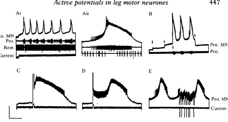
Fig. 1. Slow active potentials in a promotor MN (Pro. MN, top trace throughout). (Ai) Endogenous membrane potential oscillations and bursting following injection of a small (<2nA) steady depolarizing current through the recording electrode. The promotor nerve recording (Pro.) contains the spike of the intracellular MN and those of at least two other promotors. The remotor nerve (Rem.) contains the spike of a single active MN which fires non-rhythmically at an average frequency of about 5 Hz. (Aii) The first burst of Ai, on a faster time-base. Note the excitation of a promotor unit with a larger spike and transient inhibition of the remotor nerve. Traces as in Ai, without current monitor. (B) Threshold for regenerative behaviour, demonstrated by injecting depolar-izing current in three small (<lnA) increments from rest (upward arrows; bridge balance only approximate). At the third step an active potential is elicited, followed by repetitive bursting, until current is switched off at the downward arrow. (C) Premature triggering of a burst by a brief depolarizing pulse of current. (D) A depolarizing pulse, delivered earlier in the interburst than in C, decreases the membrane potential to a quasi-stable, depolarized level, followed later by a regenerative burst. (E) Retarding the onset of a burst by repeated, brief hyperpolarizing pulses. All records are from the same preparation. Calibrations: voltage Ai, 20mV; Aii, 12mV; B-E, 8mV; current 20nA; time Ai, 40s; Aii, C,D, 2s; B, 25s; E, 4s.
about 30 s but gradually lengthened (Fig. lAi). This bursting, which was maintained for the duration of the current injection, differed from that recorded in the rhythmic motor pattern, because the remotor nerve root was tonically active throughout (apart from transient inhibition in the first burst, shown on a faster time-base in Fig. lAii). This indicated that the activity was probably not synaptically driven by the rhythm-generating network. Further evidence for an endogenous, cellular mechanism is the finding that, during stepwise increments of positive current from rest, the rapid depolarization and burst was not initiated until a threshold was reached (Fig. IB).
The active potentials showed bistable behaviour, since during repetitive bursting a full-blown response could be elicited immediately by a brief pulse of depolarizing current (Fig. 1C), causing premature burst initiation. Hyperpolarizing pulses, in contrast, could retard burst onset (Fig. IE). The effectiveness of current pulses depended on their timing: for example, a brief positive pulse given soon after a burst triggered a quasi-stable depolarized membrane potential that only later developed into the full burst (Fig. ID). This endogenous bursting activity could affect the spike frequencies of other MNs. At least two other promotor MNs were excited to spike during the depolarization of the bursting MN (e.g. larger action potential in promotor nerve, Fig. lAii), whereas the tonic spiking of a remotor MN was transiently inhibited (Fig. lAii) (similar effects are seen in Fig. 2Ai). Such actions are presumably mediated via the central excitatory coupling between synergistic MNs and reciprocal inhibition between antagonists (Skorupski, 1985; Skorupski, Sillar & Bush, 1984).
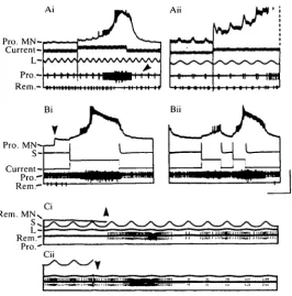
Afferent inputs interacted with the active potentials (Fig. 2A). Stretching the TCMRO sinusoidally evoked excitation of promotor MNs (a resistance reflex) which was subthreshold in the plateauing MN in the absence of bursting. When regenerative behaviour was induced by injecting long pulses of depolarizing current, two modulatory changes occurred in the reflex response. First, the depolarizing postsynaptic potential (EPSP) evoked by TCMRO stretch (Sillar & Skorupski, 1986) steadily increased in amplitude during the active depolarization (Fig. 2Aii). Second, the number of spikes elicited by the reflex in a different promotor MN increased during the depolarizing phase of the burst, and decreased during the repolarizing phase (e.g. Fig. 2Ai, arrowhead). The increase in PSP amplitude with postsynaptic depolarization is unusual, because the synaptic input from the TCMRO is probably a chemically mediated, conductance increase response (Skorupski, 1985; K. T. Sillar & P. Skorupski, unpublished observation) as in crabs (Blight & Llinas, 1980).
Pro. MN-
[image:5.451.85.352.59.329.2]Current-iri ;; "II.."I III." Ml,,,...'
pulse (e.g. Fig. 2Bii). The reaction of the MN again depended on the time at which the stimulus was given: depolarizing the afferent soon after a previous burst evoked a small depolarization and few spikes in the MN, whereas a similar stimulus later elicited a long-lasting regenerative response (Fig. 2Bii). Similar responses occurred in a remotor MN in a different preparation (Fig. 2C). Here, remotors were excited by stretching the TCMRO, in an assistance reflex, and the MN received sub-threshold depolarizing input at each stretch. Injecting a steady depolarizing current into the MN not only brought the reflex response above threshold (monitored in the motor nerve), but also evoked a regenerative, apparently endogenous burst whose termination was accompanied by a transient reduction in reflex MN excitability (Fig. 2Ci). The S-fibre tonically inhibits remotor (Skorupski & Sillar, 1986). Hyperpolarizing this afferent was sufficient to release another burst in the remotor MN (Fig. 2Cii).
DISCUSSION
The potentials described here have characteristics in common with active poten-tials in lobster stomatogastric (Miller & Selverston, 1982; Moulins & Nagy, 1983; Hartline & Russell, 1984) and crab cardiac (Tazaki & Cooke, 1983) MNs: they show threshold and refractory-like behaviour, can be triggered by brief depolarization and display relatively rapid depolarizing and hyperpolarizing trajectories. Whilst these properties are consistent with intrinsic mechanisms, they do not exclude the possibility that they arise from feedback networks of neurones. However, a network explanation is unlikely in view of the absence of phasic patterned output in other motor roots. As the MNs interact with an unknown number of other neurones in the thoracic ganglia, a rigorous demonstration of endogenous mechanisms would require the blocking of all synaptic traffic.
The extent of regenerative behaviour in walking-leg MNs is not yet clear. Although we have observed slow active potentials on only 14 occasions, this number is probably artificially low. First, most preparations are rhythmically active and such potentials cannot easily be studied independently of patterned synaptic inputs. Second, these potentials may require a tonic depolarization for their initiation (see Fig. 1 A), which may not be present in some quiescent preparations. Third, they may occur in a restricted portion of a MN pool, further reducing the chances of recording them with probing microelectrodes. Among the MNs of a motor nerve, active potentials seem to occur in the more tonic, lower-threshold cells, but not obviously in the more phasic MNs. However, synaptic interactions between MNs will ensure that the effects of such potentials are not confined to the cells in which they arise.
arise, and to burst formation and coordination among other, synergistic or an-tagonistic MNs, via motoneuronal interactions. In this way intrinsic motoneuronal properties would amplify the output of the central rhythm generator. The central rhythm also phasically modulates proprioceptive reflexes elicited by the TCMRO (Skorupski & Sillar, 1986). One feature of this is the augmentation of excitatory reflex input from the TCMRO during the depolarized phase of activity in a MN. The enhancement of TCMRO input seen during a plateau-like potential could provide a novel postsynaptic mechanism for central reflex gain control. MN-MN interactions would serve to spread these effects among synergists and antagonists.
The afferent neurones of the TCMRO may themselves trigger the onset of an endogenous burst (as shown for the S-fibre, Fig. 2). Similarly, in the segmentally homologous swimmeret system, two non-spiking stretch receptor neurones (Heitler, 1982) have been implicated in the control of plateau 'flip-flop' behaviour in MNs (Heitler, 1984). In the thoracic ganglia, the TCMRO influences the timing of the motor rhythm: periodic stretch entrains the motor bursts while brief current pulses into single afferents may trigger the switch between antagonistic bursts, and thus between the two phases of a cycle (Elson, Sillar & Skorupski, 1986; Sillar, Skorupski, Elson & Bush, 1986). These timing effects may be due in part to the ability of changes in afferent membrane potential to release long-lasting motor bursts
via active potentials. Endogenous membrane potential oscillations between two
voltage levels have been found recently in spinal neurones active in lamprey swimming (Sigvardt, Grillner, Wallen & van Dongen, 1985). These, and the present results, suggest that intrinsic mechanisms may be important for rhythmic activity not only in molluscan burster neurones and crustacean cardiac or stomatogastric ganglia, but also in the generation of locomotor patterns.
Supported by SERC Research Grant GR/C/24244 to B. M. H. Bush. We thank Sue Maskell for secretarial help and Jane Robbins for technical assistance. RCE is an SERC Postdoctoral Fellow.
REFERENCES
BENSON, J. A. (1980). Burst reset and frequency control of the neuronal oscillators in the cardiac ganglion of the crab, Portunus sanguinolentus. J'. exp. Biol. 87, 285-313.
BENSON, J. A. & COOKE, I. M. (1984). Driver potentials and the organization of rhythmic bursting in crustacean ganglia. Trends Neurosci. 7, 85-91.
BLIGHT, A. R. & LLINAS, R. (1980). The non-impulsive stretch-receptor complex of the crab: a study of depolarization-release coupling at a tonic sensorimotor synapse. Phil. Trans. R. Soc.
Ser. B 290, 219-276.
BUSH, B. M. H. & CANNONE, A. J. (1985). How do crabs control their muscle receptors? In
Feedback and Motor Control in Invertebrates and Vertebrates (ed. W. J. P. Barnes & M.
Gladden), pp. 145—166. London: Croom Helm.
DELCOMYN, F. (1980). Neural basis of rhythmic behaviour in animals. Science 210, 492-498. ELSON, R. C , SILLAR, K. T . & SKORUPSKI, P. (1986). Stretch reflexes from the thoracic-coxal
muscle receptor organ entrain a central locomotor rhythm of the crayfish. J . Physiol., Land. 373, 13P.
HEITLER, W. (1982). Non-spiking stretch-receptors in the crayfish swimmeret system.X exp. Biol. 96, 355-366.
HEITLER, W. (1984). Sensorimotor integration in the crayfish swimmeret system. Soc. Neurosci.
Abstr. 10, 628.
LEGENDRE, P., COOKE, I. M. & VINCENT, J.-D. (1982). Regenerative responses of long duration recorded intracellularly from dispersed cell cultures of fetal mouse hypothalamus.
Jf. Neurophysiol. 48, 1121 -1141.
LLINAS, R. & SUGIMORI, M. (1980). Electrophysiological properties of in vitro Purkinje cell dendrites in mammalian cerebellar slices. J. Physiol., Land. 305, 197—213.
MILLER, J. P. & SELVERSTON, A. I. (1982). Mechanisms underlying pattern generation in the lobster stomatogastric ganglion as determined by selective inactivation of identified neurons. II. Oscillatory properties of pyloric neurons. J . Neurophysiol. 48, 1378-1391.
MOULINS, M. & NAGY, F . (1983). Control of integration by extrinsic inputs in the crustacean pyloric circuit. J . Physiol., Paris 78, 755-764.
RUSSELL, D. F . & HARTLINE, D . K. (1978). Bursting neural networks: a re-examination. Science 200, 453-456.
RUSSELL, D. F . & HARTLINE, D . K. (1982). Slow active potentials and bursting motor patterns in pyloric network of the lobster, Panulirus interruptus.j. Neurophysiol. 48, 914-937.
SIGVARDT, K. A., GRILLNER, S., WALLEN, P. & VAN DONGEN, P. A. M. (1985). Activation of NMDA receptors elicits fictive locomotion and bistable membrane properties in the lamprey spinal cord. Brain Res. 336, 390-395.
SILLAR, K. T . & SKORUPSKI, P. (1986). Central input to primary afferent neurones in crayfish
Pacifastacus leniusculus, is correlated with rhythmic motor output of thoracic ganglia. J. Neurophysiol. 55, 678-689.
SILLAR, K. T . , SKORUPSKI, P., ELSON, R. C. & BUSH, B. M. H. (1986). Two identified afferent neurones entrain a central locomotor rhythm generator. Nature, Land, (in press).
SKORUPSKI, P. (1985). Central nervous and proprioceptive control of crayfish walking leg motoneurones: an intracellular microelectrode study of the isolated 4th thoracic ganglion. Ph.D. thesis, University of Bristol, U K .
SKORUPSKI, P. & SILLAR, K. T . (1986). Phase-dependent reversal of reflexes mediated by the thoraco-coxal muscle receptor organ in the crayfish, Pacifastacus leniusculus. J. Neurophysiol. 55, 690-695.
SKORUPSKI, P., SILLAR, K. T . & BUSH, B. M. H. (1984). Central modulation of proprioceptive input to the isolated 4th thoracic ganglion of the crayfish. Soc. Neurosci. Abstr. 10, 627. STEWART, W. W. (1978). Functional connections between cells as revealed by dye coupling with a
highly fluorescent naphthalimide tracer. Cell 14, 741-751.