warwick.ac.uk/lib-publications
A Thesis Submitted for the Degree of PhD at the University of Warwick
Permanent WRAP URL:
http://wrap.warwick.ac.uk/88011
Copyright and reuse:
This thesis is made available online and is protected by original copyright. Please scroll down to view the document itself.
Please refer to the repository record for this item for information to help you to cite it. Our policy information is available from the repository home page.
The Role Of QRFP & GPR103
In Human Prostate Cancer
Mohamed A.B Kawan
A
thesis submitted in partial fulfilment of the requirements for
the degree of Doctor of Philosophy (PhD)
October 2016
2
PUBLICATIONS: -
Hu J, Kyrou I, Tan BK, Dimitriadis GK, Ramanjaneya M, Tripathi G,
Patel V, James S, Kawan M, Chen J, Randeva HS, “Short-chain
Fatty Acid Acetate Stimulates Adipogenesis and Mitochondrial
Biogenesis via GPR43 in Brown Adipocytes”,
Manjunath Ramanjaneya, Bee K. Tan, Marcin Rucinski, Mohamed
Kawan, Jiamiao Hu, Jaspreet Kaur, Vanlata H. Patel, Ludwik K.
Malendowicz, Hanna Komarowska, Hendrik Lehnert, and Harpal S.
Randeva, “Nesfatin-1 inhibits proliferation and enhances apoptosis
of human adrenocortical H295R cells,” Journal of Endocrinology, vol. 226, no. 1, pp. 1–11, 2015.
POSTER PARTICIPATIONS: -
World Congress on Cancer and Prevention Methods, August
27-29, 2015, Dubai, UAE. Mohamed Kawan, Ramanjaneya M and
Harpal HS, “QRFP and its receptor, GPR103, have a role in
prostate tumorigenesis”.
The 8th Dubai International Conference for Medical Sciences,
2014. Mohamed Kawan, Manjunath Ramanjaneya, and Harpal S.
Randeva, “The role of QRFP and GPR103 in Human Prostate
Cancer”.
Warwick Medical School Postgraduate Research Student
Symposium 2013. Awarded Highly Commended Poster Symposium
3
Contents
DECLARATION ... 15
ACKNOWLEDGEMENTS ... 16
ABBREVIATIONS... 17
ABSTRACT ... 19
CHAPTER 1; GENERAL INTRODUCTION ... 21
1 BACKGROUND ... 22
1.1 THE NORMAL PROSTATE GLAND ... 22
1.1.1 ANDROGENS: ... 25
1.2 PRODTATE CANCER (PCa) ... 28
1.2.1EPIDEMIOLOGY ... 28
1.2.2 ETIOLOGY ... 30
1.2.3 AGE ... 30
1.2.4 ETHNICITY ... 30
1.2.5 GENETIC FACTORS... 31
1.2.6 ENDOGENOUS HORMONE PROFILE ... 32
1.2.7 DIET ... 32
1.2.8 PATHOLOGIC MARKERS ... 33
1.2.9 TUMOURIGENESIS; CELL PROLIFERATION, THE CELL CYCLE ... 35
1.2.10 APOPTOSIS AND ITS REGULATION ... 40
1.2.11 BCL-2 ... 44
4
1.3.1 EXTRACELLULAR SIGNAL-REGULATED PROTEIN
KINASES (ERK1/2) ... 47
1.3.2 p38 MAPKs ... 48
1.3.3 c-Jun N-TERMINAL KINASE OR STRESS-ACTIVATED PROTEIN KINASES (JNK/SAPK) ... 49
1.3.4 AKT/PKB KINASE ... 50
1.4 OBESITY RELATED TO PROSTATE CANCER ... 51
1.4.1 ROLE OF DYSFUNCTIONAL ADIPOSE TISSUE ... 55
1.5 ADIPOKINES AND PTOSTATE CANCER ... 56
1.5.1 LEPTIN: ... 57
1.5.2 ADEPONECTIN: ... 59
1.5.3 VISFATIN: ... 61
1.6 QRFP:... 63
1.6.1RFamide PEPTIDE RECEPTORS ... 64
1.6.2 QRFP PEPTIDE RECEPTOR (GPR103) ... 65
1.7 HYPOTH ESIS ... 69
1.8 AIMS ... 69
CHAPTER 2; MATERIAL AND METHODS ... 70
2.1 MATERIALS ... 71
2.1.1 EQUIPMENT ... 71
2.1.2 BIOCHEMICAL REAGENTS AND KITS: ... 71
2.1.3 CELL CULTURE MEDIA AND CELL LINES ... 74
5
2.2.1 COLLECTION OF HUMAN SAMPLES ... 75
2.2.2 CELL CULTURE ... 76
2.2.3 SUBCULTURE OF ADHERENT CELLS: ... 76
2.2.4 RESERVATION OF CELLS: ... 77
2.2.5 CELL COUNTS USING HEMOCYTOMETER: ... 77
2.3 TOTAL RNA EXTRACTION AND COMPLEMENTARY DEOXYRIBONUCLIC ACID (cDNA) SYNTHESIS ... 78
2.3.1 REAL TIME QUANTITATIVE PCR ... 79
2.3.2 SEQUENCE ANALYSIS ... 80
2.4 WESTERN BLOTTING ... 81
2.4.1 DETERMINATION OF PROTEIN CONCENTRATION... 81
2.4.2 MAKING: SDS-PAGE gel ... 81
2.4.3 WESTERN BLOTTING OF CELL LYSATES AND CONDITIONED MEDIA ... 82
2.4.4 MAPK- (ERK1/2, p38 and JNK1/2 MAPK) AND AKT ACTIVATION ANALYSIS ... 84
2.4.5 STRIPOING AND RE-PROBING MEMBRANES WITH TOTAL ANTIBODIES ... 85
2.5 PHOSPHO-KINASE ARRAY ... 85
2.5.1 SAMPLES PREOARARTION ... 86
2.5.2 ARRAY PROCEDURE ... 86
2.6 PROLIFERATION, MIGRATION AND INVASION ASSAY ... 87
2.6.1 MTS PROLIFERATION ASSAY ... 88
6
2.6.3 xCELLigence INVASION ASSAY ... 89
2.6.4 xCELLigence MIGRATION ASSAY ... 90
2.7 APOPTOSIS ASSAY ... 91
2.7.1 Vybrant® APOPTOSIS ASSAY ... 91
2.7.2 DNA RAGMENTATION ELISA ... 92
2.8 SERUM COLLECTION ... 95
2.9 STATISTICAL ANALYSIS ... 96
CHAPTER 3; THE EXPRESSION OF QRFP AND ITS RECEPTOR (GPR103) IN PROSTATE CANCER CELL LINES AND TISSUE ... 97
3 INTRODUCTION ... 98
3.1 HYPOTHESIS ... 102
3.2 AIMS ... 102
3.3 RESULTS ... 103
3.3.1 EXPRESSION OF QRFP AND GPR103 mRNA IN PROSTATE CANCER CELL LINES AND HUMAN PROSTATE TISSUE ... 103
3.3.2 QRFP AND GPR103 PROTEIN EXPRESSION IN HUMAN PROSTATE TISSUE AND CELL LINES ... 105
3.3.3 DETERMINING QUANTITATIVE EXPRESSION OF QRFP & GPR103 IN HUMAN PROTATE CANCER ... 106
3.3.4 CIRCULATING QRFP LEVELS IN PROSTATE CANCER ... 110
3.4 DISCUSSION ... 111
CHAPTER 4; FUNCTIONAL EFFECTS OF QRFP ON PROSTATE CANCER CELL LINES ... 116
7
4.1 AIMS ... 118
4.2 RESULTS ... 119
4.2.1 REGULATION OF QRFP ON PROSTATE CANCER CELL PROLIFERATION ... 119
4.2.2 CELL PROLIFERATION ASSAY USING xCELLigence SYSTEM ... 120
4.2.3 EFFECTS OF QRFP INCUBATION ON PC3 AND DU145 CELL MIGRATION AND INVASION ... 125
4.2.3.1 CELL MIGRATIO ASSAY... 125
4.2.3.2 CELL INVASION ASSAY ... 125
4.2.4 EFFECTS OF QRFP ON APOPTOSIS ASSAYS ... 129
4.3 DISCUSSION ... 137
CHAPTER 5; THE MECHANISM OF QRFP ON SIGNAL TRANSDUCTION PATHWAYS IN PROSTATE CANCER ... 143
5 INTRODUCTION ... 144
5.1 HYPOTHESIS ... 146
5.2 AIMS ... 147
5.3 RESULTS ... 148
5.3.1 THE EFFECT OF QRFP ON PHOSPHORLATION 43 SIGNALLING MOLUCULES IN PC3 CELLS ... 148
5.3.2 THE EFFECT OF QRFP ON MAPK EXPRESSION IN PC3 and DU145 CELLS ... 153
8
CHAPTER 6; THE ROLE OF QRFP ONAGR-2,
METALLOPROTEINASES (MMP), CASPASE-3 AND AMPK IN
PROSTATE CANCER ... 174
6 INTRODUCTION ... 175
6.1 HYPOTHESIS ... 180
6.2 AIMS ... 181
6.3 RESULTS ... 182
6.3.1 METHODS FOR ASSESSING THE EFFECTS OF QRFP ON AGR2 EXPRESSION ... 182
6.3.2 THE EFFECT OF QRFP ON MMP-2 PROTEIN EXPRESSION IN PC3 and DU145 CELLS ... 184
6.3.3 THE EFFECT OF QRFP ON TOTAL CASPASE-3 ACTIVITY IN PC3 and DU145 CELLS ... 187
6.3.4 THE EFFECT OF QRFP ON AMPK ACTIVITY IN PC3 and DU145 CELLS ... 190
6.3.5 GPR103 GENES SILENCING INHIBITTS p-AKT PATHWAY IN PC3 cells ... 193
6.3.6 THE EFFECT OF QRFP WITH MAPK, PI3K/AKT AND AMPK IINHIBITORS IN PC3 AND DU145 CELLS. ... 196
6.4 DISCUSSION ... 198
CHAPTER 7; THE REGULATION OF QRFP & GPR103 EXPRESSION BY ADIPOKINES IN PROSTATE CANCER ... 207
7 INTRODUCTION ... 208
7.1 HYPOTHESIS ... 211
9
7.3 RESULTS ... 212
7.3.1 REGULATION OF QRFP & GPR103 EXPRESSION BY ADIPOKINES AND HORMONES IN PROSTATE CANCER ... 212
7.3.2 THE EFFECT OF LEPTIN ON REGULATION OF QRFP & GPR103 mRNA EXPRESSION IN PROSTATE CANCER ... 214
7.3.3 LEPTIN REGULATES QRFP & GPR103 GENE EXPRESSION IN PROSTATE CANCER CELLS VIA MAPK AND PI3 KINASE PATHWAY ... 218
7.4 DISCUSSION ... 220
CHAPTER 8; GENERAL DISCUSION ... 223
GENERAL DISCUSSION ... 224
10
FIGURES
Figure Title
Page
1.1 Diagram view of the cell types in the interior a human prostatic duct. 25
1.2 Hormonal regulation of the male reproductive system 27
1.3 The extrinsic and intrinsic pathways of apoptosis. 44
1.4 Schematic representation of the MAPK cascade activation and
potential cross talk signals.
47
3.1 RT-PCR analysis for QRFP and GPR103 gene expression in both
prostate cancer cell lines PC3 and LNCaP
105
3.2 Western blot analysis revealed protein expression for QRFP and
GPR103 in the three prostate cancer cell lines (PC3, DU145 and
LNCaP).
106
3.3 Bar chart representing quantification QRFP mRNA expression in
benign prostate hyperplasia and tumour resection.
108
3.4 Bar chart representing quantification GPR103 mRNA expression in
benign prostate hyperplasia and tumour resection.
108
3.5 Quantification of the western blot showing protein expression levels
of QRFP and GPR103 in human benign and malignant tissue
109
3.6 Bar chart representing QRFP protein quantification expression in
benign and malignant prostate tissue.
110
3.7 Bar chart representing GPR103 protein quantification expression in
benign and malignant prostate tissue.
110
3.8 Graphical representation of QRFP serum levels in controls
and patients.
111
4.1 MTS assay demonstrating the effects of QRFP on PC3 cell
proliferation for 24 hr.
122
4.2 MTS assay demonstrating the effects of QRFP on PC3 cell
proliferation for 48 hr.
11
4.3 MTS assay demonstrating the effects of QRFP on LNCaP cell
proliferation for 24 hr.
123
4.4 MTS assay demonstrating the effects of QRFP on LNCaP cell
proliferation for 48 hr.
123
4.5 The effect of QRFP on PC3 cell proliferation using xCELLigence
system for 24 hr.
124
4.6 The effect of QRFP on PC3 cell proliferation using xCELLigence
system for 48 hr.
124
4.7 The effect of QRFP on DU145 cell proliferation using xCELLigence
system for 24 hr.
125
4.8 The effect of QRFP on DU145 cell proliferation using xCELLigence
system for 48 hr.
125
4.9 The effect of QRFP on PC3 cell migration using xCELLigence
system.
128
4.10 The effect of QRFP on DU145 cell migration using xCELLigence
system.
128
4.11 The effect of QRFP on PC3 cell invasion using xCELLigence
system.
129
4.12 The effect of QRFP on DU145 cell invasion using xCELLigence
system.
129
4.13 Yo pro-1 and propidium iodide used to measurement of PC3 cell
apoptosis for 24 hr.
132
4.14 Yo pro-1 and propidium iodide used to measurement of PC3 cell
apoptosis for 48 hr.
132
4.15 Yo pro-1 and propidium iodide used to measurement of LNCaP cell
apoptosis for 24 hr.
133
4.16 Yo pro-1 and propidium iodide used to measurement of LNCaP cell
apoptosis for 48 hr.
133
4.17 Assessment of apoptosis by TUNEL assay on PC3 cells for 24 hr. 134
4.18 Assessment of apoptosis by TUNEL assay on PC3 cells for 48 hr. 134
4.19 Assessment of apoptosis by TUNEL assay on DU145 cells for 24hr 135
12
4.21 Assessment of apoptosis by TUNEL assay on LNCaP cells for 24hr 136
4.22 Assessment of apoptosis by TUNEL assay on LNCaP cells for 48hr 136
5.1 The effects of QRFP on phosphorylated signaling proteins in PC3
cells.
150
5.2 The effects of QRFP on phosphorylated signaling proteins in PC3
cells for 60 min.
151
5.3 The effects of QRFP on phosphorylated signaling proteins in PC3
cells for 5 and 25 min.
153
5.4 The effect of QRFP on (ERK 1/2) MAPK expression in PC3 cells. 156
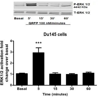
5.5 The effect of QRFP on (ERK 1/2) MAPK expression in DU145 cells. 157
5.6 The effect of QRFP on (p38) MAPK expression in PC3 cells. 158
5.7 The effect of QRFP on (p38) MAPK expression in DU145 cells. 159
5.8 The effect of QRFP on (JNK1/2) MAPK expression in PC3 cells. 160
5.9 The effect of QRFP on (JNK1/2) MAPK expression in DU145 cells. 161
5.10 The effect of QRFP on (AKT) MAPK expression in PC3 cells. 162
5.11 The effect of QRFP on (AKT) MAPK expression in DU145 cells. 163
6.1 The effect of QRFP on AGR 2 protein expression in PC3 cells. 184
6.2 The effect of QRFP on MMP-2 protein expression in PC3 cells. 186
6.3 The effect of QRFP on MMP-2 protein expression in DU145 cells. 187
6.4 The effect of QRFP on Caspase-3 protein expression in PC3 cells. 189
6.5 The effect of QRFP on Caspase-3 protein expression in DU145
cells.
190
6.6 The effect of QRFP on AMPK protein expression in PC3 cells. 192
6.7 The effect of QRFP on Caspase-3 protein expression in DU145
cells.
193
6.8 siRNA mediated knockdown of GPR103 receptor PC3 cells. 195
6.9 Effect of GPR103 receptor knockdown on QRFP induced AKT
phosphorylation in PC3 cells.
196
6.10 The effects of QRFP and combination inhibitors on migration
signalling pathways in PC3 cells.
13
6.11 The effects of QRFP and combination inhibitors on migration
signalling pathways in DU145 cells.
198
7.1 Effects of adipokines and hormones on GPR103 expression in PC3
prostate cell lines.
214
7.2 Effects of adipokines and hormones on mRNA QRFP expression in
prostate cell lines.
214
7.3 Effects of Leptin stimulation on GPR103 genes expression in pC3
cells.
216
7.4 Effects of Leptin stimulation on QRFP genes expression in pC3
cells.
216
7.5 Effects of Leptin on GPR103 expression in PC3 cells. 217
7.6 Effects of Leptin on QRFP expression in PC3 cells. 217
7.7 Effects of Leptin on GPR103 expression in PC3 cells. 218
7.8 Effects of Leptin on QRFP expression in PC3 cells. 218
7.9 Effects of Leptin and MAPK inhibitors on mRNA GPR103
expression in PC3 cells.
220
7.10 Effects of Leptin and inhibitors on mRNA QRFP expression in
prostate PC3 cells.
14
TABLES
Table Title Page
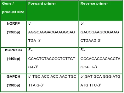
2.1 Primer Sequences of the sense and anti-sense primers. 81
2.2 Table demonstrating antibodies used for western
blotting with concentrations and relevant secondary
antibodies.
84
2.3 The Human Phospho-Kinase Array coordinates. 95
4.1 Table illustrates the overall effects of QRFP on cellular
apoptosis in PC3, DU145 and LNCaP cells.
15
DECLARATION
This thesis is submitted to the University of Warwick in support of my
application for the degree of Doctor of Philosophy. It has been composed
by myself and has not been submitted in any previous application for any
16
ACKNOWLEDGEMENTS
First of all, I would like to thank the almighty God (Allah) for establishing
me to complete my PhD. I fully express my appreciation to thank my
supervisor Dr. Harpal Randeva for supporting me during this past four
years’ patience of guidance and advise me as he has been a remarkable
mentor for me to enhance my expert knowledge. He has been
encouraging me throughout these years with my research and allowing
me to grow as a research scientist. Also I am a grateful to thank
co-supervisor Dr. Manjunath Ramanjaneya for his assistance and guidance
practically expertise in the lab. I would to thank Dr. Kevin Williams who
collected the human prostate tissue samples.
I would like to take this opportunity to thank specially my parents for their
support and care. Words cannot express how grateful them for all of the
sacrifices that they have made for me to be where I am, their prayers for
me was what sustained me to achieve my goals. I would like to thank all
of my family for their support. I great my wife and children the once I love
the most, they have showed me all the love, care and warmth. Finally, I
recognize that this study would not achieved without funding from my
17
ABBREVIATIONS
AI androgen-insensitive
AR
BMI
androgen receptor
body mass index
BPH benign prostatic hypertrophy
BSA bovine serum albumin
DHT Dihydroxytestosterone
cDNA complementary deoxyribonucleic acid
DNA deoxyribonucleic acid
ELISA
ERK
ECM
enzyme linked immunosorbentassay
extracellular signal related kinase
extracellular matrix
FBS
GPR103
fetal bovine serum
G-protein coupled receptor 103
H2O2
JNK
hydrogen peroxide
c-Jun N-terminal kinase.
MAPK
MMP
mitogen-activated protein kinase
matrix metalloproteinase
M-MuLV moloneymurineleukemia virus
18 p38
AGR 2
protein 38
anterior gradient protein 2 homolog
PBS
PI3K
PCa
phosphate buffered saline
phosphatidylinositide 3-kinases
Prostate cancer
PCR Polymerase Chain Reaction
PSA prostate specific antigen
PVDF
QRFP
Polyvinylidenedifluoride
Pyroglutamylated RFamide Peptide
RIPA radioimmunoprecipitationlysis buffer
RNA ribonucleic acid17
RT-PCR reverse transcriptase polymerase chain reaction
SDS sodium dodecylsulfate
SDS-PAGE sodium dodecylsulfate-polyacrylamide gel electrophoresis
TEMED N, N, N’, N’- tetramethylethylenediamine
Tris tris (Sonn et al.) aminomethane
19
ABSTRACT
Prostate cancer (Choudhury et al., 2014) is the leading cause of
non-cutaneous malignancy and is the second most commonly diagnosed
cancer among men after lung cancer. QRFP is a secreted protein that is
extensively expressed in the brain with highest expression levels in the
medulla, pituitary gland, cerebellum, retina, and vestibular nucleus; in the
periphery it is expressed in the adipose tissue prostate gland, bladder,
colon, testis and in the parathyroid and thyroid gland. QRFP is a member
of the RF amide neuropeptide family. This family might be implicated in
extensive array of biological activities for instance, food intake,
cardiovascular functioning, blood pressure, analgesia, aldosterone
secretion and locomotor activity, resulting in orexinergic activities. It is
suggested that obesity is one of the contributing factors for aggressive form
of prostate cancer. There is a strong association between adipokines and
aggressive form of various cancers. QRFP has been recently described as
an adipokine that exerts its functions via activation of the G protein coupled
receptor GPR103. At present the role of the QRFP and GPR103 in prostate
cancer has not been explored in detail. I studied the potential role of the
adipokine QRFP in prostate cancer. The three prostate cancer cell lines:
PC3, DU145 (Orbetzova, 2012) and LNCaP (androgen-sensitive) were
used as models for my studies. Nevertheless, no studies to date have
20
prompted me to examine the expression and function of QRFP and
GPR103 in the human prostate cells. Also I sought to identify the role of
QRFP and GPR103 in prostate cancer. The novel data presented in this
study demonstrates that QRFP & GPR103 genes and protein are
expressed in both human prostate tissue and prostate cancer cell lines.
Expression of both QRFP and GPR103 were higher in PCa tissues
compared to controls. Moreover, an ELISA detected circulate QRFP serum
levels were lower in human cancer patients compared to benign and
healthy group. Stimulation with QRFP induced MAPK signalling cascades,
AKT, AGR-2 expression, MMP 2, Caspase-3 and AMPK in PC3 & DU145
cells. QRFP also increased PC3 & DU145 cells migration and invasion,
nonetheless QRFP showed induced suppression cell proliferation in PC3,
21
CHAPTER 1
22
1 BACKGROUND
1.1 THE NORMAL PROSTATE GLAND
The prostate gland is a part of the male reproductive system involved in
production of seminal fluid, an alkaline liquid which is rich in spermine,
cholesterol, fibrinogens, fibrinolysin, zinc and acid phosphatase,
phospholipids, citric acid and some proteins, however the exact function of
the prostate gland still remains unclear. This gland is important in
protecting the lower urinary tract from infection and inflammation, and for
men fertility. The growth and activity of prostate is dependent on male
hormones; androgens (Crawford, 2005).
Anatomically prostate gland is situated deep in the pelvis surrounding the
proximal urethra, beneath the urinary bladder and facade the rectum. The
size of prostate gland differs by age. Normal adult human prostate is
typically slightly larger than a walnut and weighs around 20-25 grams. The
prostate is comprises of four essential zones based on the glandular
components, embryonic origins, and form a patho-physiological basis of
benign prostatic hyperplasia (BPH) and prostate cancer (Alshaker et al.).
The peripheral zone (PZ): Extends along the posterior and lateral
surfaces of the gland, consists of 70 % of prostatic volume and is
23
The central zone (Leongamornlert et al.): The area surrounding the
utricle and contains the ejaculatory ducts. This constitutes up to
25% of the normal prostate volume, CZ has more smooth muscle
than the PZ, which makes this zone comparatively resistant to
inflammation and hyperplasia.
The transition zone (Venkateswaran and Klotz): This covered only
5% of the total prostatic volume. It surrounds the proximal urethra
and is the state of the prostate, contains sub- mucosal and mucosal
glands that grows throughout life and is responsible for the benign
prostatic hypertrophy (BPH).
The anterior fibro-muscular zone: situated anteriorly and contains
no glandular tissue and is made up of only muscle and fibrous
tissue. Therefore it does not become hyperplastic (Lee et al., 2011).
PZ and CZ are referred to as external prostate while TZ and
anterior fibro-muscular zone are termed as interior prostate. These
zones contain essentially no glandular tissue (Iremashvili et al.,
2012b).
Histologically the prostate is divided into two parts glandular tissues and
duct, glandular tissue is involved in secretion of fluid and ducts, which
consist of 7 % of the gland and the remaining 30 % non-glandular tissue,
which is fibromuscularstroma. The fibromuscularstroma covers prostate
24
glands and connective tissue comprising smooth muscle. The gland
padded essentially with two main forms of epithelial cells: Basal cells and
secretory cells within various cells types mediated among basal cells and
secretory cells. Basal cells are located at the exterior of the gland under
the secretory cells. Secretory cells are cuboid formed with pale to clear
cytoplasm. This deep coat is a main feature, as it is missing in malignancy
(Iremashvili et al., 2012a) & (Reese, 2016).
Figure 1.1 Diagram view of the cell types in the interior a human prostatic
25
1.1.1 ANDROGENS:
Testosterone is the main androgen steroid hormone secreted by the
testicular leydig cells which is controlled by a complex chain of signals
referred to as the hypothalamic-pituitary-gonadal axis. This accounts for
the majority of circulating androgen response to luteinizing hormone
(Hoogland et al.) about 94 %- 97 %; the remaining androgen production
arises essentially from the adrenals and peripheral conversion of estrogen
by aromatase (Hiort et al., 1998). Testosterone that circulates in the
plasma is essentially bound to one of two kinds of plasma protein
sex-hormone binding globulin (SHBGs) or with albumin and only testosterone
set free can enters into prostate cells through diffusion. However, the
mechanism for testosterone carrying from the leydig cell to the blood
stream or lymph is not completely identified (Rove et al., 2012).
Testosterone is irreversibly converted into an active metabolic 5 α-
dihydrotestosterone (DHT), via enzyme 5- α. After being formed, DHT has
a higher binding affinity to the androgen receptor (AR) to control prostatic
cellular proliferation and survival, or might be additional metabolized along
a number of alternative pathways. The transformation of testosterone to
DHT is depending on organs, for instance in prostrate, since DHT is the
major biological active androgen. Hydroxysteroid dehydrogenases control
ligand the binding of AR in the human prostate (Godoy et al., 2011) &
26
FSH LH
[image:27.595.143.467.97.425.2]
FSH
LH
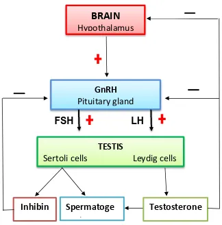
Figure 1.2 Hormonal regulation of the male reproductive system
At the hypothalamic pituitary gonadal axis, GnRH stimulates the release of follicle-stimulating hormone (FSH) which act on the testes to begin spermatogenesis and luteinizing hormone (LH) that secrete testosterone. In turn, the testes production of testosterone and the hormone inhibin prevent the release of GnRH, FSH, and LH in a negative feedback.
Furthermore, testosterone aromatization into estrogen act as a main role
in prostate growth by actin interaction with oestrogen receptor B and
androgen are essential to regulate the proliferative and apoptosis change
that happen throughout normal prostate growth and differentiation.
Concentrations of estrogen in prostate stromal tissue are evidently
increased in cases of benign prostate hyperplasia (BPH) (Green et al.,
BRAIN
Hypothalamus
GnRH
Pituitary gland
TESTIS
Sertoli cells Leydig cells
Testosterone
Spermatoge
27
2012). The main role of androgen in normal prostate is to stimulate
differentiation of luminal epithelial cells and to control the transcription of
genes encoding proteins required for prostate function, for instance
prostate specific antigen (PSA). Measuring PSA levels commonly
employed for detection and diagnosis of neoplastic malignancies of
prostate tissues and response to therapeutic intervention (Green et al.,
2012) & (Crawford, 2005). Conversely, PSA is not specific for PCa. Several
other conditions can be associated with an elevated PSA level, including
benign prostatic hyperplasia (BPH) and prostatitis. Though a PSA level
higher than 4.0 ng/mL is generally used as an indication for prostate
biopsy, the sensitivity and specificity of the PSA test limits its use as a
prostate cancer screening method (Ochiai et al., 2005). In prostate cancer
the main function of AR is less obvious, though possibly AR activity
modulates the expression of genes related with cell cycle regulation,
growth and survival. Irrespective of the mechanism, it is strongly observed
that AR signalling plays a critical role in the growth and progression of PCa
(Balk and Knudsen, 2008) & (Green et al., 2012). AR ablation therapy has
been recently used as an approach to treatment advanced prostate cancer
(PCa) and is initially effective in reducing the development of the disease.
Nevertheless, it has been observed frequently, PCa appears again as an
androgen- tumour with a related life expectancy of only15-20 months
28
1.2 PROSTATE CANCER (PCa)
1.2.1 EPIDEMIOLOGY
PCa is the leading cause of non-cutaneous malignancy and the second
most common diagnosed cancer among men after lung cancer, and is the
fifth most frequent cancer globally (rsted et al., 2011). Worldwide around
1,111,689 new cases of PCa were diagnosed in 2012 (Van Rij et al.,
2016).The high rates of prostate cancer are recorded in developed
countries approximately 3/4 of the cases appear in Australia, New Zealand,
Western and Northern Europe and Canada. In 2009 has documented that
approximately 192,280 new cases of PCa diagnosed and
approximately 27,360 recorded died in Northern America which becomes
the highest incidence and significant death (Rosenthal and Sandler, 2010).
There are evident differences between the incidences of clinically
significant disease universally, nevertheless incident non-clinically
pertinent disease is still equal. There are significant variance among
northern and southern countries in Europe, with PCa incidence and death
being higher in England and Wales and lower in Mediterranean countries
(Van Rij et al., 2016). The occurrence rates are comparatively high in
certain developing regions such as the Caribbean, South America and
sub-Saharan Africa countries. Conversely the lowest incidence is expected in
south Asian countries (Sankaranarayanan et al., 2010). The rise in
29
factors. Since the routine examination of transurethral prostatectomy
(TURP) histology specimens and practice of serum analysis for prostate
specific antigen (PSA) as a sign for PCa has more role to play in the
increased incidence mainly in the more developed (Djavan et al., 2011) &
(Giles, 2014). However, around 258,000 deaths in 2008, and the sixth
leading cause of cancer death. The figure of deaths from prostate cancer
is nearly similar in developed and developing regions. Death rates are
mostly higher in mostly black populated regions such as Caribbean and
sub-Saharan Africa, in contrast it is very lower in Asia. The PCa globally is
expected to grow at 1.7 million new cases and 499 000 new deaths in 2030
(Center et al., 2012). Nevertheless, lots of men have prostate cancer with
no symptoms therefore does not undergo any specific treatment and finally
die of further causes. PCa shares a figure of lineaments with benign
prostatic hyperplasia (BPH) and the putative signs of cancer, prostatic
intraepithelial neoplasia (Martin et al.). All rise with in spread by men age,
all need androgens for proliferate and development, and all react to
androgen prevention treatment (Bostwick et al., 2004). The high-grade PIN
and early stage cancer is categorized by, aberrations in markers of
secretory differentiation, rising nuclear, progressive basal cell layer
disorder and nuclear variations, increasing cell proliferation, variation in
DNA sequent, and increasing genetic instability. A number of biomarkers
demonstrate up-regulation or increase in the development from benign
down-30
regulated or missing. Present data show that more biomarkers are up
regulated; however, the comparative importance of each is unknown.
There is a considerable increase in micro-vessel intensity in PIN and
carcinoma comparison with normal prostate tissue (Jemal et al., 2003).
1.2.2 ETIOLOGY
The main causes of PCa are still unidentified, however there are several
risk factors related with the development of cancers and PCa is no
exclusion to this and risk factors are divided into two sections: Endogenous
risk factors including (family history, hormones and race) and exogenous
risk factors including (diet, environmental agents and other factors) are all
involved (Venkateswaran and Klotz, 2010) & (Bostwick et al., 2004).
1.2.3 AGE
Age is one of the significant risk factors; there is a strong association
between incidences and mortality of PCa with old age. Appearance of PCa
in men under 50 years old is very unusual, with about 60% of cases
diagnosed in men over 70 years of age (Macefield et al., 2009). Recent
studies confirmed that PCa has a long induction period, and that many men
carry the diseases in their 20s and 30s (Gann, 2002).
1
.2.4 ETHNICITYThe highest incidence rates of PCa in the USA are in black American men.
31
white American men. Asia is the continent with the lowest incidence
universally; Countries for instance China and Japan have comparatively
low rates of PCa in native people (Bostwick et al., 2004). The causes for
this rates 60% higher risk within African Americans and 38% lower risk
within Asian Americans in compare to white Caucasians are still unclear
(Ferris-i-Tortajada et al., 2011).
1.2.5 GENETIC FACTORS
Prostate cancer like other cancers is a genetic disorder that begin by the
accumulation of chromosomal alterations produced by the clone selection
of aggressive cells; about 5-10% of PCa are hereditary. Recent studies,
have identified around 100 genetic variants which linked with prostate
cancer disease (Stegeman et al., 2015). Some of these genes such as
BRCA1, BRCA2, or HOXB13 genes provide proteins that suppression
tumor. Inherited alterations in these specific genes, have been linked with
increased possibilities of many types of cancers including hereditary
prostate cancer (Leongamornlert et al., 2014). Prostate cancer family
history increase risk 2 to 3 times for men with first-degree relative by PCa,
families with 3 consecutive affected generations, or families with two
relations below the 55 ages would be considered for recommendation
surgery. It is evident that genetics play a significant role in prostate cancer
aetiology (Guy, 2009). On Caucasian families, found that seven
32
HPC1, PCAP, CAPB on (chromosome1), HPC2 on (chromosome17), and
HPCX on (chromosome 8 and X chromosome) and HPC20 on
(chromosome20) (Gronberg, 2003), (Lange, 2010) & (Goh et al., 2012).
1.2.6 ENDOGENOUS HORMONE PROFILE
Androgens have long been involved in prostate carcinogenesis, elevated
levels of circulating androgen were found in African-American men
compared to white American men. This would make a compelling
hypothesis, conversely, the relationship between circulating and
intraprostatic levels of androgens and PCa is still under debate (Wirén and
Stattin, 2008). Insulin-like growth factor (IGF-1) which was found to have
mutagenic and anti-apoptotic effects, are strongly associated with PCa risk
(Crowe et al., 2008). Hormone levels might be affected both by exogenous
factors; exposure to environmental chemicals and endogenous factors
such as genetics can affect hormone activity (Bostwick et al., 2004).
1.2.7 DIET
Diverse dietary factors have been associated in the development of PCa
according to epidemiologic literature of migrant populations and
geographic variations (Wolk, 2005). However, some prospective studies
found no clear association between dietary fat consumption and risk PCa
(Crowe et al., 2008) & (Bosire et al., 2013). Many dietary factors and some
dietary patterns hold potential in prevent or/ enhance progression risk of
33
(Lin et al., 2015). Several mechanisms have been proposed to explain
which dietary fat intake may be possibly linked with PCa: Pro-inflammatory
fatty acid metabolites, IGF-1 levels in the serum, free radicals, variation of
serum androgens. The high concentration of zinc in normal prostate
reduced >90% risk of PCa, but the relation of dietary intake of zinc on PCa
is uncertain. An antioxidant found in tomatoes, broccoli, and asparagus,
vitamin E, selenium, and green tea regular intake might decrease risk of
PCa (Sonn et al., 2005).
1.2.8 PATHOLOGIC MARKERS
95% of the prostate tumors are adenocarcinomas, only around 4% of
cases have transitional cell morphology and are thought to arise from the
epithelial lining of the prostatic urethra. Few cases have neuroendocrine
morphology. These cells are believed to arise from the neuroendocrine
stem cells normally present in the prostate (Dan Theodorescu et al., 2009).
PCa is different in clinical staging, histopathological tumor growth forms,
and survival. Thus, individual assessment of a tumor’s aggressive potential
is critical for clinical decision-making in men with prostate cancer. Sufficient
therapy of PCa fundamentally depends on their single histological kind,
stage and grade of malignancy. High grades are related with early and
rapid tumor development and consequent metastasis (Hoogland et al.,
2014). Low grade and low stage cancers can be treated locally by curative
34
subjected to an Active Surveillance (AS) strategy (Böcking et al., 2014).
However, the relationship between biopsy and radical prostatectomy
specimens still remains a difficult, particularly and notably the
under-grading of the biopsy specimen. Grading the malignant prospective of
different cancers of the prostate presently is conducted according to the
modified Gleason-score according to the International Society for Urologic
Pathology (ISUP), on histological sections of biopsies or resected tissue
(Fine et al., 2012).
Staging of PCa: Staging of prostate cancer divided into two parts
pathological and clinical staging. The tumor, node, metastasis staging
(TNM) is widely used staging system for prostate cancer to clinically
describe the anatomical range of cancer spread. It divided on 3 main
sections of information: T- assesses the tumour, N- spread the malignancy
to region and lymph nodes and secondary and M-cancer metastases
outside of prostate gland (Cheng et al., 2012).
Gleason Score: The Gleason system is the most prevalent applied
pathological grading for PCa as an essential prognostic factor in predicting
detections in biochemical failure, radical prostatectomy, lymph node local
recurrences, or distant metastasis in patients without treatment, radiation
therapy and other therapies, including high-intensity focal ultrasound
therapy and cryo-therapy. Moreover, using hematoxylin-eosin (H&E)
staining to describe the histological pattern of carcinoma cells (Martin et
35
The Gleason score is calculated based on the dominant histologic grades,
from grade 1 to grade 5. Adding the two most prevalent pattern grades,
yielding a score ranging from 2 to 10, derives the typical score (Veltri et al.,
2012). Since there is some indication that the least-differentiated element
of the specimen may provide independent prognostic information, the
score is normally provided by its separate components such Gleason
scores 3 + 4 = 7; or 4 + 3 = 7. Needle Biopsy, first pattern with the highest
grade must be recorded and ignoring secondary pattern. Radical
Prostatectomy, Gleason score must be depending the primary and
secondary patterns with a statement on the tertiary pattern (Blume-Jensen
et al., 2015).
1.2.9 TUMOURIGENESIS; CELL PROLIFERATION, THE CELL CYCLE
The complicated balance conserved between cell growth and proliferation
factors and apoptosis promoting factors are essential to the regulation of
prostate growth. Disturbances in this homeostasis commonly cause the
absence of apoptosis and the over activation of factors promoting cell
survival and proliferation (Hanahan and Weinberg, 2011). The unavoidable
consequence is a dysfunctional signaling pathway leading to
tumorigenesis and cancer. Furthermore, cancer cells increase the
capability to migration and invasion heterotopic tissues, through bone
being the common site of human PCa metastasis (Bononi et al., 2011).
36
by many growth factor pathways which classified into three distinct groups;
insulin-like growth factor-1 (IGF-1), fibroblast growth factor (FGF) both are
mostly stimulators of cell proliferation, survival, differentiation, migration
and invasiveness and transforming growth factor-β (TGF-β) responsible
apoptosis which is inhibitor prostate growth (Dasgupta et al., 2012). The
Ras/Raf/MEK/ERK and Ras/PI3K/PTEN/Akt/ mTOR signaling pathways
have been shown to play significant roles in the transmission of
proliferative signals from membrane bound receptors (McCubrey et al.,
2008). Mutations may appear in the genes encoding pathway components
including (RAF, RAS, PTEN, PIK3CA, TSC2, AKT, TSC1) or in upstream
receptors that activate these pathways. These pathways spread this
information by interactions with many other proteins to the nucleus to
regulator gene expression (Steelman et al., 2011). Cell proliferation is
induced and initiated by the act of growth factors. Cell division comprises
of two consequent processes essentially characterized by DNA replication
and isolation of replicated chromosomes into two separate cells called cell
cycle (Williams and Stoeber, 2012). The cell cycle consists of four distinct
phases: G1 phase in which the cell grows and prepares to synthesize DNA,
S phase (DNA synthesis), G2 phase (premyotic) proteins are synthesized
in preparation for mitosis, and M phase (mitosis) in which cell division
occurs. Quiescent phase or G0 is a resting phase where the cell has left
the cycle and has stopped dividing (Słonina et al.). Replication of the
37
achieved once in each cell cycle. Genetic disorders in the control of the
cell cycle are occurrence in almost human cancers. Main molecules that
are implicated cell cycle regulation and apoptosis in PCa cells such as
cyclin-depenent kinases (CDKs), p53, p21 and p27 (Koljonen et al., 2006).
Through proliferation a cell needs to grow and replicate its genome prior to
dividing to generate two daughter cells. Though in cancer cells is
essentially a disease in which cells have absent their normal checks on
cell proliferation. Cancer cells in order to satisfy the increased supplies of
proliferation, often display essential changes in pathways of energy
metabolism and nutrient uptake. Many distinct oncogenes such as (AKT
src, c-Myc, and H-ras) promot anabolic metabolism in transformed cells
(Jones and Thompson, 2009). Prostate cancer initiates when the
semen-secreting prostate organ cells transform into cancer cells, proliferating at
higher mitotic stages. Originally, the prostate cells begin to proliferate
leading to tumor development in the peripheral zone of the prostate gland
(da Silva et al., 2013). By the time these cancer cells finally proliferate to
further invade neighboring organs, such as the, bladder, seminal vesicles,
rectum, and urethra. Through the early metastatic stages, malignant cells
from the initial tumor separate from their original location and migrate
through blood and lymphatic vessels. In the advanced stages, PCa cells
eventually spread to more terminal organs, including bones, liver, and lung.
Metastasis is one of the main causes of death in cancer. Metastasis of PCa
38
intravasation, circulation and extravasation of tumor cells and then
angiogenesis (Jin et al., 2011). Furthermore, metastasis to bone is the
most common prostate cancer metastatic (Altieri et al., 2009). Cell
migration is crucial steps in several biological processes, including tissue
repair and regeneration, inflammation, immune surveillance and
embryonic morphogenesis (Condeelis et al., 2005).Abnormal regulation of
cell migration leads to progression of several diseases, such as cancer
invasion and metastasis (Yamaguchi and Condeelis, 2007). Invasive
carcinoma cells gain a migratory phenotype related with increased
activation of several genes implicated in cell motility (Bozzuto et al., 2010).
Thus, understanding the essential mechanisms of cell migration and
invasion are critical for comprehension of both essential biology and the
pathology of disease (Yamaguchi et al., 2005). Cancer cells invade
throughout the basement membrane and extracellular matrix is the feature
of the metastatic process. Each stage in the metastatic process requires
distinct molecular interactions between the tumor cells, the cells of the
stroma and the extracellular matrix (ECM) (Bozzuto et al., 2010). The
interface between normal prostate epithelial cells and the ECM is critical
for migration cell proliferation and survival (Jin et al., 2011). Cell migration
and invasion are stimulated via a number of chemo attractants, which
stimulate intracellular signaling pathways that control reorganization of the
actin cytoskeleton. Fundamental to this process is the structural and
39
are identified as focal adhesions (Chen et al., 2013c). Matrix
metalloproteinases (MMPs) have been known act to degrade the ECM.
The level of ratios of MMP-2/-9 to tissue inhibitor of metalloproteinases-1
(TIMP-1) In primary tumor prostrate tissues is increased relative to normal
prostate tissues (Brehmer et al., 2003) & (Clarke et al., 2009). However,
loss of TIMP-1 was associated with up-regulation of MMPs in malignant
prostate cancer tissues. Additional, has been found that high
concentrations of MMP-2/9 have been detected in plasma patients with
metastatic disease (Morgia et al., 2005). Binding of FN to integrin results
in MMP-2/9 over-activation through the FAK/ILK/ERK/PI3K/NF-κB
pathways, and thus driving to ECM degradation and cancer invasion.
Moreover, there are several intracellular molecules including Notch, Sonic
Hedgehog, Wnt, NF-κB, Ras/Raf/MEK/MAPK, in addition the ERK/AKT
pathways that regulator each side of each of the stages of cancer invasion
(Stivarou and Patsavoudi, 2015). Several chemotactic such as EGF
influences have been shown to stimulate intracellular signaling pathways
that drive to thorny end formation through these mechanisms. Focal
adhesion kinase (FAK) is a crucial mechanism of focal adhesion turnover,
and is lead to increasing cell migration. A high expresser of FAK and EGFR
in prostate cancer PC3 cell lines also associates with increased metastatic
possible (Chen et al., 2007) & (Tremblay et al., 1996). PI3K, Rho and Ras
activated by Src/ FAK-mediated lead to increased cell proliferation,
40
migration by different molecules including interleukin 8 (IL-8). Inhibition of
Src blocked IL-8 prompted migration in prostate cancer LNCaP cells (Lee
et al., 2004). There is an accumulation data describing the function of
hepatocyte growth factor (HGF) and its receptor c-Met in PCa
development. Interaction of HGF with its receptor and other factors
including TGF and TG-b have been demonstrated to modulate tumor cell
interaction, cell proliferation, cell migration, cell matrix adhesion, cell
invasion, and angiogenesis in PCa cells (Nagle and Cress, 2011). In the
regulation of cancer invasion, it has been stated that neuropeptides such
as bombesin and vasoactive intestinal polypeptide (VIP) increased the
invasive capability of prostate cancer cells PC 3 and LNCaP (Hoosein et
al., 1993). However, the mechanism of the influence of these
neuropeptides on tumor invasion remains ambiguous. Though, several
neuropeptides other than VIP and bombesin are existent in the prostate
gland, their influence on cancer invasion has yet to be specified
(Nagakawa et al., 1998).
1.2.10 APOPTOSIS AND ITS REGULATION
One of the highly significant advances has been the detection that
resistance to cell death, specifically apoptosis cell death, is critical aspect
of both tumorgenesis and progress of resistance anti-cancer drug
(Johnstone et al., 2008). Although, different apparatuses such as
41
tumor suppressor down-regulation, loss of cell cycle control and influence
of tumor microenvironment result in progress of tumor resistance to cell
death (apoptosis) (Hanahan and Weinberg, 2011). Kerr, Wyllie, and Currie
in 1972 were first used the term of apoptosis to define a morphologically
distinct form of cell death, however some components of the apoptosis
notion had been clearly described many years before (Elmore, 2007).
Apoptosis known as a programmed cell death, which occurs through the
activation of cell-intrinsic or extrinsic dependent mechanism, e.g. exposure
to irradiation or treatment with cytotoxic drugs, by DNA damage, or by
development death signals (Hassan et al., 2014). Apoptosis happens
routinely during growth and aged and as a homeostatic mechanism to
preserve cell an inhabitants in tissues (Ouyang et al., 2012). Many
morphological changes happens during apoptosis and are characterized
by membrane cell shrinking, membrane blebbing, nuclear fragmentation,
chromatin aggregation, and degradation of DNA fragmentation (Elmore,
2007). The mechanisms of apoptosis and the associated signalling
pathways are extremely complicated which can be initiated in a cell through
activation of intrinsic pathway or the extrinsic pathway. The extrinsic
pathway is initiated via the activation of the transmembrane death
receptors on the objective cell to stimulate apoptosis, for example the
ligands of CD95/TNF family induce apoptosis through multi-protein
complexes and involves activation of the multiple cascade pathways and
42
In contrast, the intrinsic pathway mediated apoptosis involves mitochondria
which is an essential player in the integration of variety of apoptotic signals,
containing loss of hypoxia, growth factors, DNA damage, and oxidative
stress (Spencer and Sorger, 2011).Caspases are the main initiators and
executioners of the apoptosis pathway. So far 14 members of caspases
have been discovered and classified into two groups, based on the lengths
of the pro-domains. The initiator caspases (caspase-1, -2, -4, -5, -8, -9 and
-10) have long pro-domains and function are involved in targeting and
regulation activation (Jager and Zwacka, 2010). While executioner
caspases (including caspase-3, 6 and 7) have short domains and are
responsible to role more downstream in targeting and regulating apoptosis
43
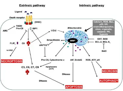
Figure 1.3 The extrinsic and intrinsic pathways of apoptosis: Death
receptor pathway (left) is initiated by the ligation of the ligands to their respective surface receptors (Indran et al., 2011).
The two main families of proteins that regulate apoptosis are P53 and
BCL-2 family members and are found to have abnormal function and expression
in PCa. The main member of apoptotic regulators is transforming growth
factor beta (TGF-β) family members that also include BCL2. TGF-β
signalling is critical for normal prostate epithelial cell proliferation and
induction of apoptosis it acts as a tumour suppressor in initial
tumorogenesis, however it promotes tumour growth in later stages of
tumour progression (Rentsch et al., 2006). The BCL2 family is one of the
[image:44.595.119.525.92.393.2]44
1.2.11 BCL-2
The BCL-2 gene family have highlighted the role of proteins as essential
regulators of the apoptotic pathway in variety of cells, which are classified
based on structural and functional features as either pro-apoptotic group
(BAX, BAK, BAD and BID) or anti-apoptotic group (BCL2, XL,
BCL-W, and MCL-1 proteins) (Tzifi et al., 2012). However, BAX families’
activation of mitochondria consequence that release of apoptogenic
factors including cytochrome C, endonuclease G, Same/Diablo, and
apoptosis including factor from the mitochondrial intermembrane space
(Du et al., 2000). In prostatic tissues both in normal and hyperplastic,
Bcl-2 protein is expressed in the cytoplasm of basal epithelial cells. The
occurrence of Bcl-2 overexpression is lesser in localized PCa compared
with hormone refractory PCa. The Over-expression of Bcl-2 may assist the
PCa cells to live in an androgen-deprived environment, and to accord
45
1.3 MITOGEN ACTIVATED PROTEIN KINASES (MAPK)
To understand the complicated pathogenesis of prostate cancer, the
interweave of signalling pathways and cell signal transmissions such as
cell survival, proliferation, migration invasion, inflammation and apoptosis,
might give a new understanding of the development, progression and
inhibition of cancers (Chen et al., 2014a). An extensive different stimuli
include growth factors, hormones, cytokines and environmental stresses
stimuli cell, through activation of receptor tyrosine kinases (RTKs), by
activation of subsequent signalling cascades which play essential roles in
cellular processes, such as proliferation, differentiation, migration,
apoptosis, cell metabolism, and cell cycle control (Osaki and Gama, 2013).
One of main signalling pathway induced by members of RTK family,
mediated through; mitogen- activated protein kinase (MAPK) (Pritchard
and Hayward, 2013). Mitogen activated protein kinases (MAPKs) family
consists of three distinguished group include; extracellular signal-regulated
kinase (ERK1/2), p38 isoforms (α/ β/γ/ /δ) and c-jun N-terminal or
46
Figure 1.4 Schematic representation of the MAPK cascade activation
and potential cross talk signals. MAPK signaling pathways mediate
intracellular signalinginitiated by extracellular or intracellular stimuli. Activated MAPKs phosphorylate many substrate proteins including transcription factors, resulting in regulation of a diversity of cellular activities including cell proliferation, differentiation, migration, inflammatory responses, and apoptosis (Osaki and Gama, 2013).
Growth
factors
Stress, cytokines, GPCR Growth factors, mitogens, GPCRStimulus
MAPKKk K MEKK 1,4MLK3,AS
K1
MEKK 2,3 TpI2
MEK 5
ERK 5 BMK 1
Downstream kinases and transcription factors
Responses Proliferation, differentiation, survival, migration, inflammatory, apoptosis
Growth factors
Raf MEK1/2K
MAPKKMAPK ERK 1/2
MILK3,TAK
MKK 3/6
p38 α,β, γ
47
1.3.1 EXTRACELLULAR SIGNAL-REGULATED PROTEIN KINASES
(ERK1/2)
The extracellular regulated kinases (ERK1/2) MAPKs are 44/42 kDa
serine/threonine kinases, has been strongly related to cell growth, survival,
proliferation, cell cycle, invasion, migration, and differentiation in both
normal and cancer cells (Saini et al., 2013). Significantly, activation of
factors of this pathway is assumed to contribute to tumorigenesis, tumor
progression and metastatic disease in a diversity of solid tumors
(Manzo-Merino et al., 2014). Nevertheless, ERK can also promote apoptotic
regulatory molecules counting bcl-2 family members and caspase-9
(Thakur et al., 2009). The ERK pathway consists of (A-Raf, B-Raf, Raf-1,
MEK1/2 and ERK1/2) are strongly activated mostly in response to growth
factors, serum, mitogens, ligands for G protein-coupled receptors (GPCRs)
and in solid tumors e.g. breast and prostate cancer (Pearson et al., 2001).
However, the ERK signaling pathway plays a role in several steps of tumor
development. Majority of the signals activating the ERK pathway are
initiated by receptor-mediated activation of the small G-protein, Ras -Raf
48
1.3.2 p38 MAPKs
The p38 MAPKs contains four members, p38α, p38β, p38γ, and p38δ
(Roux and Blenis, 2004). These kinases play roles in cell differentiation,
growth inhibition, cell survival, proliferation, and apoptosis (Risco and
Cuenda, 2012). Mainly, all p38MAPKs are strongly activated in cells in
response to verity of environmental and stress signals, or by
pro-inflammatory (TNFα, IL-6 or IL-1) or anti-pro-inflammatory (EGF, TGF-β)
cytokines and are weakly activated by growth factors or serum. Activated
p38 phosphorylates and controls various transcription factors such as
(p53, Elk-1, ATF-2, NF-κB, MEF-2, Max, Mac, or Stat1) and apoptotic
mediators as Cdc25A and Bcl-2 (Thornton and Rincon, 2009). p38 has
been revealed to promote cell survival in response to stress stimuli, such
as in response to DNA damage. Nevertheless, p38α may also have
oncogenic functions that are mediated by its involvement in key processes
of cancer progression, such as invasion, inflammation and angiogenesis.
Several propose that p38 play significant role in several cancers such as
49
1.3.3 c-Jun N-TERMINAL KINASE OR STRESS-ACTIVATED PROTEIN
KINASES (JNK/SAPK)
The first three mammalian JNK genes were discovered from rat livers
injected with cycloheximide JNK1, JNK2, and JNK3 also known as
(SAPK-γ, SAPK-α, and SAPK-β) respectively within implicate in growth,
morphogenesis, and cell differentiation (Bogoyevitch et al., 2010). JNK
proteins, share a threonine-proline-tyrosine (TPY) motif, are activated in
response to a diversity of extracellular stimuli, counting, growth factor
deprivation, inflammation cytokines (IL-6, IL-1, and TNF), UV irradiation,
DNA-damaging agents and mitogens (Pritchard and Hayward, 2013).
Depending on the cellular stimulus, JNKs phosphorylate varied substrates,
counting transcription factors (p53, AP-1, ATF-2, c-Myc, Elk-1, MLK2) and
certain members of the Bcl-2 family (Bode and Dong, 2007). Several
studies propose that JNK activity is chronically altered in many cancer
types include breast, prostate, lymphoma pancreas and lung cancer
(Zhang and Selim, 2012). Nevertheless, studies into the role of JNK in
human prostate tissues are rare (Takahashi et al., 2010). Depending on
cellular stimulus and types or even JNK isoform, JNKs have opposite
function. So, JNKs can induce apoptosis by reduced tumor formation and
malignant progression, however also may promote cell survival and
proliferation, also involved in regulation of the cell cycle
50
1.3.4 AKT/PKB KINASE
The serine/threonine kinase Akt also named protein kinase B (PKB) has
three isoforms revealed in mammals (Akt1/2/3 -2) encoded by genes
(Werden and McFadden, 2010). Akt is a main regulator of multiple cellular
processes; include glucose metabolism, proliferation, cell survival, and
protein synthesis (Zheng et al., 2012). Therefore many studies proposed
that AKT might associate with tumorigenesis by activated
phosphatidylinositol 3-kinase (PI3K), which transmits signals from
cytokines, growth factors and oncoproteins (da Silva et al., 2013).
Furthermore, Akt pathway activated in PCa cells acts as contributing to
cancer progression by both stimulation of cellular proliferation, migration,
invasion and inhibit apoptosis pathway by inactivate pro-apoptotic factors
51
1.4 OBESITY RELATED TO PROSTATE CANCER
Obesity is currently a major worldwide concern. It is estimated that above
of 2.1 billion people around 30% of world population with overweight or
obese and 3.4 million deaths each year worldwide. If its incidence stays on
its current curve, approximately 1/2 of adult population global will be
overweight or obese by 2030. In Western Europe UK comes behind only
Iceland of the highest levels of obesity or overweight 74% of men and 61%
of female are either overweight or obese. There are several countries
worldwide, including Arabic countries (Libya, Kuwait, Qatar and Egypt)
where more than half the female population is obese (Musaiger et al.,
2011) & (Musaiger, 2011).Body mass index (BMI) is predictably used
measurement in clinical medicine like obesity and population health.
However, the issues related to weight gain beyond the accurate capacity
of other markers to determine the incidence of obesity have been
suggested with body fat percentage being one of the most practical due to
its superior capability to stratify patients according to their cardiovascular
risks and metabolic (Campbell, 2014).Obesity leads to increases the risk
of developing various diseases. Type 2-diabetes, certain types of cancers
and cardiovascular disease risk increases due to overweight elevating
hypertension and dyslipidemia. 90 % of people with type-2 diabetes are
overweight and patients with diabetes are leads to cause of early death,
52
considered one of the main significant features in the progress of insulin
resistance, and insulin resistance might lead to type 2-diabetes. Though
several studies have reported associations between BMI and risk of
individual cancers including that of the prostate, pancreatic, ovarian, breast
and colon cancer. There is strong evidence the obesity is an important
cause of unnecessary suffering and mortality from many forms of cancer.
Also obesity is related with worse prognostic and malignant transformation
of epithelial cells (Campbell, 2014) & (Spangler et al., 2007). There are
some of biological processes common that could lead to the relation BMI
and aggressive PCa. Several hormones, adipokines signaling and
androgens implicated in obesity moreover play a role in the initiation and
promotion of cancer both at systemic level and a cellular paracrine
(Freedland and Aronson, 2004).
Although, other studies have found no association between BMI and PCa
(Schuurman et al., 2000). To understand the link between obesity and PCa
are complicated since obesity is associated not only with excess BMI,
however also with altered serum levels of many hormones, as estrogen,
testosterone, insulin and insulin-like growth factor (IGF)-1, all of which have
to some degree been related to PCa. Furthermore, obesity is highly
associated with dietary intake in terms of the quantity of calories in addition
to the amount of dietary fat, both of which have been linked to prostate
cancer. Obesity might effect on sex hormone binding globulin, testosterone
53
Rehman, 2014). High levels of metabolic factors, individually or combined,
are not associated to the development of PCa, however are associated to
an increased risk of disease progression, although with no proof of synergy
between the metabolic factors (Haggstrom et al., 2012). Several studies
suggested obese men have lower PSA values. Some men with obese
produce less PSA than normal weight men and thus could have late
diagnosis of PCa (Kim et al., 2013).However, some studies concluded that
BMI is not affected the capability of the PSA level for predict adverse
pathologic features, such as seminal vesicle invasion, extracapsular
extension, and positive surgical margins across increasing BMI (Banez et
al., 2009). Otherwise, obesity has a significant positive associated with
prostate volume (PV) in several study populations. Furthermore, obesity is
essentially produced by an excess accumulation of adipocyte tissue, and
adipocytes are specified for the storage and synthesis of fatty acids (Kwan
et al., 2015). Two main types of adipose tissue are known, white and
brown, with different origins and functions. White adipose tissue (WAT)
plays a role that related to maintaining energy homeostasis through storing
triglycerides and releasing fatty acids for energy synthesis, also controls a
wide range of functions including glucose and lipid homeostasis, food
intake control, immune and inflammatory regulation, or metabolism by
secreting a great number of adipokines (Falcao-Pires et al., 2012). The
main role of brown adipose tissue BAT is particularly in thermogenesis, the