warwick.ac.uk/lib-publications
A Thesis Submitted for the Degree of PhD at the University of Warwick
Permanent WRAP URL:
http://wrap.warwick.ac.uk/108883
Copyright and reuse:
This thesis is made available online and is protected by original copyright.
Please scroll down to view the document itself.
Please refer to the repository record for this item for information to help you to cite it.
Our policy information is available from the repository home page.
TITLE
TH E B R IT IS H L IB R A R Y
BRITISH THESIS SERVICE
ATTACHMENT OF BACTERIA TO GLASS SURFACES
IN PURE CULTURE AND IN MIXED SUSPENSIONS
AND THE EFFECT OF GROWTH CONDITIONS ON
THAT ATTACHMENT.
AUTHOR
JAMES NEILSOND EG R EE...
AW ARDING BODY
DATE...
UNIVERSITY OF WARWICK AUGUST 1991
THESIS
NUMBER
T H I S T H E S I S H A S B E E N M IC R O F IL M E D E X A C T L Y A S R E C E I V E D
The quality of this reproduction is dependent upon the quality of the original thesis submitted for microfilming. Every effort has been made to ensure the highest quality of reproduction.
Some pages may have indistinct print, especially if the original papers were poorly produced or if the awarding body sent an inferior copy.
If pages are missing, please contact the awarding body which granted the degree. Previously copyrighted materials (journal articles, published texts, etc.) are not filmed.
T h is c o p y o f the th e sis has b een su p p lie d on c o n d itio n th a t a n yo n e w h o c o n s u lts it is u n d e rsto o d to re co g n ise th a t its c o p y rig h t re s ts w ith its a u t h o r a n d t h .it no in fo rm a tio n d e riv e d fro m it m a y b e p u b lish e d w ith o u t th e a u t h o r ’s p r io r w ritte n co n se n t.
Reproduction of this thesis, other than as permitted under the United Kingdom Copyright Designs and Patents Act 1988, or under specific agreement with the copyright holder, is prohibited.
1 1
ATTACHMENT OF BACTERIA TO GLASS SURFACES
IN PURE CULTURE AND IN MIXED SUSPENSIONS
AND THE EFFECT OF GROWTH CONDITIONS ON
THAT ATTACHMENT.
JAMES NEILSON
A THESIS SUBMITTED FOR THE DEGREE OF DOCTOR OF PHILOSOPHY.
UNIVERSITY OF WARWICK DEPARTMENT OF BIOLOGICAL SCIENCES.
coiTErrs
CHAPTER OIE - IITRODPCTIOI PAGE
1. 1 Biofilm Importance 1
1.2 Why Bacteria Attach to Surfaces 5
1.2. 1 Comparison Between Attached and Free-Living Bacteria 5
1.2.2 Survival and Attachment 0
1.3 Attachment Process 11
1.3. 1 The Solid Surface 11
1.3. 1. (A) Physico-chemical Characteristics 11
1.3. 1. (B) Physical Characteristics 16
1.3.2 The Bacterium and Attachment 10
1.3.2. (A) The Bacterial Surface 10
1.3.2. <B> Bacterial External Appendages 23
1.3.2. <C> The Attachment Sequence 26
1.3.3 The Liquid Environment 28
1.4 Alms 20
CHAPTER TWO
2.1 Alms 30
2.2 Introduction 30
2.3 Materials and Methods 30
2.3. 1 Sterilisation 30
2.3.2 Media Preparation 30
2.3.3 Isolation 30
2.3.4 Identification 31
PAGE
(2) Bacterial Motility 31
(3) Oxidase Test 32
(4) Catalase Test 32
(5) Huge and Lelfson's Test 32
<e>
Indole Test 33(7) Methyl Red Test 33
(8) Voges-Proskauer Test 34
(9) Citrate Utilization Test 34
<10> Hydrogen Sulphide Production (Combined with Gelatin 35
Liquefaction)
(11) Fermentation Tests 36
(12) Urease Test 37
(13) Pigment Production 37
(14) Dihydrolase and Decarboxylase Activity 38
(15) Polymer Hydrolysis 39
(16) Growth on Milk Agar (10% HaCl) 40
(17) Spore Stain 40
2 . 4
Results 412.5 Discussion 41
CHAPTER THREE - An Investigation into the Attachment Abilities af
3.1
the Bacterial Isolates
Alms 46
3.2 Introduction 46
3.3 Materials and Methods 46
PAGE
3.3.2 Glass Coverslips 47
3.3.3 Absorbance versus Viable Counts 48
3.3.4 Attachment Experiments 48
3.3.5 Detachment 49
3.3.6 The Effects of Subculture on Bacterial Attachment 50
3.3.7 The Effect of Culture Age on Bacterial Attachment 51
3.3.8 The Effect of Bacterial Concentration on Attachment 51
3.4 Results 52
3.4.1 Attachment Abilities of Isolates 52
3.4.2 Detachment 52
3.4.3 The Effect of Subculture on Bacterial Attachment 55
3.4.4 The Effect of Culture Age on Bacterial Attachment 55
3.4.5 The Effect of Bacterial Concentration on Attachment 60
3.5 Discussion 63
3.5.1 The Bacterial Cell and Attachment 63
3.5.2 Detachment 66
3.5.3 Experimental Procedure and Attachment 68
3.5.4 Physico-chemical Properties of Substratum 72
CHAPTER FOUR - The Effect nf Carbon Source. Carbon Concentration.
Carbon-To-Eltrogen Ratio and Temperature on the
At.tarihMBnt of Bacteria
4.1 Aims 74
4.2 Introduction 74
4.3 Material and Methods 75
PAGE
4.3.2 Culture Conditions 76
4.3.3 Attachment Experiments 77
4.3.4 Effect of Temperature on Bacterial Attachment Ability 79
4.3.5 Statistical Analysis 79
4.4 Results 80
4.4.1 Bacterial Attachment In Batch Culture Grown Cells 80
4.4.2 Bacterial Attachment In Continuous Culture Grown Cells 84
4.4.3 Effects of Temperature on the Attachment of Batch 88
Culture Grown Cell
4.4.4 Effects of Temperature on the Attachment of Continuous 92
Culture Grown Cells
4.5 Discussion 93
4.5.1 Changes In the Bacterial Surface Characteristics with 93
Butrient Conditions
4.5.2 Butrient Source and Effects on Developing Blofilms 97
4.5.3 Temperature and Attachment 100
CHAPTER f i v e - Effect of pH and Electrolyte Type and Concentration
m> the Attachment. of Bacteria
5.1 Aim 105
5.2 Introduction 105
5.3 Materials and Methods 108
5.3.1 Bacteria and Inoculation 108
5.3.2 pH and Attachment 108
5.3.3 Electrolyte Type and Concentration and Attachment 109
5.4.1 The Effect of pH on the Attachment of Bacteria 110
5.4.2 Electrical Double-Layer Thickness (l/K) and Attachment 110
5.5 Discussion 114
5.5.1 pH and Attachment 114
5.5.2 Electric Double-Layer Thickness and Attachment 117
CHAPTER SIX - Attachment of a Bacterium When in a Mixed Suspension
with Another Bacterium
6.1 Aims 120
6.2 Introduction 120
6.3 Material and Methods 121
6.3.1 Bacteria and Inoculation 121
6.3.2 The Effect of Carbon Source, Carbon Concentration and 121
Carbon-to-Nitrogen Ratio on the Attachment of Bacteria
in Mixed Suspensions
6.3.3 Effect of Temperature on the Attachment of Bacteria in 122
Mixed Suspensions
6.3.4 pH and Attachment 122
6.3.5 Effect of Electrolyte Type and Concentration on the 122
Attachment of Bacteria in Mixed Suspensions
6.4 Results 123
6.4.1 Effect of Growth Conditions on the Attachment of 123
Bacteria in Mixed Suspensions
6.4.2 Attachment of Bacteria Grown in Continuous Culture 127
in Mixed Suspensions
PAGE
6.4.3 Effect of Temperature on the Attachment of Bacteria 133
in Mixed Suspensions
6.4.4 The Effect of pH on the Attachment of Bacteria in 142
Mixed Suspensions
6.4.5 The Effect of Blectrolyte Type and Concentration on 149
the Attachment of Bacteria in Mixed Suspension
6.5 Discussion 153
CHAPTER SEVE* - Attachment of Pure Bacterial Suspensions and Mixed
Bacterial Suspensions In Vivo Oslng a Model System
7.1 Aims 156
7.2 Introduction 156
7.3 Materials and Methods 157
7.3. 1 Bacteria 157
7.3.2 Model System 157
7.3.3 Experimental Conditions 159
7.3.4 Immunofluorescence Microscopy 159
7.4 Results 161
7.4. 1 Pure Culture vrs Mixed Suspension Attachment 161
7.4. 1.(A) Two-Membered Suspension 161
7.4. 1. <B> Three-Membered Suspensions 163
7.4.1. <C> Four-Membered Suspension 167
7.4. 1. <D> Five-Membered Suspension 167
7.4. 1.(E> Slx-Membered Suspension 171
7.4.2 Effects Temperature on Attachment 171
PAGE
7.4.4 Immunofluorescence Studies 175
7.5 Discussion 175
7.5.1 Blofilm and its Environment 175
7.5.2 Microorganisms in Aquatic Environments 179
CHAPTER BIGHT - An Investigation into the Inhibition of Bacterial
Attachment by Aclaatabactar. and the affects cf
Different Molecular Weight Lake Water Fractions on
the Attachment, nf Bacteria
8. 1 Alms 184
8.2 Introduction 184
8.3 Materials and Methods 185
8.3.1 Bacteria 185
8.3.2. <A> Inhibition of Bacterial Attachment by Aclnetobacter 185
8.3.2. (B) Effect of Lake Water Molecular Weight Fractions on 188
Bacterial Attachment
8.4 Results 190
8.4.1. (A) Bacterial Attachment Inhibition by Ac!netnhact.Br 190
8.4.1. <B> Effects of Vater Fractions on Bacterial Attachment 193
8.5 Discussion 197
BIBLIOGRAPHY 203
T*m.ws PAGE
Identification results and Bergey's mamnual of
Determinative Bacteriology.
TABLE 3.1 Attachment abilities of the bacterial isolates under 53
different nutrient conditions.
TABLE 3.2 The detachment of selected bacteria by 50 ppm Tween 20 54
in different media in a rotary incubator at 150 rpm
at 37°C.
TABLE 3.3 The optimum concentrations for attachment in nutrient 54
and glucose medium.
TABLE 3.4 The attachment of selected bacteria in nutrient broth 56
after subculture on nutrient agar.
TABLE 3.5 The attachment abilities of selected bacteria after 57
subculture on glucose agar and attachment in glucose
medium.
TABLE 3.6 The attachment abilities of selected bacteria after 58
storage and attachment in nutrient broth.
TABLE 3.7 The attachment abilities of bacteria after storage 59
and attachment in glucose medium.
TABLE 3.8 The attachment abilities of bacteria after storage 61
on nutrient agar and attachment in nutrient broth.
TABLE 3.9 The attachment abilities of bacteria after storage 62
on glucose agar and attachment in glucose medium.
s
TABLE 7.1 Attachment of bacteria in pure culture and in a
mixed suspension with one other bacterial species
In the model system. The temperature during these
experiments were within the range 8°C to 15°C.
TABLE 7.2 Attachment of bacteria in pure culture and in a
mixed suspension with two other bacterial species
in the model system. The temperatures during these
experiments were within the range 8°C to 15°C.
TABLE 7.3 Attachment of bacteria in pure culture and in a
mixed suspension with three other bacterial species
in the model system. The temperatures during these
experiments were within the range 8°C to 15°C.
TABLE 7.4 Attachment of bacteria in pure culture and in a
mixed suspension with four other bacterial species
in the model system. The temperatures during these
experiments were within the range 8°C to 15°C.
TABLE 7.5 Attachment of bacteria in pure culture and in a
mixed suspension with five other bacterial species
in the model system. The temperatures during these
experiments were within the range 8°C to 15°C.
TABLE 7.6 Attachment of bacteria in pure culture and in a
mixed suspension with different numbers of other
bacterial species in the model system. The
temperatures during these experiments were within
the range 1°C to 5°C.
170
172
[image:12.610.13.582.15.674.2]PAGE
glucose Medium 6 and In glucose Medium 6 with lake
water fractions (a), (b) or <c).
FIGURES
FIGURE 1.1 A model of the Gram-negative cell envelope or outer 20
membrane.
FIGURE 4.1 The continuous culture system. 78
FIGURE 4.2 The attachment of Aeroaonas, ChroaabactarlUffl, the 81
coryneform and Staphylococcus after batch growth and +
attachment In glucose, mannose and sucrose media 82
at 15°C.
FIGURE 4.3 The attachment of Aeromonas. Chromobacterium, the 85
coryneform and Staphylococcus in batch at 15°C, 25°C +
and 37°C after continuous culture growth In glucose, 86
mannose and sucrose media at 15°C.
FIGURE 4.4 The attachment of Aeromonas. Chromobacterium, the 89
coryneform and Staphylococcus in batch at 15°C, 25°C +
and 37°C after batch growth in glucose, mannose and 90
sucrose media at 15°C.
FIGURE 5.1 The potential energy of interaction between two 107
particles at large 1/K.
FIGURE 5.2 The potential energy of Interaction between two 107
[image:13.598.13.576.16.680.2]particles at small 1/K.
PAGE
FIGURE 5.3 The attachment of AeromonaR. the coryneform 111
and Staphylococcus in a mixed buffer system.
FIGURE 5.4 The effect of the electrical double-layer thickness 112
on the attachment of AercmnnaR. the coryneform and
Staphylococcus.
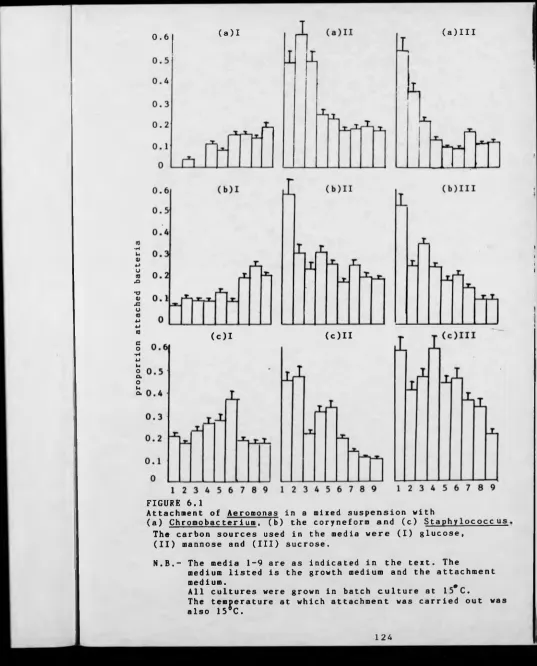
FIGURE 6.1 The attachment of Aeromonas in a mixed suspension 124
with Chromobacterium, the coryneform or
Staphvlococcus in different media.
FIGURE 6.2 The attachment of Chromobacter 1 nm in a mixed 125
suspension with Aeromonas. the coryneform or
Staphylococcus in different media.
FIGURE 6.3 The attachment of the coryneform in a mixed 128
suspension with Aeroaonas, Chromobacterium or
Staphylococcus in different media.
FIGURE 6.4 The attachment of Staphylococcus in a mixed 129
suspension with Aeroaoaas, Chromobacterium or
the coryneform in different media.
FIGURE 6.5 The attachment of Aeromonas in a mixed suspension 130
with Chromobacterlum. the coryneform or +
Staphylococcus in different media and at 131
different temperatures after all cells were grown
PAGE
suspension with AernninnaK. the coryneform or +
Staphylococcus in different media at different 135
temperatures after all cells were grown in batch
culture.
FIGURE 6.7 The attachment of the coryneform in a mixed 137
suspension with Aeromonas. Chromobacterium or
Staphylococcus in different media at different
temperatures after all cells were grown in batch culture.
FIGURE 6.8 The attachment of Staphylococcus in a mixed 139
suspension with Aeromonas. Chromobacterium or
the coryneform in different media at different
temperatures after all cells were grown in batch
culture.
FIGURE 6.9 The attachment of Aeromonas in a mixed suspension 140
with r.hrnranbacterium. the coryneform or
Staphylococcus in different media and at
different temperatures after all cells were grown
in continuous culture.
FIGURE 6.10 The attachment of Chromobacterium in a mixed 141
suspension with Aermnrmag. the coryneform or
Staphylococcus in different media at different
temperatures after all cells were grown in continuous
[image:15.601.22.577.11.673.2]culture.
PAGE
FIGURE 6.11 The attachment of the coryneform In a mixed
suspension with Aerofflonas, Chromobacterium or
Staphylococcus In different media at different
temperatures after all cells were grown In continuous
culture.
FIGURE 6.12 The attachment of Staphv1ococcus In a mixed
suspension with AerOBPnas, Chromobacterium or
the coryneform In different media at different
temperatures after all cells were grown in continuous
culture.
FIGURE 6.13 The attachment of Aeromonas In a mixed suspension
with the coryneform or Staphylococcus in a mixed
buffer system.
FIGURE 6.14 The attachment of the coryneform in a mixed
suspension with Aeromonas or Staphy1ococcus in a
mixed buffer system.
FIGURE 6.15 The attachment of Staphylococcus in a mixed
suspension with Aarninnag or the coryneform in
a mixed buffer system.
FIGURE 6.16 The effect of the electrical double-layer thickness
on the attachment of Aarnmnnag in pure culture
and In a mixed suspension with the coryneform
or Staphylncncous.
143
144
146
148
148
FIGURE
FIGURE
FIGURE
FIGURE
FIGURE
FIGURE
FIGURE
6.17 The effect of the electrical double-layer thickness
on the attachment of the coryneform In pure culture
and In a mixed suspension with Aernmnnns or
St.aphylococeus.
6.18 The effect of the electrical double-layer thickness
on the attachment of Staphylococcus In pure culture
and In a mixed suspension with Aeromonas or the
coryneform.
7. 1 The model system.
8. 1 The filtration process employed to obtain water
fractions <a>, (b> and (c>.
8.2 The attachment of Aeromonas. Chromobacterium or
Staphylococcus In glucose Medium 6, In glucose
Medium 6 while In mixed suspension with
Acinetobacter. In glucose Medium 6 with the
experimental supernatant and In glucose Medium 6
with killed Acinetobacter cells.
8.3 The growth and attachment of Aernmnnas.
Chromnbacterlum or Staphylococcus In glucose
Medium 6 and In glucose Medium 6 with water
fractions <a>, (b) or (c>.
8.4 The attachment of Aerononas. Chromobacterium
or Staphylococcus In glucose Medium 6 and In
glucose Medium 6 with water fractions (a), (b>
or <c> after all bacteria were grown in batch
culture In glucose Medium 6.
152 PAGE
154
158
189
191
194
APPEHDICBS PAGE
APPEHDIX TABLES
TABLE 1.1 Gram stain, cell morphology, motility and pigmentation 233
of selected bacteria.
TABLE 1.2 Indole, methyl red, V. P. and citrate utilisation 234
reactions of selected bacteria.
TABLE 1.3 Catalase, urease, H2S and oxidase reactions of 235
selected bacteria.
TABLE 1.4 Hugh and Lelfson's and King's A + B reaction of 236
selected bacteria.
TABLE 1.5 Hydrolysis of starch, cellulose, casein and DNA by 237
selected bacteria.
TABLE 1.6 Dlhydrolase and Decarboxylase activity of selected 238
bacteria.
TABLE 1.7 Fermentation of glucose, maltose, mannitol and 239
arablnose by selected bacteria.
TABLE 1.8 Fermentation of Xylose, lactose, Inositol and 240
sucrose by selected bacteria.
TABLE 1.9 Fermentation of malonate, adonltol and dulcltol by 241
selected bacteria.
TABLE 1.10 Spore stain and milk agar reaction by selected 242
bacteria.
TABLE 2.1- Detachment of Chromobacterium in Tween-20 at 15°C. 243
2.3
TABLE 2.4- Detachment of the coryneform in Tween-20 at 15°C. 244
PAGE
2.9
TABLE 2.10- Detachment of the coryneform in EGTA at 15°C. 246
2 . 1 2
TABLE 2.13- Detachment of Chromobacterium in Tween-20 at 37°C. 247
2. 15
TABLE 2.16- Detachment of the coryneform in Tween-20 at 37°C. 248
2.18
TABLE 2.19- Detachment of Chromobacterium in EGTA at 37°C. 249
2 . 2 1
TABLE 2.22- Detachment of the coryneform in EGTA at 37°C. 250
2.24
TABLE 3.1 The effect of slllcoset on bacterial cell viability, 251
growth and attachment ability.
APPENDIX FIGURBS
FIGURE 1 (a> The biofllm obtained after the growth and 252
attachment of Aeromonas in glucose Medium 4.
(b) The biofllm obtained after the growth in
continuous culture and attachment in batch
[image:19.606.23.576.16.673.2]of Aeromonas in mannose Medium 6.
PAGE
FIGURE 2
FIGURE 3
(a) The biofilm obtained after the attachment of a
mixed suspension of Chromobacterium with the
coryneform.
(b> The blofllm obtained after the attachment of a
mixed suspension of the coryneform with
Staphylococcus.
(a) The blofllm obtained after the attachment of
Aeroaonas. Chromobacterium and the coryneform
in the model system.
<b> The biofllm obtained after the attachment of
AeroaonaSi Chromobacterium, the coryneform and
Aclnetobacter in the model system.
253
ACKMOWLEDGBMBMTS
I would like to express my thanks to Dr M Fletcher for her
supervision and guidance during the early part of this project, and also to Dr K Flint who provided supervision, guidanace and constant support during the latter stages of the project.
Greatful thanks are also due to my fellow researchers and technicians in the Department of Environmental Sciences, at Warwick University, for their kindness and encouragement.
nH T .i.»B «T inw
I declare that this thesis Is a report of the research undertaken by myself In the Department of Biological Sciences at Warwick University, under the supervision of Dr X Fletcher and Dr K Flint. It is my own work and Includes nothing which is the outcome of work done in collaboration.
SUMMARY
The attachment of selected freshwater bacteria, Aai-nmnnaK.
Chminnhacterlum. a coryneform and Staphylococcus In pure culture and in mixed suspensions with one other bacterium to glass surfaces was investigated in the laboratory.
Changes in the nutrient conditions of the growth medium during growth and attachment and the temperature, pH and electrolyte type and concentration present in the attachment solution during attachment experiments all influenced bacterial attachment. The pure culture attachment results obtained depended on the bacterial species being investigated. When bacterial species were attached in the presence of one other species the growth conditions still had a profound affect on attachment. The results obtained depended on the bacterial species present in the mixed suspension, with some bacterial species having a more profound affect on the attachment of other species than others.
The bacterial species used to study bacterial attachment in the laboratory were used along with Aclnetobacter and PBPiirinmnnaR to investigate bacterial attachment in vivo using a model system. The attachment of these bacteria were investigated in two- to six-membered suspensions. The blofllms obtained in the model system consisted of smaller bacterial cells in a more densely packed blofilm. These blofilms could still be influenced by growth conditions as temperature was seen to influence the bacterial blofllm obtained. During these attachment experiments the Acinetobacter which did not attach itself had a profound affect on the attachment of other bacterial species when present in the liquid phase.
The mechanism by which Acinetobacter inhibited the attachment of other bacteria was investigated and it appeared Acinetobacter did not excreate a chemical to influence the attachment of other species, but the Acinetobacter cells themselves had to be present in the liquid phase to influence the attachment of other species.
CHAPTER ONE
INTRODUCTION
1.1 BIOFILN IMPORTANCE
Bacterial attachment Is an Important and widespread phenomenon and
research has shown that only a few surfaces In the natural environment
cannot be colonised by bacteria. These are rare and are always surfaces
of plants or animals, e.g. Sieburth (1975), showed that the body of a
hydra was free of attached bacteria but the stalk was colonised by
numerous bacteria. The apical tips of the brown algae Ascophyllum
nodosum have also been shown to be free of attached bacteria whereas old
growth was colonised by bacteria (Cundell, 1977). Variations in
bacterial attachment to these living surfaces were thought to be due to
modifications of the living tissue being used as a surface as it ages.
Bacterial blofllms present on a surface can have detrimental
consequences for humans. Sphaerotllus natans has been found growing on
surfaces In water delivery systems such as the surfaces of Industrial
heat exchanges (McCoy et al. 1982). These biofilms have been shown to
cause energy loss due to heat transfer, accelerated corrosion and
clogging In the heat exchange systems (Pedersen, 1982). During oil
recovery in the petroleum industry, water is sometimes Injected into
reservoirs to enhance oil recovery. The bacteria present In this water
have been known to cause plugging of pipelines used In this process
(Cerini et al■ 1946; Clementz et al. 1982). This is due to fluid
frictional resistances, a result of which Is that the cross sectional
area available for flow Is reduced due to the attached bacteria and the
Investigating how plant pathogens cause disease In their fruit hosts.
These diseases result In the fruits being unsuitable for human
consumption and this will be Important when considering agricultural
economics (Jones, 1990).
There are numerous examples of bacterial diseases resulting from the
adherence of microorganisms to the surfaces of cells, disorders such as
cholera, gonorrhoea and endocarditis, all need specific attachment of
the pathogen to the host tissues (Lankford, I960; Ward et a l . 1972;
Gould et al. 1975). One of the most common examples of bacterial
attachment In humans is the interactions of oral steptococcl and the
surfaces of teeth. These bacteria Initiate dental diseases (carles) by
the demineralisation of the teeth due to acid production In a localised
area (Hsieh et al■ 1985; Doyle et al. 1982; Kolenbrander, 1990). Other
medical problems can arise due to bacterial adherence to prosthetic
devices such as cardiac valves, pacemakers, catheters and Joint
prostheses. The colonisation of these devices by bacteria, such as
Staphylococcus species, will usually lead to an Infection (Costerton,
1978; Jacques et al■ 1987).
The attachment phenonemon can also be a problem during
microbiological experiments. A good example of this Is the attachment of
an adhesive mutant to the surface of the vessel In an artlflcal culture
system like a chemostat which then becomes the dominant bacterial
population by ousting the parental organism from the system. Further
bacteria originating in this culture will be derived mainly from wall
growth by sloughing or propagation into the medium (Larsen et al. 1964).
Bacterial blofllms, however, can also be advantageous to humans.
include those in trickling filters which are used to treat wastewater,
leading to the removal of enteric bacteria and viruses and also to the
decomposition of organic matter, allowing the discharge of relatively
clean sewage effluent into the river system (Mack etal, 1975). Vater
from water treatment plants must also be monitored. Stewart (1990) has
shown that bacteria attached to activated carbon are very resistant to
disinfection and these bacteria could cause problems if they were to get
into effluent from the water treatment plant. Attached bacteria can be
exploited for their metabolic capabilities, a good example is the
exploitation of bacterial methanogenic activity for methane production
(Harvey et al. 1984). Bacterial enzymes can also be more efficient in
industrial applications if the bacterium is attached. An example of this
is in the production of chemical feedstocks through the enzymatic
hydrolysis of plant biomass and other waste materials (Duff et al.
1985).
As the field of bacterial blofilm development is so large this
Introduction will look mainly at blofllm development on solid surfaces
in freshwater and marine environments. Research has shown that microbial
colonisation of a solid surface in a marine or a freshwater environment
follows a distinct succession. This suggests that as the blofilm
develops there is a change in the abundance and the species composition
of the bacteria present in the biofllm.
Coplotrophic bacteria which cannot grow in low nutrient conditions
but can react quickly to high nutrient levels, (Poindexter, 1981), are
the first group of bacteria found in a developing blofllm (Marshall el
al. 1971b; Corpe, 1973). Good examples of these primary colonisers are
surrounded by a polymeric matrix in a biofilm, and this polymeric
material can represent in some cases a large component of the blofllm
(Corpe, 1973; Dempsey, 19B1). Bacterial colonisation will continue with
the appearance of oligotrophic bacteria which can grow in low nutrient
environments, examples of these types of bacteria are, Hvphnmlcrnhi m
species and Caulobacter species (Corps, 1973). Photosynthetic algae and
diatoms as well as larval forms of macro-organisms embedded in the
polymeric matrix have been reported at this early stage (Crisp et a l .
1960).
As the succession continues, the biomass, numbers and diversity of
the attached microorganisms Increase (Jordan et al. 1976). The quantity
of polymeric matrix also increases during biofilm development, (Bryers
et al. 1981), therefore the composition of the blofllm becomes more
complex with time. In the latter stages of biofllm development and of
the succession, microalgae followed by fungi, diatoms and protozoa are
seen to appear (Jordan at a l . 1976; Karszalek et al. 1979). Therefore in
these later stages of blofilm development, there are primary producers
present as well as the heterotrophlc consumers and this could allow for
the possible interaction between the different blofllm components.
These studies demonstrate how a developing blofllm can become a
complex community in aquatic environments. This thesis is concerned with
investigating one component of this blofllm, the attachment of bacteria
to solid surfaces in freshwater aquatic environments. As bacteria are
the initial colonisers of solid surfaces when Introduced into aquatic
environments, then these studies are concerned with the initial stages
1.2 WHY BACTERIA ATTACH TO SURFACES
Research has demonstrated that attached bacteria can be more
competitive in aquatic environments than free-living bacteria (Fletcher,
1979). However attachment may not always be benefical to a bacterium, as
seen when bacteria attached to suspended particles in some aquatic
environments are lost from the aquatic environment as these particles
settle out of the liquid phase of the aquatic environment (Lovell,
1985). To aid in the understanding of why bacteria attach, a comparison
of attached and free-living bacteria was made.
1.2.1 Comparison Between Attached and Free-Living Bacteria
In the open ocean, free-living bacteria predominate (Ferguson, 1976;
Vibe et al. 1972), whilst in fresh and estuarine waters the numbers of
attached bacteria have been shown to vary greatly (Bell et al. 1982;
Geesey, 1979). The contribution made by attached bacteria and free-
living bacteria to the total microbial process in the aquatic ecosystems
is therefore difficult to evaluate due to the variations in the numbers
of attached and free-living bacteria present in the system. Attached
bacteria in estuarine environments can be seen to be active for longer
periods of time compared to free-living bacteria. These attached
bacteria also remain in estuarine environments longer than free-living
bacteria. This could be due to the attached bacteria being trapped
within the turbidity zone of the estuary, where suspended particles are
concentrated due to a complex series of events within the estuary, while
free-living bacteria can be dispersed by natural forces within the
estuary. This could result in the free-living bacteria being flushed discussed I n detail, the reasons why bacteria attach to solid surfaces
from the estuarine environment (Clarke, 1980). Therefore free-living
bacteria which play a role in the biological activity of the estuary
would be lost from the estuarine environment as they are washed into the
sea.. The fate of the bacteria lost from the estuary would depend on how
good these bacteria were at adapting to their new environment. It is
possible that these free-living bacteria would be more competitive in
the new environment than attached bacteria. This could also occur when
large numbers of bacteria are attached to particles. These particles
could settle out and so another fraction of the aquatic bacterial
population could be lost to the sediment. This will be of most
significance in water systems with high particle content or if large
amounts of particles are introduced into an aquatic system (Lovell,
1985).
Bacteria which are attached in aquatic environments are said to be
more active than free-living bacteria (Fletcher, 1979; Harvey, 1980).
Jeffrey (1986) demonstrated that the activity of attached bacteria is
greater than that of free-living bacteria in ollgotrophlc environments,
while glucose and glutamate Incorporation by attached bacteria was
greater than that observed for free-living bacteria in different coastal
environments (Klrchman, 1982). Glucose assimilation by attached cells
and the respiration of this glucose was also found to be greater for
attached cells than free-living cells (Fletcher, 1986). These
experiments also indicated that glucose assimilation by detached cells
was greater than the assimilation by attached cells. This could be due
to the cells having a bigger surface area for glucose assimilation when
they are detached. Attached cells were activated or changed by the
actively. It Is possible that the glucose transport system was modified
by the attachment process (Fletcher, 1986* Iriberri, 1990). However,
these differences between attached and detached cells have not been
reported by other researchers (Bright, 1983), so their relevance is
questionable.
Attached cells are thought to be less sensitive to change in
nutrient conditions. If nutrients suddenly become plentiful in an
aquatic system attached bacteria are seen to be less active than free-
living bacteria. This could be due to the attached cells having less
surface area for nutrient uptake than free-living bacteria (Jeffrey,
1986). Attached cells tend not to decrease in size like free-living
bacteria responding to nutrient starvation. If marine bacteria are
starved, they decrease their size, so that the surface area to volume
ratio increases enormously (up to 50% higher than for comparable non-
starved cells). Attached cells do not do this, because nutrient
accumulation at surfaces tends not to lead to starvation conditions.
Therefore, on detachment these cells have a larger surface area but a
lower surface area-to-volume ratio. It is this ratio that is most
important in determining uptake rates by bacteria. This greater activity
by unattached bacteria has also been reported by Hattori et al (1960 +
1961). Higher succinate oxidation rates were observed for unattached
Escherichia coll and Azotobacter agile compared to the values obtained
for the attached species. The presence of inert particles in a liquid
medium has been shown to increase bacterial activity (Jannasch, 1977) or
decrease bacterial activity (Gordon, 1983). Therefore, the influence of
surfaces on bacterial activity is unclear. It is likely that external
presence of a surface, with the net result that attachment can be
beneficial or detrimental to the bacterium.
Bell (1982) demonstrated that salinity, heterotrophlc uptake and
particle load were the main factors Influencing differences between
attached bacteria and free-living bacteria In different aquatic
environments. Substrate and substrate concentration can also account for
differences in the activity between attached bacteria and free-living
bacteria (Bright, 1983; Melnhard, 1985). Seasonal variations within
water bodies and the resulting environmental changes will influence the
activity of attached and free-living bacteria (Xelnhard, 1985). These
results suggest that at different times of the year and In the presence
of different substrates It Is possible to get different parts of the
bacterial community becoming active. Therefore the nutritional and
environmental conditions present In a water body could select for the
active bacterial population, and could possibly select for the bacterial
populations attaching to solid surfaces introduced Into this aquatic
environment. This thesis will look Into this proposal In greater detail
and Investigate If changing the nutritional and environmental conditions
during bacterial attachment will influence the bacterial species
attaching to solid surfaces In aquatic environments.
There are many reports of the shape and size of a bacterium varying
when It becomes attached compared with Its shape and size in the liquid
phase (Fletcher, 1982; Neinhard, 1985). Bacteria have been shown to be
larger in size when attached compared to free-living cells, which could
account for any increase In activity. However, bacterial cells can also
be smaller In size when attached compared to the liquid phase and this
1.2.2 Survival and Attachment
The adhesion of bacteria to surfaces has often been described as a
survival tactic. Dawson (1981) has shown that the number of cells of a
Vibrio species attaching to a surface, Increased with exposure to
starvation. The presence of Inert beads in a medium with a low nutrient
concentration allowed attached bacteria to grow whereas bacteria In the
liquid phase could not grow (Jannasch, 1972). Even the absence of a
specific substrate can Influence adhesion, a carbon-limited medium can
Increase attachment (Brown, 1977). Therefore bacterial attachment may be
involved in overcoming starvation especially when a specific nutrient is
involved. Stevenson (1978) said that attachment was an important
"fitness trait" for aquatic bacteria. The ability of bacteria to attach
to a surface could provide them with a new microenvironment which could
be higher In nutrients than the surrounding environment, enabling them
to survive starvation conditions. The ability of a bacterium to scavenge
the nutrients associated with a surface is related to its ability to
attach to the surface, which In turn will be influenced by other
nutritional and environmental factors (Kjelleberg, 1983). As indicated
in many bacterial attachment experiments, external factors will
influence the attachment, and the same factors will affect the
starvation process of bacteria.
The actual starvation process Is Important when studying attachment.
Two stages can be observed in the starvation response In aquatic
environments. The Initial process is dwarfing which is marked by a
decrease in cell volume which is the result of reductive division present on a solid surface have been utilized by the attached bacteria
(Novitsky, 1977) or utilisation of cellular products (Koch, 1971). This
initial process is followed by a more stabilised starvation stage which
is seen during long-term exposure to low nutrient conditions
(KJelleberg, 1983j Beverley, 1983). These dwarf cells both on the
surface and in liquid media are capable of surviving long periods of
time without nutrients (Novitsky, 1977). During starvation, the
nutrients present on a surface will be used up very quickly. Therefore
the surfaces will become nutrient limited. Studies have shown that the
mucopeptide in the cell diminishes and the outer membrane of the
bacterium changes during attachment under starvation conditions
(Beverley, 1983). These changes in the outer components of the cell
could be due to the attached bacteria utilising some of their own
surface components. Perhaps attached bacteria can so this more
efficiently than free-living bacteria, and therefore can survive
starvation conditions better than free-living bacteria (Malmcroma-
Friberg, 1986). Alternatively the bacteria could change their outer cell
components to aid in the hunt for nutrients. Vrangstadh (1988) has
demonstrated that a Pseudomonas species attached to a solid surface
produced a polymer on the onset of starvation, which resulted in the
PKeiidnrannas detaching from the solid surface. This polymer was thought
to be part of a mechanism by which a bacterium could be detached from a
surface and then go onto scavenge other sites on the surface for
nutrients.
One of the main cell surface characteristics which can influence
attachment and survival is the hydrophobicity of the cell surface.
KJelleberg (1983) has shown that the binding of dwarf cells was affected
nutrients were limited compared to hydrophobic bacteria (Humphrey,
1983). Hydrophobic bacteria can still utilise nutrients at a surface.
This could be due to changes in the cell surface during starvation which
could then enable the hydrophobic bacteria to utilise the surface bound
nutrients (Kefford, 1982). The importance of cell hydrophobicity on
attachment will be discussed in detail later in this chapter.
These studies demonstrate that it is not always benefical for
bacteria to attach to solid surfaces in aquatic environments. However,
it is clear that attachment has a role to play when considering
bacterial populations in aquatic environments.
1.3 ATTACHJtHIT PROCESS
There are three components of the aquatic attachment process which
should be considered, the solid surface, the attached bacteria and the
liquid environment. This thesis in part investigates how changing
environmental and nutritional conditions in the liquid environment
Influence the attachment of bacteria to a surface. To aid in the
understanding of the results obtained from these investigations it is
important to have a good knowledge of the solid surface and bacteria
involved in these attachment studies.
1.3.1 The Solid Surface
The characteristics of the solid surface Involved in attachment
experiments are important as they will Influence the attachment process.
The characteristics of a solid surface to be considered Include the
physico-chemical or physical characteristics of the solid surface.
1.3.1.(A) Physico-chemical Characteristics
surface. Surface free energy is important as it includes all surface
forces which could interact with forces in the other components of the
attachment process e.g. the liquid environment or the bacteria. Vhen
considering this thermodynamic approach to the attachment process, free
energy is the 'available' energy within a system. Vhen considering a
surface, free energy is a result of the molecules, groups or atoms
which are unable to Interact with other such molecules, groups or atoms
which approach the surface (Fletcher, 1982). The significance of surface
free energy in bacterial attachment can only been seen when the free
energy within the system is considered;
^ p a d h = ^ b « _ y l m _ y b l
Vhere hF*“1M is the change in free energy of adhesion and where yto", y 1“
and ybl are the bacterium/solid, llquld/solld and bacterlum/liquid
interfacial energies respectively (Neumann, 1979; Fletcher, 1982). The
interfacial energies in each case are determined by the surface free
energies of the two phases. Generally, the larger the difference in
surface energies between two phases, the larger the interfacial
energies. Vhen considering the system as a whole adhesion is favoured by
a reduction in free energy e.g. a negative value for nF*"3^. Gerson
(1980) demonstrated that this thermodynamic model for bacterial
attachment did work as the attachment of Serratia murraeceiis and two
Staphylococcus species Increased when the nFBC"3 value was negative
compared to when the value was positive.
In the laboratory the aF“dH value is hard to obtain, however,
Neumann (1979) calculated nF*c""' as a function of the surface tension of
the surface (y.^>. Therefore free energy (y„> of a surface could be
tension. This can be done by using the equatlon-of-state approach
outlined by Eeumann <1974). Problems, experimental and theoretical,
arise when evaluating the surface tensions of liquids, solids and the
bacterial surface. Theoretically liquid should not interact with the
solid surface, however, the molecules of the liquid and the bacterial
cell can interact e.g. hydrogen bonding. Other problems can arise when
measuring the surface tension of bacterial cells in the laboratory as
decribed by Pethica (1980). These problems include cellular components
being transferred to the liquid which alters the liquid surface tension.
Vhen the attachment process is considered as a whole other factors will
affect attachment such as entropy (the degree of disorder within the
system) and free energy changes in the bacterial surface must be
considered (Fletcher, 1982). Therefore the use of surface tension to
obtain the AF“'”’ can be questionable.
Vhen two surfaces come together during the attachment process, the
interaction between the free energy of the surfaces is not the only
factor to be considered. When two surfaces come into contact, the
interactive groups on the two surfaces must be complementary. Therefore
a surface with few of polar groups would be unlikely to be attached to
by a bacterial surface with little polar groups. Thermodynamic models
applied to cellular adhesion also assume that electrical charges in the
system can be ignored. Research has shown that changing the ionic
strengh of a medium can change the levels of bacterial attachment
(Glngell, 1978). These results indicated that electrostatic interactions
could be involved in the attachment process.
Electrostatic attraction or repulsion can occur when the charged
contact. Vhen the groups on each surface bear opposite charge, binding
of the surfaces can occur. However, if similar charges are present then
repulsion can occur. In Chapter 5, the primary and secondary minima are
described when a bacterial surface and a solid surface come into
contact. As bacteria and solid surfaces usually bear a net negative
charge, the long range forces between them will tend to be repulsive.
Adsorption can still occur with two like surfaces when the long range
repulsion forces are balanced by van der Vaal's attraction forces. Two
distances of separation between the surfaces become apparant at which
there is a net attraction, the primary and secondary minima (Chapter 5
and FIGURE 5.2). These two areas of attraction are separated by an area
where repulsive forces will be hard for the bacteria to overcome.
The primary and secondary minima are thought to be important in
bacterial attachment to a solid surface and will be discussed in greater
detail in Chapter 5. However, in relation to the interactions being
discussed here, It is thought that at the secondary minimum, bacteria
are held a distance from the surface and that long range forces may be
the interactions involved in interacting with the solid surface. When
the primary minimum is considered however, short range forces are
thought to be more important in interactions between the bacterial
surface and the solid surface (Marshall, 1971; Absolom, 1983; Fletcher,
1985). These short range interactions include;
(a) van der Vaals dispersion interactions, weak interactions caused
by fluctuations in the spatial concentration and distribution
of electrons in molecules or atoms.
(b) electrostatic interactions between charged groups.
dipoles.
(d) chemical bonding e.g. Ionic, covalent and hydrogen bonding.
(e> hydrophobic bonding.
When considering bacterial attachment to a solid surface, the bacterium
must overcome the repulsion barrier and allow short range forces to play
a part In the attachment process. This might not be possible with some
bacteria, however, some bacterial species will have external appendages
which will enable them to overcome these repulsion barriers and make
contact with the solid surface. These appendages Include pill and
flagella which will be discussed In this chapter.
Of the short range forces investigated In the laboratory,
hydrophobic bonding has been shown to be Important In bacterial
attachment to a solid surface. Hydrophobic bonding In aqueous solutions
Involves the Interaction of non-polar groups of opposing surfaces. This
1s thought to occur due to the exculslon of non-polar groups from water
due to the strong attraction between molecules of water rather than the
attraction forces between the non-polar groups themselves (Tanford,
1979). Research has shown that marine and freshwater bacteria prefer to
attach to hydrophobic (low energy) surfaces than hydrophilic (high
energy) surfaces (Fletcher, 1979; Pringle, 1983). These results suggest
that hydrophobic bonding could be Involved In bacterial attachment to
solid surfaces.
Fletcher (1983) demonstrated that a range of bacteria from
freshwater attached least to the most hydrophobic and hydrophilic
surfaces. These results question the Importance of hydrophobic bonding
In bacterial attachment to solid surfaces. As the bacterial surface can
enter Into numerous interactions with the solid surface as Indicated In
this chapter, it is unlikely that only hydrophobic interactions will be
involved in bacterial attachment. In these experiments (Fletcher, 1983),
the attachment of bacteria to different solid surfaces varied with the
bacterial species being investigated. When considering the bacterial
attachment process as a whole it is not surprising that these
interactions will be Influenced by differences in bacterial surface
characteristics. Other solid surface factors will also Influence the
attachment of bacteria. These Include changes in the solid surface due
to physical changes such as surface roughness or due to surface
conditioning when introduced to the liquid environment.
1.3.1.<B> Physical Characteristics
Physico-chemical characteristics have been shown to Influence the
attachment of bacteria to solid surfaces. Therefore different surfaces
with different physico-chemical characteristics will have varying
numbers of attached bacteria. The attachment of bacteria to solid
surfaces will also be Influenced by physical factors such as surface
roughness (Velse, 1978), which is discussed in Chapter 3. Another factor
which must be considered when solid surfaces are Immersed in liquid
environment 16 the spontaneous adsorption of molecules at the solid
surface Interface which results in the formation of a surface
conditioning film (Baler, 1981). This conditioning film could provide an
area where nutrients are concentrated at the surface and are accessible
to bacteria (Characklis, 1983). These nutrient could be used up very
quickly by bacteria so this form of conditioning film could be only of
6hort term importance with regard to the attachment process. These
being held at the surface by strong Interaction forces. Faison (1990)
has demonstrated surface energy to be Important In determining the
amount and the availability to bacteria of a protein at a surface. These
nutrients may not be utilised by bacteria If they do not fit in with the
nutritional characteristics of the bacteria concerned (Fletcher, 1984).
Therefore these molecules could remain on the solid surface and
Influence the attachment of bacteria to these surfaces by other means
such as changing the solid surface characteristics In some way.
When macromolecules are present in the conditioning film, they are
usually irreversibly bound to the solid surface. These macromolecules
will be less accessible to bacteria to be used as nutrients as bacterial
enzymes will be required to break these macromolecules down before they
could be utilised by bacteria. These macromolecules Include proteins,
glycoproteins, proteoglycans and polysaccharides. These molecules are
often found in low concentrations in natural environments (Fletcher,
1982). Therefore the macromolecules present In these conditioning films
could originate from other sources such as bacterial or other organisms
excretory products or from the breakdown of dead cells. These
macromolecules when present In the conditioning films have been shown to
change the physico-chemical characteristics of a solid surface (Baler,
1981), therefore they will influence the attachment of bacteria to solid
surfaces.
Horde (1978) suggested that the formation of a conditioning film
will depend on the molecules and solid surfaces being investigated.
Therefore the affinity of a given protein for a negatively charged
surface will be influenced by protein characteristics such as;
Cb) Structural rearrangements of the protein.
Cc) The number of positively charged groups located at the surface.
The affinity of the solid surface for the protein will also be
important. This affinity would Increase with;
(a) Increasing hydrophobicity of the surface.
(b> Lowering of the surface charge.
<c> Screening of the solid surface charge by specifically adsorbed
cations.
The result of a conditioning film forming at a surface is that the
surface will assume the net charge and characteristics of the outermost
portion of the protein. These changes in the characteristics of the
solid surface will lead to an increase or decrease in bacterial
attachment to the solid surface.
Research has shown conditioning films do not always promote
bacterial attachment to solid surfaces (Marshall, 1985). Fletcher (1984)
demonstrated that various proteins inhibited the attachment of bacteria
to solid surfaces when present on the solid surface. There is, however,
alot of evidence to suggest that conditioning films aid or encourage
bacteria to attach to solid surface. Bacteria have been shown to utilize
compounds adsorbed onto solid surfaces (Kefford, 1982; Gordon, 1985) and
the presence of these compounds on a solid surface could lead to an
Increase in attached bacteria on the surface. It has also been suggested
that conditioning films may attract bacteria to surface, due to a
bacterial chemotactlc response (Harris, 1973). In these cases bacteria
are not using the conditioning film compounds as nutrients, but the
chemical compound themselves are enhancing bacterial attachment by some
1.3.2 The Bacterium and Attachment
The latter discussion has demonstrated that the solid surface Itself
can Influence the attachment of bacteria to that solid surface. Research
has also shown that different bacterial species under the same
nutritional and environmental conditions vary in their ability to attach
to the same solid surface (KcEldowney, 1986). These results suggest that
these bacterial species attach differently to the solid surface due to
differences in the bacteria themselves.
Bacterial parameters which can influence bacterial attachment to
solid surfaces are discussed throughout this thesis, however, when a
bacterium comes into close proximity to a solid surface the bacterial
surface would be expected to be important in the attachment process.
1.3.2.<A) The Bacterial Surface
The surface characteristics of a bacterial surface could be
important in determining whether or not a particular bacterium attaches
to a solid surface (Section 1.3.1.(A>>. The surface components of the
bacterial surface itself will determine these characteristics. The outer
most layer of the outer membrane of a Gram-negative bacterium is the
lipopolysaccharide layer (FIGURE 1.1). The outer most part of this is
the oligosaccharide-containing variable region, which one could
speculate would be the most important portion for attachment. Attached
to the outer oligosaccharide core is an 'O' antigen polysaccharide side
chain, and a glucosamine-containing lipid (Schlegel, 1986). Costerton
(1974), has shown that parts of this complex are exculslvely located on
the outer most portion of the bacterial membrane and are therefore
probably involved in attachment. Gram-negative bacteria, even in the
porin
lipopolysocchoride
FIGURE 1.1
A model of the Gram-negative cell envelope or outer membrane (ON) .
The cytoplasmic membrane (CM) is surrounded by the murein layer (M). The periplasmic space is the area separating the CM and M. Lipoproteins are embedded in a lipid layer which contains phospholipids and the lipid A zone of lipopolysaccharides.
A detailed structure of the lipopolysaccharide is shown on the right.
Glc - glucose
Glc-N - glucosamine,
Glc-NAc - N-acetyl glucosamine, Gal - galactose.
Hep - heptose,
KDO - 2-keto-3-deoxyoctanoic acid.
[image:43.609.21.579.24.679.2]therefore their potential to attach to a solid surface could also vary
(Shands, 1966).
The outer most layers of Gram-positive bacteria are different from
those of the Gram-negative bacteria. In Gram-negative bacteria, the
mureln Is present as a single layer and represents less than 10% of the
cell wall dry weight. In Gram-positive bacteria, murein represents 30-
70% of the cell dry weight and consists of about 40 layers. In Gram-
positive bacteria, the tetrapeptlde side chains of the muramic acid are
connected by lnterpeptlde chains and the amino acids present In these
chains vary with the bacteria species being investigated. These
different amino acids could transfer different surface characteristics
to the different bacterial species. Teicholc acid Is also present In
Gram-positive outer layers, this is not found in Gram-negative outer
layers. Teichoic acids consist of 8-50 glycerol or ribitol molecules
connected by phosphate ester bridges. Teichoic acid can also vary with
the Gram-positive bacteria being investigated. Sometimes teichoic acid
can contain erythrltol or mannitol or in some instances teichoic acid
may be bound to the mureln (Schlegel, 1986).
This short study into the outer layers of Gram-negative and Gram
positive bacteria indicates that these outer layers vary in their
composition, and variations in composition between bacterial species
within these groups is also possible. Such variations in the outer layer
composition could change the characteristics of the cell surface to
increase or decrease the potential of these bacteria to attach to solid
surfaces.
Exterior to the cell wall other bacterial structures can be found,
important to note that even if capsules and exocellular polymers are
present, the components of the cell wall can still be Involved in
attachment. These cell wall components could be exposed due to the
polymer material not covering the bacterium completely, or due to parts
of the wall being exposed by cell wall components breaching the polymer
wall.
This polymer material is usually polysaccharide in nature,
homopolysaccharides such as glucans can sometimes be present, however,
heteropolysaccharides such as uronic acid are usually the most common
polysaccharides found (Ward et al. 1980). Costerton (1978), suggested
that the carbohydrate slimes produced by bacteria were involved in
attachment. Other studies have indicated that polymers are critical for
the Irreversible binding of marine microorganisms to surfaces (Corpe,
1973). Research has shown that many polymers can be produced when
bacteria are not attached, therefore, polymers could be Involved in
other functions other than attachment (Uhlllnger, 1983). Mittelman
(1984), has shown that one of the other functions of polymers is the
binding of copper to bacteria prior to uptake. Many other surface
polymers could be involved in the attachment of nutrients to the cell
surface prior to transport to the cell.
Polymers have been shown to be affected by the nutritional
components in the liquid phase. Bacteria attached to a surface in the
presence of polymers produced in glucose-limited media, but not with
polymers produced in nitrogen-limited media (Brown et a l ■ 1977). Indeed
the polymer produced in nitrogen-limited media, appeared to be
Inhibiting the lntlal attachment of the bacteria to the surface. This
reactions such as Ionic repulsion between the surface and the polymer,
or by the polymer saturating the binding sights on the bacterial surface
so that the bacterium was unable to attach to the surface. This
situation can be further complicated, as was shown by Fletcher (1972),
who Indicated that more than one polymer was produced by a bacterium.
These polymers were seen to differ in both their appearance and their
function. Marshall (1989) has also demonstrated that the polymers
produced during the attachment of Psmirtnunnas to solid surfaces can
differ In structure and so could differ In their function.
This conflicting evidence questions the importance of polymers in
relation to attachment. Research into the importance of electrostatic
Interactions on the attachment of bacteria to solid surfaces suggests
that for a bacterium to enter into irreversible attachment, the
bacterium must overcome the repulsion barrier present at the solid
surface (Section 1.3.1.(A)). Bacterial external appendages such as pill
and flagella may then be important when considering bacterial
attachment. These appendages could help the bacteria overcome the
repulsion energy at the solid surface and allow the bacterium to enter
into irreversible attachment.
1.3.2.(B> Bacterial External Appendages
Bacterial attachment has been studied to a great extent and has
provided us with information on the different adhesive mechanisms of
bacteria. Experiments using the electron microscope found structures on
the surface of bacteria such as pill, genetically coded for by a
plasmid, and fimbriae, the genetic code for which is found on the
bacterial chromosome (Corpe et a l ■ 1975). Research suggests pill and
their long, straight and thin structure, which would allow then to
overcone the electrostatic repulsion barrier at a solid surface
interface and so help facilitate adhesion. These structures could act as
anchors in the attachment of bacteria to surfaces. Pill have also been
shown to contain surface polymers of the bacterial cell which can be
involved in adhesion, or could influence such factors as hydrophobicity,
which can influence attachment (Fadar et a l ■ 1984) Handley et al. 1984).
Ekback, (1986) has shown pill to be important in the attachment of
E. coll in urinary tract infections. Other E. coll studies have shown
that fimbriae can be associated with lectins which can be specific for
sugars, and this can bring about haemagglutination (Duguid et a l . 1957 +
1979). Fimbriae have also been shown to be important in the attachment
of Actinomyces vlscosus to lectins (Heeb et al. 1982). This evidence
does suggest that pill and fimbriae may be important in attachment and
their possible significance to attachment in aqueous habitats cannot be
ignored.
The Importance of flagella in the attachment to solid substrata has
again been indicated (Piette, 1991). Flagella can be involved in
overcoming the repulsion barrier at a solid surface Interface, and have
also been shown to be Involved in determining the cell orientation at a
surface. Therefore, they could Influence the potential of a bacterium to
become irreversibily attached (Moore et al ■ 1981). Hvphomicroblum
attachment can occur at the same pole as the flagellum, after attachment
the flagellum is subsesquently lost. During the attachment process the
Hvphnmlcroblum produces a polymer at the same pole as the flagellum. It
is possible that the flagellum initiates this polymer production, or the