50TH ANNIVERSARY
PERSPECTIVES
Neuroradiology Back to the Future: Spine Imaging
E.G. Hoeffner S.K. Mukherji A. Srinivasan D.J. Quint
SUMMARY:Although radiography of the spine began shortly after Roentgen’s discovery in 1895, there was little written in the medical literature about spine imaging until nearly 25 years later with the development of myelography, first by using air and then a variety of positive contrast agents. The history of spine imaging before CT and MR imaging is, in large part, a history of the development of contrast agents for intrathecal use. The advent of CT and, more important, MR imaging revolutionized spine imaging. The spinal cord and its surrounding structures could now be noninvasively visualized in great detail. In situations in which myelography is still necessary, advances in contrast agents have made the procedure less painful with fewer side effects. In this historical review, we will trace the evolution of spine imaging that has led to less invasive techniques for the evaluation of the spine and its contents and has resulted in more rapid, more specific diagnosis, therapy, and improved outcomes.
ABBREVIATION:LP⫽lumbar puncture
T
here was very little in the medical literature about spine imaging until nearly 25 years after Roentgen’s discovery of the x-ray in 1895. The development of contrast studies of the spine in the 1920s, first by using air and later various ra-diopaque contrast agents, was the first major development. During the next 50 years, a variety of contrast agents was in-troduced with the goal of improving diagnostic specificity with less toxicity. The advent of CT and MR imaging dramat-ically changed the way the spine was imaged. These discoveries have advanced the field of neuroradiology and improved the lives of patients via easier, safer, more rapid diagnosis and treatment.The Beginning: Spine X-Rays
Unlike imaging of the skull and brain, about which textbooks were published as early as 1912, there is very little in the med-ical literature on spine imaging until the 1920s.1Historic re-views indicate spine x-rays came into use shortly after Roent-gen’s discovery in 1895.2 The main use was to identify fractures and foreign bodies.3,4As early as 1897, the noted neurosurgeon Harvey Cushing, in his first publication, re-ported on a patient with Brown-Sequard syndrome after a gunshot wound, in which spine x-rays showed the bullet lodged in the C6 vertebra.5It took at least 10 –15 minutes, if not longer, to obtain an exposure of the spine, and there was no way to angle the x-ray tube or eliminate scattered radia-tion.6-8The reason for the dearth of literature on spine imag-ing before approximately 1920 is not entirely clear. Bull7 sug-gests that clinical localization of spinal lesions presented less of a dilemma than intracranial lesions; thus, there was little im-petus to develop imaging of the spine beyond x-rays. Others suggest, however, that little useful information was obtained from early spine x-rays beyond identifying fractures and
for-eign bodies.2Tomography was introduced as early as 1914.9 This allowed the detection of subtler abnormalities such as complex fractures, bone fragments within the spinal canal, and cortical erosions; however, it was still only osseous changes that were detectable.8,10 Despite these limitations, spine x-rays have remained a mainstay of imaging for a variety of traumatic and nontraumatic conditions of the osseous spine, unlike skull x-rays, which are now virtually obsolete.
Contrast Studies of the Spinal Canal
Walter E. Dandy, the noted neurosurgeon, published the first description of pneumoencephalography and its use in diag-nosing intracranial tumors and hydrocephalus in 1919.11In this article, he noted that the normal spinal cord could be seen outlined by the air injected into the spinal canal. He postulated that the same technique could be used to localize spinal cord tumors with the air column extending up to the level of the lesion. However, he did not publish any more on this topic until 1925.12In 1921, the injection of air into the subarachnoid space followed by x-ray examination was described indepen-dently by 2 Scandinavian physicians. Hans Christian Jaco-baeus, a Swedish internist, reported on the use of pneumomy-elography to diagnose spinal cord tumors.13This development evolved from his earlier unsuccessful attempts to treat tuber-culous meningitis by replacing 100 mL of CSF with air.13,14 Sofus Widero¨e, a Norwegian surgeon, described a similar pro-cedure to diagnose a spinal cord tumor.15
A year later, French physician, Jean-Athanese Sicard, and his student, Jacques Forestier, reported on the intrathecal use of iodized poppy seed oil, Lipiodol (Andre Guerbet, Aulnay-sous-Bois, France), for diagnosing spinal masses.14,16This was a somewhat fortuitous development. Although it was known at the time that Lipiodol was radiopaque, Sicard injected it into lumbar muscles or the epidural space to treat sciatica and other neuralgias.16,17Occaionally, he took x-rays after the epi-dural injection of Lipiodol to assess tumor or infection.16It was by accident that a student of Sicard injected the Lipiodol into the thecal sac.17 After ensuring that there were no ill-effects to the patient, Sicard looked at the patient’s spine on the fluorescent screen and saw that the Lipiodol had descended to the bottom of the spinal canal. Sicard then placed the patient
From the Division of Neuroradiology, Department of Radiology, University of Michigan Health System, Ann Arbor, Michigan.
Please address correspondence to Ellen G. Hoeffner, MD, Division of Neuroradiology, Department of Radiology, University of Michigan Health System, 1500 East Medical Center Dr, UH B2A 209G, Ann Arbor, MI 48109; e-mail: hoeffner@umich.edu
Indicates open access to non-subscribers at www.ajnr.org
http://dx.doi.org/10.3174/ajnr.A3129
50TH
ANNIVERSARY
in the Trendelenburg position and was able to see the cranial flow of Lipiodol within the dural sac.14,17A year later, Sicard and Forestier began injecting Lipiodol into the subarachnoid space by cisterna magna puncture.17In 1932, Sicard and For-estier published a book on the diagnostic and therapeutic uses of Lipiodol, which, in addition to myelography, included im-aging of the respiratory, gastrointestinal, and genitourinary systems.18
In 1925, Dandy reported on more than 30 spinal cord le-sions that had been localized by gas myelography; however, he was aware of Sicard’s discovery and even indicates in the arti-cle that he had used Lipiodol himself via both LP and cisternal puncture.12Dandy preferred injecting the Lipiodol by cister-nal puncture because he thought it was more comfortable for the patient compared with LP, for which the patient had to lie head down.12The same year, an American neurosurgeon, Wil-liam Mixter, also reported on the use of Lipiodol in diagnosing spinal cord tumors.19
Lipiodol was originally developed in 1901 for therapeutic use, which in addition to the aforementioned treatment of sciatica and neuralgias, included syphilis, cardiovascular and respiratory diseases, leprosy, and goiter.20Its use for myelog-raphy was widely accepted in France after the publication of Sicard and Forestier’s article. However, its acceptance in other countries was tempered by concern over its safety.21Even in their original article, Sicard and Forestier noted that patients experienced pain for 2–3 days after intrathecal injection of Lipiodol.16Later, it became known that it induced inflamma-tory changes and arachnoiditis.17,21Because of its slow absorp-tion by the body, there was also the concern that the retained Lipiodol could become encapsulated and form pseudotu-mors.21Lipiodol also separated into globules when mixed with CSF, which led to fragmentation of the contrast column.8 De-spite these drawbacks, Lipiodol continued to be used mainly
to diagnose spinal cord tumors and cord compression in the late 1920s and early 1930s.22Because these conditions were relatively rare, many clinicians in the United States at the time thought that the benefits of making a correct diagnosis with Lipiodol myelography likely outweighed the complications in these infrequent situations.22,23
Introduction of Additional Contrast Agents
In 1931, Christian Georg Schmorl published his study on disk degeneration by using both radiographic and pathologic find-ings.24Three years later, Mixter and Barr published their clas-sic article on ruptured (their preferred terminology) interver-tebral disks as a cause of radicular symptoms and sciatica, with surgery being the preferred treatment.25Before this, radicular symptoms were generally thought to be due to cartilaginous neoplasms. Most of the patients in their series had a Lipiodol myelogram before the operation. Two years later, Hampton and Robinson published their classic article on the myelo-graphic findings of ruptured intervertebral disks (Fig 1).26 These developments led to an increased demand for myelog-raphy, for which Lipiodol was the only available positive con-trast agent.22,23In 1941, Kubik and Hampton published the first description of removing iodized oil by lumbar puncture after myelography to prevent its irritating effects. The authors hoped this technique would alleviate some of the trepidation associated with myelography and allow more patients with disk herniations to be correctly diagnosed and treated.27
During this time period, there were additional attempts to develop a myelographic contrast agent that would provide good visualization without side effects or the need for re-moval.23In the late 1930s and early 1940s, articles by William Nosik proposed using thorium dioxide (Thorotrast Chem-ische Fabrik von Heyden, Dresden, Germany) for myelogra-phy followed by what was termed forced cerebrospinal
[image:2.594.137.451.45.286.2]age of the CSF containing thorium dioxide.28 The use of thorium dioxide for myelography never gained wide accep-tance because of concerns over it nonabsorbable radioactive properties.22 There are case reports of serious unfortunate complications associated with its use for myelography, includ-ing severe arachnoiditis and spinal tumors, includinclud-ing menin-gioma and schwannoma.29,30In Sweden, the iodinated water-soluble contrast agent methiodal (Abrodil, Schering, Berlin-Wedding, Germany) was introduced in 1931; however, it was so irritating that it could only be used in the lumbar region and then only under spinal anesthesia.22,31
Because of the relatively poor options for positive myelo-graphic contrast agents, gas myelography remained in use in many places, especially Sweden.32CSF needed to be drained from the spinal canal and up to 90 mL of air introduced, to obtain optimal results.14Gas and, in some instances, oxygen, because it was rapidly absorbed from the subarachnoid space, were used in myelographic examinations into the early 1970s.32,33While some thought gas myelography was safer than contrast myelography, it was not totally pain-free for the patient and the diagnostic information obtained was more limited compared with that for positive contrast agents.23
Effort to find a safer myelographic contrast agent resulted in the introduction of iophendylate (Pantopaque, Lafayette Pharmacal, Lafayette, Indiana) in the early 1940s.34It was less viscous and less prone to globule formation than Lipiodol, thus easier to inject and manipulate throughout the spinal canal and even into the basal cisterns.8When it was first intro-duced, it was thought to be slowly absorbable by the body, up to 3 mL/year, though even at that time, aspiration of the agent from the spinal canal was recommended at the end of the procedure.34Later, it was determined that for all practical pur-poses, it was not absorbable.22It was thought somewhat safer than Lipiodol because it could be more easily removed.22 Those who performed myelograms with iophendylate have described the difficulty in removing it because it had to be pooled into 1 globule by gravity and then aspirated with a lumbar puncture needle. The negative pressure on the needle required to withdraw the contrast often resulted in traction on, and sometimes frank aspiration of, a nerve root, resulting in significant patient discomfort.35Within a few years of its introduction, reports of complications related to iophendylate myelography began to appear, including hypersensitivity re-actions, meningitis, and arachnoiditis, which, in some cases, resulted in significant morbidity and even mortality.22Blood mixing with the iophendylate increased the risk of arachnoid-itis, and a bloody spinal tap became a relative contraindication to its use.8However, some estimate that the frequency of clin-ically significant arachnoiditis was rare, possibly a fraction of 1 percent, and iophendylate continued to be used for 30 years because no safer agents were developed.14,22
Introduction of Water-Soluble Contrast Agents
The 1960s saw the introduction of new ionic water-soluble contrast agents. Meglumine iothalamate (Conray, Mallinckrodt, St. Louis, Missouri) and meglumine iocarmate (Dimer X, Laboratories AndreGuerbet, Paris, France) could both be used for myelography without spinal or general anes-thesia, unlike the earlier used methiodol.36,37Compared with iophendylate (Pantopaque), these agents provided better
fill-ing of the nerve root sheaths.37While still neurotoxic, they were less so than other ionic water-soluble agents introduced around the same time. Acute side effects associated with their use included muscle spasm, paresthesias, and seizures.37As with the older contrast agents, adhesive arachnoiditis was a long-term complication.38Their use was mainly limited to lumbar myelography, they never gained wide acceptance, and the use of Pantopaque and Abrodil continued in the United States and Europe, respectively, into the 1970s. Inadvertent use of other ionic water-soluble contrast agents has resulted in severe complications, including severe muscle spasms, sei-zures, cerebral edema and hemorrhage, coma, paralysis, hypo-tension, hyperthermia, rhabdomyolysis, multisystem organ failure, and death.39Sporadic reports of such inadvertent use have continued into the 21st century. Radiologists must re-main vigilant when performing myelography to ensure that only the appropriate contrast agents are used.
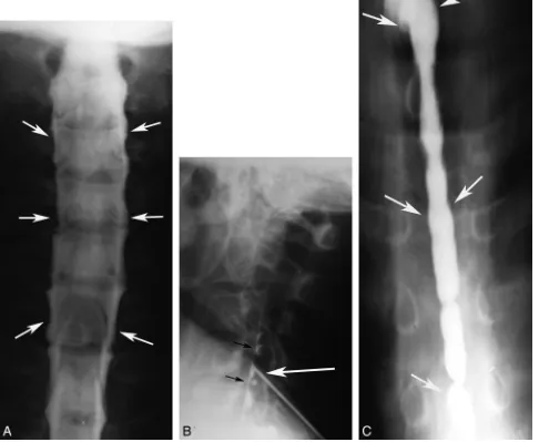
Metrizamide (Amipaque, Nyegaard and Company, Oslo, Norway), the first nonionic water-soluble contrast medium for myelography, was introduced in the early 1970s.38 Side effects were milder than those with the ionic water-soluble agents, consisting of mainly nausea and vomiting, though there were infrequent reports of more serious side effects, in-cluding seizures, hallucinations, and aseptic meningitis. Un-like Pantopaque, it was not associated with arachnoiditis.38 Use of this agent resulted in excellent delineation of the nerve root sleeves and greater accuracy in the diagnosis of lumbar disk disease compared with Pantopaque.40 It could also be used for cervical and thoracic myelograms (Fig 2). It was ab-sorbed into the blood stream, so it did not need to be removed at the end of the procedure.38
The following decade saw the introduction of newer non-ionic water-soluble agents that are still used today, including
[image:3.594.340.493.47.282.2]iohexol (Omnipaque; Nycomed, Princeton, New Jersey) and iopamidol (Isovue; Bristol-Myers Squibb, Princeton, New Jer-sey).41,42These are associated with less toxicity than metriz-amide.43Although not without risk, the side effects are gener-ally milder, consisting mainly of headache. A small percentage of patients develop confusion, radicular pain, and meningis-mus following their use, and seizures have rarely been reported.42-44
Endomyelography
If a myelogram showed cord enlargement, particularly if the enlarged cord was collapsible or partially collapsible, suggest-ing a cystic or partially cystic mass, a cord puncture could be performed at the widest part of the cord enlargement. If a cystic mass was encountered, the fluid would be drained and air or positive contrast could be injected. This was then fol-lowed by radiography and tomography while the patient’s
po-sitioned was varied. This was referred to as endomyelography or myelocystography.33,45The extent of the cyst and the char-acter of the cyst wall could be assessed. If mural nodules were seen in the cyst, these suggested a neoplastic cyst, whereas a smooth cyst wall implied a non-neoplastic process.33,45,46This procedure was apparently relatively pain free if a syrinx was punctured, perhaps related to extreme thinning of the cord, while puncture of tumoral cysts could be painful.46 Publica-tions pertaining to this procedure indicate that there were no significant complications. Careful myelography needed to be performed first to assess any abnormally enlarged vessels which, in conjunction with an enlarged cord, would suggest an hemangioblastoma or AVM, which was considered a contra-indication to performing this procedure.33,46The first descrip-tion of this procedure was in 1928, and though it does not appear to have been performed frequently, there are case re-ports of its use up until the 1980s (Fig 3).46
[image:4.594.53.539.60.459.2]Spinal Angiography
Although cerebral angiography was first successfully per-formed in 1927 by Egas Moniz, it was nearly another 40 years before spinal angiography was attempted.47Before this, my-elography was the main diagnostic technique to diagnose spi-nal vascular malformations, which were seen as serpiginous filling defects in the contrast column.48Initial attempts to vi-sualize spinal vascular lesions or the artery of Adamkeiwicz used injections into the subclavian artery or aorta, depending on the location of the suspected lesion.49Subsequently, selec-tive catheterization of the vertebral, intercostal, and lumbar arteries was described using the Seldinger technique.50 Sub-traction techniques that were developed in the 1960s helped in the visualization of the small abnormal vessels associated with spinal vascular malformations.48Many patients experienced lower extremity spasms during these procedures, which was thought to be related to the neurotoxic effects and hypertonic-ity of the early ionic water-soluble contrast agents.50,51The complication rate following spinal cord angiography, includ-ing spinal cord injury, was initially high; however, with ad-vances in angiographic techniques and contrast agent formu-lation, complication rates are now similar to those for cerebral angiography.51 Although CTA and MRA techniques of the spine are now possible, spinal angiography remains the defin-itive test for diagnosing and, in many instances, guiding the treatment of these lesions (Fig 4).52
Cross-Sectional Imaging of the Spine
The first CT scanner introduced in 1973 was only capable of imaging the head.53,54The following year, Robert Ledley, a
dentist by training with an MA in physics, who was a professor of physiology, biophysics, and radiology at Georgetown Uni-versity, developed the first whole-body CT scanner.55Ledley, who founded the National Biomedical Research Foundation in 1960, had recently lost his National Institutes of Health funding and was looking for a project to maintain his lab. He was shown a brochure of the original Electric and Musical Industries CT scanner (ACTA Scanner, Pfizer, New York, New York) by a neurosurgeon at Georgetown University.56 Frus-trated by the limitations of this scanner and aware of Allan Cormack’s work, he determined that he could build a CT scan-ner by using the convolution image-reconstruction technique that could image the whole body.17,56The prototype scanner was built with the help of a local machine shop and Cadillac car dealership.56The first articles on this technique appeared in late 1974 and early 1975, including an article on its use in the diagnosis of syringomyelia by Ledley and neuroradiologist Giovanni Di Chiro.57 The axial images of the spine were 7.5 mm thick with a 2-mm gap.
Additional articles on CT of the spine soon followed, which included descriptions of congenital anomalies, spinal canal stenosis, degenerative changes, spinal canal and spinal cord masses, vascular lesions, bone destruction, and fractures.58 The first report of a postmyelogram CT, referred to as com-puter-assisted myelography, was in 1976 by Di Chiro and Shellinger, who indicated that the spinal cord outline could be more easily seen in this manner than with plain CT (Fig 5).59 During the next several years, articles describing the efficacy of combining myelography with CT in a variety of pathologic conditions were published.60In the early 1980s, lumbar spine
[image:5.594.133.454.43.344.2]CT after IV contrast administration was used to aid in the diagnosis of degenerative disease, especially in the cervical spine, in the postoperative lumbar spine to differentiate recur-rent disk from scar, and in tumors and inflammatory diseases of the spine.8
Although the history of MR imaging dates back to the 1940s, the first commercial MR imaging scanner was intro-duced in 1980 and the first superconducting magnet was put in clinical use in 1981.61Publications on the clinical use of MR imaging of the spine began appearing in the literature in 1983. These initial studies were performed on 0.15T-0.6T magnets, and spatial resolution and signal-to-noise ratios were poor by today’s standards. The minimal section thickness for MR im-aging was 1–1.5 cm, and a T2-weighted sequence could take up to 40 minutes to perform. However, the tissue characteriza-tion and multiplanar capability allowed delineacharacteriza-tion of a vari-ety of intramedullary, intradural-extramedullary, and extra-dural processes without the need for contrast or ionizing radiation.62-64 Technical advances came rapidly, including multisection multiecho techniques, sequence optimization, surface coils, and higher field strength units, which resulted in improved contrast and spatial resolution.65Gadolinium con-trast agents were first suggested in the early 1980s, with FDA approval of gadolinium chelates in 1988, which improved the detection and characterization of many diseases involving the spine.66For the first time, the spinal cord and cord pathology could be directly visualized rather than the margins of the cord simply being outlined by contrast.
Advances in CT and MR Imaging of the Spine
Technologic advances in CT and MR imaging have continued unabated since their introduction into clinical use. Advances in CT technology have resulted in thinner sections, improved spatial resolution, faster imaging, fewer motion artifacts, and larger areas of coverage.67-69Although 3D reconstruction of CT images was first attempted in the 1970s, improved spatial resolution with isotropic voxels allowing high-quality multi-planar and 3D reconstructions is a more recent development. Faster scanning allows improved contrast use with angio-graphic imaging and dynamic scanning.68,69These advances have resulted in CT of the spine with multiplanar reformatted images having essentially replaced conventional x-rays for the evaluation of spinal trauma, especially in the cervical spine in
patients with high risk of injury, due to the improved sensitiv-ity and specificsensitiv-ity of CT compared with x-rays.70CT is also the technique of choice when evaluating the spine for nontrau-matic osseous abnormalities. CT myelography remains an im-portant tool in the diagnosis of spinal disease and not just in patients who cannot have MR imaging. CT myelography is useful in the postoperative spine, with fewer artifacts related to surgical hardware than MR imaging, and in assessing spinal canal stenosis, foraminal stenosis, and nerve root compres-sion.71,72Myelography can be performed in a dynamic fash-ion, which may be helpful in the diagnosis of spinal stenosis and in evaluating CSF leaks.71CTA of the spine is now possible and may be helpful in diagnosing and guiding the further management of spinal vascular malformations.73
The advances in CT technology resulting in increasing use have led to concerns over the radiation exposure patients re-ceive during these studies.74 In light of these concerns, CT manufacturers have developed dose modulation and iterative reconstruction techniques that decrease patient dose without sacrificing image quality.75,76Radiologists need to be advo-cates for patient safety by using protocols that limit radiation exposure and, when appropriate, substituting examinations without ionizing radiation, such as MR imaging.74
Spinal MR imaging research aimed at acquiring images faster with better contrast and spatial resolution. This led to developments such as fast spin-echo and gradient-echo se-quences, echo-planar imaging,k-space substitution, parallel imaging, and phased-array coil technology.77,78Spatial reso-lution improved, and 3D imaging with MR imaging became possible. As described in the earlier article in this series on the history of neuroradiology, MRA, DWI, DTI, CSF flow studies, MR spectroscopy, fMRI, and perfusion MR imaging were all introduced. More recently 3T scanners have been introduced into routine clinical work.79Many of these newer techniques have had limited application in the spine due to technical dif-ficulties that limit image quality, including the high magnetic susceptibility of the structures surrounding the spinal canal, the relatively small size of spinal canal structures, the large craniocaudal extent of the spine, CSF and vascular pulsation, respiration, and swallowing.80DWI has been applied to the spinal cord and may be helpful in detecting acute cord isch-emia as well as other cord lesions.81MRA of the spine has been shown to be helpful in assessing the spinal vasculature and
[image:6.594.134.454.44.200.2]vascular malformations, particularly as a guide for further en-dovascular assessment and follow-up after treatment.82Fetal MR imaging is now routinely used to further evaluate abnor-malities seen on prenatal sonography and, in conjunction with fetal surgery, has led to improved outcomes in infants with myelomeningoceles (Fig 6).83,84 These developments have made MR imaging the primary technique used to assess spinal disease.
Conclusions
To some, imaging of the spine may not be as enticing and imaging advances may not be as dramatic as those of the brain; nevertheless it remains a commonly used (some may even say overused) procedure in all imaging departments. As with im-aging of the brain, profound advances have been made in spi-nal imaging, which have had a positive impact on the diagnosis and treatment of patients. We expect that progress will con-tinue in the future that will lead to even less invasive, safer, and faster more specific diagnostic techniques, resulting in even earlier diagnosis and treatment with a continuing positive im-pact on patient outcome.
Disclosures: Suresh K. Mukherji—UNRELATED: Consultancy: Philips Medical Systems,
Comment: Consultant. Douglas J. Quint—UNRELATED:Expert Testimony: Medical legal.
References
1. Schu¨ller A.Ro¨entgen-diagnostik der erkrankugen des kopfas.Vienna, Austria: Holder; 1912
2. Dewing SB.Modern Radiology in Historical Perspective. Springfield, Illinois: Charles C. Thomas; 1962:94 –96
3. Cirillo VJ.The Spanish-American War and military radiology.AJR Am J Roentgenol2000;174:1233–39
4. Bull JW.History of neuroradiology: the presidential address, delivered at the British Institute of Radiology on October 20, 1960.Br J Radiol1961;34:69 – 84 5. Cushing H.Haematomyelia from gunshot wound of the cervical spine.Bull
Johns Hopkins Hosp1897;8:195–97
6. Dewing SB.Modern Radiology in Historical Perspective.Springfield, Illinois: Charles C. Thomas; 1962:89
7. Bull JW.The history of neuroradiology.Proc R Soc Med.1970;63:637– 43 8. Hesselink JR.Spine imaging: history, achievements, remaining frontiers.AJR
Am J Roentgenol1988;150:1223–29
9. Andrews J.Planigraphy: introduction and history.AJR Am J Roentgenol
1936;36:575– 87
10. Emch TM, Modic MT.Imaging of lumbar degenerative disc disease: history and current state.Skeletal Radiol2011;40:1175– 89
11. Dandy WE.Ro¨ntgenography of brain after the injection of air into the spinal canal.Ann Surg1919;70:397– 403
12. Dandy WE.The diagnosis and localization of spinal cord tumors.Ann Surg
1925;81:223–54
13. Jacobaeus HC.On insufflation of air into the spinal canal for diagnostic pur-poses in cases of tumors of the spinal canal.Acta Med Scand1921;55:555– 64 14. Bull J.Myelography.Neuroradiology1971;2:1–2
15. Widero¨e S.U¨ ber die diagnostische bedeutung der interspinalen luftinjek-tioned bei Ruckenmarkslieden, besonders bei geschwulsten.Z Chir1921;48: 394 –97
16. Sicard JA, Forestier JE.Methode generale d’exploration radiologique par l’huile iode´e (Lipiodol).Bull Soc Med Hop Paris1922;46:463– 8
17. Wolpert SM.In re: Di Chiro G, Schellinger D— computed tomography of spinal cord after lumbar intrathecal introduction of metrizamide (computer-assisted myelography).AJNR Am J Neuroradiol2001;22:219 –21
18. Sicard JA, Forestier J.The Use of Lipiodol in Diagnosis and Treatment.London, UK: Oxford University Press; 1932
19. Mixter WJ.The use of lipiodol in tumor of the spinal cord.Arch Neurol
1925;14:35– 45
20. Bonnemain B, Guerbet M.The history of Lipiodol (1901–1994) or how a med-ication may evolve with the times.Rev Hist Pharm (Paris)1995;42:159 –70 21. Globus JH, Strauss I.Intraspinal iodolography: subarachnoid injection of
io-dized oil as an aid in the detection and localization of lesions compressing the cord.Arch Neurol Psychiatry1929;21:1331– 86
22. Peterson HO.The hazards of myelography.Radiology1975;115:237–39 23. Camp JD.Contrast myelography past and present.Radiology1950;54:477–506 24. Schmorl G.Uber die pathologishe anatomie der wirbelbandscheiben. Beitr Klin
Chir1931;151:360 – 68
25. Mixter WJ, Barr JS.Rupture of the intervertebral disc with involvement of the spinal canal.N Engl J Med1934;211:210 –15
26. Hampton A, Robinson M.Roentgenographic demonstration of rupture of the intervertebral disk into the spinal canal after the injection of Lipiodol.AJR Am J Roentgenol1936;36:782– 803
27. Kubik CS, Hampton AO.Removal of iodized oil by lumbar puncture.N Engl J Med1941;224:455–57
28. Nichols BH, Nosik WA.Myelography with the use of thorium dioxide solution (Thorotrast) as a contrast medium.Radiology1940;35:459 – 66
29. Maltby GL.Progressive thorium dioxide myelopathy.N Engl J Med1964;270: 490 –96
30. Meyer MW, Powell HC, Wagner M, et al.Thorotrast induced adhesive arach-noiditis associated with meningioma and schwannoma.Hum Pathol1978;9: 366 –70
31. Arnell S, Lidstro¨m F.Myelography with Skiodan (Abrodil).Acta Radiol1931; 12:287– 88
32. Lindgren E.On the diagnosis of tumors of the spinal cord by the aid of gas myelography.Acta Chir Scand1939;82:303–18
33. Quencer RM, Tenner MS, Rothman LM.Percutaneous spinal cord puncture and myelocystography.Radiology1976;118:637– 44
34. Ramsey GH, French JD, Strain WH.Iodinated organic compounds as contrast media for radiographic diagnosis. IV. Pantopaque myelography.Radiology
1944;43:236 – 40
35. Huckman MS.Neuroradiology without benefit of computers: a memoir. AJNR Am J Neuroradiol2010;31:783– 86
36. Campbell RL, Campbell JA, Heimberger RF, et al.Ventriculography and my-elography with absorbable radiopaque medium.Radiology1964;82:286 – 89 37. Gonsette R.An experimental and clinical assessment of water-soluble
con-trast medium in neuroradiology a new medium: Dimer X.Clin Radiol
1971;22:44 –56
38. Skalpe IO, Amundsen P.Lumbar radiculography with metrizamide.Radiology
1975;115:91–95
39. van der Leede H, Jorens PG, Parizel P, et al.Inadvertent intrathecal use of ionic contrast agents.Eur Radiol2002;12:S86 –93
40. Herkowitz HN, Romeyn RL, Rothman RH.The indication for metrizamide myelography: relationship with complications after myelography.J Bone Joint Surg Am1983;65:1144 – 49
41. Eldevik OP, Nakstad P, Kendall BE, et al.Iohexol in lumbar myelography; preliminary results of an open, noncomparative multicenter study.AJNR Am J Neuroradiol1983;4:299 –301
42. Witwer G, Cacayorin ED, Bernstein AD, et al.Iopamidol and metrizamide for myelography: prospective double-blind clinical trial.AJR Am J Roentgenol
1984;143:869 –73
43. Stevens JM.Imaging of the spinal cord.J Neurol Neurosurg Psychiatry1995;58: 403–16
44. Altschuler EM, Segal R.Generalized seizures following myelography with io-hexol.J Spinal Disord1990;3:59 – 61
45. Kendall B, Symon L.Cyst puncture and endomyelography in cystic tumours of the spinal cord.Br J Radiol1973;46:198 –204
46. Dietemann JL, Babin E, Wackenheim A, et al.Percutaneous puncture of spinal cysts in the diagnosis and therapy of syringomyelia and cystic tumors. Neuro-radiology1982;24:59 – 63
47. Moniz E.L’encephalographie arterielle, son importance dans la localization des tumeurs cerebrales.Rev Neurol (Paris)1927;2:72–90
48. Baker HL, Love JG, Layton DD.Angiographic and surgical aspects of spinal cord vascular anomalies.Radiology1967;88:1078 – 85
[image:7.594.90.247.43.170.2]49. Houdart R, Djinjian R.Vascular disorders of the spinal cord.Proc R Soc Med
1966;59:787–90
50. Di Chiro G, Doppman J, Ommaya AK.Selective arteriography of arterio-venous malformations of spinal cord.Radiology1967;88:1065–77
51. Forbes G, Nichols DA, Jack Jr CR, et al.Complications of spinal arteriography: prospective assessment of risk for diagnostic procedures.Radiology1988;169: 479 – 84
52. Chen J, Gailloud P.Safety of spinal angiography: complication rate analysis in 302 diagnostic angiograms.Neurology2011;77:1235– 40
53. Cormack AM.Representation of a function by its line integrals, with some radiological applications.J Appl Physics1963;34:2722–27
54. Cormack AM.Representation of a function by its line integrals, with some radiological applications.II. J Appl Physics1964;35:2908 –13
55. Ledley RS, Di Chiro G, Luessenhop AJ, et al.Computer transaxial x-ray tomog-raphy of the human body: a new tomographic instrument is able to distin-guish between soft tissues everywhere in the body.Science1974;186:207–12 56. Sittig DF, Ash JS, Ledley RS.The story behind the development of the first
whole-body computerized tomography scanner as told by Robert S. Ledley. J Am Med Inform Assoc2006;13:465– 69
57. Di Chiro G, Axelbaum SP, Schellinger D, et al.Computerized axial tomography in syringomyelia.N Engl J Med1975;292:13– 6
58. Lee BCP, Kazam E, Newman AD.Computed tomography of the spine and spinal cord.Radiology1978;128:95–102
59. Di Chiro G, Shellinger D.Computed tomography of the spinal cord after lum-bar intrathecal introduction of metrizamide (computer assisted myelogra-phy).Radiology1976;120:101– 04
60. Dublin AB, McGahan JP, Reid MH.The value of computed tomography me-trizamide myelography in the neuroradiologic evaluation of the spine. Radi-ology1983;146:79 – 86
61. Taveras JM.Neuroradiology: past, present and future.Radiology1990;175: 593– 602
62. Crooks LE, Ortendahl DA, Kaufman L, et al.Clinical efficacy of nuclear mag-netic resonance imaging.Radiology1983;146:123– 8
63. Modic MT, Weinstein MA, Pavlicek W, et al.Nuclear magnetic resonance im-aging of the spine.Radiology1983;148:757– 62
64. Modic MT, Pavlicek W, Weinstein MA, et al.Magnetic resonance imaging of intervertebral disc disease: clinical and pulse sequence considerations. Radi-ology1984;152:103–11
65. Modic MT, Masaryk TJ, Ross JS.Magnetic Resonance Imaging of the Spine.2nd ed. St. Louis: Mosby; 1994:xi.
66. Modic MT.Advances in contrast-enhanced MR imaging. Neurologic applica-tions.AJR Am J Roentgenol1991;156:239 – 45.
67. Wesolowski JR, Lev MH.CT: history, technology and clinical aspects.Semin Ultrasound CT MR2005;26:376 –79
68. Goldman LW.Principles of CT and CT technology.J Nucl Med Technol2007; 35:115–28
69. Heiken JP, Brink JA, Vannier MW.Spiral (helical) CT.Radiology1993;189: 647–56
70. Kokabi N, Raper DMS, Xing M, et al.Application of imaging guidelines in patients with suspected cervical spine trauma: retrospective analysis and lit-erature review.Emerg Radiol2011;18:31–38
71. Harreld JH, McMenamy JM, Toomay SM, et al.Myelography: a primer.Curr Probl Diagn Radiol2011;40:149 –57
72. Song KJ, Choi BW, Kim GH, et al.Clinical usefulness of CT-myelogram com-paring with the MRI in degenerative cervical spine disorders: is CTM still useful for primary diagnostic tool?J Spinal Disord Tech2009;22:353–57 73. Si-jia G, Meng-wei Z, Xi-ping L, et al.The clinical application studies of CT
spinal angiography with 64-detector row spiral CT in diagnosing spinal vas-cular malformations.Eur J Radiol2009;71:22–28
74. Fazel R, Krumholz HM, Wang Y, et al.Exposure to low-dose ionizing radiation from medical imaging procedures.N Engl J Med2009;361:849 –57 75. Smith AB, Dillon WP, Lau BC, et al.Radiation dose reduction strategies for CT
protocols: successful implementation in neuroradiology section.Radiology
2008;247:499 –506
76. Hara AK, Paden RG, Silva AC, et al.Iterative reconstruction technique for reducing body radiation dose at CT: feasibility study.AJR Am J Roentgenol
2009;193:764 –71
77. Magnetic resonance: a peer-reviewed, critical introduction. http://www. magnetic-resonance.org/MagRes%20Chapters/20_15.htm. Accessed February 25, 2012
78. Roemer PB, Edelstein WA, Hayse Ce, et al.The NMR phase array.Magn Reson Med1990;16:192–225
79. Hoeffner EG, Mukherji SK, Srinivasan A, et al.Neuroradiology back to the future: brain imaging.AJNR Am J Neuroradiol2012;33:5–11
80. Vertinsky AT, Krasnokutsky MV, Augustin M, et al.Cutting-edge imaging of the spine.Neuroimaging Clin N Am2007;17:117–36
81. Tanenbaum LN.Diffusion imaging in the spine.Appl Radiol2011;40:9 –15 82. Backes WH, Nijenhuis RJ.Advances in spinal cord MR angiography.AJNR
Am J Neuroradiol2008;29:619 –31
83. Glenn OA, Coakley FA.MRI of the fetal central nervous system and body.Clin Perinatol2009;36:273–300