Dr Kumar Satya Prakash et al JMSCR Volume 06 Issue 07 July 2018 Page 717
Original Research Article
Cutaneous manifestations in patients with diabetes mellitus in a tertiary
care hospital at Bihar, India: An Observational Study
Authors
Dr Kumar Satya Prakash
1, Dr Ichchhit Bharat
2*1
Assistant Professor, Department of Dermatology, Mata Gujri Memorial Medical College & LSK Hospital, Purabbali, Dinajpur Road, Kishanganj, Bihar 855108
2
Senior Resident, Department of Dermatology, Mata Gujri Memorial Medical College & LSK Hospital, Purabbali, Dinajpur Road, Kishanganj, Bihar 855108
*Corresponding Author
Dr Ichchhit Bharat
Senior Resident, Department of Dermatology, Mata Gujri Memorial Medical College & LSK Hospital, Purabbali, Dinajpur Road, Kishanganj, Bihar 855108, India
Email: drichchhitbharat@gmail.com
Abstract
Background: Almost all diabetic patients eventually develop skin complications from the long-term effects of diabetes mellitus on the microcirculation and on skin collagen. Cutaneous infections are more common in type 2 diabetes, whereas autoimmune-related lesions are more common in type 1.
Methodology:This observational study was conducted with patients in the Diabetic OPD of a tertiary care teaching hospital in Eastern India from January 2016 to December 2016. Two hundred patients satisfying the diagnostic criteria for diabetes were taken. A detailed history was elicited in each case with particular reference to cutaneous complaints and including details regarding duration, history of evolution, progression and treatment modalites, if any.
Results:Of the study population 178 (89%) were cases of type 2 diabetes, 19 (9.5%) type 1 diabetes and 03 (1.5%) were cases of gestational diabetes. Out of 200 cases, 111 (55.5%) patients had some associated cutaneous manifestations comprising 69 (62.16%) males and 42 (37.84%) females. Cutaneous infections formed the largest group of dermatoses were observed in 61 (54.95%).Among the bacterial infection which were seen in 21 (18.92%) cases, impetigo contagiosa 7 (6.31%), folliculitis and boils 6 (5.41%), and two cases of erythrasma (1.8%). Dermatophytoses were seen in 22 (19.82%) patients, comprising tinea corporis et curis 9 (8.1%), tinea unguium in 3 (2.7%), tinea pedis 8 (7.21%) and tinea incognito in 2 (1.8%). Herpes
zoster was seen in 2 patients (1.8%). Dermatoses or non-infectious skin lesions were seen in 58 (52.25%)
diabetic cases.
Conclusion:Diabetes mellitus can be complicated by a variety of cutaneous manifestations. As the duration of diabetes increased, the likelihood of developing skin manifestations also increases. Early referral to the dermatologist may help to detect complications of the skin in diabetes at an early stage and may prevent disability caused by these complications.
Keywords: Diabetes mellitus, Cutaneous manifestations, Fungal infections, Dermatophytic infections, Dermatoses.
www.jmscr.igmpublication.org Impact Factor (SJIF): 6.379
Dr Kumar Satya Prakash et al JMSCR Volume 06 Issue 07 July 2018 Page 718
Introduction
Diabetes mellitus, more simply called diabetes, is a chronic condition that occurs when there are raised levels of glucose in the blood because the body cannot produce any or enough of the hormone insulin or use insulin effectively. Type 1 diabetes is caused by an autoimmune reaction where the body’s immune system attacks the insulin-producing beta cells in the islets of the pancreas gland. Type 2 diabetes is the most common type of diabetes, accounting for around 90% of all cases of diabetes. In type 2 diabetes, hyperglycaemia is the result of an inadequate production of insulin and inability of the body to respond fully to insulin, defined as insulin resistance.2,3 Global estimates of type 2 diabetics in the year 2030 is likely to be 552 million. The International Diabetes Federation (IDF) documents the total number of diabetic subjects to be around 61.3 million in India and this is further set to raise to 101.2 million by the year 2030.4 It exhibits a variety of multisystem complications involving the blood vessels, skin, eye, kidney, and the nervous system during the course of the disease process. Abnormal carbohydrate metabolism, other altered metabolic pathways, atherosclerosis, microangiopathy, neurone degeneration, and impaired host mechanisms all play roles.5 More than one third of diabetic patients have some type of dermatologic manifestations during the course of their chronic disease.6 Abnormal carbohydrate metabolism, atherosclerosis, microangiopathy, neuron degeneration and impaired host mechanism all play roles in the pathogenesis of cutaneous complications.7
There no epidemiologic data related to skin disorders in diabetics reported from Eastern India especially from Bihar. This study was designed to analyze the prevalence and pattern of skin disorders among diabetic patients from this region.
Materials & Methods
This observational study was conducted with patients in the Diabetic OPD of a tertiary care teaching hospital in Eastern India from January 2016 to December 2016. Two hundred patients satisfying the diagnostic criteria for diabetes were taken. The data were collected prospectively and systematically in a pre-established proforma after an informed written consent was obtained from all subjects. History, including age, sex, type of diabetes, duration, complications and treatment modalities were noted. All the patients were subjected to complete and thorough dermatological examination. A detailed history was elicited in each case with particular reference to cutaneous complaints and including details regarding duration, history of evolution, progression and treatment modalites, if any. Apart from routine haematological investigations, fasting and post prandial blood sugar levels were done in all the patients. Glycosylated Haemoglobin levels were done in patients by Ion exchange resin method. The fungal infections were confirmed by wet mount in 10%-40% Potassium hydroxide solution. Bacterial infections were confirmed by smear examination in gram stain and bacterial culture. Serum lipid profile, serum creatinine, and fundus examination were done to detect complications. Relevant microbiological and histopathological investigations were done wherever necessary to confirm the clinical diagnosis. Findings were recorded and results obtained were tabulated and statistically analyzed.
Results
Dr Kumar Satya Prakash et al JMSCR Volume 06 Issue 07 July 2018 Page 719 years in 164 patients (82%) and 19 (9.5%) had
>10 years of diabetes. Seventeen patients (8.5%) were newly diagnosed as diabetics. The maximum number of patients was in the age group of 41 to 60 years 93 (46.5%) followed by 20 to 40 years 68 (34%). In diabetic cases, the mean body mass
index was found to be 26.1±3.5 (mean ± SD). Out of 200 cases, 111 (55.5%) patients had some associated cutaneous manifestations comprising 69 (62.16%) males and 42 (37.84%) females [Table 1].
Table 1: Demographic and clinical characteristics of diabetic patients [n=200]
Characteristics No. of patients Percentage/ Mean±SD
Type 1 diabetes Males
Females
19 13 6
9.5% 68.42%
9.5% Type 2 diabetes
Males Females
178 117 61
89% 65.73% 34.67%
Gestational diabetes 03 1.5%
Mean age [years] 200 48.7±7.9
Age groups [years] < 20
20-40 41-60 >60
9 68 93 30
4.5% 34% 46.5%
15% Duration of diabetes
Newly onset 1-10 yrs >10 years
17 164
19
8.5 82 9.5
Mean body mass index 200 26.1±3.5
Cutaneous manifestations Males
Females
111 69 42
55.5% 62.16% 37.84% Cutaneous manifestations with duration
of diabetes < 1 year 1-5 years 6-10 years > 10 years
6 34 24 47
5.41% 30.63% 21.62% 42.34% Associated disorders with cutaneous
manifestations Hypertension Nephropathy Neuropathy Retinopathy
33 4 5 4
29.73% 3.60% 4.50% 3.60% HbA1c with cutaneous manifestations
Controlled Uncontrolled
60 51
54.05% 45.95% The cutaneous manifestations were most prevalent
in the age group of 41- 60 years. The majority of patients 34 (30.63%) with cutaneous manifestations were having disease duration between 1-5 years, followed by 6-10 years 24 (21.62%), > 10 years 47 (42.34%) and less than one year 6 (5.41%). In diabetic patients with cutaneous manifestations, hypertension was present in 33 (29.73%), nephropathy in 4 (3.60%), neuropathy in 5 (4.50%), and retinopathy in 4
Dr Kumar Satya Prakash et al JMSCR Volume 06 Issue 07 July 2018 Page 720
Table 2: Distribution of different infective skin lesions in diabetic patients [n=111]
Cutaneous manifestations No. of patients
Percentage
Infectious skin lesions (total) 61 54.95
Fungal infection (total) Candidiasis
Dermatophytic infections Tinea corporis et curis Tinea unguium Tinea pedis Tinea incognito
36 14 22 9 3 8 2
32.43 12.61 19.82 8.11
2.7 7.21
1.8 Bacterial infection (total)
Impetigo contagiosa Boils
Folliculitis Erythrasma
21 7 6 6 2
18.92 6.31 5.41 5.41 1.8
Infestation (Scabies) 01 0.9
Viral infection 02 1.8
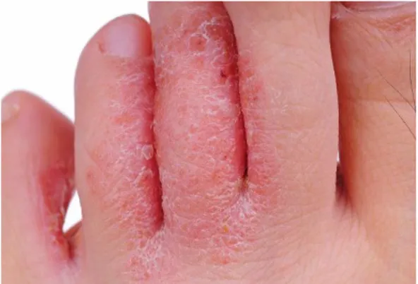
Cutaneous infections formed the largest group of dermatoses were observed in 61 (54.95%). Among the bacterial infection which were seen in 21 (18.92%) cases, impetigo contagiosa 7 (6.31%), folliculitis and boils 6 (5.41%), and two cases of erythrasma (1.8%). Dermatophytoses [Figure 1] were seen in 22 (19.82%) patients, comprising tinea corporis et curis 9 (8.1%), tinea unguium in 3 (2.7%), tinea pedis 8 (7.21%) and tinea incognito in 2 (1.8%). Herpes zoster was seen in 2 patients (1.8%) [Table 2].
Table 3: Distribution of different non-infective skin lesions in diabetic patients [n=111]
Cutaneous manifestations No. of patients Percentage Non-infectious skin lesions (total) 58 52.25
Xerosis 16 14.41
Lipodystrophy 4 3.6
Skin rash 2 1.8
Hyperpigmentation 2 1.8
Lichen planus 1 0.9
Vitiligo 2 1.8
Prurigo nodularis 2 1.8
Xanthoma 4 3.6
Acanthosis nigricans 7 6.31
Thickening of skin 2 1.8
Bullous diabeticorum 1 0.9
Psoriasis 1 0.9
Granuloma annulare 0 0
Sclerederma diabeticorum 0 0
Rubeosis facei 2 1.8
Epidermal Necrolysis/Stevens Johnson Syndrome
0 0
Onychodystrophy 3 2.7
Periungual telangectasias 1 0.9
Seborrheic keratosis 4 3.6
Eczema 3 2.7
Melasma 0 0
Keloid 1 0.9
Cutaneous amyloidosis 0 0
Dermatoses or non-infectious skin lesions were seen in 58 (52.25%) diabetic cases [Table 3]. It was found that xerosis (14.41%), acanthosis nigricans (6.31%), seborrheic keratosis (3.6%), lipodystrophy (3.6%), xanthoma [Figure 2] (3.6%), skin rash (1.8%), and hyperpigmentation (1.8%) were reported as non infectious skin manifestations in patients with diabetes mellitus [Table 3]. However, the pattern of lesions was different in Type 1 and Type 2 diabetics. Nine patients out of the 19 (47.37%) type 1 diabetics demonstrated skin lesions, the commonest being diabetic xerosis, infections and diabetic hand. In Type 2 diabetics 101 (90.99%) showed skin lesions followed by one patient (0.9%) in gestational diabetes. Dermatoses, having a possible association with diabetes mellitus were the first to appear, followed by cutaneous infections. Dermatoses, having a strong association with diabetes mellitus were found to occur in patients with a longer duration of diabetes.
Figure 1: Dermatophytic infections
Dr Kumar Satya Prakash et al JMSCR Volume 06 Issue 07 July 2018 Page 721
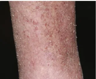
Figure 3: Xerosis in diabetic subjects
Discussion
The skin is affected by both the acute metabolic derangements and the chronic degenerative complications of diabetes.5 Although the mechanism for many diabetes associated skin conditions remains unknown, the pathogenesis of others is linked to abnormal carbohydrate metabolism, other altered metabolic pathways, atherosclerosis, microangiopathy, neuron degeneration and impaired host mechanisms.8 The association of certain skin diseases with diabetes mellitus has been fairly well recognized with an incidence rate ranging from 11.4 to 71%.[9-11] In our study, the majority of patients were between 41- 50 years and the mean age of patients was 48.7±7.9 years. Females outnumbered males and female: male ratio was 1.86:1. The duration of diabetes was 1-10 years in 164 patients (82%) and 19 (9.5%) had >10 years of diabetes. Seventeen patients (8.5%) were newly diagnosed as diabetics. In Phulari YJ et al study12, majority 50.58% were <5 years, 27.05% in 6 to 10 years, 11.76% in 16 to 20 years and 10.85% in 11 to 15 years. Study by Kumar et al13 showed that the duration of diabetes was < 10 years in 30 patients, 17 had 11-20 years and 3 had > 20 years of diabetes.
In the present study the commonest cutaneous feature of diabetes was cutaneous infections (Table 2), seen in 61 (54.95%) patients and dermatophytosis seen in 22 (19.82%) patients. Among the bacterial infection which were seen in
Dr Kumar Satya Prakash et al JMSCR Volume 06 Issue 07 July 2018 Page 722 skin lesions. However the mean HbA1C level was
higher in patients with infective lesions (8.7 ± 1.4 in contrast to 7.2 ± 1.3). Study by Chatterjee N14 revealed 73.9% of diabetic patients had one or more cutaneous manifestations. Mahajan et al.5, reported cutaneous infections in 54.69% of diabetics in their study group.
Acanthosis nigricans is likely the most readily recognized skin manifestation of diabetes (3). It is present in up to 74% of obese adult patients and can be predictive of the existence of hyperinsulinemia.22 Raised levels of insulin act on insulin-like growth factor receptors leading to the development of acanthosis nigricans.23 The proportion of acanthosis nigricans among the diabetics in the study is similar to studies done on Indian patients. [24-26] Grandhe et al27 found acanthosis nigricans to be an independent cutaneous marker of type 2 diabetes mellitus. Diabetic Dermopathy presents as small (<1cm), well-demarcated, atrophic depressions, macules, or papules on the pretibia and is considered to be a sign of insulin resistance. Diabetic dermopathy probably represents post-traumatic atrophy and post-inflammatory hyperpigmentation in poorly vascularized skin.28 Eruptive xanthoma (EX) [Figure 2] presents on the buttocks, elbows, and knees as sudden onset crops of yellow papules with an erythematous base. It is rare and occurs more often in patients with poorly controlled type 2 diabetes.29
Stevens-Johnson syndrome is a rare mucocutaneous necrotizing condition. The dipeptidyl peptidase-4 inhibitor sitagliptin has been associated with cases of Stevens-Johnson Syndrome.30 It could present with fever, headache, rhinitis, cough, malaise, burning eyes, and dysphagia.30 Vitiligo affects 0.3–0.5% of world population, making it the most common depigmenting disorder. A 2009 study of 50 patients with type 1 diabetes reported that 4% of subjects had vitiligo.5 Vitiligo has been associated with both type 1 and type 2 diabetes mellitus and there is an increased association of vitiligo with a proven or presumed autoimmune disease.15, 31
Patients with vitiligo, lichen planus, and cutaneous perforating dermatosis are reported to have an increased incidence of abnormal glucose tolerance or frank diabetes mellitus.32 Xerosis [Figure 3] is another name for dry skin. It is the second most common skin manifestation in people with diabetes.
Yosipovitch et al.33 and Sawhney et al.34 observed that majority of the patients with cutaneous lesions in their respective studies were under poor glycemic control. In Sanad et al35 study, cutaneous infections included fungal (22%), bacterial (16%), and viral (2%) infections. Diabetic dermopathy probably represents post-traumatic atrophy and post-inflammatory hyperpigmentation in poorly vascularized skin.28 In the present study diabetic patients with cutaneous manifestations, hypertension was present in 33 (29.73%), nephropathy in 4 (3.60%), neuropathy in 5 (4.50%), and retinopathy in 4 (3.60%) cases. High prevalence of systemic complications has been reported in diabetic patients with cutaneous involvement as compared to diabetics without cutaneous manifestations.25, 28 According to an Indian study in 2008, no correlation was demonstrated between glucose control and dermatologic manifestations.15 But an Iranian study36 as well as a study by Rayfield et al37 showed definite relationship between foot ulcers and fungal infections with HbA1C levels. Study by Ghosh K et al38 showed that uncontrolled DM or higher HB1AC is associated with diabetic bulla, scleredema, lichen planus and signs of insulin resistance. There is no association with any kind of skin manifestations and microangiopathic complications though often cited as an etiopathogenic factor was found.
Conclusion
Dr Kumar Satya Prakash et al JMSCR Volume 06 Issue 07 July 2018 Page 723 was higher among the diabetics than among
nondiabetics. Patients with poorly controlled diabetes had more number of cutaneous infections and diabetes associated dermatoses. Impaired diabetic control as evidenced by higher HbA1C levels was found among patients with infections. To conclude, diabetes mellitus involves the skin quite often and whenever patients present with multiple skin manifestations their diabetic status should be checked.
References
1. DeFronzo RA, Ferrannini E, Zimmet P, et al. International Textbook of Diabetes Mellitus, 2 Volume Set, 4th Edition. Wiley-Blackwell, 2015.
2. Evans JM, Newton RW, Ruta DA, et al. Socio-economic status, obesity and prevalence of Type 1 and Type 2 diabetes mellitus. Diabet Med J Br Diabet Assoc 2000; 17: 478–80.
3. Holman N, Young B, Gadsby R. Current prevalence of Type 1 and Type 2 diabetes in adults and children in the UK. Diabet Med J Br Diabet Assoc 2015; 32: 1119– 20.
4. Sicree R, Shaw J, Zimmet P. Diabetes and impaired glucose tolerance. In: Gan D, editor. Diabetes atlas. International Diabetes Federation. 5th ed. Belgium: International Diabetes Federation; 2011. p. 15-103.
5. Mahajan S, Koranne R V, Sharma S K. Cutaneous manifestation of diabetes melitus. Indian J Dermatol Venereol Leprol 2003;69:105-8.
6. Greenwood AM. A study of skin in 500 diabetics. JAMA 1927; 89: 774- 779. 7. Wani MA, Hassan F, Bhat MH, Ahmed
QM. Cutaneous Manifestations of Diabetes mellitus: A Hospital Based Study in Kashmir, India. Egyptian Dermatology Online Journal December 2009; 5(2): 1-6. 8. Yosipovitch G, Hodak E, Vardi P, Shraga
J, Karp M, Sprecher E, et al. The
prevalence of cutaneous manifestations in IDDM patients and their association with diabetes risk factors and microvascular complications. Diabetes care 1998; 21: 506-509.
9. Neerja Puri. A study on the cutaneous manifestations of diabetes mellitus. Our Dermatol Online. 2012; 3(2): 83-86. 10.Nigam PK, Pande S. Pattern of dermatoses
in diabetes. Indian J Dermatol Venereol Leprol 2003; 69: 83-85.
11.Wahid Z, Kanjee A. Cutaneous manifestations of diabetes mellitus. J Pak Med Assoc 1998; 48: 304-305.
12.Phulari YJ, Kaushik V. Study of cutaneous manifestations of type 2 diabetes mellitus. Int J Res Dermatol 2018;4:8-13.
13.Kumar AA, Ansari SH, Gupta V. A study on cutaneous manifestations in patients of type 2 diabetes mellitus in a tertiary care hospital. Int J Med Res Prof. 2016;2(1):125-7.
14.Chatterjee N, Chattopadhyay C, Sengupta N, Das C, Sarma N, Pal SK. An observational study of cutaneous manifestations in diabetes mellitus in a tertiary care Hospital of Eastern India. Indian J Endocr Metab 2014;18:217-20. 15.Timshina DK, Thappa DM, Agrawal A. A
clinical study of dermatoses in diabetes to establish its markers. Indian J Dermatol 2012;57:20-5.
16.Giligor RS, Lazarus G S. Skin manifestations of diabetes mellitus. In, Diabetes Mellitus, eds Rifkin H, Raskin P, Brady co, Louana 1981, 313. 321.
17.Sehgal VN, Sanker P Some aspects of skin diseases and diadetes mellitus. Indian J Dermatol Venereal 1965;31:264 - 269. 18.Romano G, Morretti G, Dibenedetto A
Dr Kumar Satya Prakash et al JMSCR Volume 06 Issue 07 July 2018 Page 724 19.Rao GS, Pai GS. Cutaneous manifestations
of diabetes mellitus: A clinical study. Indian J Dermatol Venereol Leprol 1997;63:232-4.
20.Demirseren DD, Emre S, Akoglu G, et al. Relationship between skin diseases and extracutaneous complications of diabetes mellitus: clinical analysis of 750 patients. Am J Clin Dermatol 2014;15:65–70. 21.Bedi BMS, Kandhari KC. Diabetes or
prediabetes and dermatosis. J Assoc Phys Ind 1965; 13: 809- 810.
22.Hud JA Jr, Cohen JB, Wagner JM, Cruz PD Jr. Prevalence and significance of acanthosis nigricans in an adult obese population. Arch Dermatol 1992;128:941– 944.
23.Burke JP, Hale DE, Hazuda HP, Stern MP. A quantitative scale of acanthosis nigricans. Diabetes Care 1999; 22:1655-9. 24.Ragunatha S, Anitha B, Inamadar AC,
Palit A, Devarmani SS. Cutaneous disorders in 500 diabetic patients attending diabetic clinic. Indian J Dermatol 2011; 56:160-4.
25.Bhat YJ, Gupta V, Kudyar RP. Cutaneous manifestations of diabetes mellitus. Int J Diabetes Dev Ctries 2006;26:152-5. 26.Girisha BS, Viswanathan N. Comparison
of cutaneous manifestations of diabetic with nondiabetic patients: A case-control study. Clin Dermatol Rev 2017; 1:9-14. 27.Grandhe NP, Bhansali A, Dogra S, Kumar
B. Acanthosis nigricans: Relation with type 2 diabetes mellitus, anthropometric variables, and body mass in Indians. Postgrad Med J 2005; 81:541-4.
28.Schemer A, Bergiman R, Linn S, Kantor Y, Friedman - Birnbaum R. Diabetic dermopathy and internal complications in diabetes mellitus. Int J Dermatol 1998; 37: 113- 115.
29.Duff M, Demidova O, Blackburn S, Shubrook J. Cutaneous manifestations of
diabetes mellitus. Clinical Diabetes Winter 2015; 33 (1):40-48.
30.Pathak R, Bridgeman MB. Dipeptidyl peptidase-4 (DPP-4) inhibitors in the
management of diabetes. P T
2010;35:509–513.
31.Sibbald RG, Schachter RK. The skin and diabetes mellitus. Int J Dermatol 1984;23:567-84.
32.Huntley AC. The cutaneous manifestations of diabetes mellitus. J Am Acad Dermatol 1982;7:427-55.
33.Yosipovitch G, Hodak E, Vardi P, Shraga I, Karp M, Sprecher E, et al. The prevalence of cutaneous manifestations in IDDM patients and their association with diabetes risk factors and microvascular complications. Diabetes Care 1998;21: 506-9.
34.Sawhney M, Talwar OP, Tutakne MA, Rajpathak SD. Diabetic dermoangiopathy: A clinico-pathological correlation. Indian J Dermatol Venereol Leprol 1992;58:173-8. 35.Sanad EM, ElFangary MM, Sorour NE,
ElNemisy NM. Skin manifestations in Egyptian diabetic patients: a case series study. Egypt J Dermatol Venerol. 2013;33:56-62.
36.Hosseini MS, Ehsani AH, Hosseinpanah F, Azizi F, Salami M, Khedmat H. Skin lesions in type 2 diabetic patients. Iran J Dermatol. 2008;11:113–7.
37.Rayfield EJ, Ault MJ, Keusch GT, Brothers MJ, Nechemias C, Smith H. Infection and diabetes: The case for glucose control. Am J Med. 1982;72:439– 50.
38.Ghosh K, Das K, Ghosh S, et al. Prevalence of Skin Changes in Diabetes Mellitus and its Correlation with Internal Diseases: A Single Center Observational Study. Indian Journal of Dermatology. 2015;60(5):465-469.
![Table 1: Demographic and clinical characteristics of diabetic patients [n=200]](https://thumb-us.123doks.com/thumbv2/123dok_us/7969530.2115996/3.892.139.739.261.939/table-demographic-clinical-characteristics-diabetic-patients-n.webp)