ABSTRACT
Castor (Casz1) is a zinc finger transcription factor that has been shown to be required for heart development in Xenopus. Casz1 has also been linked to high blood pressure and
hypertension in humans through a recent Genome Wide Association Study. The purpose of this
research was to determine the role of Casz1 in cardiac development and how Casz1 fits into the cardiac transcription program. A mouse model was used because of the high genomic
conservation between mice and humans. To determine the spatial and temporal expression of
Casz1 mRNA during murine heart development, I utilized in situ hybridization. In addition, to confirm that CASZ1 protein is present in the developing heart, I performed
immunohistochemistry. My studies showed that Casz1 is expressed in the atria and left ventricle of the developing heart. To determine the role of Casz1 in cardiac development, I examined the cardiomyocyte mitotic index of wild type embryos and Casz1 mutant embryos, and demonstrated that cardiomyocytes lacking Casz1 over-proliferate. This indicates that Casz1 regulates
cardiomyocyte proliferation. Future studies are aimed at identifying genes that Casz1 regulates, providing further insight into the cardiac gene program. These studies hold implications for
understanding congenital heart defects by giving us further insight into the molecular
mechanisms that regulate cardiac development.
INTRODUCTION
Congenital heart defects are the most common type of birth defect, occurring in about 1%
of all live births. Congenital heart defects can take an emotional and financial toll on victims and
their families, so it is important to research the mechanisms behind these defects in order to
development. By studying these mechanisms, we come closer to understanding how to provide
treatment for congenital heart defects.
The heart is the first organ to form during development. Two groups of cells, termed the
primary and secondary heart fields, contribute to the formation of the heart. Cells from the
primary heart field will form a structure known as the cardiac crescent at stage E7.0. The cardiac
crescent develops into the linear heart tube at stage E8.0, which then undergoes looping to
become the classic 4-chambered heart around stage E9.5. Primary heart field cells give rise to
the left ventricle and the two atria, whereas secondary heart field cells give rise to the right
ventricle and the outflow tract1.
Castor (Casz1) is a zinc finger transcription factor that had been shown to be required for heart development in Xenopus. The depletion of Casz1 in Xenopus embryos causes
malformation of the primitive heart tube, which inhibits proper looping of the heart tube to form
the mature heart and is lethal to the embryo. Casz1 has also been linked to high blood pressure and hypertension in humans through a recent Genome Wide Association Study (GWAS)2. The
implications from this GWAS make it important to study the mechanisms through which Casz1
affects cardiomyocyte differentiation and proliferation as a possible means of future treatment
for heart defects. We hypothesize that Casz1 is necessary for proper formation of structures that arise from the primary heart field.
Casz1 is expressed in the mouse, but its role in mammalian cardiac development is poorly understood. There is high genomic conservation between mice and humans, with 87%
conservation of cDNA identity and 90% conservation of protein identity, which makes the
how Casz1 fits into the cardiac transcription program. An important step in doing this is to examine the effects of deleting CASZ1 in the mammalian embryo. To address this, a conditional
mutation was made in mouse using CreLoxP technology. This allowed Casz1 to be ablated at specific times in development in cells expressing Nkx2.5, an early cardiac marker. Casz1
conditional knockout mice begin to show a degenerative phenotype at stage E12.5 and are
embryonic lethal by stage E14.5.
My first goal was to identify the cardiac cell types and structures that express Casz1. To accomplish this, I examined the spatial and temporal expression of Casz1 mRNA in the
developing wild type embryo. Casz1 mRNA is expressed in the developing heart at embryonic stage E8.0 and is dramatically decreased by stage E10.54. Casz1 mRNA was visualized by performing in situ hybridization on mouse embryos at stages E8.0 through E10.5 and mouse hearts at stage E13.5. The expression of Casz1 mRNA was compared to the expression of Nkx2.5
mRNA. Nkx2.5 is a well-characterized cardiac-specific gene5. Therefore, comparing spatial and temporal expression of the Nkx2.5 and Casz1 genes can give insight into the expression of
Casz1.
My studies showed that Casz1 mRNA is expressed in the developing myocardium, particularly in the left ventricle. Through a series of antigen retrieval experiments, I
demonstrated that CASZ1 protein shows similar spatial expression as mRNA. My results are
consistent with the hypothesis that Casz1 may be necessary for the proper formation of structures that arise from primary heart field cells.
Work from our lab has shown that depletion of Casz1 in Nkx2.5-positive cells results in defects in the myocardial wall. These results suggest that Casz1 has a role in regulating
and Casz1 wild type mouse hearts using the proliferation marker Phosphohistone H3 (PHH3). I used the sodium-citrate based antigen retrieval method at stages E10.5 and E12.5. Cell counts
were performed to determine the mitotic index in all heart cells and in cardiomyocytes.
Cardiomyocytes were stained using tropomyosin (Tmy), a cardiomyocyte-specific cytosolic
marker.
Through a series of antigen retrieval experiments, I demonstrated that the left and right
ventricles of Casz1 mutant embryos had increased proliferation compared to wild type embryos at stage E10.5. This is consistent with the studies done in Xenopus, which show an increased proliferation in Casz1 mutant Xenopus embryos. The embryos at stage E12.5 do not show a statistically significant difference in proliferation between mutant ventricles and wild type
ventricles, but it has been shown that there is a decrease in the total number of cardiomyocytes in
the mutant ventricles at stage E12.5. This decrease is likely due to necrosis.
Interestingly, recent RNA Sequencing data has shown that genes found in secondary
heart field cells are up-regulated in Casz1 mutant embryos at stage E10.5. Because previous data showed that Casz1 is expressed in heart structures that developed from primary heart field cells, we are currently testing the hypothesis that Casz1 down-regulates these proteins in primary heart field cells. This down-regulation may keep the primary heart field cells from developing into
secondary heart field structures. Thus, when Casz1 is depleted, the primary heart field cells may begin to express secondary heart field proteins. Further studies will determine how Casz1 fits into the transcriptional pathways involved in cardiomyocyte differentiation. Studying the
mechanisms behind cardiac development will allow us to develop new therapies for congenital
METHODS
In Situ Hybridization
The in situ hybridization probes were made of Casz1 exons 6-7 and Nkx2.5. The probes were synthesized using T7 polymerase from BamHI linearized plasmid DNA from an adult
mouse heart. The DNA was incubated at 37˚C overnight to linearize. 0.5 µl sodium dodecyl
sulfate and 50 µg/ml proteinase K were added to the DNA, which was incubated at 50˚C for 30
minutes to degrade nucleases. The linearized DNA was then incubated in 5X transcription
buffer, DIG RNA labeling mix, dithiothreitol, RNAse inhibitor, and RNA polymerase for 2
hours at 37˚C. 10X DNAse buffer and DNAse were added and the DNA was re-incubated at
37˚C for another 30 minutes. The DNA probes were then purified using the Qiagen RNEasy Kit
and its standard procedure.
Embryos were dissected and fixed in 4% paraformaldehyde, then dehydrated with
methanol and stored at -20˚C. For the whole mount in situ, embryos were rehydrated through 1XPBS (0.14M NaCl, 3mM KCl, 5mM Na2HPO4, 2mM KH2PO4), 0.1% tween, and methanol.
Embryos were incubated in 6% hydrogen peroxide for one hour at room temperature and then in
0.56 µl/ml proteinase K for 10 minutes. The embryos were then fixed in 4% paraformaldehyde/
0.1% glutaraldehyde for 30 minutes at room temperature. Embryos were incubated for 1 hour at
70˚C in 50% formamide, 1.3X SSC, 5mM EDTA, 0.5% CHAPS, 100 µg/mL heparin, 0.2%
Tween-20, and 50 µg/mL yeast torula RNA. The embryos were then incubated in 10 µl/mL of
the Casz1 or Nkx2.5 probe at 70˚C overnight.
The following day, the embryos were incubated for 15 minutes in 100mM maleic acid,
100mM NaCl, and 0.1% Tween-20, with a pH of 7.6. Afterwards, the embryos were incubated
Blocking Reagent/20% heat-inactivated lamb serum/MABT for 1 hour. The embryos were then
incubated in 0.05% anti-DIG antibody overnight at 4˚C. The following day, the embryos were
incubated for 1 hour in 100 mM NaCl, 100 mM Tris pH9.5, 50 mM MgCl2, 0.1% Tween-20, and
1.2 mg/mL Levamisol. They were then incubated in BM Purple AP Substrate in 20-minute
increments until sufficient staining was observed. The embryos were then post-fixed in 4%
paraformaldehyde/0.1% glutaraldehyde for 2 hours. The embryos were placed in 10XPBS
(1.4M NaCl, 30mM KCl, 50mM Na2HPO4, 20mM KH2PO4), and 0.1% Tween-20 for storage.
Embryos were embedded in gelatin and sectioned in 30µm sections using a vibratome.
Immunohistochemistry
Paraffin sections were done on E10.5, E12.5, and E14.5 embryos. Embryos were fixed in
4% PFA, 1XPBS overnight. They were stepped into methanol and stored in 100% methanol at
4˚C. The embryos were washed twice in ethanol for 10 minutes, then twice in 50% ethanol, 50%
xylene for 10 minutes. Then the embryos were washed twice in 100% xylene for 10 minutes,
once in 50% xylene, 50% paraffin for 30 minutes at 56˚C, and three times in 100% paraffin at
56˚C for 30 minutes. The tissue was then embedded in a paraffin mold and stored at 4˚C.
Embryos were sectioned using a microtome at 8µm and sections were stored at 4˚C. Before
antigen retrieval was performed, sections were deparaffinized and rehydrated through two 10
minute washes in 100% xylene, one 10 minute wash in 100% ethanol, one 5 minute wash in 95%
ethanol, one 5 minute wash in 70% ethanol, and two 5 minute washes in ddH2O.
Cryosectioning was performed on E7.5, E10.5, E11.5, and E12.5 embryos. The embryos
were then embedded in OCT and cut on a cryostat at 12µm. The sections were baked at 55˚C for
10 minutes and stored at -20˚C.
Antigen retrieval of both frozen and paraffin sections was performed using a 10mM
sodium citrate buffer (pH 6.0) to break protein crosslinks. Sections were blocked in 10%
heat-inactivated calf serum, 1% TritonX-100, 1XPBS for 1 hour in a humid chamber, then incubated
in primary antibody in 1% Heat-inactivated calf serum, 0.1% TritonX-100, 1XPBSovernight at
4˚C. Slides were incubated in secondary antibody in 1% Heat-inactivated calf serum, 0.1%
TritonX-100, 1XPBS for 1 hour at room temperature. Sections were stained with DAPI in
ethanol for 10 minutes.
The primary antibodies used were:
Castor (Casz1):
Casz1 e14 (Santa Cruz) SC-135453 rabbit IgG (1:1000)
Tropomyosin (Tmy):
CH1 (DSHB) mouse monoclonal IgG1 (1:50 concentration)
Phosphohistone H3
PHH3 millipore 06-570 rabbit (1:200 concentration)
The secondary antibodies used were:
Donkey-anti-rabbit Alexa 488 (green) (1:1000 concentration)
Goat-anti-mouse Alexa 546 (red) (1:1000 concentration)
Cell Counting
All pictures were taken using a Zeiss 700 confocal microscope. For proliferation studies,
antibody staining was performed on anterior, mid, and posterior sections of both a Casz1 null embryonic mouse heart and wild type embryonic mouse heart. For each section, two pictures
were taken of each of the following; the left atria, left ventricle, right atria, and right ventricle.
Cell counts were taken for the total number of cells, the number of proliferating cells, the total
number of cardiomyocytes, and the number of proliferating cardiomyocytes. The mitotic index
was calculated by dividing the number of proliferating cells by the number of overall cells. All
calculations were done using the Microsoft Office Excel software.
RESULTS
Casz1 mRNA is Expressed in the Primitive Left Ventricle and Atrial Areas in the Primitive Heart Tube (E8.0)
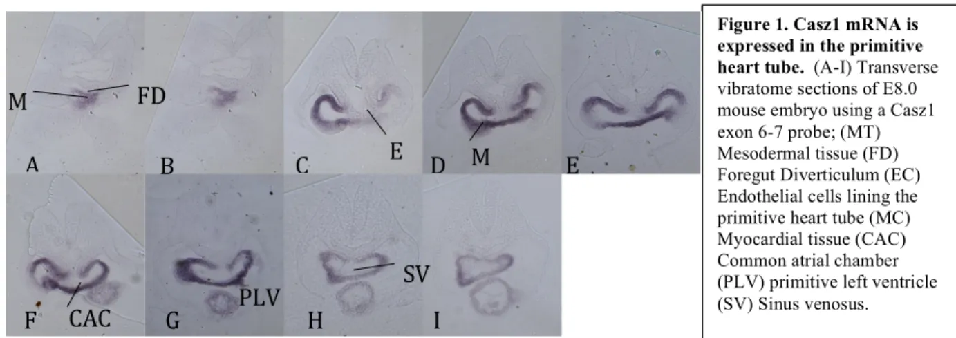
In situ hybridization was performed on E8.0 mouse embryos using a Casz1 exon 6-7 probe to determine Casz1 expression in the primitive heart tube. Transverse sections imaged using an Olympus IX81 microscope show that Casz1 is expressed throughout the primitive heart tube. Specifically, Casz1 expression in the primitive heart tube is strongest in the primitive left ventricle and primitive atrial sections (Fig.1 C-G), as well as in the bulbus cordis (Fig.1 A,B) and
sinus venosus (Fig.1 H,I). Casz1 expression was found in myocardial tissue, which makes up the outside of the primitive heart tube and the bulbus cordis
Figure 1.Casz1 mRNA is expressed in the primitive heart tube. (A-I) Transverse vibratome sections of E8.0 mouse embryo using a Casz1 exon 6-7 probe; (MT) Mesodermal tissue (FD) Foregut Diverticulum (EC) Endothelial cells lining the primitive heart tube (MC) Myocardial tissue (CAC) Common atrial chamber (PLV) primitive left ventricle (SV) Sinus venosus.
a b c d e
M
T
FD
E
C MC
CAC
SV
F G H I
A B C D E
Casz1 mRNA is Expressed in the Common Ventricular Chamber and Common Atrial Chamber in the Looped Heart (E9.5)
In situ hybridization was performed on E9.5 mouse embryos using a Casz1 exon 6-7 probe and an Nkx2.5 probe to determine Casz1 and Nkx2.5 mRNA expression in the looped heart. Transverse sections of an embryo stained for Casz1 (Fig.2 A-F) show that Casz1 is
Figure 2. Casz1 and Nkx2.5 are expressed in the looped heart at stage E9.5. (A-F) Transverse vibratome sections of E9.5 mouse embryo using a Casz1 exon 6-7 probe. (H-M) Transverse vibratome sections of E9.5 embryo using an Nkx2.5 probe. (FD) Foregut diverticulum (NL) Neural lumen (AS) Aortic Sac (RDA) Right dorsal aorta (LDA) left dorsal aorta (MC) Myocardial tissue. (RA) Right component of common atrial chamber (LA) Left component of common atrial chamber (CVC) Common ventricular chamber of heart (EC) Endocardial tissue (G) Whole mount E9.5 mouse embryo using a Casz1 exon 6-7 probe. (N) Whole mount E9.5 mouse embryo using an Nkx2.5 probe. (FBA) First brachial arch (FBG) First brachial groove (SBA) Second brachial arch (SBG) Second brachial groove (TBA) Third brachial arch (TBG) Third brachial groove (RA) Right atria (LV) Left ventricle (OT) Outflow tract
G FD
NL
RDA LDA
RA LA
CVC EC
AS
MC
A B C
D E F
FD NL
AS
LDA
RDA
MC
LA
RA
CVC
EC
K L M N
expressed in the aortic sac (Fig.2 A,B), the left and right sides of the common atrial chamber
(Fig.2 D-F), and the common ventricular chamber of the heart (Fig.2 D-F). Casz1 was also expressed in the neural tissue of the neural lumen (Fig.2A).
Transverse sections of an Nkx2.5 embryo (Fig.2 H-M) show that Nkx2. 5 is expressed in the aortic sac (Fig.2 H,I), the left and right sides of the common atrial chamber (Fig.2 K-M), and
the common ventricular chamber of the heart (Fig.3 K-M). Nkx2.5 expression was strongest in the myocardial tissue. Nkx2.5 expression was not observed in neural tissue (Fig.2 A).
Whole mount images of E9.5 mouse embryos show expression of both Casz1 and Nkx2.5
mRNA in the right side of the common atrial chamber, the left ventricle, and the outflow tract.
Casz1 is expressed in the neural tube (Figure 2 g). Nkx2.5 is expressed in the first, second, and third brachial arches (Figure 2 n).
Casz1 mRNA is Expressed in the Common Atrial Chamber and Left Ventricle in the Looped Heart (E10.5)
In situ hybridization was performed on E10.5 mouse embryos using a Casz1 exon 6-7 probe and an Nkx2.5 probe to determine Casz1 and
Nkx2.5 expression at a later stage in the looped heart. Whole mount images of an E10.5 Nkx2.5
embryo (Fig.3 A,B) show expression of Nkx2.5 in the left and right components of the common atrial
Figure 3. Casz1 mRNA is expressed in the looped heart at stage E10.5. (A,B) Whole mount E10.5 mouse embryo using an Nkx2.5 probe. (C,D) Whole mount E10.5 mouse embryo using a Casz1 exon 6-7 probe. (FBA) First brachial arch. (SBA) Second brachial arch (TBA) Third brachial arch (RA) Right component of common atrial chamber (LV) Left ventricle (O) Otocyst (LA) Left component of common atrial chamber (NT) Neural tube (HB) Hindbrain.
A B
chamber and in the left ventricle. Whole mount images of an E10.5 Casz1 embryo (Fig.3 C,D) show expression of Casz1 in the left side of the common atrial chamber and the left ventricle.
Casz1 is expressed in the neural tube and hindbrain at this stage. Both Nkx2.5 and Casz1 were expressed in the otocyst of the E10.5 embryos. This is unprecedented expression of both Nkx2.5
and Casz1, and is likely due to dye trapping in the embryo.
Casz1 and Nkx2.5 mRNA is Expressed in the Left Ventricle of the Looped Heart (E13.5) In situ hybridization was performed on
E13.5 mouse hearts using a Casz1 exon 6-7 probe and an Nkx2.5 probe to determine Casz1
and Nkx2.5 mRNA expression at a later
stage in the looped
heart. Nkx2.5 is expressed in the outer
curvature of both the
left and right ventricles. Expression of Nkx2.5 is also seen in the left and right atria and in the interventricular septum (Fig.4 A,B). Casz1 is expressed in the outer curvature of the left ventricle, the interventricular septum, and the bulbar cushion tissue. Casz1 also is lightly expressed in the left and right atria (Fig.4 C,D).
Figure 4. Casz1 and
Nkx2.5 are expressed in the looped heart at stage E13.5. (A,B) Image of whole E13.5 mouse heart using an Nkx2.5 probe. (C,D) Image of whole E13.5 mouse heart using a Casz1 exon 6-7 probe.
(RA) Right atria (LA) Left atria (BCT) Bulbar cushion tissue (RV) Right ventricle (LV) Left ventricle (IVS) Interventricular septum
A B
CASZ1 Protein is Expressed in the Aortic Sac and the Left Side of the Common Ventricular Chamber
Confocal imaging showed nuclear staining of CASZ1 protein in cardiomyocytes.
Nuclear staining was shown in
both the aortic sac (Fig.5 A-C) and
common ventricular chamber
(Fig.5 D-F). CASZ1 staining was
highly specific to the
cardiomyocytes and was present in
almost all cardiomyocytes
photographed in these sections.
Both the aortic sac and common
ventricular chamber showed
similar amounts of staining.
Figure 5. CASZ1 is expressed in cardiomyocytes of E10.5 embryos. Transverse cryosections of E10.5 embryos stained with CASZ1 antibody (A,D); CASZ1 antibody and DAPI (B,E); and CASZ1 antibody, CH1 antibody, and DAPI (C,F). Photographs were taken of the aortic sac (A-C) and the common ventricular chamber (D-F). Colors indicate CASZ1 (green), Tmy (red), DAPI (blue)
A
B
C
D
E
Casz1 Mutant Hearts Have a Higher Mitotic Index at Stage E10.5 Than Wild Type Hearts
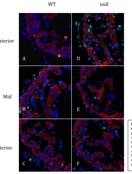
Representative sections of the left ventricles from Casz1 null and wild type hearts at stage E10.5 are shown in Figure 6. PHH3 nuclear staining is seen in each section. The cardiomyocyte
mitotic index was calculated for
each mouse using the
cardiomyocyte proliferation data
obtained for each individual
section. At stage E10.5, the
Casz1 null hearts have an average cardiomyocyte mitotic index of
0.2568, compared to a mitotic
index of 0.1764 in the wild type
cardiomyocytes.
WT null
Anterior
Mid
Posterior A
B
C F
E D
Figure 6. PHH3 is expressed in E10.5 wild type and Casz1 null hearts. Transverse paraffin sections of E10.5 embryos stained with PHH3 antibody (green), TMY antibody (red) and DAPI (blue). Photographs were taken of the Left ventricles of wild type anterior (A), middle (B), and posterior (C) heart sections; and left ventricles of Casz1 null anterior (D), middle (E), and posterior (F) heart sections.
WT null
Anterior
Mid
Posterior
A D
B
C
E
F Casz1 Mutant and Wild Type Hearts have Similar Mitotic Indexes at Stage E12.5
Representative sections of the left ventricles from the Casz1 null and wild type hearts at stage E12.5 are shown in Figure 7. PHH3 nuclear staining is seen in each section. The
cardiomyocyte mitotic index was calculated for each mouse using the cardiomyocyte
proliferation data obtained
for each individual section.
At stage E12.5, the Casz1
null hearts have an average
cardiomyocyte mitotic
index of 0.3326, compared
to a mitotic index of 0.3343
in the wild type
cardiomyocytes.
Figure 7. PHH3 is expressed in E12.5 wild type and Casz1 null hearts.
There is Increased Cardiomyocyte Proliferation in the Ventricles of Casz1 Mutant Mice at E10.5
There is a statistically significant difference in proliferation in the ventricles of wild type
hearts compared to mutant hearts at stage E10.5. The left and right ventricles of the wild type
heart at E10.5 had a combined mitotic index of
0.381, whereas the ventricles of the mutant heart
at this stage had a mitotic index of 0.526. The
ventricles of the E12.5 hearts did not show a
statistically significant difference in mitotic indexes between the wild type and mutant embryos
(Graph 1). It is important to note, however, that despite the increase in proliferation in mutant
ventricles at stage E10.5, there is a decrease in
the overall number of cardiomyocytes in the
ventricles of the mutant heart at stage E12.5
(Graph 2).
DISCUSSION
By performing in situ hybridization, it was possible to distinguish the types of tissue in which Casz1 is regularly expressed, as well as the structures in which it is expressed. Casz1
expression was seen most notably in myocardial tissue. Myocardial tissue makes up the outer
0 0.2 0.4 0.6 0.8
E10.5 wt E10.5
null E12.5 wt E12.5 null
Mitotic
In
dex
Combined Ventricle Cardiomyocyte Mitotic Index
Graph 2. Decrease in number of cardiomyocytes in mutant heart at E12.5. The number of
cardiomyocytes in the ventricles of mutant and wild type embryos at stages E10.5 and E12.5.
0 500 1000 1500 2000 2500
E10.5 E12.5
N u m b er o f C ar d io m yo cy te s
Total Number of Cardiomyocytes in Mutant and Wild Type Hearts
WT Null
components of the atria and ventricles, as well as their primitive counterparts. This expression of
Casz1 in the atria and left ventricle is in accordance with the hypothesis that cardiac progenitor cells lacking Casz1 cannot contribute to structural descendants of the primary heart field. The expression of Casz1 in the neural tissue was in accordance with existing data on the expression of Casz123 4.
The decrease in Casz1 mRNA expression in the heart at stage E10.5 corresponds to previous evidence that Casz1 is important for the looping of the primitive heart tube and the formation of the heart chambers, both of which occur before stage E10.514. This temporally
linked expression suggests that the role Casz1 plays in cell development is carried on without the transcription of new Casz1 mRNA at stage E10.5. This is concurrent with the hypothesis that
Casz1 is required for early cardiac cell fate decisions.
By stage E13.5, all four chambers of the mouse heart have formed, but are not yet fully
developed. In E13.5 mouse hearts, Casz1 mRNA is once again expressed in the outer curvature of the left ventricle. This is in accordance with the hypothesis that Casz1 contributes to the formation of the left ventricle. Casz1 is also expressed in the interventricular septum and the bulbar cushion tissue, which will form the bulbar septum. This suggests that Casz1 may function in the development of this septal tissue. The increased expression of Casz1 in the E13.5 embryo compared to the E10.5 embryo suggests that Casz1 functions in the maturation of the looped heart.
Experimental data was collected from in situ hybridizations performed using mouse embryos at stages E8.0 to E10.5 and mouse hearts at stage E13.5. This data supports the
arise from primary heart field cells. In order to test this hypothesis further, I used
immunohistochemistry to look at the expression of CASZ1 protein.
Immunohistochemistry experiments performed using E10.5 mouse embryos showed
nuclear expression of CASZ1 in most cardiomyocytes in the common ventricular chamber and
aortic sac. The spatial expression of CASZ1 protein corresponds to the expression of Casz1
mRNA that was observed through the in situ hybridization experiments. It would be beneficial in the future to repeat this experiment and to determine the differences between those
cardiomyocytes that express CASZ1 in the E10.5 embryo and those that do not, to see if there is
any significance in this spatial dissonance. It is possible that differences in the identities of the
cardiomyocyte progenitor cells play a role in this distinction. To further characterize CASZ1,
proliferation studies were done in mutant and wildtype mice.
In Xenopus, it has been shown that ablation of Casz1 results in increased proliferation at the ventral midline, but the role of Casz1 in mammalian cardiomyocyte proliferation is
unknown2. Using PHH3 staining in mouse embryos, it has now been shown that mice at
embryonic stage E10.5 also display increased proliferation in Casz1 null cardiomyocytes compared to wild type cardiomyocytes
Stage E10.5 mice showed a significant increase in cardiomyocyte proliferation in the
ventricles of Casz1 null embryos compared to those of wild type embryos. Stage E12.5 mice showed no significant difference in proliferation between the two conditions. Interestingly, there
was a decrease in the number of cardiomyocytes in the ventricles of Casz1 mutant embryos at stage E12.5 compared to E10.5, despite the increase in proliferation seen at stage E10.5. This is
required for cardiac development. I have also shown that Casz1 is another transcription factor that we can add to the mammalian cardiac program.
From the data obtained, it appears that Casz1 is primarily associated with the left
ventricle and the atria. Recent RNA sequencing data showed that secondary heart field proteins
are up-regulated in Casz1 mutant embryos at stage E10.5. We believe that Casz1 may be responsible for keeping secondary heart field proteins from being aberrantly expressed in
primary heart field cells.
Further studies are currently being done to determine if these secondary heart field
proteins and CASZ1 are expressed in mutually exclusive cardiomyocytes. We believe the
increase in the amount of these proteins may occur because they are being improperly expressed
in primary heart field cells. It is therefore necessary to determine if these secondary heart field
proteins are being up-regulated specifically in primary heart field cells when Casz1 is removed. In the future, it will be important to determine which genes are specifically being targeted
by Casz1, and which genes are downstream targets of the molecular pathway. My studies on cardiomyocyte proliferation take us one step closer to decoding the molecular mechanisms
behind heart development. These mechanisms will be important in the future as we develop new
References
1. Buckingham, M., S. Meilhac, and S. Zaffran. "Building the Mammalian Heart From Two
Sources of Myocardial Cells." Nature Reviews. 6. (2005): 826-835. Print.
2. Christine, K.S., and F.L. Conlon. "Vertebrate CASTOR Is Required for Differentiation of
Cardiac Precursor Cells at the Ventral Midline." Developmental Cell. 14. (2008): 616-623. Print.
3. Liu, Z., Yang, X., et al. “Molecular cloning and characterization of human Castor, a
novel human gene upegulated during cell differentiation.” Biochemical and Biophysical Research Communications. 344. (2006): 834-844. Print.
4. Komuro, I., and S. Izumo. "Csx: A murine homeobox-containing gene specifically
expressed in the developing heart." Proceedings of the National Academy of Sciences USA. 90. (1993): 8145-8149. Print.
5. Lints, T.J., L.M. Parsons, L. Hartley, I. Lyons, and R.P. Harvey. "Nkx2.5: a novel murine
homeobox gene expressed in early heart progenitor cells and their myogenic