Sentinel Lymph Node Biopsies in Cutaneous Melanoma: A systematic review of the literature
By Sasha Jenkins
A Master’s Paper submitted to the faculty of the
University of North Carolina at Chapel Hill in partial fulfillment of the Requirements for the degree of Master of Public Health in the
Public Health Leadership Program
Chapel Hill 2008
Abstract
Cutaneous melanoma has become a growing health problem in the United States, affecting all age groups, ethnic groups, including both genders. The incidence rate of melanoma is rising faster than any other malignancy, with the American Cancer Society projecting nearly 60,000 new cases and over 8,000 deaths due to melanoma for the year 2007. One of the main components of diagnosis and management of cutaneous
melanoma is staging of the patient. Given that the metastasis status of the lymph nodes is of great prognostic significance for melanoma, a new minimally invasive procedure called sentinel lymph node biopsy has become the preferred method of care for patients diagnosed with melanoma. However, to date, there has been no cumulative research looking at the evidence of sentinel lymph node biopsies in terms of overall survival and disease free survival. In addition, the theory that sentinel lymph node biopsies might increase the risk of in-transit metastases has been postulated. A systematic review was done evaluating the role of the routine use of sentinel lymph node biopsy in staging of melanoma to see if there is an overall benefit to patients diagnosed with melanoma in performing SLN biopsies.
Table of Contents Introduction
Burden of Disease……….4
Staging………..7
History of Staging of Cutaneous Melanoma………9
Sentinel Lymph Node Biopsy………11
Methods Search Strategy………...14
Inclusion Criteria………15
Data Extraction………...16
Results………16
Does SLN biopsy increase the risk of in-transit metastases?...18
Is the survival in favor of patients undergoing SLN biopsy?...26
Discussion………..…33
References………..45
Table 1:Studiesmeasuring ITM recurrence as outcome………...51
Introduction
Burden of Disease:
Cutaneous melanoma has become a growing health problem in the United States, affecting all age groups, ethnic groups, including both genders. Melanoma is a type of cancer that arises from specific cells in the skin called melanocytes. Malignant melanoma develops in these cells when melanocytes stop responding to normal cellular growth control mechanisms and become capable of local invasion or spreading to other organs. The incidence rate of cutaneous melanoma is on the rise, with the American Cancer Society projecting nearly 60,000 new cases and over 8,000 deaths due to melanoma for the year 2007.1 The incidence rate of melanoma is rising faster than any other
than most solid tumors. Thus, cutaneous melanoma has become an increasingly common form of cancer.
The mortality rate from melanoma in men and women increased from 1975-1990, but since 1990 there has been no change in mortality among men and a decrease in mortality among women. The age-adjusted mortality rate for cutaneous melanoma from 2000-2004 was 2.6% per 100,000 men and women per year. In specific age groups, the highest mortality rate is seen in the age group 75 to 84, with 23.1% of men and women dying from melanoma.3 The relative survival rates for melanoma are fairly good, compared to other malignancies. The five-year relative survival rate has increased since 1975 for all ages, races, and gender. Based on data from 1996-2003, the overall 5-year relative survival was 91.1%.3 Although the relative survival rate for primary cutaneous melanoma is fairly good, it still represents a huge burden of disease among people in the United States, as well as around the world.
Screening, diagnosis, management and treatment of melanoma varies
considerably among healthcare providers. While some melanoma care is delivered via local physicians in community-based strategies, others are delivered via a
multidisciplinary based approach. The Michigan Multidisciplinary Melanoma Clinic (MDMC) found that patients treated within the MDMC compared to patients treated within other sites in the Michigan community would save a third party payer roughly $1600 per patient, with equivalent rates of surgical morbidity, length of hospitalization, and long-term survival.6 However, many patients are not treated in a multi-disciplinary based environment, and the costs of melanoma healthcare is high. If the incidence of melanoma continues to increase annually, the estimation of cost for melanoma treatment by Medicare may exceed $5 billion by the year 2010.7
Staging
Bothenvironmental and genetic factors contribute to the development of
malignant melanoma. The main environmental factor is sun exposure. There are clinical signs and symptoms that increase suspicions of possible melanoma. The ABCDE
criterion for gross inspection of the skin is an acronym used by physicians and other health care professionals to aid in the early recognition of potential cutaneous malignant melanomas. Inspecting suspicious nevi lesions for asymmetry, border irregularity, color variegation, greater than 6 mm diameter, and evolving lesions over time for changes in size, shape, symptom, surface, and shades of color, aid in screening and diagnosis of malignant melanoma.8
Despite these clinical signs, the only way to accurately diagnose melanoma is by skin biopsy. After biopsy confirms melanoma, the next step is staging the patient.4 In 2002, the American Joint Committee on Cancer published its 6th edition of the T,N,M melanoma staging system. Given that staging is associated with prognosis and treatment, it is critical to establish if there is local, regional, or distant metastasis at the time of diagnosis. The T classification is based on Breslow thickness (mm) and histologic evidence of ulceration, two of the most important prognostic indictors of the primary tumor.4 However, Breslow thickness, measured from the top of the granular level of the epidermis to the deepest point of tumor penetration, is the best indicator of outcome.7 The Clark level of invasion, or the level of depth penetration from the epidermis to the
History of Staging of Cutaneous Melanoma
Cutaneous melanoma commonly metastasizes to regional lymph nodes, with the regional nodal basin usually being the first site of metastasis. Before the acceptance of sentinel lymph node biopsy (SLN) as a staging mechanism, either elective lymph node dissection (ELND) or delayed lymphadenectomy with clinical palpable nodes was performed on patients with melanoma.9 ELND has been the routine management of melanoma for the past twenty-five years because it was a way to stage the nodal basin. The major downside was that the majority of patients did not have metastatic disease. These patients had to go through the unnecessary procedure, suffering considerable morbidity and complications, without any therapeutic benefit. With these invasive procedures came many complications, such as lymphedema, nerve damage, and acute wound problems.9 In addition, randomized control trials comparing ELND with delayed lymph node dissection at the time of clinical recurrence showed no significant overall survival benefit in patients undergoing ELND. 11
Sentinel Lymph Node Biopsy
The SLN biopsy is a procedure in which the sentinel lymph node is identified and removed; usually it is the first lymph node in the pathway from the primary tumor to the nodal basin. If the SLN is positive for tumor cells via histopathologic testing, other nodes in the basin are probably affected and need to be treated with a complete
lymphadenectomy. If the node is negative, it is likely that the other nodes in the basin will not be positive and no further surgical intervention is warranted.9 There are three methods used in the sentinel lymph node biopsy procedure: a preoperative
lymphoscintigram, a blue-dye injection at the primary melanoma site immediately pre-operatively, and finally a intraoperative use of a gamma probe. 21 The intraoperative hand held gamma probe is beneficial because it allows for an easier search for the SLN resulting in a smaller incision site and shorter operating time, as well as ensuring a more complete removal of the SLN.14 Using data from the Sunbelt Melanoma Trial,
McMasters et al found the detection of sentinel nodes is best achieved by removing all nodes that stain blue and all nodes that show radioactivity greater than 10% of the count in the hottest node. 22, 23
only way to reliably stage and gather prognostic information is to see if the regional lymph nodes have disease, the SLN biopsy procedure has rapidly gained acceptance.10
Even though the rate for minor complications following SLN biopsy is small, there is a concern that the SLN biopsy procedure might increase the risk of in-transit metastases (ITM) by entrapment of tumor cells in the dermal lymphatic vessels during the procedure.27 ITM’s are subcutaneous metastases located between the site of the primary cutaneous melanoma and the regional lymph node basin. These represent a serious clinical problem as they are difficult to manage and very hard to eradicate. 28 A literature review of all studies calculating local/ITM recurrence as an outcome following SLN procedure by Thomas and Clark showed that the overall recurrence rate was 9.0%. They concluded that patients who had the SLN biopsy had double the incidence of local/ITM recurrence compared to patients treated solely with wide local excision; and patients who had the SLN biopsy followed by selective lymphadenectomy had four times the incidence of local/ITM recurrence compared to patients treated solely with wide local excision.29 However, this review only looked at ITM recurrence in patients who had sentinel lymph node biopsies, not studies directly comparing the recurrence to other staging procedures. Thus, there remains a concern for an increased risk of ITM recurrence following SNL biopsy.
Even though lymphatic mapping with sentinel lymph node biopsy has become the procedure of choice for staging cutaneous melanomas of intermediate thickness, the procedure is underutilized. Between 1998 and 2000, Baxter and Tuttle, using
Similar results were found by Stitzenberg et al where only 48% of patients with intermediate thickness melanomas underwent lymphatic mapping and sentinel
lymphadenectomy in North Carolina.31 Thus, even though sentinel lymph node biopsy has become the preferred method for management of melanoma, its use is far from being widespread.
Methods
Search Strategy:
To identify relevant studies of the outcome of sentinel lymph node biopsies in staging of cutaneous melanoma, a MEDLINE database search using the MeSH terms “melanoma,” “sentinel lymph node biopsy,” “lymph nodes” and “biopsy” was completed. Since the introduction of sentinel lymph node biopsy was introduced in the early 90’s by Dr. Morton, dates were limited from 1990 to present. Sentinel lymph node biopsy did not become a MeSH term until 2001 so the MeSH terms “lymph node” and “biopsy” had to
be used to identify all relevant articles prior to 2001. Articles were limited to English and Humans. A MEDLINE search using the terms “melanoma” and “sentinel lymph node biopsy” as keywords with limits on studies being added to Medline in the last 180 days was also completed. The Cochrane database library was also searched using the search term “melanoma”. References of all relevant papers found in the searches were
Inclusion Criteria:
Randomized control trials, case-control trials, and observational and retrospective reviews that compared sentinel lymph node biopsies to either another form of staging of the regional lymph nodes or no dissection for staging in patients with primary cutaneous melanoma were included.. All types of trials were included, regardless of the number of participants or study duration. All patients with a diagnosis of primary cutaneous melanoma with no evidence of metastases at distant sites, regardless of their breslow thickness were also included. Trials that included only patients with melanoma of the head and neck were excluded. Trials whose patients were children or adolescents were also excluded.
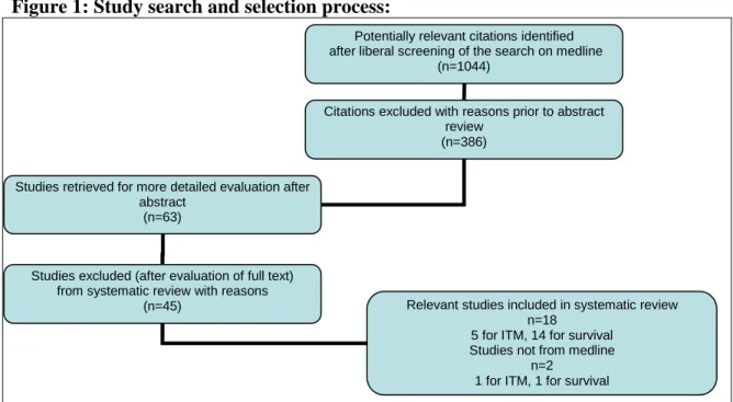
Figure 1: Study search and selection process:
Potentially relevant citations identified after liberal screening of the search on medline
(n=1044)
Citations excluded with reasons prior to abstract review
(n=386)
Studies retrieved for more detailed evaluation after abstract
(n=63)
Studies excluded (after evaluation of full text) from systematic review with reasons
(n=45) Relevant studies included in systematic review
n=18
5 for ITM, 14 for survival Studies not from medline
Data Extraction
Only one investigator independently extracted the data from relevant articles onto a standardized data collection form that included the following information: year of publication, inclusion and exclusion criteria, number of patients randomized or number of patients in study, number of patients lost to follow-up, staging procedure of melanoma, number of recurrences, survival, and mortality. The randomized control trials, articles were graded via quality grading of good, fair, or poor via the predefined criteria developed by the USPSTF and the National Health Service Centre for Reviews and Dissemination. For all other studies, quality was based on selection of cases or cohorts and controls, adjustment for confounders, methods of outcome assessment, length of follow-up and statistical analysis.
Results
Figure 1 illustrates the results of the study search and selection process of the included articles. Six articles were identified: one observation33, four retrospective reviews34-37 and one randomized control trial38, addressing the risk of in-transit
recurrence in patients who had either WLE only, WLE plus SLN biopsy or elective lymph node dissection.34, 37
Fifteen studies were identified, only one of which was a randomized control trial, that addressed survival of patients undergoing SLN biopsy compared to WLE only, DLND, and/or ELND. The majority of the studies were retrospective database reviews. The majority of the studies included patients who were either stage I and/or stage II. There was one study that included patients who were stage III39 and one study that
included patients who had thin melanomas with a breslow thickness of 0.76-1mm only.40 The studies reported on different outcome measures of survival, including overall
Does SLN biopsy increase the risk of in-transit metastases?
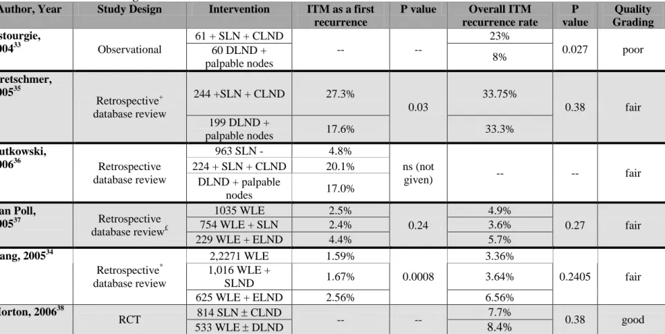
The six articles that addressed the rate of in-transit metastases are displayed in Table 1. 33-38 One of these studies was observation, one was a randomized control trial and the rest were done by retrospective data review from various melanoma databases. Only one study showed that the overall rate of ITM recurrence during the study period was statistically higher for patients who received a positive SLN followed by a complete lymphadenectomy compared to patients with clinically palpable nodes receiving a delayed lymph node dissection.33 Another study showed a statistically significant higher rate of ITM as a first recurrence in patients receiving a SLN biopsy followed by CLND compared to patients with clinically palpable nodes receiving a DLND, but a non-significant higher rate of overall ITM recurrence rate.35 Of the two studies looking at WLE, WLE + SLN biopsies, and ELND, only one study showed a statistically significant higher rate of ITM as a first recurrence in SLN compared with ELND34, but both studies showed no significant difference in the overall ITM recurrence rate.34, 37 The
randomized control trial showed a non-significant overall rate of ITM recurrence of observation plus DLND compared to SLN.38
Given that all but two of the studies were retrospective, there is inherent selection bias and confounding to these studies. There was no randomization of groups so there will always be some confounding factors present that cannot be controlled for, even with statistical analysis. Some of the studies gave inclusion and exclusion criteria to
abstracted the outcomes from the cases. Bias would be limited if an independent reviewer measuring outcome was blinded to which surgical procedure the patient obtained.
Even though there was only one study that showed a significant higher rate of overall ITM recurrence during five years in patients with a positive SLN biopsy followed by a CLND (23% vs 8%, p=0.027), the overall quality of the study was poor.33 This study suffered significantly from selection bias, confounding, and measurement bias. The observational study failed to reveal how the patients were selected to be included in the study. They stated they excluded patients with melanoma of the head and neck, and included patients who were Stage I and II. Also, there was no mention of patients who were potentially lost to follow-up. There was no mention of the procedure protocol for SLN biopsy, of the success rate of the SLN procedure, of how they assessed the histology of the lymph node biopsies, and if the same surgeon performed all surgical procedures. The two groups were statistically different at baseline with respect to breslow thickness and ulceration status of the primary lesion. Although the group who underwent SLN biopsy plus CLND had a deeper Breslow thickness as well as a greater percentage of ulcerations, logistic regression analysis did not show that Breslow thickness, age, sex, tumor location, tumor histology, or ulceration status had any statistically significant prognostic value. They found that the rate of ITM recurrence was increased in patients undergoing SLN biopsy + CLND compared to patients who underwent a delayed lymph node dissection. However, the patients who had SLN biopsy + CLND had more
they did not state their success rate of the SLN procedure, they did state that one surgeon performed 80% of all surgeries. Even though the authors assessed the two groups for initial comparability in regards to many prognostic factors, there is the potential for other confounders that they did not control for introducing bias.
This study is also generalizable to patients diagnosed with Stage I or II cutaneous melanoma, excluding those with palpable lymph nodes or neck lymph node dissections. They found that the median interval between primary tumor excision and palpable nodal metastases was 12 months in the DLND group whereas the median time interval to the occurrence of a first distant or nodal recurrence was 47.5 months in the positive SLN group. Thus, the time during which ITM may manifest as a first recurrence is almost four times longer if nodal recurrences are avoided by using the SLN biopsy procedure. Given that the overall ITM reoccurrence rate was not different between the groups, they
concluded that sentinel lymph node biopsy does not increase the risk of ITM.
The final study that compared ITM recurrence of positive SLN plus CLND with DLND in patients with clinically palpable nodes showed no significant difference in ITM as a first recurrence (20.1% vs 17.0%, no p value reported). 36 However, they found a significant lower rate of ITM recurrence in patients who received DLND compared to SLN plus CLND when only comparing patients who relapsed (25.9% vs 37.3%, p=0.02). The difference in the median time to ITM recurrence was not significant in the CLND group (231 days) vs. the DLND group (240 days). Given that the study specified
metastases. The bias from measurement was reduced by having the Pathology department confirm measurement of both Clark level and Breslow thickness. Also, the SLN biopsy procedure was according to standard protocol and the false-negative rate of the procedure was 4.9%, similar to other studies. They also defined ITM and excluded patients from the DLND group who already had ITM, which limits measurement bias. Although they used statistical analysis to find which prognostic factors influenced the rate of ITM in the entire SLN group, they did not use statistical analysis to control for potential confounders between the SLN group and the DLND group, which were different at baseline.
Furthermore, the results of this study are only applicable to patient with melanoma with a breslow thickness ≥ 0.75mm or Clarks level ≥ IV. Given that there was no significant difference in overall survival using Kaplan Meier graphs between DLND and CLND in patients who developed ITM and there was no difference in rate of ITM as a first reoccurrence in the groups, the authors concluded that the SNL biopsy procedure does not increase the risk of ITM. Furthermore, the estimated overall 3 year survival (from the date of relapse) in ITM patients was better when compared to other types of relapses after CLND and DLND.
also controlled for confounding by matching patients from each treatment group by age, sex, breslow depth, and primary site location. Again, from the analysis of 1,680 patients (560 in each group) they found no significant difference in ITM overall or as a first recurrence in the treatment groups. From their Kaplan-Meier survival curves, there was no significant treatment-related differences in rate of ITM as a first recurrence, however, patients with WLE and SLND did better with respect to overall ITM than the ELND group. With matching of patients for T stage and again for potential confounders, there was no statistical difference between the two groups on Kaplan Meier curves.
Given that this study used matching of potential prognostic factors, confounding was minimized, but not eliminated. Selection bias was high because the study did not state specific inclusion or exclusion criteria. There was also incomplete data for the patients on ulceration status of primary lesion, with 45.9% melanoma of unknown
ulceration. This is of great significance because ulceration status is a prognostic factor for melanoma. Since almost half of the ulceration status of the melanomas was unknown, this prognostic factor could not be controlled for in statistical analysis. The three groups were not comparable at baseline, with the SLN biopsy group having the highest
percentage of patients over 50 years of age and the ELND group having a greater
histology of the SLN, or success rate of the SLN procedure, leading to potential measurement bias. This study is only generalizable to patients with a stage I or II cutaneous melanoma, since this was the only inclusion criteria for the study. In
conclusion, the authors state that there is not an increased incidence of ITM after SLN biops vs WLE or ELND and there is no survival disadvantage, either disease free or overall, in those SLND patients who did develop ITM.
The final study by Van Poll et al also showed no significant difference in ITM as a first recurrence between patients in WLE, SLND, and ELND upon univariate
analysis.37 Even after multivariate analysis adjusting for Breslow thickness, ulceration, age, sex, and follow-up time there was no difference in ITM recurrence. When
comparing nodal disease in the groups, univariate analysis showed that tumor negative SLN group had a significantly lower rate of ITM as a first recurrence (1.7% vs 4.5%, p=0.03) and overall ITM (2.5% vs 5.5% p=0.04) than tumor negative ELND. However, for tumor positive disease there was no significant difference. The authors performed another subgroup analysis in patients with evidence of regional metastatic disease, a group with WLE followed by a delayed lymph node dissection of clinically palpable metastatic nodes to a group with WLE with SLN biopsy followed by a completion lymph node dissection within 3 to 4 weeks. From this analysis, the total ITM rate was
significantly lower in the SLN with CLND compared to DLND (10.8% vs 24.3% p=0.008). Although the authors minimized selection bias by stating inclusion and exclusion criteria prior to database review, the groups were not initially comparable. The WLE group had a higher proportion of men and the patients were older at age of
ELND group. The authors stated that the treatment for all three groups used the same surgical protocols over the 10 year study period and defined their outcome measurement definitions of local, in-transit, regional, and distant melanoma recurrences. Even though the groups had varying prognostic factors as baseline, the authors did control for many potential confounders (age, sex, tumor thickness, ulceration status, Clark’s level, primary tumor site location, and follow-up period) via multivariate analysis and multiple
regression. This study is generalizable to patients diagnosed with cutaneous melanoma with a breslow thickness >1mm. The group of patients who received immediate regional lymph node dissection because of a positive SLN biopsy had a lower incidence of ITM compared to patients treated with WLE followed by delayed lymph node dissection at time of clinically palpable lymph node metastases. Thus, the authors concluded that their results did not support the hypothesis that mechanical entrapment of tumor cells in
lymphatic channels due to surgical interference with the regional nodes causes ITM. The risk of developing ITM was not increased by SLN biopsy or ELND.
Is the survival in favor of patients undergoing SLN biopsy?
The fifteen articles that address survival in patients who underwent sentinel lymph node biopsy are displayed in Table 2. The majority of the articles found were retrospective database reviews comparing SLN biopsy patients to either ELND, DLND, or WLE and observation only. Given the limitations and bias inherent to retrospective database reviews, there is not strong evidence to make a valid conclusion on SLN biopsies and overall survival and disease free survival from the studies analyzed in this review. There was one randomized control trial38 that looked at the primary outcome of overall survival. This RCT was only given a quality rating of fair as well due to bias in its internal validity.
Five retrospective database review articles were identified comparing SLN biopsies to ELND41-43, 50, 51. Three of these studies reported overall survival (OS) as their primary endpoint41-43 and two studies reported disease free or recurrence free survival (DFS/RFS) as their endpoints50, 51. The study by Essner et al found no significant
difference in OS at five years when comparing all patients who received a SLN CLND compared to patients receiving an ELND, nor was there a significant OS when comparing only node positive SLN patients + CLND to node positive patients who received
ELND43. This analysis was after a matched-pair analysis on age, gender, location of primary lesion and breslow thickness. However, the analysis failed to control for ulceration status, Clarks level, and type of histological melanoma, all of which affect prognosis.
multivariate analysis, the type of operation (SLN vs ELND) had no significant affect on patient survival (p=0.24). In this study, the two groups were different from baseline, with the SLN group being older, but having fewer ulcerative lesions and not as high of a breslow thickness. However, the study performed by Dessureault et al found a significant OS difference in patients treated with SLN vs ELND vs observation alone (p<0.0001) at five years. However, this study was rated as poor, as data was collected from 12
institutions with no standardization of diagnosis, staging, or treatment techniques and there was a difference in follow-up time. In addition, there was no baseline comparison of the groups and no statistical analysis controlling for potential confounders. From the results of these three trials, it seems that there is poor to fair evidence resulting from retrospective reviews that performing SLN biopsies results in no difference in overall survival compared to ELND.
with several limitations, such as short follow-up period and its design. From the five studies comparing ELND to SLN biopsy, it seems there is no difference in OS or DFS. Although, it seems that there might be a negative risk by decreasing DFS in node negative patients by performing SLN biopsy.
The literature search identified four studies comparing SLN biopsy to
observation. Three of these studies, all retrospective, reported OS and one of the studies, an observational cohort, reported relative risk. The studies by Gutzmer et al44, Koskivuo et al45, and Starz et al40, found no significant OS difference in patients treated with SLN biopsy vs observation. However, in a subgroup analysis performed by Kosviuo of patients with a positive SLN compared to observation, there was a significant difference in OS, with more patients benefiting from observation. These studies also point towards SLN biopsies offering a significantly better DFS compared to observation (see table 2). The study performed by Mohrle et al52 found that the relative risk of melanoma related death to be 0.8, but was not statistically significant, comparing patients who had undergone SLN to observation. The relative risk did not change when comparing patients who had positive or negative SLN biopsies to observation. In conclusion, these overall fair quality articles point towards no difference in OS for performing SLN
tumor excision, for patients treated with positive SLN biopsy plus CLND compared to DLND. However, they found a significant OS difference favoring SLN biopsy plus CLND when calculated from the time of lymphadenectomy (48% vs 38%, p=0.02). This article does not support an improved OS, or even a DFS, in patients who are stage III and have clinical disease in lymph nodes undergoing SLN biopsy plus CLND compared to DLND. Van Akkooi et al also found that there was no significant difference in OS at 5 years, when calculated from date of primary tumor excision, for patients treated with SLN biopsy plus CLND vs. DLND49. Even when patients with nodes containing submicrometastases were excluded from analysis, there was still no difference in OS. The authors are in agreement with Rutkowski et al and conclude there is no survival benefit in performing SLN biopsies. The main bias in the both of these studiess is that the two study groups were different at baseline, and they do not factor differences during their statistical analysis for OS survival.
On the other hand, Kretschmer et al found a significant 13% OS difference at 5 years, calculated from time of primary tumor excision, in patients treated with positive SLN biopsy plus CLND compared to DLND. This study represented cases from five different clinical centers, with one of the centers not performing DLND. One of the main bias with this study is that the data came from 5 different centers. There was no way of making sure everyone was treated the same, and thus the measurements were most likely not equal, valid, and reliable given that many different surgeons performed the
retrospective review, a matched pair analysis was performed on 287 patients, matched for pT stage, ulceration, sex, age, and total number of tumor-involved nodes, using a
computer program. This analysis revealed 5, 10, and 15 year significant OS, calculated from time of primary tumor excision, in favor of positive SLN biopsy plus CLND compared to DLND. The authors conclude that the result from this study indicate that melanoma behaves according to the incubator hypothesis, where melanoma metastasis first to lymph nodes where it remains latent before metastasizing further (as opposed to the marker hypothesis, where melanoma metastases via lymphatics and blood
simultaneously. Thus, finding tumors cells in the SLN is merely a marker that the melanoma has already metastasized and removal of tumor in the lymph nodes is unlikely to have a therapeutic effect) Thus, there is a clinical window where the tumor can be removed before it spreads. The authors calculated the proportion of patients who would benefit from an early lymphadenectomy based on the matched pair analysis (if these patients represented the entire population of patients with melanoma). They calculated 7.4% of patients would benefit from early lymphadenectomy.
Finally, the study by Starz et al compared SLN biopsy CLND to DLND in melanoma patients with breslow thickness greater than 0.75mm48. For all patients who had undergone SLN compared to DLND, there was a significant OS difference favoring SLN biopsy. When comparing only patients who had positive SLN biopsy plus CLND compared to positive DLND, there was barely a significant difference in OS favoring SLN plus CLND (p=0.0419). Differences in follow-up times were not taken into
was significantly better in the SLN biopsy group compared to DLND (p=0.0076). The authors conclude that early removal of lymph nodes is beneficial in the management of melanoma.
In summary, there are two retrospective reviews which show no overall survival benefit for performing SLN biopsies and three retrospective studies showing a potential overall survival benefit in performing SLN biopsies. Given that these are retrospective reviews, they suffer from confounding, selection bias, and measurement bias. Many of the studies do not control for all potential confounders, such as age, differences in length of follow-up time, histology, breslow thickness, site of primary lesion, and ulceration status. Given that many of the studies do not contain equal comparison groups, it would be important to control for differences, although some studies do not. Many suffer from selection bias because they do not state guidelines as to how patients were selected. Many also are subject to measurement bias because some do not explain techniques of
performing SLN biopsies or the SLN success rate. Thus, many authors conclude in their studies that a randomized control trial is needed to answer the question of an overall survival benefit.
to patients who had observation only (78.31.6% vs 73.12.1% 95%CI of 0.59-0.93, p=0,009). The subgroup analysis comparing patients who had a positive SLN biopsy with CLND to patients with WLE and DLND also showed a significantly better five year survival rate for patients who received the SLN biopsy (72.34.6% and 52.45.9%, 95% CI of 0.32-0.81, p=0.004). There was no significant difference in overall survival between the two groups at the 3rd interim analysis. Perhaps one of the reasons that the SLN group showed no difference in overall survival was because they had more distant recurrences compared to the observation group. At the 4th of the 5th planned interim analysis, melanoma specific survival is now significant (HR 0.74, p < 0.001) and the DFS remains significant (HR 0.74, p < 0.001). This is the first RCT to look at SLN vs DLND. At the 3rd interim analysis there was significant difference in DFS, but not OS. In the 4th interim analysis there seems to be a significant difference in OS as well.
Even though this was a well-designed RCT, the study suffered from several bias’ and resulted in a overall quality grading of fair. The main problem with the RCT by Morton was their analysis of the post-hoc subgroup survival benefit. When they did this subgroup analysis, the randomization of patient characteristics for the two groups was lost, thus confounding and selection bias were introduced. Another potential bias is they did not include patients with false-negative results in the group who underwent
Discussion
Sentinel lymph node biopsy was adopted as the preferred method of care in the United States by the World Health Organization and by National Comprehensive Cancer Network treatment guidelines32. The reason is because studies show patients who have a negative sentinel lymph node biopsy have a better prognosis compared to patients who have a positive sentinel lymph node. Thus, SLN biopsy offers a minimally invasive staging procedure resulting in a prognostic indictor. Due to the morbidity and lack of overall survival advantage of elective lymph node dissection, sentinel lymph node biopsies replaced ELND as a staging procedure and became the preferred method even though there were no studies showing a therapeutic advantage of SLN biopsy.
the widespread use of SLN. From my review, it seems that ITM should not be a concern of mechanical disruption but perhaps of the biology of ITM.
Another question raised with the implementation of SLN biopsies was if the procedure resulted in a subsequent increase in disease free survival and overall survival. From the fifteen articles found during this comprehensive literature search, SLN provides a significantly better disease free survival but perhaps not an overall survival benefit. There were three retrospective reviews pointing towards an OS benefit in patients
undergoing SLN biopsy compared to DLND, but two retrospective reviews that lacked to find this same benefit. The one randomized control trial, considered to be the gold standard but only given a fair quality grade, showed a significant improvement in DFS and the newest results show an improvement in OS as well. However, this RCT only shows the results from a small subgroup of the entire patients included in the MSLT-1 trial. Biologically, it makes sense that patients undergoing SLN biopsies with subsequent complete lymphadenectomy would have a prolonged disease free survival period because the patient is given an early stage III diagnosis with detection and removal of metastasis in the lymph nodes. However, strong evidence is lacking in the possibility that
performing SLN biopsies results in an overall survival benefit.
Perhaps one reason that studies have failed to show an overall survival for patients treated with SLN biopsy is because not all melanoma metastasis are present in the regional lymph nodes. In about 2/3rd of cases of melanoma, metastatic disease
in this study appeared to be an early event in metastatic spread and could have occurred via hematogenous spread, resulting in support of the marker hypothesis.53 Thus, SLN biopsy might not show a significant improved overall survival in studies because
hematogenous spread has already occurred in a majority of patients. Perhaps SLN biopsy will become only one part of the staging technique in the future where other modalities might be able to identify metastasis that have bypassed the regional lymph nodes.
Another possible reason the studies failed to show an improvement in overall survival with SLN biopsy is because it represents a lead-time bias. The studies identified showed an improvement in disease free survival, but not of overall survival, except for the newest results of the 4th interim analysis of the MSLT-1 Trial. The SLN biopsy procedure is detecting melanoma metastases early in the asymptomatic period. Thus, the patients who get a SLN biopsy procedure are being upstaged earlier than patients who have a delayed lymphadenectomy. However, since the overall survival between the two groups is the same, the SLN biopsy patients are not actually living longer than the observation patients but are merely finding out about their disease at an earlier point.
disease if not removed early. Starz et al concluded that perhaps only deposits greater than 1 mm (SIII) were of adverse prognostic significance. 48 In this study, patients’ S
classification, or the maximum distance from the interior margin of the lymph node capsule, provided better prognostic information compared to the mere presence of a positive SLN, as patients with an SIII classification had a significantly worse overall survival rate. Another study suggested that perhaps only micrometastases found by immunohistochemical analysis were not of prognostic significance.54 There is evidence that not all occult nodal disease will progress to overt clinical disease; some
micrometastases in the SLN will either be destroyed by host-immune processes or become dormant.55 Thus, research is still being done to help shed light on prognostic factors to determine which patients would be better suited for the SLN biopsy procedure.
more studies need to be performed to definitely answer this question. A recent retrospective study by Roka et al attempted to identify clinico-pathological features to predict which positive SLN patients will have additional disease present in CLND in hopes of identifying high-risk patients.58 They concluded that clinico-pathological features can not reliably be used to identify patients who will have additional disease on CLND, and thus all patients with a positive SLN biopsy require a CLND. The
therapeutic utility of CLND after a positive SLN is still largely unknown and is currently being tested in the Multicenter Selective Lymphadenectomy Trial (MSLT II) randomized control trial. If there is a survival benefit for CLND after SLN biopsy, then it would favor performing SLN biopsies on all patients with melanoma.
If sentinel lymph node biopsy with completion lymphadenectomy does not confer any sort of survival advantage and there is no conclusive evidence that adjuvant treatment is beneficial, then why use this procedure on patients diagnosed with melanoma? Given that SLN biopsy is minimally-invasive with minor side effects, some patients may wish to go through this procedure simply to know their lymph node status, and thus their prognostic status. Patients may feel reassured knowing that if they have a negative sentinel lymph node, their survival will be better than if they are positive. Even though there is roughly a 4% failure rate and being sentinel lymph node negative is not a 100% guarantee for no recurrences (around 13% of SLN negative patients will develop
recurrence by 3 years18), patients will still have a better idea of their long-term outcome. On the other hand, if a patient has a false-positive SLN, this information can be
devastating and lead to unnecessary completion lymphadenectomy and/or adjuvant therapy.
In a questionnaire survey with patients who underwent SLN biopsy, 91% believed that they gained some benefit from the procedure, such as peace of mind or the ability to plan for the future, both of which were independent of the outcome of the biopsy.61 However, this benefit seemed to be only short-term. Regardless, the majority of patients approved of the procedure and would recommend it to others. One advantage of the SLN biopsy, even though there is still not universal agreement that it has a therapeutic advantage, is that it may provide patients with psychosocial benefits necessary to cope with the diagnosis of cutaneous melanoma.
scintigraphy as possible alternatives. However, none of these modalities are as sensitive as SLN biopsy at detecting microscopic positive nodal metastasis.62-65 There has been further research on other screening alternatives. One study compared methallothioneines over-expression to SLN biopsy and found that it was comparable to SLN as a prognostic marker, but cheaper and easier.66 Other research has looked at the prognostic
information of tumor-infiltrating lymphocytes67 and p-cadherin68. The information that SLN biopsy, a one time procedure, provides may be considered important to both the clinician and patient in terms of management options and psychosocial factors. Yet there is still further research being done on other potential alternatives.
The use of ultrasound with fine needle aspiration in management of cutaneous melanoma has shown potential promise for an additional role in melanoma management. Given that ultrasound has a lower sensitivity and high false negative rate, ultrasound cannot completely replace SLN biopsies. However, studies have shown using US with FNA allows 10%-16% of patients to be spared the SLN biopsy procedure. 69, 70 Another newly published study highlight the potential promise of use of inductively coupled plasma-mass spectrometry (ICP-MS) with FNA as a means of nonsurgical evaluation of sentinel lymph nodes.71 Perhaps by using US with FNA, the SLN biopsy procedure can be limited to only high risk patients, or even using a non-surgical alternative such as ICP-MS, thus reducing the number of unnecessary SLN biopsies.
one-diagnosed with melanoma received a SLN biopsy, and the non-universal adherence to standards did not appear to have an effect on overall survival, although the study was not powered to show this outcome. 72 Concerns have been raised that this procedure might not be feasible for management of all patients presenting with melanoma. Given that SLN is a multi-disciplinary approach, with the need for surgeons, pathologists, and nuclear medicine physicians, it is not surprising that the national standards have not been universally met.
In light that the evidence for SLN biopsy is one of clinical equipoise, the cost-effectiveness of this procedure must also be considered. For melanomas less than 1 mm in thickness, there is much controversy over whether or not SLN biopsy should be performed, given only a very small percentage of patients would benefit from this procedure. The SLN biopsy procedure costs $10,096 to $15,223, compared to $1,000 to $1,720 for wide local excision alone. In one quantitative method analysis of cost-effectiveness for performing SLN biopsies in patients with melanomas less than 1.2 mm in thickness found that a large number of SLN biopsies would need to be performed to identify one patient with regional disease. The estimated the cost per life saved ranged between $627,000 to $931,000 for melanomas less than 1.2mm and even greater for melanomas less than 1mm, up to $153,00 annual cost per life saved. These authors conclude by raising the question as to whether or not it is cost-effective to perform SLN biopsies in this population group. 76
Even if the SLN biopsy procedure does not drastically improve survival, it is a diagnostic procedure that allows for nodal staging of melanoma. At this stage, it seems highly unlikely that the AJCC will revert back to clinical staging of nodal disease because the SLN biopsy procedure does provide better control of regional disease, thus improving disease free survival. Wide local excision with delayed lymphadenectomy when a patient has clinically palpable disease is not the best option for management of regional nodal disease of cutaneous melanoma. By the time the DLND is performed, extracapsular extension, invasion of neurovascular structures makes regional disease control more difficult than performing a SLN biopsy +/- complete lymphadenectomy.78 Thus, even though there is not widespread agreement that SLN improves overall survival, it does control local disease, providing patients with a significantly better chance of remaining disease free without recurrences. SLN biopsy remains the best option currently
available. Perhaps with further research on other modalities, such as ultrasound and/or molecular markers, the use of SLN biopsy can be limited to only high-risk patients, thus minimizing unnecessary procedures.
SLN biopsy can identify the 15-20% of patients with melanoma that have clinically negative lymph nodes but occult regional nodal metastasis, thus who would might benefit from a lymphadenectomy. The majority of patients will undergo this procedure and not have negative SLN biopsies. Many studies have recently been done attempting to create a model that incorporates the factors that are important in
ability to predict the presence of SLN metastasis, which can help with individual patient risk estimation and decision making to help the patient make an informed decision on if SLN is right for them. One of these models was a nomogram that when tested against predictions based on the AJCC clinical staging system was found to be more accurate and discriminating. 79 Hopefully, models such as these can help reduce the number of SLN biopsies so that patients only at high risk for regional nodal mets will be subject to this minimally invasive procedure.
Given that all the important questions surrounding the use of SLN melanoma are still unanswered and data is currently being collected for the MSLT I and II trials, I think it is important for physicians to have a discussion with their patients who are diagnosed with cutaneous melanoma. I think it is important for the patient to know that the sentinel lymph node biopsy is stated as the preferred method of care by the AJCC and the WHO, but there are still questions that remain unanswered. Patients need to be aware that SLN biopsy does not increase the risk of in-transit metastases. They also need to be aware that undergoing the SLN biopsy probably does not improve overall survival, as provided by the results of current studies. The patient also needs to be aware of all the facts
and cons of each option, discuss them with their physician provider and family/friends, and make their own decision given that this is a preference-sensitive decision with
References
1. Society AC. Cancer facts and figures.
www.cancer.org/downloads/STT/CAFF2007PWSecured.pdf. Accessed January 24, 2008.
2. Geller AC, Swetter SM, Brooks K, Demierre MF, Yaroch AL. Screening, early detection, and trends for melanoma: current status (2000-2006) and future directions. J Am Acad Dermatol. Oct 2007;57(4):555-572; quiz 573-556. 3. Ries LAG MD, Krapcho M, Mariotto A, Miller BA, Feuer EJ, Clegg L, Horner
MJ, Howlader N, Eisner MP, Reichman M, Edwards BK (eds). SEER Cancer Statistics Review, 1975-2004. http://seer.cancer.gov/csr/1975_2004. Accessed January 24, 2008.
4. Petro A, Schwartz J, Johnson T. Current melanoma staging. Clin Dermatol. May-Jun 2004;22(3):223-227.
5. Tsao H, Rogers GS, Sober AJ. An estimate of the annual direct cost of treating cutaneous melanoma. J Am Acad Dermatol. May 1998;38(5 Pt 1):669-680. 6. Fader DJ, Wise CG, Normolle DP, Johnson TM. The multidisciplinary melanoma
clinic: a cost outcomes analysis of specialty care. J Am Acad Dermatol. May 1998;38(5 Pt 1):742-751.
7. Rigel DS, Friedman RJ, Kopf AW. The incidence of malignant melanoma in the United States: issues as we approach the 21st century. J Am Acad Dermatol. May 1996;34(5 Pt 1):839-847.
8. Abbasi NR, Shaw HM, Rigel DS, et al. Early diagnosis of cutaneous melanoma: revisiting the ABCD criteria. JAMA. Dec 8 2004;292(22):2771-2776.
9. Landry CS, McMasters KM, Scoggins CR. The evolution of the management of regional lymph nodes in melanoma. J Surg Oncol. Sep 15 2007;96(4):316-321. 10. Aldredge LM. The role of sentinel node biopsy in patients with cutaneous
melanoma. Nurs Clin North Am. Sep 2007;42(3):379-392, v-vi.
11. Lens MB, Dawes M, Goodacre T, Newton-Bishop JA. Elective lymph node dissection in patients with melanoma: systematic review and meta-analysis of randomized controlled trials. Arch Surg. Apr 2002;137(4):458-461.
12. Fee HJ, Robinson DS, Sample WF, Graham LS, Holmes EC, Morton DL. The determination of lymph shed by colloidal gold scanning in patients with malignant melanoma: a preliminary study. Surgery. Nov 1978;84(5):626-632. 13. Morton DL, Wen DR, Wong JH, et al. Technical details of intraoperative
lymphatic mapping for early stage melanoma. Arch Surg. Apr 1992;127(4):392-399.
14. Landi G, Polverelli M, Moscatelli G, et al. Sentinel lymph node biopsy in patients with primary cutaneous melanoma: study of 455 cases. J Eur Acad Dermatol Venereol. Jan 2000;14(1):35-45.
17. Temple CL, Scilley CG, Engel CJ, et al. Sentinel node biopsy in melanoma using technetium-99m rhenium colloid: the London experience. Ann Plast Surg. Nov 2000;45(5):491-499.
18. Gershenwald JE, Thompson W, Mansfield PF, et al. Multi-institutional melanoma lymphatic mapping experience: the prognostic value of sentinel lymph node status in 612 stage I or II melanoma patients. J Clin Oncol. Mar 1999;17(3):976-983. 19. Zettersten E, Shaikh L, Ramirez R, Kashani-Sabet M. Prognostic factors in
primary cutaneous melanoma. Surg Clin North Am. Feb 2003;83(1):61-75. 20. Jansen L, Nieweg OE, Peterse JL, Hoefnagel CA, Olmos RA, Kroon BB.
Reliability of sentinel lymph node biopsy for staging melanoma. Br J Surg. Apr 2000;87(4):484-489.
21. Thompson JF, Scolyer RA, Uren RF. Surgical management of primary cutaneous melanoma: excision margins and the role of sentinel lymph node examination. Surg Oncol Clin N Am. Apr 2006;15(2):301-318.
22. McMasters KM, Reintgen DS, Ross MI, et al. Sentinel lymph node biopsy for melanoma: how many radioactive nodes should be removed? Ann Surg Oncol. Apr 2001;8(3):192-197.
23. McMasters KM, Noyes RD, Reintgen DS, et al. Lessons learned from the Sunbelt Melanoma Trial. J Surg Oncol. Jul 1 2004;86(4):212-223.
24. Leong SP, Donegan E, Heffernon W, Dean S, Katz JA. Adverse reactions to isosulfan blue during selective sentinel lymph node dissection in melanoma. Ann Surg Oncol. Jun 2000;7(5):361-366.
25. Roaten JB, Pearlman N, Gonzalez R, McCarter MD. Identifying risk factors for complications following sentinel lymph node biopsy for melanoma. Arch Surg. Jan 2005;140(1):85-89.
26. Wrightson WR, Wong SL, Edwards MJ, et al. Complications associated with sentinel lymph node biopsy for melanoma. Ann Surg Oncol. Jul 2003;10(6):676-680.
27. Thomas JM, Patocskai EJ. The argument against sentinel node biopsy for malignant melanoma. BMJ. Jul 1 2000;321(7252):3-4.
28. Hayes AJ, Clark MA, Harries M, Thomas JM. Management of in-transit
metastases from cutaneous malignant melanoma. Br J Surg. Jun 2004;91(6):673-682.
29. Thomas JM, Clark MA. Selective lymphadenectomy in sentinel node-positive patients may increase the risk of local/in-transit recurrence in malignant melanoma. Eur J Surg Oncol. Aug 2004;30(6):686-691.
30. Baxter N, Tuttle T. Increased utilization of regional lymph node staging for melanoma. Ann Surg Oncol. 2004;11(supplement 2):S121.
31. Stitzenberg KB, Thomas NE, Beskow LM, Ollila DW. Population-based analysis of lymphatic mapping and sentinel lymphadenectomy utilization for intermediate thickness melanoma. J Surg Oncol. Feb 1 2006;93(2):100-107; discussion 107-108.
32. Network NCC. Clinical Practice Guidelines in Oncology
33. Estourgie SH, Nieweg OE, Kroon BB. High incidence of in-transit metastases after sentinel node biopsy in patients with melanoma. Br J Surg. Oct
2004;91(10):1370-1371.
34. Kang JC, Wanek LA, Essner R, Faries MB, Foshag LJ, Morton DL. Sentinel lymphadenectomy does not increase the incidence of in-transit metastases in primary melanoma. J Clin Oncol. Jul 20 2005;23(21):4764-4770.
35. Kretschmer L, Beckmann I, Thoms KM, Haenssle H, Bertsch HP, Neumann C. Sentinel lymphonodectomy does not increase the risk of loco-regional cutaneous metastases of malignant melanomas. Eur J Cancer. Mar 2005;41(4):531-538. 36. Rutkowski P, Nowecki ZI, Zurawski Z, et al. In transit/local recurrences in
melanoma patients after sentinel node biopsy and therapeutic lymph node dissection. Eur J Cancer. Jan 2006;42(2):159-164.
37. van Poll D, Thompson JF, Colman MH, et al. A sentinel node biopsy does not increase the incidence of in-transit metastasis in patients with primary cutaneous melanoma. Ann Surg Oncol. Aug 2005;12(8):597-608.
38. Morton DL, Thompson JF, Cochran AJ, et al. Sentinel-node biopsy or nodal observation in melanoma. N Engl J Med. Sep 28 2006;355(13):1307-1317. 39. Rutkowski P, Nowecki ZI, Nasierowska-Guttmejer A, Ruka W. Lymph node
status and survival in cutaneous malignant melanoma--sentinel lymph node biopsy impact. Eur J Surg Oncol. Sep 2003;29(7):611-618.
40. Starz H, Balda BR. Benefit of sentinel lymphadenectomy for patients with
nonulcerated cutaneous melanomas in the Breslow range between 0.76 and 1 mm: a follow-up study of 148 patients. Int J Cancer. Aug 1 2007;121(3):689-693. 41. Dessureault S, Soong SJ, Ross MI, et al. Improved staging of node-negative
patients with intermediate to thick melanomas (>1 mm) with the use of lymphatic mapping and sentinel lymph node biopsy. Ann Surg Oncol. Dec 2001;8(10):766-770.
42. Doubrovsky A, De Wilt JH, Scolyer RA, McCarthy WH, Thompson JF. Sentinel node biopsy provides more accurate staging than elective lymph node dissection in patients with cutaneous melanoma. Ann Surg Oncol. Sep 2004;11(9):829-836. 43. Essner R, Conforti A, Kelley MC, et al. Efficacy of lymphatic mapping, sentinel lymphadenectomy, and selective complete lymph node dissection as a therapeutic procedure for early-stage melanoma. Ann Surg Oncol. Jul-Aug 1999;6(5):442-449.
44. Gutzmer R, Al Ghazal M, Geerlings H, Kapp A. Sentinel node biopsy in melanoma delays recurrence but does not change melanoma-related survival: a retrospective analysis of 673 patients. Br J Dermatol. Dec 2005;153(6):1137-1141.
45. Koskivuo I, Talve L, Vihinen P, Maki M, Vahlberg T, Suominen E. Sentinel lymph node biopsy in cutaneous melanoma: a case-control study. Ann Surg Oncol. Dec 2007;14(12):3566-3574.
of nodal microanatomy and molecular staging for improving the accuracy of detection of nodal micrometastases. Ann Surg. Oct 2003;238(4):538-549; discussion 549-550.
48. Starz H, Siedlecki K, Balda BR. Sentinel lymphonodectomy and s-classification: a successful strategy for better prediction and improvement of outcome of
melanoma. Ann Surg Oncol. Mar 2004;11(3 Suppl):162S-168S.
49. van Akkooi AC, Bouwhuis MG, de Wilt JH, Kliffen M, Schmitz PI, Eggermont AM. Multivariable analysis comparing outcome after sentinel node biopsy or therapeutic lymph node dissection in patients with melanoma. Br J Surg. Oct 2007;94(10):1293-1299.
50. Tsutsumida A, Furukawa H, Yamamoto Y, et al. Sentinel node biopsy versus elective lymph node dissection in patients with cutaneous melanoma in a Japanese population. Int J Clin Oncol. Aug 2007;12(4):245-249.
51. Clary BM, Mann B, Brady MS, Lewis JJ, Coit DG. Early recurrence after lymphatic mapping and sentinel node biopsy in patients with primary extremity melanoma: a comparison with elective lymph node dissection. Ann Surg Oncol. May 2001;8(4):328-337.
52. Mohrle M, Schippert W, Rassner G, Garbe C, Breuninger H. Is sentinel lymph node biopsy of therapeutic relevance for melanoma? Dermatology.
2004;209(1):5-13.
53. Meier F, Will S, Ellwanger U, et al. Metastatic pathways and time courses in the orderly progression of cutaneous melanoma. Br J Dermatol. Jul 2002;147(1):62-70.
54. Spanknebel K, Coit DG, Bieligk SC, Gonen M, Rosai J, Klimstra DS.
Characterization of micrometastatic disease in melanoma sentinel lymph nodes by enhanced pathology: recommendations for standardizing pathologic analysis. Am J Surg Pathol. Mar 2005;29(3):305-317.
55. De Giorgi V, Leporatti G, Massi D, et al. Sentinel lymph nodes in melanoma patients: evaluating the evidence. Oncology. 2006;71(5-6):460-462.
56. Wong SL, Morton DL, Thompson JF, et al. Melanoma patients with positive sentinel nodes who did not undergo completion lymphadenectomy: a multi-institutional study. Ann Surg Oncol. Jun 2006;13(6):809-816.
57. Morton DL, Cochran AJ, Thompson JF, et al. Sentinel node biopsy for early-stage melanoma: accuracy and morbidity in MSLT-I, an international multicenter trial. Ann Surg. Sep 2005;242(3):302-311; discussion 311-303.
58. Roka F, Mastan P, Binder M, et al. Prediction of non-sentinel node status and outcome in sentinel node-positive melanoma patients. Eur J Surg Oncol. Jan 2008;34(1):82-88.
59. Verma S, Quirt I, McCready D, Bak K, Charette M, Iscoe N. Systematic review of systemic adjuvant therapy for patients at high risk for recurrent melanoma. Cancer. Apr 1 2006;106(7):1431-1442.
60. Tsai KY. Systemic adjuvant therapy for patients with high-risk melanoma. Arch Dermatol. Jun 2007;143(6):779-782.
62. Kell MR, Ridge JA, Joseph N, Sigurdson ER. PET CT imaging in patients undergoing sentinel node biopsy for melanoma. Eur J Surg Oncol. Sep 2007;33(7):911-913.
63. Longo MI, Lazaro P, Bueno C, Carreras JL, Montz R. Fluorodeoxyglucose-positron emission tomography imaging versus sentinel node biopsy in the primary staging of melanoma patients. Dermatol Surg. Mar 2003;29(3):245-248.
64. Vereecken P, Laporte M, Petein M, Steels E, Heenen M. Evaluation of extensive initial staging procedure in intermediate/high-risk melanoma patients. J Eur Acad Dermatol Venereol. Jan 2005;19(1):66-73.
65. Alonso O, Martinez M, Delgado L, et al. Comparison of 99mTc-MIBI scintigraphy and sentinel node biopsy in the detection of occult lymph node metastases from cutaneous melanoma. Eur J Dermatol. Sep-Oct 2003;13(5):449-454.
66. Weinlich G, Topar G, Eisendle K, Fritsch PO, Zelger B. Comparison of
metallothionein-overexpression with sentinel lymph node biopsy as prognostic factors in melanoma. J Eur Acad Dermatol Venereol. May 2007;21(5):669-677. 67. Taylor RC, Patel A, Panageas KS, Busam KJ, Brady MS. Tumor-infiltrating
lymphocytes predict sentinel lymph node positivity in patients with cutaneous melanoma. J Clin Oncol. Mar 1 2007;25(7):869-875.
68. Pacifico MD, Grover R, Richman PI, Buffa F, Daley FM, Wilson GD.
Identification of P-cadherin in primary melanoma using a tissue microarrayer: prognostic implications in a patient cohort with long-term follow up. Ann Plast Surg. Sep 2005;55(3):316-320.
69. Rossi CR, Mocellin S, Scagnet B, et al. The role of preoperative ultrasound scan in detecting lymph node metastasis before sentinel node biopsy in melanoma patients. J Surg Oncol. Jun 2003;83(2):80-84.
70. Voit C, Kron M, Schafer G, et al. Ultrasound-guided fine needle aspiration cytology prior to sentinel lymph node biopsy in melanoma patients. Ann Surg Oncol. Dec 2006;13(12):1682-1689.
71. Beavis A, Dawson M, Doble P, et al. Confirmation of sentinel lymph node identity by analysis of fine-needle biopsy samples using inductively coupled plasma-mass spectrometry. Ann Surg Oncol. Mar 2008;15(3):934-940. 72. Scott JD, McKinley BP, Bishop A, Trocha SD. Treatment and outcomes of
melanoma with a Breslow's depth greater than or equal to one millimeter in a regional teaching hospital. Am Surg. Mar 2005;71(3):198-201.
73. Ross GL, Shoaib T, Scott J, Soutar DS, Gray HW, MacKie R. The learning curve for sentinel node biopsy in malignant melanoma. Br J Plast Surg. Jun
2002;55(4):298-301.
74. Tafra L. The learning curve and sentinel node biopsy. Am J Surg. Oct 2001;182(4):347-350.
77. Wilson LS, Reyes CM, Lu C, Lu M, Yen C. Modelling the cost-effectiveness of sentinel lymph node mapping and adjuvant interferon treatment for stage II melanoma. Melanoma Res. Dec 2002;12(6):607-617.
78. McMasters KM. What good is sentinel lymph node biopsy for melanoma if it does not improve survival? Ann Surg Oncol. Sep 2004;11(9):810-812.
79. Wong SL, Kattan MW, McMasters KM, Coit DG. A nomogram that predicts the presence of sentinel node metastasis in melanoma with better discrimination than the American Joint Committee on Cancer staging system. Ann Surg Oncol. Apr 2005;12(4):282-288.
80. Sondak VK, Taylor JM, Sabel MS, et al. Mitotic rate and younger age are predictors of sentinel lymph node positivity: lessons learned from the generation of a probabilistic model. Ann Surg Oncol. Mar 2004;11(3):247-258.
81. Mocellin S, Ambrosi A, Montesco MC, et al. Support vector machine learning model for the prediction of sentinel node status in patients with cutaneous melanoma. Ann Surg Oncol. Aug 2006;13(8):1113-1122.
82. Kruper LL, Spitz FR, Czerniecki BJ, et al. Predicting sentinel node status in AJCC stage I/II primary cutaneous melanoma. Cancer. Nov 15
2006;107(10):2436-2445.
Table 1: Studies measuring ITM recurrence as outcome
Author, Year Study Design Intervention ITM as a first
recurrence
P value Overall ITM
recurrence rate P value Quality Grading Estourgie,
200433 Observational
61 + SLN + CLND
-- --
23%
0.027 poor
60 DLND +
palpable nodes 8%
Kretschmer, 200535
Retrospective+ database review
244 +SLN + CLND 27.3%
0.03
33.75%
0.38 fair
199 DLND +
palpable nodes 17.6% 33.3%
Rutkowski,
200636 Retrospective
database review
963 SLN - 4.8%
ns (not
given) -- -- fair
224 + SLN + CLND 20.1%
DLND + palpable
nodes 17.0%
Van Poll,
200537 Retrospective
database review£
1035 WLE 2.5%
0.24
4.9%
0.27 fair
754 WLE + SLN 2.4% 3.6%
229 WLE + ELND 4.4% 5.7%
Kang, 200534
Retrospective* database review
2,2271 WLE 1.59%
0.0008
3.36%
0.2405 fair
1,016 WLE +
SLND 1.67% 3.64%
625 WLE + ELND 2.56% 6.56%
Morton, 200638
RCT 814 SLN CLND -- -- 7.7% 0.38 good
533 WLE DLND 8.4%
* Results show in the table are for unmatched analysis. Overall incidence and ITM as first recurrence rates for matched analysis are not shown in table.
Table 2: Studies measuring survival: overall survival (OS), disease free survival (DFS), recurrence free survival (RFS), melanoma specific survival (MSS)
Note: ~ refers to my interpretation of the Kaplan Meier Curve if not stated in the study
Author, Year Study Design AJCC stage
Intervention Type of Survival Survival (%) P value Type of Survival
Survival P value Time period Quality Grading Essner, 199943 Retrospective Database review Stage I
267* SLN
CLND OS
-- 0.98 DFS -- 0.25
5 year+ fair 267* ELND
Node positive patients
42+SLN/CLND OS -- 0.08
£ 32 ELND Clary, 200151 Retrospective Database review Stage I/II 152 SLN RFS 71%
0.12 MSS -- 0.87
3 year
fair
329 ELND 80%
All high risk patients
SLN RFS for
breslow > 3mm
~ 47%
0.04
RFS for age > 50 years
~ 65% 0.01
ELND ~ 75% ~ 82%
node negative high risk∫
90 SLN RFS
~ 70%
0.04
167 ELND ~ 82%
Dessureault, 200141 Retrospective Database review Stage IB/IIA€
5, 156 - observe
OS
69.8%
<
0.0001 5 year poor
2,032 - SLN 90.5%
Doubrovsky, 200442 Retrospective database review > 1.5-mm thick
672 SLN
CLND OS -- 0.139 5 year fair
793 ELND
Tsutsumida,
200750 Retrospective
> 1.5 mm 30 SLN DFS 87.2% 0.280
3 year fair
72 ELND 72.7%
12 + SLN 82.5%
0.90
19 + ELND 72.2%
Rutkowski, 200339 Retrospective database review Stage III
145 +SLN +
CLND OS 41% ns DFS 35% ns 5 year fair
205 DLND 42% 31%
Morton, 200347 Retrospective database review Stage I/II 287 SLN+CLND OS§ 73% / 69%/ 69% < 0.001 5/10/15
year fair
287 DLND
51% / 37% / 32% Kretschmer, 200446 Retrospective database review Stage I/II 314
SLN+CLND OS 62.5% 0.002 5 year fair
623 DLND 50.2%
Van Akooi, 200749 Retrospective database Stage I/II
64 SLN +
CLND OS 13%
difference 0.1115 5 year fair
124 DLND
Retrospective Breslow
324 SLN
CLND
57 + DLND ~ 35% ~ 38%
Morton,
200638 RCT
1.2-3.5 mm
814 SLN CLND
DFS
78.31.6%
0.009
Melanoma specific survival
87.11.3%
0.58
5 year fair 533 WLE
DLND 73.12.1% 87.11.3%
SLN+CLND 72.34.6%
0.004
WLE+DLND 52.45.9%
Möhrle, 200452 Observational cohort Stage I/II
271 +/-SLN RR of
melanoma-related
death
0.8 0.37
fair 2,617 observe
238 - SLN
RR 0.75 0.36
2,617 observe 33 + SLN
RR 0.73 0.38
246 observe Gutzmer, 200544 Retrospective database review Stage I/II 296 SLN
OS -- 0.32 RFS
~ 78%
0.0064 4 year poor
377 no SLN ~ 65%
Koskivuo, 200745 Retrospective case-control cohort Stage I/II 305 SLN OS 87.8%
0.66 DFS 85.1% 0.42
5 year fair
616 control 82.5% 79.0%
+ SLN ~ 76% <
0.001
control ~ 82%
Starz, 200740 retrospective
Breslow 0.76-1mm
87 WLE + SLN
OS 0.99
Melanoma specific survival
0.03
fair
*The 267 patients were matched based on gender, age, site of primary tumor, and the breslow thickness.
+ The difference in follow-up times for the two groups was statistically significant (p=0.001) but was controlled for via statistical methods. ∫ There was no sig difference b/w ELND and SLN/CLND for patients that were node positive.
£ Nonsignificant trend favoring of SLN/CLND