For many the subject of vascular malformations remains shrouded in mystery and confusion resulting from a lack of understanding of the nature of the pathology compounded by the confusing nomenclature still associat-ed with these lesions. For example, the term ‘haemangioma’ has been used to describe a variety of vascular lesions with different aetiologies and natural histories. Names utilising a mixture of descriptive and histologic terms, such as ‘strawberry angioma’, ‘nevus flam-mus’, or ‘cavernous haemangioma’ still abound in current medical usage. The classification of vascular anomalies outside of the central nervous system (CNS) is based on the seminal works of Mulliken and Glowacki.1,2 They
divided vascular anomalies into those exhibiting rapid endothelial cell growth (proliferation) followed by a period of stabilisation (quiescence) and finally spontaneous involution, termed haemangiomas, and those having a rate of growth
commensu-rate with the growth of the individual termed vascular malformations (VMs). As opposed to haeman-giomas, VMs reach stability by adult-hood or continue to grow by hyper-trophy rather than cellular prolifera-tion. The current classification of CNS vascular anomalies remains primarily a pathoanatomically-based one and is based on the works of McCormick et al.3-5and Russel and Rubenstein.6
In this review we will present a syn-opsis of CNS and non-CNS vascular anomalies based on the abovemen-tioned classification schemes in the hope that this will dispel much of the confusion surrounding the subject of vascular malformations or anomalies.
Classification of
CNS vascular
malformations
The current classification is based on the pathoanatomic works of McCormick et al.3-5 and Russel and
Rubenstein.6 This assumes vascular
malformations to be congenital or developmental hamartomas rather than neoplasms, and is based on the microscopic and gross pathologic fea-tures of these lesions (Table I). Already confounding this classification is the identification of mixed malformation
types,7together with the identification
of increased endothelial cell prolifera-tion within CNS vascular malforma-tions further blurring the distinction between malformation (hamartoma) and tumour (proliferation).8
Although intracranial arteriove-nous shunts are similar angioarchitec-turally to AV shunts elsewhere in the body, the cerebral and spinal cav-ernous malformation (cavernoma) does not have a similar counterpart in the rest of the body. Analysis of struc-tural proteins within the wall of caver-nomas shows these lesions to be developmentally immature unlike the walls of venous malformations else-where in the body. Furthermore the so-called venous angiomas or devel-opmental venous anomalies (DVAs) of the brain actually represent persis-tent normal embryological venous drainage of regions of the brain rather than true vascular malformations.9
Although most CNS vascular mal-formations occur in isolation, some are found in association with cuta-neous and mesodermal vascular anomalies. This mirrors the develop-ment of both the skin and nervous system from the ectodermal plate and neural crest of the early embryo, often
Vascular
malforma-tions part 2 —
current
classifica-tion of vascular
malformations
Ian C Duncan
FFRad (D)
Pieter A Fourie
M Med Rad(D)
Unitas Interventional Unit PO Box 14031
Lytlelton 0410
Table I. Classification of CNS vascular malformations
Arteriovenous shunts Classical AV Malformations Pial AV fistulas
Dural AV shunts Galenic shunts
Cavernous malformations Capillary telangiectasias Venous malformations
Developmental venous anomalies
also involving the adjacent subecto-dermal mesoderm. The most classic examples of such syndromes would include the Wyburn-Mason syn-drome (or Bonnet-Dechaume-Blanc syndrome) and the Sturge-Weber syndrome. The Wyburn-Mason syn-drome is an association of cerebral (diencephalic), retinal and maxillofa-cial arteriovenous malformations, hence its alternative name of ‘unilat-eral retinocephalic vascular malfor-mation’.10 Recognition of the
metameric nature of this disorder has led to a revised classification of such craniofacial vascular malformation syndromes as the cerebrofacial arteri-ovenous metameric syndromes (CAMS).11,12A similar developmental
metameric distribution would also explain the development of the rare type III or juvenile spinal AVM syndromes (cutaneomeningospinal angiomatosis, Cobb syndrome), now termed the spinal arteriovenous metameric syndromes (SAMS).13The
Sturge-Weber syndrome, also ‘encephalo-trigeminal angiomatosis’, involves capillary-venous malforma-tions of the brain, usually affecting a single cerebral hemisphere, associated with retinal vascular malformations and cutaneous facial capillary malfor-mations (previously nevus flammeus, cutaneous angioma or port-wine stain). Again the association of a telencephalic venous malformation with facial involvement usually within the V1 distribution suggests a venous metameric syndrome (cerebrofacial venous metameric syndrome (CVMS) — Lasjuanias, personal communication). However, often the cutaneous malformation involves the V2 or V3 distributions as well or may even extend to the neck and upper trunk, thus showing a spectrum of
expressivity. Forty-five per cent of children having V1 and V2 involve-ment will develop glaucoma due to associated venous malformations affecting the canal of Schlemm or other disruption of the ocular venous drainage.14
Other associations between CNS and other vascular malformations include the association of craniofacial vascular malformations such as cuta-neous capillary malformations and venous and lymphatic malformations with intracranial DVAs,15,16 and the
heritable association between cerebral cavernous malformations and cuta-neous or other systemic venous mal-formations.17,18 Finally there is the
development of CNS arteriovenous malformations as part of the spec-trum of multisystemic vascular mal-formation development in hereditary haemorrhagic telangiectasia.19 These
associations with non-CNS vascular malformations suggest a particular phenotypic expression peculiar to the environment of the brain and spinal cord of the same underlying vascular malformative processes found else-where in the body, hinting that the classic pathoanatomical classification of CNS vascular malformations may well later be revised along more bio-logically-orientated lines.20
Non-CNS
vascular
malformations
As mentioned above the current classification of these disorders as adopted by the International Society for the Study of Vascular Anomalies (ISSVA) is that described by Mulliken and Glowacki. Initially they differen-tiated between true vascular malfor-mations and haemangiomas, based
upon endothelial cell growth charac-teristics.1Haemangiomas show initial
rapid endothelial cell proliferation more typical of a tumour followed later by variable spontaneous regres-sion in many cases. Conversely VMs show no (or rather minimal) endothelial cell proliferation and are characterised by a growth rate com-mensurate with the rate of growth and development of the child. They then divided the VMs into high-flow (arteriovenous shunts) and low-flow VMs. The low-flow VMs were further subdivided according to their histo-logical origins into the capillary, venous, lymphatic or mixed malfor-mations (Table II).
Haemangiomas
The term haemangioma has tradi-tionally been used as a generic term to describe a large variety of vascular lesions with different aetiologies.1
Currently the term haemangioma is restricted to a distinct proliferative lesion of early childhood being more a tumour than a vascular malforma-tion. Haemangiomas are the most common vascular tumour, with an incidence of 1 - 2.6%.21Thirty-three
Table II. Classification of non-CNS vascular malformations
Haemangiomas (proliferative lesions or tumours)
Vascular malformations (non-prolifer-ative)
High flow
• Arteriovenous malformations Low flow:
• Capillary malformations • Venous malformations • Lymphatic malformations • Mixed malformations
to 40% of lesions are apparent at birth, with 70 - 90% appearing during the first 4 weeks of life and none beyond the age of 5 years. Haemangiomas may be superficial (cutaneous), deep (covered by normal overlying skin) or visceral.22They can
occur in any race, but are more com-mon in girls (3:1), light-skinned whites and premature infants (20%) especially those with a birth weight < 1500 g.23The superficial cutaneous
lesions tend to show a bright red colour initially that deepens during the first year of life (Fig. 1a).
Haemangiomas tend to show a firm, rubbery consistency compared with venous malformations that are com-pressible.22 The prevalence of
hae-mangiomas is approximately 1 - 3% of neonates, increasing to about 10% of white children and 1.4% of black children at 1 year of age.21Sixty
per-cent of haemangiomas occur in the head and neck region, with 25% on the trunk and 15% on the extremities. Occasionally they may involve oral or genital mucosa. In about 15 - 20% of cases the lesions are multiple (benign neonatal haemangiomatosis). They
initially present with an erythematous patch or pink macule often referred to as a ‘herald spot’. They then undergo a proliferative (growth) phase of 3 - 12 months followed by a variable plateau or stabilisation phase and finally by a phase of involution. All lesions inevitably show some degree of spon-taneous involution or regression.1,22
Approximately 30% of infantile haemangiomas will regress by the third birthday, about 50% by the fifth, and 70% by the seventh.24,25
If a haemangioma fails to show evi-dence of regression by 5 - 6 years of age, then it is unlikely to regress com-pletely. Lesions exhibiting early changes of involution are associated with more rapid disappearance and a better cosmetic result. Regression is heralded by the appearance of white streaks (fibrosis) on the surface of the lesion. Occasionally haemangiomas may ulcerate or bleed or develop sec-ondary infections, mainly during the proliferative phase.22 The tumour in
the proliferative phase is characterised by endothelial cell proliferation pro-ducing plump, hyperplastic endothe-lial cells together with a variable num-ber of mast cells. During the involu-tional phase there is a conversion from proliferation to apoptosis of the endothelial cells with progressive deposition of perivascular and inter-lobular fibrous tissue.26An increase in
basic fibroblast growth factor (BFGF), proliferating cell nuclear antigen, type IV collagenase, E-selectin and mono-cytic chemoattractant protein is seen during the proliferative phase, where-as during the involutive phwhere-ase there is an increase in metalloproteinase tissue inhibitor and a fall in BFGF levels.27
BFGF can thus be used as marker to assess the cessation of proliferation and onset of involution.
Most haemangiomas are recog-nised clinically. Imaging is only required in cases where there is diag-nostic uncertainty or where interven-tion is required. Ultrasound (with doppler) will rapidly distinguish solid haemangiomas from vascular malfor-mations. Computed tomography (CT) and magnetic resonance imag-ing (MRI) will help to assess the depth and extent of lesions. Angiography, while showing a characteristic homo-geneous contrast blush without evi-dence of AV shunting, should rather be reserved for therapeutic purposes.27
In most cases no treatment is required because of spontaneous regression. However 10 - 20% of lesions will require treatment because of impend-ing (or established) loss of function or a threat to life (Fig. 1b). Treatment for cosmetic reasons alone is a controver-sial indication given the natural histo-ry of these lesions. More often than not the parents are more affected by appearance than the children them-selves except as the children approach school age. Examples of indications for active treatment (i.e. ‘alarming’ Fig. 1a. Small cutaneous haemangioma on the
cheek of an infant.
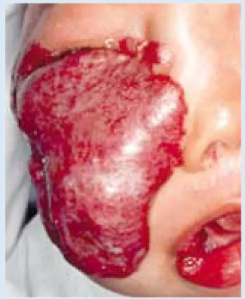
Fig. 1b. Large maxillary and periorbital cutaneous haemangioma. Its anatomical position threatens the function of the eye.
haemangiomas) would include:27,28
(i) periocular lesions threatening vision; (ii) lesions obstructing the air-way; (iii) large lesions associated with high-output cardiac failure; (iv) facial lesions with rapid growth and distor-tion; (v) lesions with severe persistent cutaneous ulceration or haemorrhage (Fig. 1c); and (vi) Kasabach-Merritt syndrome.
The Kasabach-Merritt phenome-non or syndrome, first described in 1940, consists of very large haeman-giomas complicated by severe throm-bocytopoenia, microangiopathic haemolytic anaemia and consump-tion coagulopathy. It is seen most fre-quently in young infants during the first week of life and carries a mortali-ty of 20-30%, thus requiring aggres-sive treatment when recognised.
Multiple cutaneous haeman-giomas may rarely co-exist with vis-ceral haemangiomas in a condition termed ‘diffuse neonatal heman-giomatosis’ or ‘disseminated haeman-giomatosis’. Visceral lesions may be found in the liver, GIT, spleen, pan-creas, adrenals, lungs, heart, skeleton, muscle, salivary glands, kidney, blad-der, testes, thymus, thyroid, bone,
meninges, brain and eyes. The mor-tality rate is high, with extensive vis-ceral involvement mainly during the first months of life and mainly due to high-output cardiac failure.22
Large facial haemangiomas may be associated with posterior fossa brain malformations, arterial developmen-tal anomalies, cardiac anomalies, aor-tic coarctation and eye anomalies in the PHACE syndrome, a strange asso-ciation of proliferative and non-pro-liferative vascular anomalies.29,30
Medical treatment for haeman-giomas includes local and systemic steroids, interferon α-2a and α-2b, and Pentoxifylline. Other options include intralesion injection of scle-rosing agents, cryotherapy, laser thera-py, embolisation, radiation therapy and surgical excision.21,22 Several
groups, including the Pretoria Vascular Malformation Group, have reported successes in treating hae-mangiomas using the antiangiogenic properties of the antimitotic antibiot-ic derivative bleomycin.31Other
treat-ment types used when other methods have failed included vincristine or radiation therapy.
Arteriovenous
(high-flow)
malformations
Arteriovenous malformations (AVMs) are high-flow lesions with direct communications between an artery (or arteries) and a vein (or veins) bypassing the capillary bed (Fig. 2). The term ‘arteriovenous fistu-la’ is often reserved for direct single hole communications usually related to trauma or other acquired condi-tion, whereas the term ‘arteriovenous malformation’ should be reserved for
congenital lesions.32 They are much
less common than low-flow malfor-mations and often become sympto-matic following trauma (including biopsy) or around puberty. Clinically they can present as a pulsatile mass with a thrill, bruit and occasionally local hyperthermia, skeletal over-growth, trophic changes with ulcera-tion or bleeding, congestive heart fail-ure or functional impairment due to arterial steal and ischaemia. The diag-nosis is generally made clinically with deep lesions identified as being high-flow with the aid of doppler ultra-sound. CT, MRI and angiography are all used to assess the extent of a lesion during the planning of therapy. AVMs are the most difficult and dan-gerous lesions to treat, and for this rea-son quiescent AVMs should be fol-lowed clinically, with treatment delayed until complications such as pain, ulceration, haemorrhage or car-diac failure intervene.21 Treatment
options include direct percutaneous and transarterial embolisation and surgical excision, with the treatment best left to those specialists well versed in the management of such lesions. Fig. 1c. Ulceration of scalp haemangioma.
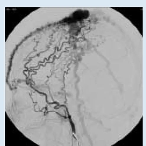
Fig. 2. High-flow (arteriovenous) malformation of the scalp. This composite digital subtraction angiogram combines both the arterial and venous phases in one image.
Venous
malformations
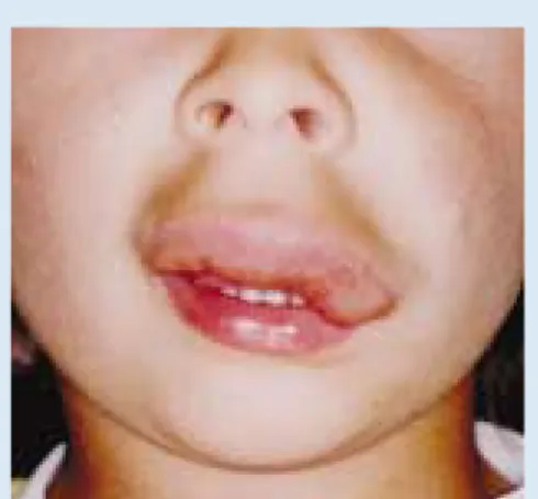
Venous malformations (VMs), often incorrectly termed cavernous haemangiomas, are low-pressure, low-flow malformations. They are present at birth, although not always visible at that stage, and undergo slow growth commensurate with the growth of the child.1,27VMs are soft,
compressible, non-pulsatile masses that expand after a Valsalva’s manoeu-vre or in a dependent position. They have a bluish colour often with nor-mal overlying skin (Figs 3a, b). Forty per cent are found in the head and neck region, 20% on the trunk and 40% on the extremities. VMs can be superficial (cutaneous) or occur in
buccal and intestinal mucosa and in other organs. Sudden enlargement can occur after trauma, haemorrhage, surgery, or with hormonal changes. VMs can become painful due to the development of thrombophlebitis or in cases with muscular or articular involvement. Calcified phleboliths may be present in the lesions, hence the previous term phlebangioma. Ultrasound features include low (or absent) flow and compressibility of the widened vascular spaces. In some lesions a biphasic flow pattern sug-gests the presence of a mixed malfor-mation such as a capillary-venous or lymphatico-capillary-venous malfor-mation.33CT and MRI are useful in
delineating the full extent of the lesion.21,34
There is no role for angiography although direct puncture venography may be useful in documenting the extent of a lesion and pattern of its draining veins.
VMs may be associated with other anomalies as a component of several complex syndromes such as: (i) Blue Rubber Bleb Nevus syndrome or Bean syndrome; (ii) Maffucci’s syndrome; (iii) Klippel-Trenaunay syndrome; or (iv) Gorham’s syndrome (Gorham-Stout syndrome).
The Blue Rubber Bleb Nevus syn-drome is a rare condition consisting of multiple VMs involving several organ systems, especially the skin and GIT. Most cases are sporadic, but some are inherited with an autosomal domi-nant inheritance pattern.35,36 The skin
lesions are present at birth but other lesions only become apparent later in life.
The Klippel-Trenaunay syndrome (KTS) is a complex constellation of VMs including abnormal
develop-ment of the normal deep and superfi-cial limb venous drainage, lymphatic malformations and limb asymmetry (hemi-hypertrophy).37The lower limb
is most commonly affected, rarely affecting the upper limb or both upper and lower limbs. The phy is mainly due to muscle hypertro-phy, thickened skin and subcutaneous fat and occasionally lymphoedema. There is usually relatively little increase in bone size.36 This indicates
a combined ectodermal and mesoder-mal mesoder-maldevelopment. Deep venous thrombosis (DVT) and pulmonary embolism are common in patients with KTS. A variation of KTS where limb hemihypertrophy is associated with a high-flow AVM within the affected limb and also in other organ systems is the Parkes-Weber syn-drome.20
VMs may be associated with osseous abnormalities e.g. in Maffucci’s syndrome and the Gorham-Stout syn-drome. VMs may be associated with a low-grade consumption coagulopa-thy.27,36,38,39Therefore prior to any
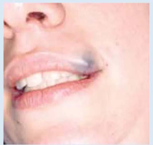
inter-vention a full assessment of coagula-bility is required. As many VMs are asymptomatic no treatment other than reassurance may be necessary in many cases. Discomfort due to localised lesions may respond to external compression, e.g. with elasti-cated stockings. More aggressive treatment is indicated in lesions pro-ducing pain, discomfort, significant cosmetic disturbance or functional impairment. This generally involves either percutaneous sclerotherapy or surgical excision. Percutaneous sclerotherapy involves the use of a number of agents including absolute alcohol, Ethibloc, Sodium Morrhuate, Sodium Tetradecyl Sulphate and Fig. 3a. Small venous malformation of an upper
lip. Note the characteristic bluish colouration.
Fig. 3a. Large venous malformation of a lower limb. No leg length discrepancy was noted.
Sotradecal.27,33,40-44 Intralesional
bleomycin injections have also recent-ly been shown to be effective in treat-ing VMs.31,45
Capillary
malformations
Cutaneous capillary malforma-tions (also termed port-wine stains or nevus flammeus) are due to ectatic vessels within the upper dermis. They are present at birth with an equal sex distribution. The are flat sharply demarcated lesions that are pink dur-ing infancy deependur-ing in colour to red in young adulthood and purple in middle age.21 Forty-five per cent of
facial lesions are restricted to one of the three trigerminal sensory areas, whereas 55% overlap dermatomes, cross the midline or occur bilaterally.46
The malformation may extend over the trunk or extremities. Port-wine stains are often associated with numerous malformative syndromes. The best known of these is the Sturge-Weber syndrome where they are asso-ciated with cerebral capillary-venous malformations (resulting in the so-called angiomatous lesions of the lep-tomeninges), retinal angiomas, cere-bral atrophy and cortical calcifica-tions. VMs may also be found in other organs, and Sturge-Weber syn-drome may also occur in association with Klippel-Trenaunay syndrome.36
Clinically these patients develop seizures that are difficult to control medically, hemiparesis, hemisensory deficit, homonymous hemianopia and mental retardation. Although the strict definition of Sturge-Weber syn-drome includes VMs of the brain, eye and upper facial skin, the disorder can present with considerable variation in expressivity, with skin lesions
fre-quently covering the entire face, neck, trunk and extremities. Overgrowth of facial soft-tissues and facial bones may occur under the area of the port-wine stain, although this overgrowth is rarely seen in black patients.14Other
variations include the occurrence of facial skin lesions with ocular anom-alies but without the intracranial abnormalities, or where the lep-tomeningeal angiomatosis occurs without the port-wine stain.47,48
Port-wine stains may also be found in asso-ciation with a number of other con-genital syndromes including Cobb syndrome (spinal metameric arteri-ovenous malformation),
Wyburn-Mason syndrome (CAMS 2), Von Hippel-Lindau disease, Proteus syn-drome, Roberts’s syndrome and thranbocytopenia-absent radius (TAR) syndrome.36Treatment of the
port-wine stains includes dermabra-sion, tattooing, laser and surgery.
Hereditary haemorrhagic telang-iectasia is an inherited multisystemic vascular dysplasia syndrome in which the most commonly encountered VMs are mucocutaneous capillary telangiectasias. (Figs 4a-d).49-51 Patients
may also develop AVMs of the brain, spinal cord, lungs and liver.
Lymphatic
malformations
There are two types of lymphatic malformations (LMs), firstly abnor-Fig. 4b. Mucosal telangiectasias seen in the
duo-denum at endoscopy.
Fig. 4a. Mucocutaneous telangiectasias on the tongue of a patient with hereditary hemorrhagic telangiectasia (HHT).
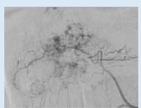
Fig. 4c. Selective facial arterial digital subtraction arteriogram showing multiple enhancing telang-iectasias in the nasal mucosa of the same patient.
Fig. 4d. Nasal endoscopic view showing multiple nasal telangiectasias seen through a septal perfo-ration that has resulted from repeated surgical procedures for intractable epistaxis.
malities of lymph vessels and nodes leading to inadequate clearance of lymph (primary lymphoedema), and secondly solitary or multiple cystic lymphatic malformations.52LMs occur
either due to defective origin of lym-phatics together with the venous sys-tem or abnormal development of the lymphatics themselves. Cystic LMs result from sequestered lymphatic sacs that fail to communicate with normal lymphatic vessels.33Most
(70-80%) occur in the head and neck region where they tend to be more cystic. These were previously referred to as cystic hygromas (Fig. 5a). Twenty per cent of cystic LMs are found in the axilla, with other uncom-mon sites being the superior medi-astinum, mesentery, retroperitoneum, pelvis and extremities. Cystic LMs can be subdivided into macrocystic, microcystic and mixed types. Microcystic LMs were previously called lymphangiomas.
Although present at birth, cystic LMs become clinically apparent later in life, usually before 2 years of age.33
Sudden enlargement may be due to bleeding or inflammation.21
LMs may be associated with a number of syndromes including Turner’s syndrome, Noonan’s drome, multiple pterygium syn-drome, fetal alcohol synsyn-drome, Klinefelter's syndrome, Down's syn-drome, and Klippel-Trenaunay and Parkes-Weber syndromes.33,53
Treatment is again dependent on the presence of functional impair-ment or cosmetic defect (Fig. 5b). An upper respiratory tract infection may frequently cause enlargement of head and neck LMs that may compromise the airway. Macrocystic lesions are probably best treated with percuta-neous sclerotherapy. Various agents have been used including alcohol, Ethibloc, hypertonic glucose, bleomycin, triamcinolone and more recently OK-432.27,31 Microcystic LMs
have a high risk of recurrence after surgery and therefore conservative management is recommended for quiescent lesions.33,54
Mixed vascular
malformations
Given the common vasculogenic origin of arteries, capillaries, veins and lymphatics it is hardly surprising that mixed or combined malformation
types occur.55 Combinations include
capillary-venous (e.g. Sturge-Weber) arteriovenous, lymphaticovenous, capillary-lymphatic, arteri-ovenous (Parkes-Weber), capillary-venous-lymphatic (Klippel-Trenaunay) and more complex combined forms.
Conclusion
Mulliken and Glowacki1 wrote: ‘A
classification is clinically appropriate only when it provides clinicians with a common language through which ideas can be exchanged. A classifica-tion is necessary because there is such a large list of clinical congenital vascu-lar lesions with a complex variability in signs, symptoms, and clinical behaviour. Each lesion has its own lit-tle story of happiness or grief, particu-larly as to whether it will require con-servative or aggressive management and whether it will become acceptable with this form of treatment. Therefore we must continue to strive for an easy yet efficient method of labelling our patients’ problems’.1 A good working
classification of any group of patholo-gies should thus be ‘a comprehensive and clinically relevant means of sim-ply and uniformly describing (vascu-lar) anomalies that occur within any human organ system’.20 Each of the
classification systems described above (CNS and non-CNS) is simple and easy to understand and follow, although each is far from complete. There are still lesions that do not com-fortably fit into these classifications such as the hepatic cavernous hae-mangioma and the spinal osseous haemangioma, both of which are technically low-flow vascular malfor-mations. And what of the aneurysmal bone cyst? These classifications will certainly change with a more thor-Fig. 5a. Macrocystic lymphatic malformation of the
neck. Previously termed cystic hygroma.
ough understanding of the genetic and molecular biology underlying the development of VMs, but for now they will suffice.
References
1. Mulliken JB, Glowacki JG. Hemangiomas and vascular malformations in infants and children: A classification based on endothelial characteris-tics.Plast Reconstr Surg1982;69:412-420. 2. Mulliken JB, Young AE. Vascular Birthmarks:
Hemangiomas and malformations. Philadelphia: WB Saunders. 1988.
3. McCormick WF. Pathology of vascular malfor-mations in the brain. In: Wilson CB, Stein BM eds. Intracranial Arteriovenous Malformations.
Baltimore: Williams and Wilkins, 1984: 44-63. 4. McCormick WF. The pathology of vascular
(‘arteriovenous’) malformations. J Neurosurg
1966;24:807-816.
5. McCormick WF, Hardman JM, Boulter TR. Vascular malformations (‘angiomas’) of the brain, with special reference to those occurring in the posterior fossa.J Neurosurg1968;28: 241-251.
6. Russel DS, Rubenstein LJ.Pathology of Tumours of the Nervous System.3rd ed. B a l t i m o r e : Williams and Wilkins, 1971: 93- 102. 7. Awad IA, Robinson JR, Mohanty S, Estes ML.
Mixed vascular malformations of the brain: Clinical and pathological considerations.
Neurosurgery1993;33:179-188.
8. Sure U, Butz N, Schlegal J,et al. Endothelial pro-liferation, neoangiogenesis, and potential de novo generation of cerebrovascular malforma-tions.J Neurosurg2001;94:972-977.
9. Lasjaunias P, Burrows P, Planet C. Develop-mental venous anomalies (DVA): the so-called venous angioma.Neurosurg Rev1986;9: 233-242.
10. Theron J, Newton TH, Hoyt WF. Unilateral retinocephalic vascular malformations. Neuro-radiology1974;7:185-196.
11. Bhattacharya JJ, Luo CB, Suh DC, Alvarez H, Rodesch G, Lasuanias P. Wyburn-Mason or Bonnet Dechaume-Blanc as cerebrofacial arteri-ovenous metameric syndromes (CAMS): A new concept and a new classification.Interventional Neuroradiology2001;7:5-17.
12. Wong IYC, Barista LL, Alvarez H, Lasjaunias PL. Craniofacial arteriovenous metameric syn-drome (CAMS) 3 - a transitional pattern between CAM 1 and 2 and spinal arteriovenous metameric syndromes.Neuroradiology2003;43:
611-615.
13. Willinsky RA. Spinal cord arteriovenous malfor-mations. In: Marks MP, Do HM, eds. Endovas-cular and Percutaneous Therapy of the Brain and Spine. Philadelphia: Lippincott Willliams and Wilkins, 2002: 415-448.
14. Mulliken JB. Capillary (port-wine) and other telangiectatic stains. In: Mulliken JB, Young AE, eds.Vascular Birthmarks: Hemangiomas and Malformations. Philadelphia: WB Saunders, 1988: 170-195.
15. Lasjaunias P. Venous anomalies and malforma-tions. In: Lasjaunias P, TerBrugge K, eds.Vascular Disease in Neonates, Infants and Children.Berlin: Springer, 1997: 445-471.
16. Boukobza M, Enjolras O, Guichard JP,et al.
Cerebral development venous anomalies
associ-ated with head and neck venous malformations.
Am J Neuroradiol1996;17:987-994.
17. Brouillard P, Vikkula M. Vascular malforma-tions: localized defects in vascular morphogene-sis.Clin Genet2003;63:340-351.
18. Vikkula M, Boon LM, Mulliken JB. Molecular genetics of vascular malformations.Matrix Biol
2001;20:327-335.
19. Shovlin CL. Genetic aspects of cerebrovascular malformations. Interventional Neuroradiology
2000;6:107-111.
20. Chaloupka JC, Huddle DC. Classification of vas-cular malformations of the central nervous sys-tem.Neuroimaging Clin N Am1998;8:295-321. 21. Van Aalst JA, Bhullar A, Sadove AM. Pediatric vascular lesions.J Craniofac Surg2003;14: 566-583.
22. Requina L, Sangueza OP. Cutaneous vascular proliferations Part II: Hyperplasias and benign neoplasms.J Am Acad Dermatol1997;37: 887-919.
23. Chiller KG, Passaro D, Freiden IJ. Hemangiomas of infancy: clinical characteristics morphologic subtypes, and their relationship to race, ethnici-ty and sex.Arch Dermatol2002;138:1567-1576 24. Bowers RE, Graham EA, Tomlinson KM. The natural history of the strawberry nevus.Arch Dermatol1960;82:667-680.
25. Nakayama H. Clinical and histological studies of the classification and the natural course of the strawberry mark.J Dermatol1960;82:667-680. 26. Razon MJ, Kraling BM, Mulliken JB, Bischoff J. Increased apoptosis coincides with onset of involution of infantile hemangioma.
Microcirculation1998;5:189-195.
27. Dubois J, Garel L. Practical aspects of interven-tion in vascular anomalies in children.Semin Intervent Radiol2002;19:73-87.
28. Enjolras O, Riche MC, Merland JJ, Escande JP. Management of alarming hemangiomas in infancy: A review of 25 cases.Pediatrics1990;85:
491-498.
29. Metry DW, Dowd CF, Barkovich AJ, Freiden JJ. The many faces of PHACE syndrome.J Pediatr
2001;139:117-123.
30. Frieden IF, Reese V, Cohen D. PHACE syn-drome. The association of posterior fossa brain malformations, hemangiomas, arterial anom-alies, coarctation of the aorta and cardiac defects, and eye abnormalities.Arch Dermatol1996;132:
307-311.
31. Muir T, Kirsten M, Fourie P, Dippenaar N, Ionesco GO. Intralesonal bleomycin injection (IBI) treatment for haemangiomas and congen-ital vascular malformations. Pediatr Surg Int
2004;19:766-773
32. Young AE. Arteriovenous malformations. In: Mulliken JB,Young AE, eds.Vascular Birthmarks: Hemangiomas and Malformations Philadelphia: WB Saunders, 1988: 228-245.
33. Trop I, Dubois J, Guibard L,et al.Soft-tissue venous malformations in pediatric and young adult patients: diagnosis with Doppler US.
Radiology1999;212:841-845.
34. Dubois J, Garel L. Imaging and therapeutic approach of hemangiomas and vascular malfor-mations in the pediatric age group. Pediatr Radiol1999;29:879-893.
35. Kassarjian A, Fishman SJ, Fox VL, Burrows PE. Imaging characteristics of Blue Rubber Bleb Nevus syndrome.Radiology2003;181: 1041-1048.
36. Reqena L, Sangueza OP. Cutaneous vascular anomalies. Part I: Hamatomas, malformations and dilatation of pre-existing vessels.J Am Acad Dermatol1997;37:523-549.
37. Berry SA. Peterson C, Mize W,et al. Klippel-Trenaunay syndrome.Am J Med Genet1998;79:
319-326.
38. Enjolras O, Mulliken JB. Vascular tumours and vascular malformations (new issues). Adv Dermatol1998;13:375-423.
39. Enjolras O, Ciabrini D, Mazoyer E, Lauriane C, Herbreteau D. Extensive pure venous malforma-tions in the upper of lower limb. A review of 27 cases.J Am Acad Dermatol1997;36:219-225. 40. Lasjaunias P, Berenstein A. Craniofacial
heman-giomas, vascular malformations and angio-matosis. In: Lasjaunias P, Berenstein A, eds.
Surgical Neuroangiography Vol 2: Endovascular Treatment of Craniofacial Lesions. Berlin, Springer-Verlag 1987: 341-397.
41. Pappas DC, Perskey MS, Berenstein A. Evaluation and treatment of head and neck venous vascular malformations.Ear Nose Throat
1998;77:914-922.
42. Berenguer B, Burrows PE, Zurakowski D, Mulliken JB. Sclerotherapy of craniofacial venous malformations: Complications and results.Plast Reconstr Surg 1999;104:1-11. 43. de Lorimier AA. Sclerotherapy for venous
mal-formations.J Pediatr Surg1995;30:185-194. 44. Suh JS, Shin KH, Na JB, Won JY, Hahn SB.
Venous malformations: Sclerotherapy with a mixture of ethanol and lipiodol.Cardiovasc Intervent Radiol1997;20:268-273.
45. Wang C, Gao Q, Fu F,et al. Treatment of heman-gioma in oral and maxillofacial region with pin-gangmycin injection (abstract).Hua Xi Kou Qiang Yi Xue Za Zhi2000;18:317-319. 46. Enjolras O, Riche MC, Merland JJ. Facial
port-wine stains and Sturge-Weber syndrome.
Pediatrics1985;76:48-51.
47. Jacobs AH. Sturge-Weber syndrome without port-wine nevus.Pediatrics1977;60:785-786. 48. Crosley CJ, Binet EF. Sturge-Weber syndrome:
presentation as a focal seizure without nevus flammeus.Clin Pediatr1978;17:606-609. 49. Guttmacher AE, Marchuk DA, White RJ.
Hereditary hemorrhagic telangeictasia.N Engl J Med1995;333:918-924.
50. Pau H, Carney AS, Murty GE. Hereditary haem-orrhagic telangiectasia (Osler-Weber-Rendu syndrome): otorhinolaryngological manifesta-tions.Clin Otolaryngol2001;26:93-98. 51. Begbie ME, Wallace GMF, Showlin CL.
Hereditary haemorrhagic telangiectasia (Osler-Weber-Rendu syndrome): a view from the 21st century.Postgrad Med J2003;79:18-24. 52. Young AE, Stewart G. Lymphatic malformations
In: Mulliken JB, Young AE, eds.Vascular Birth-marks: Hemangiomas and Malformations. Philadelphia, WB Saunders 1988: 215-227. 53. Greenlee R, Hoyme H, Witte M, Crowe P, Witte
C. Developmental disorders of the lymphatic system.Lymphology1993;26:156-168. 54. Sanlialp I, Karnak I, Tanyel FC, Senocak ME,
Buyukpamukcu N. Sclerotherapy for lymphan-gioma in children.Int J Pediatr Otorhinolaryngol
2003;67:795-800.
55. Young AE, Ackroyd J, Baskerville P. Combined vascular malformations. In: Mulliken JB, Young AE, eds.Vascular Birthmarks:Hemangiomas and Malformations. Philadelphia. WB Saunders, 1988: 246-274.