*Correspondence to Author: A. Kılıç
Department of Microbiology, Sivrice Vocational High School, University of Firat, 23119 Elazig-Turkey.
Tel.: +90-424 237 0000/4096; Fax: +90-424 236 08 46.
How to cite this article:
Ayşe KILIÇ, Nurgül BİRBEN, Fat-ma TÜRKARSLAN AKBABA, Mu-hammed Fatih TURSUN, Osman KOÇ, Aslıhan ARSLAN. The Inves-tigation of the Infectious Agalactiae Infection in Sheep and Goat Milk Samples. International Journal of Animal Research, 2018; 2:19.
eSciPub LLC, Houston, TX USA. Website: http://escipub.com/
Ayse Kilic et al, IJAR, 2018; 2:19
International Journal of Animal Research
(ISSN:2575-7822)
Research Article IJAR (2017) 2:19
The Investigation of the Infectious Agalactiae Infection in Sheep and
Goat Milk Samples
Many infections are known to be responsible for ruminants of Mycoplasma, especially in Europe and North America all over the world, mainly in cattle, goats and sheep cultivation causes great economic losses. Stress, immune system failure, incorrect antibiotic treatment, animal transport, breeding, artificial insemi-nation with sperm infected with mycoplasmosis in the increase of cases of importance.
In this study, Elazig and Malatya lots of sheep and goats belong-ing to 300 milk collected from sample Agalaksi Contagious (Con-tagious agalactia) disease molecular methods to detect the pres-ence of (specific PCR). Molecular diagnosis of 300 in the case of milk as a result of specific PCR rate of 45% found positive for Mycoplasma sp. 135 the Mycoplasma sp. positive milk sample was found Mycoplasma agalactiae at 99.
Keywords: Sheep, Goat, Mycoplasma, Milk, PCR
Ayşe KILIÇ1, Nurgül BİRBEN2, Fatma TÜRKARSLAN AKBABA3, Muhammed Fatih TURSUN2, Osman KOÇ2, Aslıhan ARSLAN2
1 Sivrice Vocational High School, University of Firat, Elazig-Turkey;
2 Department of Microbiology, Veterinary Control Institute, Elazig- Turkey; 3 Konya Veterinary Control Institute, Konya-Turkey
ABSTRACT
Introduction
Infectious agalactia, caused by Mycoplasma agalactiae, commonly seen in small ruminants, causes mortality in sheep and goats, as well as diminishing milk yield and causing abortions in pregnancy, leading to significant economic loss. Mycoplasma is present all over the world and also leads to diseases in our country, which lead to severe clinical signs and deaths in cattle, sheep and goats. Although many clinical signs of infectious agalactia are present, the disease is characterized by inflammation of the breast, joints and eyes. This disease is the most serious disease of small milk ruminants. Initially, it was considered that the primary etiologic agent of the infectious Agalactic disease was M. agalactiae (Ma); the large colony, M. capricolum subsp. capricolum and M. mycoides subsp capri. It has been reported that Mycoplasma mycoides cluster group, including capri (Mmc), is a member of the cluster group (Arda et al., 1982, Kumar et al., 2014, Becker et al. 2012). The entry of the disease into the body occurs via the intake of contaminating water and feedstuffs, and sometimes via the conjunctival route (Keskin, 2013). Healthy animals can also be transmitted with the hands of the milkers. Milking, maintenance, feeding and hygiene conditions are not good business in the disease is more frequently encountered. In addition to sick animals, animals called carriers (clinically unspecified) are also effective in spreading the disease to healthy herds (Amores et al., 2010, Gómez-Martín et al., 2011). The main source of M. agalactiae infection is feed and water contaminated with the agent, milk, urine and stool of infected animals, and nasal discharge or tear flow. The incubation period of this disease is between 7 and 56 days. Most cases of infections occur during the summer months, during pregnancy, and throughout the peak level of lactation (Khezri et al., 2012).Contagious agalactia is a serious infection with three distinct clinical manifestations such as mastitis, arthritis and
keratoconjunctivitis (Khezri et al., 2012; Bergonier et al., 1996). Causing infectious agalactia mycoplasma species the diagnosis and the identification, molecular diagnostic methods proved its effectiveness. PCR methods are useful in direct diagnosis of M. agalactiae in milk samples taken from sheep and reduce the duration of diagnosis in excess samples (Tola et al., 1996).
This study was carried out in Elazığ and Malatya sheep and goat flocks The presence of contagious agalactia (Contagious agalactia) a total of 300 milk samples identify with molecular methods .This study was carried out to identify the presence of contagious agalactia (Contagious agalactia) in 300 sheep and goat milk samples belonging to Elazig and Malatya by molecular methods.
Material and methods
This study material is consisted milk samples taken 150 sheep and 150 goat from 5 sheep and 5 goat herds (totaling 30 animals in each herd) and without clinical symptoms and showing mastitis findings, one of the symptoms of infectious agalactia in Elazığ and Malatya provinces. All milk samples were obtained from rapidly, It has been studied in terms of Mycoplasma sp., Mycoplasma agalactiae and Mycoplasma mycoides subsp capri by the direct PCR method in the lab. In this study, while the collected milk samples were examined, we tried to optimize the extraction method from direct milk in order to speed up the identification. For this purpose, milk samples brought to the laboratory were centrifuged for 15 minutes at 3,000 rpm. At the end of the centrifugation, 300 μl was taken from the clear and DNA extraction was performed according to the procedure of the commercial extraction kit (QIAamp DNA mini kit Qiagen, France). The purified DNA was then stored at -200 C for processing in the PCR.
Mycoplasma sp. Group Specific PCR
denaturation, 30 sec at 53 ºC. hybridization and 1 min at 72 ºC. synthesis a total of 35 PCR cycles were performed . The last cycle is 5 min at 72 ºC. extra synthesis was performed. The final products obtained from the Thermal Cycler were run on a 1.5% agarose gel in an electrophoresis apparatus for 90 minutes at 100 volts.
Final products from Thermal Cycler are
obtained from the
device Cycler 1.5% strength agarose gel electr ophoresis device was run at 100 volts for 90 minutes. Then The gel was transferred to the Carestream Gel Logic 212 PRO imaging system; Mycoplasma sp. bands for
280 bp length were searched for positivity. To determine the molecular weight of the resulting tape 100 bp DNA ladder was used . Reference vaccine strains were used as positive control for All PCR applications (Pendik Veterinary Control Institute, Turkey) and distilled water was used as a negative control.
MycoplasmaagalactiaeSpecific PCR
In order to investigate existence M. agalactiae from DNA obtained from milk isolates using a pair of primers specific to the MgA F and MgA R genes reported by Pankaj et al. (2011) (Table 1)PCR was performed. PCR amplification at 95 ºC for 5 min Following denaturation step at ,94 ºC for 1 min . denaturation , 1 min at 57 ºC . hybridization and 1 min at 68 ºC . synthesis . a total of 40 PCR cycles were performed.The last cycle is 10 min at70 ºC . extra synthesis was performed ( Pankaj et al ., 2011). Thermal End products were obtained from the device Cycler 1.5% strength agarose gel electrophoresis device was run at 100 volts for 90 minutes. The gel were then transferred to the Carestream Gel Logic 212 PRO imaging system; bands in the length of 360 bp were searched for Mycoplasma agalactiae positivity,. in all PCR applications were used Reference Infectious agalactia vaccine strain as a positive control (Pendik Veterinary Control Institute, Turkey)
and distilled water was used as a negative control.
Mycoplasma mycoides subsp. capri
Specific PCR
M. mycoides subsp . capri PCR was performed using a pair of primers specific for P4 and P6 genes (Table 1) were reported by Pankaj et al. (2011).
At PCR Following at 94 ºC for 1 min the pre-denaturation step, at 94 ºC for 1 min. denaturation, 1 min at 46 ºC. hybridization and 2 min at 72 ºC. synthesis as a total of 30 PCR cycles were performed. The last cycle was extra synthesis 5 min at 72 ºC was applied ( Pankaj et al ., 2011). The final products were obtained from the thermal Cycler at 1.5% strength agarose gel electrophoresis device was run at 100 volts for 90 minutes. Then, in the Carestream Gel Logic 212 PRO imaging system; Mycoplasma mycoides subsp. capri 194 bp bands were searched for positivity . The reference strain as a positive control in PCR applications (Pendik Veterinary Control Institute, Turkey) and the distilled water was used as a negative control.
Results
Mycoplasma sp. Group Specific PCR
Findings From milk samples
DNA samples were collected from the milk obtained were subjected to PCR amplification using Mycoplasma sp. specific primers, Of the 300 PCR products, 135 (45%) were isolated from Mycoplasma sp. positivity was found (Table 2). 49 (16.33%) milk samples in sheep and 86 (28.66%) goat milk samples were found to be mycoplasma sp positivity (Table 2 ).According to PCR results, milk samples found positive 53 (35.33%) of the Elazığ and 82 (54.66%) were of Malatya was found to be Mycoplasma sp. positive reason (Table 2).
flocks and 92 (61.33%) belonged to patient flocks (Table 3).
PCR Results of Mycoplasma agalactiae Specific Milk Samples
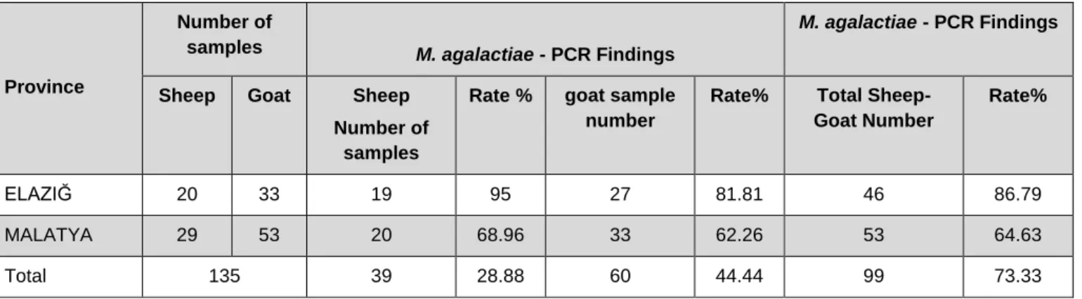
135 milk samples of Mycoplasma sp. Positive, 99 (73.33%) M. agalactiae were detected (Figure 1). 39 (28.88%) of the sheep and the 60 (44.44%) goat samples were found
positive in which was determined to belong to M. agalactiae positive 99 milk samples (Table 4).According to the PCR results, 46 (86.79%) of the 53 positive milk samples of Elazığ province Mycoplasma sp. 53 (64.63%) of the 82 milk samples of Malatya province were found to be positive for Mycoplasma agalactiae (Table 4).
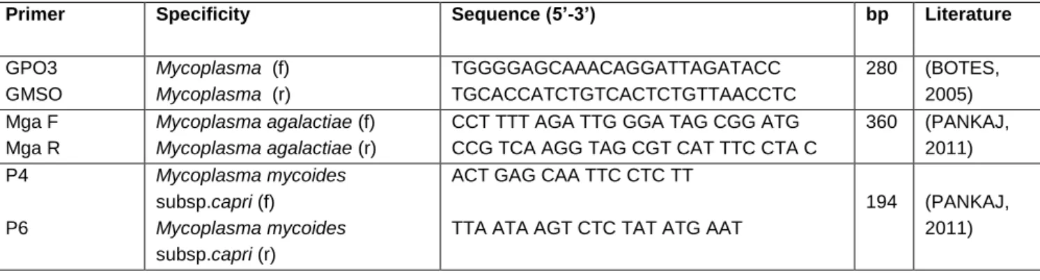
Table 1. Oligonucleotide primers specifically used in the genes investigated in the PCR analyzes
Primer Specificity Sequence (5’-3’) bp Literature
GPO3 GMSO
Mycoplasma (f)
Mycoplasma (r)
TGGGGAGCAAACAGGATTAGATACC TGCACCATCTGTCACTCTGTTAACCTC
280 (BOTES, 2005) Mga F
Mga R
Mycoplasma agalactiae (f)
Mycoplasma agalactiae (r)
CCT TTT AGA TTG GGA TAG CGG ATG CCG TCA AGG TAG CGT CAT TTC CTA C
360 (PANKAJ, 2011) P4
P6
Mycoplasma mycoides
subsp.capri (f)
Mycoplasma mycoides
subsp.capri (r)
ACT GAG CAA TTC CTC TT
TTA ATA AGT CTC TAT ATG AAT
194 (PANKAJ, 2011)
Table 2. Milk samples from sheep and goats were collected from Mycoplasma sp. Specific PCR findings
Province
Number of samples
Mycoplasma sp.-PCR Results Mycoplasma sp.-PCR Results
Sheep Goat Sheep
Number of samples
Rate% Goat number
of samples
Rate% Total sheep
goat sample number
Rate%
ELAZIĞ 90 60 20 22.22 33 55 53 35.33
MALATYA 60 90 29 48.33 53 58.88 82 54.66
Total 300 49 16.33 86 28.66 135 45
Table 3. In Patient and healthy sheep and goat milk samples Mycoplasma sp. Specific PCR findings
Province
Number of healthy flock
samples
Mycoplasma sp.-PCR Findings Patient Herd Sample Number
Mycoplasma sp.-PCR Findings
Sheep Goat Number
of sheep sample
Rate% Number of goat sample
Rate% Sheep Goat Sheep sample number
Rate% Goat sample number
Rate %
ELAZIĞ 60 30 13 21.66 7 23.33 30 30 7 23.33 26 86.66
MALATYA 30 30 12 40 11 36.66 30 60 17 56.66 42 70
Table 4. Mycoplasma sp. specific PCR results of positive milk PCR products in terms of M. agalactiae
Province
Number of
samples M. agalactiae - PCR Findings
M. agalactiae - PCR Findings
Sheep Goat Sheep
Number of samples
Rate % goat sample
number
Rate% Total
Sheep-Goat Number
Rate%
ELAZIĞ 20 33 19 95 27 81.81 46 86.79
MALATYA 29 53 20 68.96 33 62.26 53 64.63
Total 135 39 28.88 60 44.44 99 73.33
It was determined that 27 (62.79%) of the total of 99 milk samples positive for M. agalactiae belonging to Elazığ and Malatya were belong to
healthy flocks and 72 (78.26%) belonged to the patient flocks (Table 5).
Table 5. The spesific PCR findings for Mycoplasma agalactiae from from milk samples Mycoplasma sp. positive which belong to sick and healthy looking sheep and goats
Province
Number of healthy flock
samples
M. agalactiae - PCR findings Patient Herd Sample Number
M. agalactiae - PCR findings
Sheep Goat Sheep
Number of sample
Rate %
Goat Number
of sample
Rate% She ep
Goat Sheep Number of sample
Rate %
Goat number
of sample
Rate %
ELAZIĞ 13 7 13 100 3 42.85 7 26 6 85.71 24 92.30
MALATYA 12 11 3 25 8 72.72 17 42 17 100 25 59.52
Total 43 27 62.79 92 72 78.26
Mycoplasma mycoides subsp. capri Specific PCR Results
135 Mycoplasma sp. positive milk, Mycoplasma mycoides subsp. capri-specific positive bands were not found.
Discussion
Today, The Mycoplasma identification is mostly done with cultural and serological methods. However, both methods are consuming and laborious. In addition, the application is also expensive methods (Corrales ve ark., 2007; Sımeckave ark., 1993). It has also been demonstrated in the study conducted by Çetinkaya et al. (2006-2008) that it is possible to obtain results in a very short time by direct PCR in cases where there are problems with
It is important to develop methods that accurately
and specifically identify mycoplasmas . In
recent years molecular methods have been used to identify mycoplasmas. The PCR method is a technique that yields rapid results and replicates a single DNA sequence millions of times within a few hours (Çetinkaya ve ark., 2006-2008).
Tola et al., (1997) 357 samples from 21 new infected infections (Group 1) and 87 (Group 2) from the past 8 infections, sheep milk sample was examined by direct PCR and culture methods for Mycoplasma agalactiae.
Tola et al., (1997), in terms of Mycoplasma agalactiae the 357 sheep milk sample (Group 1) from 21 new infected herds and the 87 sheep milk sample (Group 2) from the 8 herds infected in the past , in terms of Mycoplasma agalactiae was examined with direct PCR and culture methods. In Group 1, 175 positive for PCR and 153 positive for culture, whereas in Group 2, milk samples were found negative by PCR and culture method. In this study, 135 (45%) Mycoplasma sp. were found positive; Mycoplasma sp. positive 99 of the PCR products (73.33%) were isolated as M. agalactiae. In this study, the PCR technique has been demonstrated that from culture is much faster and in a shorter time from culture results. It has also been shown that the PCR technique can routinely be used to diagnose infectious agalactia caused by Mycoplasma agalactiae.
In a study by Göçmen et al. (2015) for investigating the infectious agalactiae disease in sheep and goats with bacteriological and PCR methods, Mycplasma sp. were identified in 29 samples of from 339 samples (162 milk samples, 147 eye swabs, 15 joint fluids, 11 nasal swabs and 4 lung tissues) collected from sheep and goats belonging to Canakkale and Edirne province. When PCR and culture data were compared, 5 milk samples and 1 lung sample were found to be positive by polC-PCR and negative by culture. The major cause of the
disease was found to be Ma, and none of the other mycoplasma agents causing the disease were found. In a study carried out by Bidhendi et al. (2011), Mycoplasma sp. were cultured positive 20 out of 367 milk samples taken from the sheep and goat flocks of the Iranian province of Kurdistan, 5 (belong to 1 goat and 4 of sheep) of these positive isolates were positive with Mycoplasma agalactiae primers. In addition, PCR was positive for Mycoplasma agalactiae in 11 of 367 milk samples. Rosetti et al. (2010) reported that real-time PCR is a specific, sensitive and rapid method for the diagnosis of Mycoplasma bovis from direct milk and tissue specimens without DNA extraction. Cai et al. (2005) concluded that direct real-time PCR of Mycoplasma bovis from lung tissue showed very high specificity (100%) and sensitivity (96.6%). In the present study, the direct PCR technique from milk was used as a rapid and economical method to detect infectious agalaxin compared to the culture method.
As a result of the study conducted, it was determined that M. agalactiae was the main cause of the disease according to the findings, and M. mycoides subsp. capri wasn’t found. The results of this study proves that infectious agalasin is common in Elazığ and Malatya provinces. M. agalactiae origin mastitis infectio ns lead to major problems and economic losses in terms of flocks in terms of small ruminants. Mycoplasma induced mastitis considering the infectious nature of the infections , effective and radical control strategies should be implemented and vaccination programs should be established in the affected diseases.
Aknowledgement
This project was supported by the Directorate General for Agricultural Research of the Turkish Ministry of Agriculture and Village Affairs (TAGEM/HSGYAD/13/A07/P02/29).
The authors declare that they have no conflict of interest.
References
1. AMORES J., GÓMEZ-MARTÍN A., CORRALES J.C., SÁNCHEZ A., CONTRERAS A., DE LA FE C (2010): Presence of contagious agalactia causing mycoplasmas in Spanish goat artifical insemination centres. Theriogenology. 75, 1265-70.
2. ARDA M., MİNBAY A., AYDIN N (1982). Özel Mikrobiyoloji, Bakteriyal İnfeksiyöz Hastalıklar, Ankara, A.Ü. Vet. Fak. Yayınları.
3. BECKER C.A., RAMOS F., SELLAL E., MOİNE S., POUMARAT F., TARDY F (2012): Development of a multiplex real-time PCR for contagious agalactia diagnosis in small ruminants. Journal of Microbiological Methods. 90, 73–79.
4. BERGONİER D., POUMARAT F (1996): Contagious agalactia of small ruminants: Epidomology, diagnosis and control. Revue Scientifiqoe et Technique. 15, 1431-1475. 5. BERGONİER D., BERTHELOT X., POUMARAT
F (1997): Contagious Agalactia of Small Ruminants: Current Knowledge Concerning Epidemiology, Diagnosis and Control. Rev Sci Tech. OIE. 16, 848–873.
6. BİDHENDİ M., KHAKİ S., LANGROUDİ P (2011): Isolation and identification of Mycoplasma agalactiae by culture and Polymerase Chain Reaction in Sheep and Goat Milk Samples in Kordestan province, Iran. Archives of Razi Institute. 66, 11-16.
7. BOTES A., PEYROT B.M., OLİVİER A.J., BURGER W.P., BELLSTEDT D.U (2005): Identification of Three Novel Mycoplasma
Species from Ostriches in South Africa. Vet Microbiol. 111, 159-169.
8. CAI H.Y., BELL-ROGERS P., PARKER L., PRESCOTT J.F (2005): Development of a real-time PCR for detection of Mycoplasma bovis in bovine milk and lung samples, J Vet Diagn Invest. 17, 537-545.
9. CORRALES J., ESNAL A., DE LA FE C., SÁNCHEZ A., ASSUNÇAO P., POVEDA J.B., CONTRERAS A (2007): Contagious Agalactia in Small Ruminants. Small Rum Res. 68, 154-166. 10. ÇETİNKAYA B., KARAHAN M., KALIN R., ATIL
E (2006-2008): Biodiversity of Ruminant Mycoplasmas in Eastern Turkey: Application for Vaccines and Control Strategies, Tübitak (PIA-553).
11. GÓMEZ-MARTÍN A., CORRALES J.C.,
AMORES J., SÁNCHEZ A., CONTRERAS A., PATERNA A., DE LA FE C (2011): Controlling contagious agalactia in artificial insemination centers for goats and detection of Mycoplasma mycoides subspecies capri in semen. Theriogenology.
12. GÖÇMEN H., ÜLGEN M., ÇARLI K.T., ÖNAT K., KAHYA S., ÖZDEMİR Ü., MAT B (2015): Koyun ve Keçilerde Bulaşıcı Agalaksi Hastalığının Bakteriyolojik ve PCR Metotları ile Araştırılması. Kafkas Üniv Vet Fak Derg. 21, 75-80.
13. KESKİN D (2013): Koyun ve Keçilerde
Mycoplasma agalactiae’nin Önemi. Tralleis
Elektronik Dergisi. 2, 1-5.
14. KUMAR A., RAHAL A., CHAKRABORTY S., VERMA A.K., DHAMA K (2014). Mycoplasma agalactiae, an Etiological Agent of Contagious Agalactia in Small Ruminants: A Review. Veterinary Medicine International. Article ID 286752, 13 pages.
15. KHEZRİ M., POURBAKHSH S.A., ASHTARİ A., ROKHZAD B., KHANBABAİE H (2012): Isolation and prevalence of Mycoplasma agalactiae in Kurdish sheep in Kurdistan, Iran. Vet World. 5, 727-731.
16. LANGFORD E.V (1975): Mycoplasma
Recovered from Bovine Male and Female Genitalia and Aborted Foeti, Proceedings of 18th Annual Meeting of American Association of Veterinary Laboratory Diagnosticians. 221-232. 17. PANKAJ K., ASHİSH R., BHARAT B.B., BHİK
C.P (2011): Isolation, identification and molecular characterization of Mycoplasma isolates from goats of Gujarat State, India. Veterinarski Arhıv. 81 , 443-458.
18. ROSSETTİ B.C., FREY J., PİLO P (2010): Direct detection of Mycoplasma bovis in milk and tissue samples by real-time PCR. Molecular and Cellular Probes. 24, 321-323.
19. SIMECKA J.W., ROSS S.E., CASSELL G.H., DAVİS J.K (1993): Interactions of Mycoplasmas
with B Cells: Antibody Production and Nonspecific Effects, Clin Infect Dis. 17, 176-182. 20. TOLA S., IDİNİ G., MANUNTA D., CASCİANO
I.G., ROCCHİGİANİ A.M., ANGİOİ A., LEORİ G (1996): Comparison of Mycoplasma agalactiae
Isolates by Pulsed Field Gel Electrophoresis, SDS - PAGE and Immunoblotting. FEMS Microbiol Letters. 143, 259-265.