Original Research Article
High resolution computed tomography in chronic obstructive
pulmonary disease
Pragati Rao D.
1, Aruna Talatam
2*, Chakradhar B.
2, Bhargavi K.
2, Bhagyaraj A.
2INTRODUCTION
Chronic obstructive pulmonary disease (COPD) is a common preventable and treatable disease characterized by persistent respiratory symptoms and airflow limitation that is due to airway and or alveolar abnormalities usually caused by significant exposure to noxious particles and
gases.1 The most common cause of COPD is tobacco
smoking. There are other risk factors like pollution, biomass exposure and certain occupations. COPD is a mixture of airway disease (bronchitis) and parenchymal destruction (emphysema). Chronic bronchitis is defined as cough with expectoration for at least three months in two consecutive years. Emphysema is permanent ABSTRACT
Background: Chronic obstructive pulmonary disease (COPD) is a common preventable and treatable disease characterised by persistent respiratory symptoms and airflow limitation with varied presentations (bronchitis and emphysema). High resolution computed tomography (HRCT) plays an important role in identifying the various morphologies thereby reducing morbidity and mortality. The aim of the present study was to evaluate the role of high resolution computed tomography in COPD patients. The Objectives of the present study was to differentiate emphysema predominant, airway predominant and mixed phenotypes and to identify other disease processes and complications.
Methods: 50 COPD patients attending Respiratory medicine Department, NRI general hospital were advised for chest x-rays and pulmonary function tests. All the patients selected were smokers with no other co-morbid illnesses. Those patients whose chest x-rays showed no other changes except for COPD changes were selected for HRCT chest.
Results: Out of 50 COPD patients emphysema predominance was present in 28 patients (56%), bronchitis predominance in 19 patients (38%) and 3(6%) patients had mixed pattern. In emphysema centriacinar pattern was commonly seen (42.9%), paraseptal in 35.71%, panacinar in 3.57% and bullae in 17.8% cases. All the patients were chronic smokers with pack years >20. All are males with average age above 45 years. Emphysema was common in elderly patients with age above 50 years. Chronic bronchitis is predominantly seen in the age group 40-50 years. Additional diagnoses like bronchiectasis, mass, ILD were identified in 28% cases.
Conclusions: HRCT plays a significant role in COPD patients in differentiating phenotypes which have different modes of therapy. Other subtle changes in lungs which cannot be identified on chest x ray are discernible on HRCT. Early identification of complications reduces morbidity and mortality.
Keywords: Bronchitis, Chronic obstructive pulmonary disease, Emphysema, High resolution computed tomography
1Department of Respiratory Medicine, M.S. Ramaiah Medical College, Bangalore, Karnataka, India
2Department of Respiratory Medicine, NRI Medical college and General Hospital, Chinakakani, Guntur, Andhra
Pradesh, India
Received: 24 June 2018
Revised: 02 July 2018
Accepted: 27 July 2018
*Correspondence:
Dr. Aruna Talatam,
E-mail: [email protected]
Copyright: © the author(s), publisher and licensee Medip Academy. This is an open-access article distributed under the terms of the Creative Commons Attribution Non-Commercial License, which permits unrestricted non-commercial use, distribution, and reproduction in any medium, provided the original work is properly cited.
dilatation and destruction of lung parenchyma distal to terminal bronchioles leading to air trapping. Emphysema is father classified int centriacinar emphysema, panacinar and paraseptal emphysema. Pathologically characterized by chronic inflammation and small airway fibrosis. These changes ultimately lead to air trapping, mucus hypersecretion, defective gas exchange and finally pulmonary hypertension. Both the entities are different pathologically, clinically and radiologically. The relative contribution of these entities in COPD patients is important because of different therapeutic interventions. COPD affects lung parenchyma, airways both central and peripheral and vasculature. All the changes in these structures are best depicted on computed tomography. High resolution computed tomography (HRCT) plays an important role in identifying the various morphologies, other associated respiratory diseases thereby reducing morbidity and mortality.
METHODS
There were 50 COPD patients attending respiratory medicine department of NRIGH for a duration of 1 year were included in this cross-sectional observational study.
Inclusion criteria
• COPD patients with history of smoking between age 35- 70 years
• Those who have given consent. Exclusion criteria
• COPD patients suffering from co-morbid illnesses like ischemic heart diseases
• Concomitant respiratory diseases like active tuberculosis, PTB sequelae, bronchiectasis, interstitial lung diseases.
All patients included in our study were males with significant smoking history >20 pack years. A thorough clinical examination and laboratory evaluation was done All patients underwent pulmonary function test and those with post bronchodilator FEV1/FVC ratio less than 0.7 and partial reversibility that is increase in FEV1 less than 200 ml and 12% with bronchodilator from baseline were considered for study. Initially chest x rays were done for all patients. Those patients whose chest x-rays showed no other changes except for COPD changes were selected for HRCT chest. In few clinically suspected cases and when radiologically pulmonary hypertension was present 2D-ECHO (echocardiography)was done. Results are expressed as percentages. Z test of proportion was applied for age groups in chronic bronchitis and emphysema patients.
CXR findings of COPD -hyperinflated lung fields, flattened diaphragms, bullae, tubular heart, increased retrosternal translucency, rapid tapering of vessels.
Parameters evaluated on HRCT
• Centriacinar emphysema: focal area of low attenuation in a secondary pulmonary lobule. Usually found in upper zones and is most commonly associated with smoking
• Panacinar emphysema: large areas of decreased attenuation evenly distributed, predominantly seen in lower lobes.
• Paraseptal emphysema: Areas of low attenuation are along edges.
• Small airways: Airways with diameter less than 2 mm.
• Mosaic attenuation: Alternate areas of high and low attenuation.
• Ground glass opacities: Areas of high attenuation with preservation of vasculature.
RESULTS
The study included 50 COPD patients all of them were chronic smokers with pack years above 25 years. All the patients included were males. 25 patients (50%) had mild COPD with FEV1> or = 80% and 25 patients had moderate COPD with FEV1 50-80%.
Z test of proportion was done between two age groups in chronic bronchitis and emphysema patients. There is a significant difference in proportion of cases between age groups of 40-50 and >50 years in chronic bronchitis patients with a higher predominance in the age group 40-50 years (p value - 0.0004). In emphysema predominant group majority were above 50 years (p value-0.0013) (Table 1).
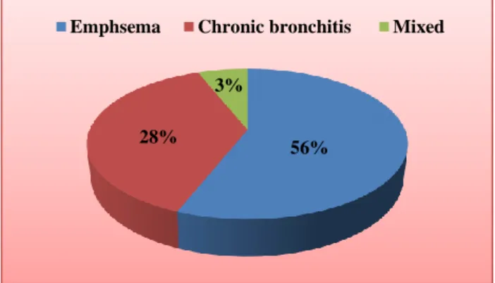
Figure 1: Various subtypes of COPD.
Table 1:Age wise of distribution of disease.
Phenotype Age group 40-
50 years >50 years
Chronic bronchitis (19) 15 4
Emphysema (28) 8 20
Mixed pattern (3) 0 3
Emphysema is identified in HRCT as low attenuation areas. Emphysema predominant pattern was present in 28
Emphsema Chronic bronchitis Mixed
56% 28%
patients (56%), chronic bronchitis predominance in 19 patients (38%) and 3 patients had mixed pattern (6%).
In emphysema, centriacinar pattern (Figure 2a) was commonly seen in 12 patients (42.9%), paraseptal (Figure 2c) in 10 patients (35.71%), panacinar in 1 patient (3.57%) (Figure 2b) and bullae in 5 patients (17.8%).
Upper lobe (65%) was most commonly involved in emphysema patients.
Majority of patients with centriacinar emphysema had mild COPD (8 cases) and only 25% had moderate COPD in our study. Chronic bronchitis patients suffered from moderate obstruction in more than 90% of cases.
Figure 2: HRCT chest showing various patterns in COPD patients; A) Centriacinar emphysema, B) Panacinar emphysema, C) Paraseptal emphysema, D) Chronic bronchitis.
Chronic bronchitis is manifested on computed tomography CT (CT) as bronchial wall thickening and those with thickness more than 2mm had more decrease in FEV1 (Figure 2d). Ground glass opacities were found in 3 cases and mosaic attenuation was present in 15 patients.
Other significant radiological features which could not be seen on chest X ray were made out on CT scan like bronchiectatic changes (Figure 3) in 10 cases, interstitial pattern in 2 cases, mass lesions in 2 cases. Therefore around 28% of patients got benefitted from CT scan due to diagnosis of additional features.
Figure 3: HRCT chest of a COPD patient showing associated traction bronchiectasis.
Vascular attenuation is thinning and reduction in number of pulmonary vessels. This feature was most commonly seen in patients with moderate COPD (70%) compared to those with mild COPD (40%). In 5 patients (10% cases) pulmonary hypertension (Figure 4) was diagnosed which correlated well with 2D-ECHO.
Figure 4: HRCT chest of a COPD patient showing pulmonary hypertension.
DISCUSSION
COPD is the fourth leading cause of death in the world currently.1 The prevalence and burden of disease is
projected to be on rise because of continued risk factors and aging of population.2 COPD is not only a disease
which involves lungs, but it leads to lot of other
consequences involving other systems. It is a systemic illness which leads to very poor quality of life. Therefore, it is important to identify the condition at an early stage to prevent complications. CT plays an important role in identifying various forms, other concomitant respiratory diseases and for screening of lung cancer.3,4
Present study included 50 subjects in the age group range between 40-60 years. In Gupta et al, study mean age was 58.55 (range 50-69) years.5 Z test of proportion applied in
chronic bronchitis patients revealed a higher predominance in the age group 40-50 years (15/19) compared to those above 50 years (4/19) (p value- 0.0004). In emphysema predominant group most of the patients were above 50 years (20/28) (p value-0.0013) which is in agreement with the study done by Koshiol J et al, in their study the mean age of chronic bronchitis was 45 and emphysema was 54.2 years.6
In present study emphysema was the commonest form 28 out of 50 patients (56%). Gupta P et al, also found emphysema the commonest phenotype in 25 patients out of 40 (62.5%).7
In present study, centrilobular emphysema was the most common pattern (42.9%) followed by paraseptal emphysema seen in 35.71% of cases. Least common was panacinar emphysema. This is in concordance with a study done by Nazia et al.8 In their study centrilobular
pattern was commonest seen in 12 patients (42.9%). In Gupta et al, study centriacinar emphysema was most common (16 cases), followed by paraseptal in 13 patients and panacinar emphysema in 11 patients.5 Klein et al,
also observed that centrilobular emphysema was the dominant pattern.9
In 3 cases (6%) mixed pattern was revealed with both emphysema and bronchitis features. Present study revealed vascular attenuation in 70% of patients with moderate COPD and 40% of mild COPD patients. Gupta P et al, found that involvement of small airways was present in 16 patients with associated features of vascular attenuation.7 Pulmonary hypertension was noted in 5
patients where main pulmonary artery diameter was more than that of aorta (10%).
In present study, bronchiectasis was present in 10 cases (20%) which is much less compared to study done by Singh et al, where bronchiectasis was revealed in significant number of patients (54.8%).10 Presence of
brochiectasis suggests that these patients are at risk of more severe infective exacerbations.
Ground glass opacities which are very non -specific were found in 3 cases. Mosaic attenuation was present in 15 patients. Interstitial lung disease in 2 cases, mass lesions in 2 cases were found additionally. Usual interstitial pneumonia and non-specific interstitial pneumonia features were present in those two cases.
Though there is a risk of radiation exposure in all patients subjected to HRCT scan the benefit -risk ratio is much higher in at least some of the cases where patients are not responding to routine therapy and those patients who are more prone for complications.11
Chest radiography has low sensitivity and specificity in identifying mild emphysema and limited ability to quantify severity of emphysema.12 CT is invaluable in
identifying the disease early, in differentiating the various forms-airway predominant form, emphysema predominant and mixed forms, each of which have different treatment approaches. It is also useful in excluding alternative diseases like interstitial lung diseases, bronchiectasis, fibrosis.
CT is also useful for lung cancer screening. With CT distribution of emphysema is also better known for lung volume reduction surgeries.13
CONCLUSION
COPD is a systemic disease with various subtypes. HRCT definitely allows us to identify various subtypes, severity and other concomitant respiratory diseases. This will help in managing COPD patients more appropriately according to the disease subtype.
ACKNOWLEDGEMENTS
Authors would like to acknowledge s Prof. Dr. Ankamma Rao, Head of the Department, Department of Radio Diagnosis, NRI Medical College and General Hospital for his invaluable contribution.
Funding: No funding sources Conflict of interest: None declared
Ethical approval: The study was approved by the Institutional Ethics Committee
REFERENCES
1. Global Initiative for Chronic Obstructive Lung Disease -Global Initiative for Chronic Obstructive Lung Disease - GOLD. 2018. Available at: http://goldcopd.org.
2. Mathers CD, Loncar D. Projections of global mortality and burden of disease from 2002 to 2030. PLoS Med. 2006 Nov 28;3(11):e442.
3. Washko GR. The role and potential of imaging in COPD. Medi Clin. 2012 Jul 1;96(4):729-43. 4. Matsuoka S, Yamashiro T, Washko GR, Kurihara
Y, Nakajima Y, Hatabu H. Quantitative CT assessment of chronic obstructive pulmonary disease. Radiographics. 2010 Jan;30(1):55-66. 5. Gupta PP, Yadav R, Verma M, Gupta KB, Agarwal
6. Koshiol J, Rotunno M, Consonni D, Pesatori AC, De Matteis S, Goldstein AM, et al. Chronic obstructive pulmonary disease and altered risk of lung cancer in a population-based case-control study. PLoS One. 2009 Oct 8;4(10):e7380.
7. Gupta PP, Yadav R, Verma M, Agarwal D, Kumar M. Correlation between high-resolution computed tomography features and patients' characteristics in chronic obstructive pulmonary disease. Ann Thoracic Med. 2008 Jul;3(3):87.
8. Mehfooz N, Bhargava R, Ahmad Z, Ahmad I, Patigaroo SA. HRCT findings in early cases of COPD-an experience. Int J Basic Applied Med Sci. 2013;3(3):20-31.
9. Klein JS, Gamsu G, Webb WR, Golden JA, Müller NL. High-resolution CT diagnosis of emphysema in symptomatic patients with normal chest radiographs and isolated low diffusing capacity. Radiol. 1992 Mar;182(3):817-21.
10. Singh A, Kumar S, Mishra AK, Kumar M, Kant S, Verma SK, et al. Correlation between clinical characteristics, spirometric indices and high
resolution computed tomography findings in patients of chronic obstructive pulmonary disease. Lung India: official organ of Indian Chest Society. 2016 Jan;33(1):42.
11. Brenner DJ. Radiation risks potentially associated with low-dose CT screening of adult smokers for lung cancer. Radiology. 2004 May;231(2):440-5. 12. Thurlbeck WM, Müller NL. Emphysema: definition,
imaging, and quantification. AJR. Am J Roentgenol. 1994 Nov;163(5):1017-25.
13. National Emphysema Treatment Trial Research Group. A randomized trial comparing lung-volume– reduction surgery with medical therapy for severe emphysema. N Eng J Med. 2003 May 22;348(21):2059-73.