COMPARISON OF MORPHOLOGICAL AND CELL
ADHESION MOLECULES MODULATION BETWEEN
PRIMARY OSTEOSARCOMA CELLS AND
OSTEOSARCOMA CELL LINES
Fadzliza Hafiza Ramli
Institute of Molecular Medicine &Biotechnology UiTM Sungai Buloh 47000 Jalan Hospital Sungai Buloh, Selangor Malaysia
Nor Faissal Yasin
Depart. Of Orthopaedic SurgeryFaculty of Medicine University of Malaya 50603 Kuala Lumpur
Malaysia
Mudiana Muhamad
Biochemistry & MolecularMedicine Department UiTM Sg buloh 47000 Jalan Hospital, Sungai Buloh, Selangor Malaysia
Tunku Kamarul
Depart. Of Orthopaedic Surgery Faculty of Medicine University of Malaya 50603 Kuala Lumpur
Malaysia [email protected]
Azura Mansor
Depart. Of Orthopaedic SurgeryFaculty of Medicine University of Malaya 50603 Kuala Lumpur
Malaysia [email protected]
Sharaniza Ab Rahim
Biochemistry & MolecularMedicine Department UiTM Sg buloh 47000 Jalan Hospital, Sungai Buloh, Selangor, Malaysia
[email protected] (corresponding author)
ABSTRACT
Osteosarcoma (OS) is a rare primary bone cancer arising among the adolescent and worsen when it metastasized. The important biomarkers involved in the invasion and metastasis of cancer cells include the cell adhesion molecules (CAMs). Therefore, this study aim to characterized the expression of CAMs proteins in metastatic OS cells isolated from OS patients. Morphological changes were observed using haematoxylin and eosin (H&E) staining in normal human osteoblast (NHOst), OS cell line (MG-63), non-metastatic and metastatic primary cell culture. The expression of VCAM-1 and ICAM-3 was determined by immunocytochemical (ICC) stain and western blot techniques. The H&E results showed distinct morphology differences in the four different cells respectively. Further analysis through IHC and western blot showed the expression of VCAM-1 is higher than ICAM-3 in all cancer cells (MG-63, metastatic and non-metastatic primary cells) but at different level of intensity. Nevertheless, both VCAM-1 and ICAM-3 were significantly under expressed in NHOst cells. In addition, this study has established the different characteristic of metastatic OS primary culture, non-metastatic OS primary cells and OS cell line. These findings are important for further exploration in understanding OS progression using primary cell culture instead of cell line. In conclusion, the two CAMs were expressed at different levels respective to the progressive state of OS.
General Terms:
biomarkers, primary cells, cell line, morphology1.
INTRODUCTION
Osteosarcoma (OS) is an aggressive primary malignant bone tumour in children and adolescents [1]. The peak incidence of osteosarcoma is associated with pubertal maturation when the growth emission is at its greatest [2, 3]. This is due to the prompt bones division to facilitate the increased growth velocity during adolescents. The stimulatory signals that are associated with pubertal growth have been suggested to summon episode in the oncogenesis of OS [1]. The OS have a high potential of being metastasized, mainly to the lungs [4]. Previous study showed that over 80% of OS patients have metastatic or micro-metastatic diseases upon diagnose with majority of them having metastasized to the lungs [5]. Prior to the development of chemotherapy and surgery-involved amputations, the survival rate in OS patients was around 20%, which also reflects the number of patients who never developed metastases. Following advents of chemotherapy surgery and radiotherapy, the 5-year survival rate for patients diagnosed with OS are improved to 60% to 70%, considering high although not completely high [6].
Metastasis progressed through several biological steps whereby malignant cells extent from the tumour primary sites before spreading to distant organs [5] that eventually being mostly deposited in the lungs. The cell adhesion molecules (CAMs) facilitate interactions of cells with each other as well as their microenvironment through cell proliferation, migration and differentiation [7]. Nevertheless, normal cell-cell and cell-matrix interactions were disrupted by the aberrant expression of CAMs, releasing cells from normal checkpoints and constraints, which then promotes the tumour development and subsequently metastasis [8].
Vascular cell adhesion molecule-1 (VCAM-1) is an immunoglobulin (I)-like adhesion molecule with seven extracellular Ig domains that are mainly expressed at the low level in endothelial cells. However, VCAM-1 proteins are highly susceptible to be induced by several inflammation cytokines [9] that mediates leukocytes adhesion on endothelial cells. In addition, VCAM-1 proteins are able to activate signalling pathways to facilitate leukocyte passage from blood to tissues. In addition to passive entrapment of tumour cells by size restriction, it has been suggested that capillary beds actively participate in the extravasation into disseminated sites of CTCs via the adhesion molecules on vascular endothelial cells [10].
Intercellular adhesion molecule 3 (ICAM-3) also known as CD50 belongs to one of the immunoglobulin (Ig) family. Its role in the preliminary steps of immune cell interactions have been documented since 1992 which stated that there was an increase in constitutive expression of ICAM-3 in the resting T-lymphocytes. Furthermore, it has been reported that ICAM-3 is restricted in cell-cell contacts during initial of cell aggregation and ICAM-3 also induce the homologous Ig-like family, ICAM-1 adhesion [11] as well as several proteins suchas integrin molecules and IkappaB kinase (IKK) proteins. In addition, ICAM-3 interacts with LFA-1 (Lymphocyte function-associated antigen-1) through its expression on leukocytes and endothelial cells, thus, involved in intercellular adhesion of leukocytes. Moreover, ICAM-3 expression on endothelial cells has been suggested to be involved in angiogenesis [12]. These findings suggest that the expression of ICAM-3 proteins could be a significant factor in tumour progression.
In this study, the different levels of expressions of ICAM-3 and VCAM-1 in the primary OS cells compared to OS cell lines (MG-63) and normal human osteoblasts (NHOst) was determined by IHC stain and western blot techniques. The results provided new insight in understanding the role of both VCAM-1 and ICAM-3 proteins within the cell lines and the primary OS cells.
2.
MATERIALS AND METHODS
2.1
Sample Collections
The bone samples were collected from osteosarcoma patients (non-metastatic and metastatic) in University Malaya Medical Centre (UMMC) upon ethical approval from UMMC ethics committee (BK-MIS-1117-E01). The tissue was transported in a sterile tube containing 10 to 20ml of phosphate buffered saline (PBS) depending on the size of the OS tissues and placed at 4˚C for further usage.
2.2
Primary Cells and Cell Lines Culture
The OS tissue samples collected were digested within 24 hours upon biopsied. Tissue was cleaned and digested with scalpel into small fragments and further incubated in Collagenase type II (Invitrogen, USA) at 37˚C overnight for cells liberation. Isolated cells were collected and allowed to
NHOst (Lonza, Switzerland) and MG-63 (ATCC, USA) were cultured in Dulbecco’s Modified Eagle Medium (DMEM, Merck, Germany) supplemented with 10% foetal bovine serum (FBS, Invitrogen, USA) and 1% of Penicillin Streptomycin (Invitrogen, USA).
2.3
Hematoxylin and Eosin Staining
Cells were seeded on glass slides at 70-80% of cell confluence. The cells were stained with Hematoxylin dye for a minute. Subsequently, the slides were fixed with clarifier for 1 minute followed by potassium acetate for a minute. Ethyl alcohol (95%) was added on the slides before application of Eosin on the slides. Following that, 95% and 100% of ethyl alcohol were applied for 1 minute respectively. Finally, the cells we treated with xylene and mounting agent before viewed under light microscope (Leica TCS SP5, Olympus).
2.4
Immunocytochemistry (ICC)
For immunocytochemistry, cells were seeded on chamber slides at 80-90% of cell confluency. Cells were fixed in paraformaldehyde for 20 min and subsequently washed in phosphate-buffered saline (PBS). Blocking buffer was applied and later removed by wicking it with Whatman filter paper (No.1) then added with desired primary antibody (VCAM-1 and ICAM-3) following incubation for three hours and then washing with PBS solution. Secondary antibody was applied after the washing and incubated for an hour before final washing. Finally, few drops of mounting agent were used to cover the cells with coverslip. Using three-point scoring system, the heterogeneity of the immunocytochemistry result was further assessed.
2.5
Western Blot (WB)
Thirty micrograms of total protein extracted from cultured cells were separated by electrophoresis using 10% SDS-PAGE gel and transferred to nitrocellulose (NC) membrane. BSA (3%) solution has been used for membrane blocking for an hour. Every stage required washing steps with TBS-Tween. Following that, NC membrane was incubated overnight at 4°C with desired primary antibody (Beta actin, VCAM-1 and ICAM-3) with gentle agitation. Prior to detection, the NC were incubated for 1 h at 4°C with secondary antibodies. Proteins were detected by using enhanced chemiluminescence (ECL, Thermo Scientific, USA) detection system and visualized on X-ray films.
3.
RESULTS
3.1
Morphology of the Cell Lines and Primary CellsThe normal human osteoblast (NHOst) cells presented plump and polarized cuboidal cells with varying and faded bone lining cells as shown in Fig. 1.
Fig. 1 Morphology of the cultured cells seen under bright field microscopy. (A) Normal human osteoblast (NHOst) cells: wide spindle cytoplasm; (B) OS cell line (MG-63): short and pointy shape cytoplasm; (C) Non-metastatic primary OS cells: elongated and wide
A) (B)
Fig. 2 Level of intensity of H&E stain in the cell lines and OS primary cells. Low intensity stain of nuclei and cytoplasm in the NHOst cells (A) and metastatic OS primary cells (D). High intensity stain of nuclei and cytoplasm in the MG-63 cells (B) and non-metastatic
OS primary cells (C)
3.1 Expression of VCAM-1 and ICAM-3
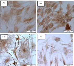
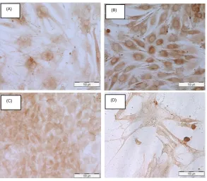
The labelling patterns for all cells were in homogenous showing various proportions of cells indicated by positive reactions toward VCAM-1 (Fig. 3) and ICAM-3 (Fig. 4) antibodies. The labelling of the cells was assessed by three-point scoring system as shown in Table 1. More than 95% of the cell populations in MG-63 and non-metastatic OS primary cells were positive for both VCAM-1 and ICAM-3. Positive labelling was also observed for both VCAM-1 and ICAM-3 were also observed in the NHOst and metastatic primary OS cells but expressed by only 50-95% of the cells.
Fig. 3 Immunocytochemical labelling results of four types of cells for VCAM-1 protein expression. (A) Normal human osteoblast (NHOst) cells; (B) MG-63 cells; (C) Non-metastatic primary OS cells; (D) Metastatic primary OS cells.
(A)( (B)
(C) (D)
(A) (B)
Fig. 4 Immunocytochemical labelling results of four types of cells for ICAM-3 protein expression. (A) Normal human osteoblast (NHOst) cells; (B) MG-63 cells; (C) Non-metastatic primary OS cells; (D) Metastatic primary OS cells.
Table 1. Results of immunocytochemical labelling against VCAM-1 and ICAM-3 antibodies. The scoring system was a follow: score 0 – less than 5% of cells positive; score 1 – between 5-50% cells positive; Score 2 – between 50-95% cell positive; sore 3 – more than
95% of cell positive.
Antibodies
Type of Cells
NHOst MG-63 Non-Metastatic Metastatic
VCAM-1 2 3 3 2
ICAM-3 2 3 3 2
Western blot analysis was further carried out to determine the expression of VCAM-1 and ICAM-3 proteins with β-actin as control. Both VCAM-1 and ICAm-3 was expressed as 70kDa respectively. Nevertheless, the expression level for VCAM-1 was higher compared to ICAM-3 across all four cells in relative to the control β-actin as shown in Fig. 5.
Fig. 5 The VCAM-1 and ICAM-3 were expressed at 70kDa in all four types of cells, with higher expression level for VCAM-1 in relative to control β-actin.
(A) (B)
(C) (D)
Β- actin
VCAM-1
ICAM-3
4.
DISCUSSIONS
The expression of cell adhesion molecules, ICAM-3 and VCAM-1 in cancer cells is important as they play a crucial role in modulatory inflammation, immune responses and tumor progression. This study of the differential expression of these biomarkers is vital for better characterization of human normal osteoblast cells in comparison to osteosarcoma cells. These characteristics can be applied as useful tools in drug design and many other therapeutic interventions for improvement of poor prognostic of osteosarcoma disease.
This study used four different types of cultured cells; normal human osteoblast cell line (NHOst), osteosarcoma cell line (MG-63), non-metastatic primary cells isolated from OS patients and metastatic primary cells isolated from OS patients with metastasis. The NHOst was presented as monolayer fibroblast cell shape with several prominent and elongated cell processes. In contrast, the MG-63 cells showed diagonal short and pointy spindle shape cells with no branching cell processes. The primary cells of non-metastatic OS showed a similar morphology with the normal human osteoblast (NHOst) cells while the metastatic primary OS cells showed similarity towards the MG-63 cell lines. However, the metastatic primary OS cells presented longer spindle shaped cell connecting to the neighboring cells.
The H&E staining results showed higher intensity of haematoxylin compared to eosin indicated by the prominent intracellular details for MG-63 cells and non-metastatic primary OS cells. Both the NHOst and metastatic primary OS cells showed low intensity of H&E stains. This could be due to the limited number of nucleic acids, glycosaminoglycans and acid glycoproteins presented in the NHOst cells. Its high passage number of 98 could also contribute to the results which accentuates the importance of primary cells. Meanwhile the nucleus of metastatic primary OS cells was not stained prominently which might due to the high number of other acidic organelles containing RNAs and ribosomes that promotes abundance of polyribosomes and thus allowed the cytoplasm to have a more distinct blue color instead of pink. Morphological understanding of these cells is vital, however, a more specific detection methods of immunocytochemistry is needed to provide a reliable comparison between these cell lines and primary cells.
Immunocytochemistry results showed higher chromogenic stain intensity of VCAM-1 compared to ICAM-3. This could be attributable to the higher expression to VCAM-1 rather than ICAM-3. This is due to its inducible monomeric cell surface glycoproteins that are expressed not only on vascular endothelium but also on vascular sites such as mesangial cells, tubular epithelial cells, bone marrow stromal cells and certain tissue macrophages. The ICAM-3 showed low expression because it supposedly expressed mainly by cells of hematopoietic lineage that present on resting lymphocytes, monocytes and neutrophils but not in endothelial cells [11]. Immunocytochemistry results of NHOst and primary OS cells showed unique labeling result of the relative extracellular matrix proteins and biomarkers involved in OS progressions. These may suggest several important distinctive criteria between the two cell types as they share some similar features.
Further analysis using Western blot for ICAM-3 and VCAM-1 was carried out to determine their protein expression level in all four type of cells. The VCAM-1 was shown to be highly expressed in all four cell types. This could be due to its cell-cell interactions within the endothelial cells whereas ICAM-3 play a role in blood hematopoietic circulation and thus found to be low in expression within endothelial cells. These findings may suggest that several alteration and modification of the cells phenotype have occurred. However, a slight decrease of expression signal was observed for both the NHOst and metastatic primary OS cells.
5.
CONCLUSIONS
This study has establishedcharacterization of the non-metastatic and metastatic primary OS cell in relative to normal human osteoblast (NHOst) and the OS cancer cell Lines (MG-63). This study has also evidently showed a significant different between OS cell lines and OS primary cells which signify the importance of selecting the right culture model in investigating the OS disease progression. Results of immunocytochemistry and western blot showed showed modulation of VCAM-1 and ICAM-3 proteins in cancer cells in comparison to normal osteoblast cell line. Theexpressions of these two proteins were differ prominently different between the non-metastatic and metastatic primary OS cells. Further study is needed to investigate the role of these proteins in OS disease progression. The protein-protein interaction understanding could contribute to the improvement of current OS poor prognosis.
6.
ACKNOWLEDGMENTS
REFERENCES
[1] J. Gill, M.K. Ahluwalia, D. Geller and R. Gorlick,“New targets and approaches in osteosarcoma”.Pharmacology &Therapeutics, vol. 137(1), pp. 89–99, Jan. 2013.
[2] J.R. Musselman, T.L. Bergemann, J.A. Ross, C. Sklar, K.A. Silverstein, E.K. Langer, S.A. Savage, R. Nagarajan, M. Krailo, D. Malkin and L.G. Spector, “Case-parent analysis of variation in pubertal hormone genes and pediatric osteosarcoma: Children’s Oncology Group (COG) study”. Int J Mol Epidemiol Genet., vol. 3(4), pp.286–293, Nov. 2012.
[3] Z. Jianwei, B. Enzhong, L. Fan, L. Jian and A. Ning, “Effects of Kruppel-like factor 6 on osteosarcoma cell biological behavior”. Tumour Biology, vol. 34(2), pp.1097–105, Apr. 2013.
[4] X. Du, J. Yang, D. Yang, W. Tian and Z. Zhu, “The genetic basis for inactivation of Wnt pathway in human osteosarcoma”. BMC Cancer, vol. 14, pp. 450-459, 2014.
[5] C. Salinas-Souza, R. De Oliveira, M.T. Alves, R.J. Garcia Filho, A.S. Petrili and S.R. Toledo,“The metastatic behavior of osteosarcoma by gene expression and cytogenetic analyses”,Human Pathology, vol 44(10), pp.2188–98, Oct. 2013.
[6] M.L. Broadhead, J.C.M. Clark, D.E. Myers, C.R. Dass and P.F.M. Choong. (2011), “The molecular pathogenesis of osteosarcoma: a review”. Sarcoma[Online],
[7] T. Okegawa, R.C. Pong, Y. Li and J.T. Hsieh, “The role of cell adhesion molecule in cancer progression and its application in cancer therapy”. Acta Biochimica Polonica, vol 51(2), pp. 445–457, 2004.
[8] C. W. Wong, D.E. Dye, and D.R. Coombe, (2012) “The role of immunoglobulin superfamily cell adhesion molecules in cancer metastasis”. International Journal of Cell Biology[Online], Available:http://dx.doi.org/10.1155/2012/340296.
[9] Q. Chen andJ. Massagué, “Molecular pathways: VCAM-1 as a potential therapeutic target in metastasis”. Clinical Cancer Research, vol. 18(20), pp.5520–5525, Oct. 2012.
[10] S. Ab-Rahim, A. Mansor, Z. Roslan, E. Omar, K.M. Zahari, M. Muhamad and T, Kamarul, (2015). “Proteome comparison between pre-chemotherapy and post-chemotherapy serum of metastatic osteosarcoma patients reveals potential novel biomarker”. Biochemistry and Molecular Biology Journal [Online], Available: http://biochem-molbio.imedpub.com/archive.php
[11] M.R. Campanero, M.A. del Pozo, A.G. Arroyo, P. Sanchez-Mateos, T. Hermandez-Casseles, A. Craig, R. Pulido and F. Sanchez-Madrid, “ICAM-3 Interacts with LFA-1 and Regulates the LFA-1/ICAM-1 Cell Adhesion Pathway”. J Cell Biol. Vol. 123(4), pp.1007–1016,Nov. 1993.

![Poly[tetraaqua μ3 pyridine 3,5 dicarboxylato strontium(II)]](data:image/gif;base64,R0lGODlhAQABAIAAAP///wAAACH5BAEAAAAALAAAAAABAAEAAAICRAEAOw==)