ARTIGO ORIGINAL
Percutaneous Endovascular Aortic Repair with Local
Anesthesia – One Day Surgery
Tratamento Endovascular do Aneurisma da Aorta
Abdominal por Via Percutânea e Anestesia Local –
One Day
Surgery
1. Serviço de Angiologia e Cirurgia Vascular. Hospital CUF Porto. Porto. Portugal. 2. Faculdade de Medicina da Universidade do Porto. Porto. Portugal.
Autor correspondente: Armando Mansilha. [email protected]
Recebido: 04 de abril de 2016 - Aceite: 21 de abril de 2016 | Copyright © Ordem dos Médicos 2016
Joel SOUSA1,2, Daniel BRANDÃO1,2, Paulo BARRETO1, Joana FERREIRA1, José ALMEIDA-LOPES1,2,
Armando MANSILHA1,2
Acta Med Port 2016 Jun;29(6):381-388 ▪ http://dx.doi.org/10.20344/amp.7715
RESUMO
Introdução: Avaliar os resultados do tratamento endovascular do aneurisma da aorta abdominal (EVAR) por via percutânea e
anes-tesia local, segundo o conceito de one day surgery.
Material e Métodos: Análise retrospetiva, unicêntrica dos doentes com doença aneurismática aorto-ilíaca, consecutivamente
subme-tidos a tratamento endovascular do aneurisma da aorta abdominal por via percutânea (pEVAR) pela técnica de Preclose, seguindo critérios de ambulatorização com pernoita após o procedimento. O sucesso técnico, exclusão do saco aneurismático, endoleak, rein-tervenção e tempo de internamento foram avaliados.
Resultados: Vinte doentes consecutivos (todos homens, idade média 74,65 anos) foram tratados por pEVAR e anestesia local, dos
quais 95% (19) apresentavam aneurisma da aorta abdominal e 5% (1) aneurisma da artéria ilíaca comum. Todos os implantes foram realizados com sucesso, com uma taxa de endoleak inicial de 10% (2), à custa de um endoleak 1a corrigido intraoperatoriamente com sucesso, e um endoleak 2a diagnosticado na primeira angio-tomografia computorizada pós-operatória, que selou espontaneamente no controlo aos 6 meses. O sucesso técnico inicial do encerramento percutâneo foi de 97,5%, com um caso reportado de pseudo--aneurisma femoral, corrigido posteriormente por injeção percutânea de trombina. A mediana de internamento foi de 1 dia [1-10], com
follow-up médio de 11,4 meses [1-36]. A reintervenção e mortalidade são de 0% no período descrito.
Conclusão: O tratamento ambulatório do aneurisma da aorta abdominal por via endovascular com acesso percutâneo segundo o
nosso modelo de one day surgery é inovador, seguro e eficaz, respeitando os critérios de seleção.
Palavras-chave: Aneurisma da Aorta Abdominal; Implante de Prótese Vascular; Procedimentos Cirúrgicos Ambulatórios;
Procedimen-tos Endovasculares.
ABSTRACT
Introduction: To evaluate the results of the abdominal aortic aneurism endovascular treatment (EVAR), percutaneously and with local
anesthesia, according to the concept of one day surgery.
Material and Methods: Unicentric, retrospective analysis of patients with aorto-iliac aneurysmal disease, consecutively treated by
EVAR with percutaneous access trough the Preclose technique (pEVAR), according to the outpatient criteria, with one overnight stay in the hospital. The technical success, exclusion of the aneurysmal sac, endoleak, re-intervention and mortality were evaluated.
Results: Twenty consecutive patients (all male; mean age 74.65 years) were treated by EVAR with percutaneous access and local
anesthesia, from which 95% (19) presented with abdominal aortic aneurysm and 5% (1) common iliac aneurysm. All implants were sucessfully performed, with an initial endoleak rate of 10% (2), determined by one type 1a endoleak successfully corrected intra-operatively and one type 2a endoleak diagnosed in the first imaging control, which sealed spontaneously on the second control. Initial technical success for percutaneous closure was 97.5%, with one case reported of femoral pseudo-aneurism, posteriorly treated by percutaneous thrombin injection. Median length of stay was one day [1-10], with a mean follow-up of 11.4 months [1-36]. Both the re-intervention and mortality rate are 0% for the selected period.
Conclusion: Our one day surgery model for the outpatient treatment of abdominal aortic aneurysm by the pEVAR technique is
innovative, safe and effective, as long as the selection criteria are respected.
Keywords: Ambulatory Surgical Procedures; Aortic Aneurysm, Abdominal; Blood Vessel Prosthesis Implantation; Endovascular
Pro-cedures.
INTRODUCTION
Endovascular aneurysm repair (EVAR) was first
undertaken by Volodos et al. in 19871and was established
by Parodi et al. as a therapeutic option for aortic abdominal
aneurysm in 1991.2 Since then, EVAR became the gold
standard treatment for this pathology over the last decade, showing a gradually increased acceptance by the clinical community and different studies described lower admission,
post-operative morbidity and mortality rates.3,4
Due to the use of relatively large sheaths, unilateral or bilateral surgical exposure of the common femoral artery was initially required in order to allow for an adequate control and manipulation of the artery during the insertion of
the graft material (open EVAR or oEVAR).5 Even though this
ARTIGO ORIGINAL surgical exposure of the femoral artery were described in 14-22% of the patients, ranging from simple groin hematoma
or lymphocele to thrombosis, arterial dissection, femoral
nerve injury, wound infection and even necrosis,6,7 reducing
postoperative ambulation, leading to impaired wound healing and subsequent longer length of stay in hospital and therefore to the search for more efficient alternatives. Percutaneous suture-mediated closure devices were initially developed in order to allow for a quick and safe haemostasis of the arterial access upon procedures requiring
the use of low-diameter sheaths (ranging 5F – 10F).8 Their
efficacy has been remarkable and these were gradually applied in larger-hole arteriotomy closure. Percutaneous
EVAR (pEVAR) was first described by Haas et al. in 1999,
showing the use of Prostar XL suture percutaneous closure
device (Abbott Vascular, Redwood City, CA) and using a
technique that became known as Preclose technique.9
This technique was subsequently described for second-generation Proglide percutaneous closure device Proglide
(Abbott Vascular, Redwood City, CA) in 2007.10
An efficient use of these devices allowed for the insertion of stent grafts without surgical exposure of the artery, with all the benefit associated with it, including lower post-operative morbidity, lower local complications and obviously shorter
length of stay in hospital.11-13
Different uni-centric and non-randomized trials were published aimed to assess the real usefulness of the pEVAR, with favourable and comparable outcomes to oEVAR, allowing for an adequate haemostatic control of the puncture site, in addition to some benefits found in this subgroup, with shorter length of stay in hospital, lower blood loss and lower rate of complications associated to the procedure, corresponding to an overall increased patient
satisfaction when compared to oEVAR.11,13-15
The first prospective, multi-centric, randomized and controlled trial was performed in 2013, aimed to identify risks and benefits of pEVAR compared to oEVAR – the
PEVAR trial.8 A 94% technical success rate was found
with Proglide closure device and 88% with ProStar XL vs.
98% with oEVAR.8Failure rates in the access closure
sub-study analyses showed noninferiority of Proglide closure device (6% failure rate) but not of Prostar XL device
(12%) vs. open femoral exposure (10% failure).8,12 Both
percutaneous devices allowed for significantly shorter times to haemostasis and procedure completion, with favourable (even though statistically not significant) outcomes in blood loss, groin pain and overall quality of life, when compared to
classical open femoral exposure.8
Even though a reduction in patient’s length of stay in hospital has been described with percutaneous closure technique when compared to the open repair, this was not as important as it was initially expectable, showing
reductions of the average length of stay from 3.5 to 2.6-2.7
days.15, 16
In order to optimise patient’s length of stay in hospital and considering that complications related to the percutaneous access mostly occurred intraoperatively or within the first hours upon the procedure, different studies aimed to assess the outcomes and safety of outpatient pEVAR have been
carried out.4,16,17
A study by Dosluoglu et al.16 aimed to assess postoperative
ambulation of patients with abdominal aortic aneurysm (AAA) who undergo EVAR found that approximately 33% of the patients can be safely discharged home after a 6-hour observation period upon an uneventful procedure with good functional capacity and no comorbidities. Choice of anaesthetic technique largely contributed to such a low rate of postoperative ambulation, as most patients (81%) were operated under general anaesthesia, with a relevant impact
on patient’s ambulation.16
Based on this and considering the possibility of late-onset arterial complications, as well as patient’s own preference, a one-day surgery protocol was implemented in our department using outpatient pEVAR with local anaesthesia and Preclose technique, involving overnight stay in the hospital.
Our study aimed to assess the results of our experience.
MATERIAL AND METHODS Design and methodology
This was a retrospective and uni-centric study of consecutive patients presenting with aortoiliac aneurysm who underwent percutaneous EVAR under local anaesthesia with Preclose technique, starting from when this technique was first used in our institution.
All patients were electively operated and patients who underwent emergency EVAR were excluded from the study. Patient’s demographic characteristics were assessed, as well as clinical presentation, aneurysm sac diameter, intraoperative complications and percutaneous closure outcome.
Exclusion of the aneurysm sac, endoleak rate, the need for re-operation and length of stay in hospital were also assessed.
Procedure
Surgical planning and selection of the correct endoprosthesis were made by experienced vascular surgeons, based on patient’s anatomical characteristics and stent’s graft manufacturer’s instructions for use (IFU) were complied with.
ARTIGO ORIGINAL
Exclusion criteria included the presence of an aneurysm of the common femoral artery or severe atherosclerotic disease with total occlusion. The presence of femoral circumference calcification was not a contraindication for the use of a percutaneous access, whenever the preoperative ultrasound showed the presence of an adequate puncture site. The arterial diameter of the vascular access was also assessed in all procedures in order to ensure that percutaneous access and closure were only applied to patients with anatomical conditions for it; overweight was not considered as an exclusion criteria.
All the patients were operated using local anaesthesia with 2% lidocaine and minimal conscious sedation. Patient’s breath-holding was asked for during intraoperative angiography.
One-day-surgery concept
According to ‘one-day-surgery’ model, all the patients stayed in the hospital overnight upon procedure, under monitoring, admitted to an intermediate care unit and re-examined by the surgical team up to 24 hours later. Patients with successful intraoperative angiographic results and clinically well on re-examination, with normal kidney function and no indication for intravenous hydration with no major medical comorbidities and without any local complication associated to percutaneous closure were discharged home and provided with support contacts and explained about alarm signs requiring for medical re-assessment.
The first re-assessment took place on average two weeks upon the procedure, with computed tomographic angiography (CTA) on the first and sixth postoperative months. In the absence of any endoleak or aneurysm sac expansion at six months, patients were re-examined annually with CTA or with Doppler vascular ultrasound according to surgeon’s decision.
Statistical analysis
SPSS 22.0 (SPSS Inc, Chicago, Ill) software was used
for data analysis. A level of p <0.05 was considered as
statistically significant.
RESULTS
In total, 20 consecutive patients were referred and admitted to our department. All the patients were male, aged on average 74.65 years [61-88]. In our group of patients, 55% (11) were smokers, 75% (15) presented with high blood pressure, 15% (3) with type-2 diabetes, 60% (12) with dyslipidaemia and 30% (6) with coronary disease (Table 1). None of the patients presented with chronic kidney disorder (TFG < 90 mL/min/1.73 m²).
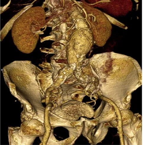
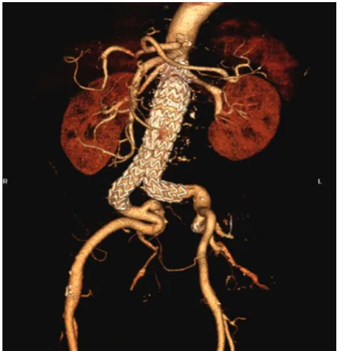
From these, 95% (19) presented with an abdominal aortic aneurysm (AAA) and 5% (1) with a common
iliac artery aneurysm and all underwent EVAR with percutaneous access (Fig. 1, 2 and 3). From all AAA, 11% (2) were saccular and the remaining (17) were fusiform. Concomitant aneurysms affecting other vascular territories were found in 20% of the patients (4) – two patients presented with a popliteal artery aneurysm, one with a thoracic aortic aneurysm and one with a superficial femoral artery aneurysm not involving the common femoral artery. The presence of femoral circumference calcification was found in 9.5% (2) of the patients, even though the ultrasound-guided access allowed for the identification of an adequate area for puncture and the application of the percutaneous closure device in all patients.
All the patients were operated under local anaesthesia with 2% lidocaine applied at the puncture site and conscious sedation.
All stent grafts were successfully inserted, with a 10% rate of initial endoleak formation (2 patients – (i) type IA endoleak successful intraoperative repair using an aortic stent graft and (ii) type IIA endoleak identified on the first postoperative imaging control spontaneously sealed in the CTA control at six months. None of these required conversion to an open correction.
All the patients remained clinically stable at 24 hours upon procedure, with no cardiac morbidity, no acute kidney failure or limb ischaemia.
Initial clinical success of percutaneous closure was obtained in 97.5% (39 access procedures) of the patients. A femoral artery pseudoaneurysm was diagnosed in one patient (ultrasound imaging) at six hours upon the procedure and the patient underwent percutaneous repair with ultrasound-guided thrombin injection technique, with full recovery and no need for surgery (type-I Clavien-Dindo complication).
No statistically significant relation was found between the presence of femoral circumference calcification and
the presence of complications (p > 0.05). No post-closure
arterial stenosis was found in any of the patients, nor any haemodynamically significant iatrogenic lesion in need for intervention.
A median 1-day length of stay in hospital was found (1-10 days) and 95% of the patients were discharged home within 24 hours. One patient was not discharged due to the abovementioned complication and stayed in hospital for 10 days.
ARTIGO ORIGINAL
Table 1
-Clinical characteristics of our group of patients
ARTIGO ORIGINAL
with a type IIA endoleak which remained in the subsequent imaging control.
An average 11.4 month follow-up period was found [1-36 months] with no drop-outs up to now. No patient was re-admitted on the follow-up period. A 0% re-operation rate was found, with no mortality from any cause up to now.
DISCUSSION
Even though there is some evidence that a reduction in patient’s length of stay in hospital will bring benefits for the patient, as well as economic advantages for the healthcare system, some indicators showed that an outpatient
ambulatory AAA treatment is safe and reproducible.17,18
In our study, a group of 20 consecutive patients underwent one-day-surgery with percutaneous EVAR under local anaesthesia, admitted to an intermediate care unit (according to a standardized protocol), with no mortality, no re-operations and with a minimal complication rate found over the abovementioned follow-up.
Unlike what was found in other case-series involving same-day discharge outpatient pEVAR in accordance with
standardized protocols,17 our model was based on a
one-day admission to the institution due to the fact that, even though complications related to percutaneous closure
ARTIGO ORIGINAL
mostly occur within the first hours upon the procedure,4 local
late complications are rare yet possible and usually lead to major complications when not promptly identified, apart from being a reason for re-admission, with all subsequent morbidity. Furthermore, even though any of the patients in our group presented with pre-operatively diagnosed chronic kidney failure, contrast-induced acute kidney injury usually shows at 24-72 hours and therefore may go unnoticed when
the patient is immediately discharged home.19
For that reason, our protocol involved one overnight stay in the hospital, ensuring greater monitoring of clinical and postoperative parameters, while keeping ambulatory benefits.
Anaesthetic modality has also contributed to outcomes, in line with what has been described in literature. The analysis of the subgroup of patients operated under local anaesthesia showed shorter surgical times, shorter length of stay in hospital and less postoperative complications in a review of 10 studies comparing local to regional
anaesthesia and involving 13,459 patients.20 Even though
local anaesthesia may be associated to patient’s discomfort with the manipulation of the device or to partial ischaemia of the limb, these were compensated with the application of conscious sedation, ensuring a greater comfort to the patient during the entire procedure. In conclusion, the use of this model significantly reduced the complications related with patient’s intubation or residual curarisation, allowing for an earlier discharge.
Finally, closure complication rate in our group of patients (2.5% - one complication) is in line with what has been found in some of the largest systematic revisions, with rates
as low as 3.6%.21
These results are largely dependent on the experience
of the operator22 as well as on patient’s adequate selection.
Percutaneous EVAR’s outcome depends on different factors, according to literature, with more or less agreement between the different studies and that should always be met in order to achieve the best possible outcome, mainly regarding ultrasound-guided puncture as well as pre-operative assessment of femoral diameter.
The importance of ultrasound-guided puncture in percutaneous procedures has been increasing in literature. Ideally, gaining access to the common femoral artery should always be performed across the anterior wall (at the 12-o’clock position), approximately 1 cm proximal to the femoral bifurcation and in an area with no atherosclerotic plaque. Not complying with these conditions greatly increase the risk for complications due to the fact that, on one hand, high punctures may be associated to the incorporation of the inguinal ligament into the suture, with the subsequent risk of rupture and potentially lethal bleeding when patient’s ambulation is started and, on the other hand, low punctures into lower-diameter superficial femoral artery can lead to artery wall’s damage and vessel’s
occlusion and ischaemia.14 For this reason,
ultrasound-guided access to the common femoral artery for pEVAR is
currently mandatory.23
In addition, femoral diameter also seems as a major determinant of the technical outcome of percutaneous closure devices, with the risk for suture of artery’s posterior
wall, usually leading to complications.14 For this reason, it is
currently considered that access vessel diameter <5mm is
Figure 2 - Three dimensional computed tomographic angiography
ARTIGO ORIGINAL
predictive of technical failure,24 which has been considered
in pre-operative patient assessment. Female are prone to higher rate of technical failure when compared to male patients, which is obviously due to vessel’s lower diameter. The use of these devices in adequately selected female patients showed overlapping outcomes to those found in
large clinical trials.25
There is a conflicting evidence as regards the impact of obesity and the calcification of the arterial wall on technical outcomes of percutaneous access, unlike other
abovementioned determinants.14 For that reason, none
of these factors was considered as exclusion criteria for pEVAR in our group of patients and limitations due to the presence of any of those were balanced by the use of ultrasound-guided access, with the identification of a non-calcified arterial area or by a better identification of the location of femoral artery in the presence of obesity.
CONCLUSION
Percutaneous EVAR using the Preclose technique is safe, efficient and associated to a low rate of local complications, provided that it is applied by trained surgeons to adequately selected patients,
The use of local anaesthesia with conscious sedation, ultrasound-guided puncture and highly trained surgical team allowed for AAA outpatient treatment with pEVAR with good outcomes and minimal associated morbidity.
The option for an overnight-stay, in accordance with a pre-defined standardized protocol, allowed for the
identification of late-onset complications, ensuring its correction over the same length of stay in hospital and preventing from re-admission with a potential subsequently increased morbidity.
Outpatient AAA treatment with pEVAR following our one-day-surgery model showed to be innovative, safe and efficient, combining the benefits associated to outpatient treatment with higher immediate postoperative monitoring, which is crucial for a complication-free postoperative period.
DATA CONFIDENTIALITY
The authors declare that they have followed the protocols of their work centre on the publication of patient
data.
HUMAN AND ANIMAL PROTECTION
The authors declare that the followed procedures were according to regulations established by the Ethics and Clinical Research Committee and according to the Helsinki Declaration of the World Medical Association.
CONFLICTS OF INTEREST
The authors declare that there were no conflicts of interest in writing this manuscript.
FINANCIAL SUPPORT
The authors declare that there was no financial support in writing this manuscript.
REFERENCES
1. Volodos NL, Karpovich IP, Troyan VI, Kalashnikova YuV, Shekhanin VE, Ternyuk NE, et al. Clinical experience of the use of self-fixing synthetic prostheses for remote endoprosthetics of the thoracic and the abdominal aorta and iliac arteries through the femoral artery and as intraoperative endprosthesis for aorta reconstruction. Vasa Suppl. 1991; 33:93–5. 2. Parodi JC, Palmaz JC, Barone HD. Transfemoral intraluminal
graft implantation for abdominal aortic aneurysms. Ann Vasc Surg. 1991;5:491-9.
3. Dua A, Kuy S, Lee CJ, Upchurch GR, Jr., Desai SS. Epidemiology of aortic aneurysm repair in the United States from 2000 to 2010. J Vasc Surg. 2014;59:1512-7.
4. Moscato VP, O’Brien-Irr MS, Dryjski ML, Dosluoglu HH, Cherr GS, Harris LM. Potential clinical feasibility and financial impact of same-day discharge in patients undergoing endovascular aortic repair for elective infrarenal aortic aneurysm. J Vasc Surg. 2015;62:855-61.
5. Morasch MD. Percutaneous techniques for aneurysm repair. J Vasc Surg. 2006;43 Suppl A:69A-72A.
6. Lonn L, Larzon T, Van Den Berg JC. From puncture to closure of the common femoral artery in endovascular aortic repair. J Cardiovasc Surg. 2010;51:791-8.
7. Maleux G, Koolen M, Heye S. Complications after endovascular aneurysm repair. Semin Intervent Radiol. 2009;26:3-9.
8. Nelson PR, Kracjer Z, Kansal N, Rao V, Bianchi C, Hashemi H, et al. A multicenter, randomized, controlled trial of totally percutaneous access versus open femoral exposure for endovascular aortic aneurysm repair (the PEVAR trial). J Vasc Surg. 2014;59:1181-93.
9. Haas PC, Krajcer Z, Diethrich EB. Closure of large percutaneous access sites using the Prostar XL Percutaneous Vascular Surgery device. J Endovasc Surg. 1999;6:168-70.
10. Dosluoglu HH, Cherr GS, Harris LM, Dryjski ML. Total percutaneous endovascular repair of abdominal aortic aneurysms using Perclose ProGlide closure devices. J Endovasc Ther. 2007;14:184-8.
11. Kauvar DS, Martin ED, Givens MD. Thirty-Day Outcomes after Elective Percutaneous or Open Endovascular Repair of Abdominal Aortic Aneurysms. Ann Vasc Surg. 2016;31:46-51.
12. Malkawi AH, Hinchliffe RJ, Holt PJ, Loftus IM, Thompson MM. Percutaneous access for endovascular aneurysm repair: a systematic review. Eur J Vasc Endovasc Surg. 2010;39:676-82.
13. Kontopodis N, Tsetis D, Kehagias E, Daskalakis N, Galanakis N, Ioannou CV. Totally Percutaneous Endovascular Aneurysm Repair Using the Preclosing Technique: Towards the Least Invasive Therapeutic Alternative. Surg Laparosc Endosc Percutan Tech. 2015;25:354-7. 14. Patel PJ, Kelly Q, Hieb RA, Lee CJ. Current Status of Percutaneous
Endografting. Semin Intervent Radiol. 2015;32:278-88.
15. van Dorp M, Ruyssers M, Amajoud Z, Lauwers P, Van Schil PE, Hendriks JM. Preclose Percutaneous Endurant Endografting with the Proglide Device: a Safe and Feasible Combination. Acta Chir Belg. 2015;115:219-23.
16. Dosluoglu HH, Lall P, Blochle R, Harris LM, Dryjski ML. Ambulatory percutaneous endovascular abdominal aortic aneurysm repair. J Vasc Surg. 2014;59:58-64.
17. Lachat ML, Pecoraro F, Mayer D, Guillet C, Glenck M, Rancic Z, et al. Outpatient endovascular aortic aneurysm repair: experience in 100 consecutive patients. Ann Surg. 2013;258:754-8; discussion 8-9. 18. Al-Zuhir N, Wong J, Nammuni I, Curran G, Tang T, Varty K. Selection,
ARTIGO ORIGINAL
(CIN) in Interventional Radiology Practice. Semin Intervent Radiol. 2010;27:348-59.
20. Karthikesalingam A, Thrumurthy SG, Young EL, Hinchliffe RJ, Holt PJ, Thompson MM. Locoregional anesthesia for endovascular aneurysm repair. J Vasc Surg. 2012;56:510-9.
21. Jaffan AA, Prince EA, Hampson CO, Murphy TP. The preclose technique in percutaneous endovascular aortic repair: a systematic literature review and meta-analysis. Cardiovasc Intervent Radiol. 2013;36:567-77.
22. Bechara CF, Barshes NR, Pisimisis G, Chen H, Pak T, Lin PH, et al. Predicting the learning curve and failures of total percutaneous endovascular aortic aneurysm repair. J Vasc Surg. 2013;57:72-6.
23. Mousa AY, Campbell JE, Broce M, Abu-Halimah S, Stone PA, Hass SM, et al. Predictors of percutaneous access failure requiring open femoral surgical conversion during endovascular aortic aneurysm repair. J Vasc Surg. 2013;58:1213-9.
24. Bensley RP, Hurks R, Huang Z, Pomposelli F, Hamdan A, Wyers M, et al. Ultrasound-guided percutaneous endovascular aneurysm repair success is predicted by access vessel diameter. J Vasc Surg. 2012;55:1554-61.