© 2020 by the Serbian Biological Society How to cite this article: Nikolić N, Popović S, Vidaković D, Subakov Simić G, 279 Krizmanić J. Genus Humidophila from caves in Serbia with an improved detailed
description of rare H. brekkaensoides. Arch Biol Sci. 2020;72(2):279-89.
Genus
Humidophila
from caves in Serbia with an improved detailed description of rare
H. brekkaensoides
Nataša Nikolić1,*, Slađana Popović2, Danijela Vidaković3, Gordana Subakov Simić1 and Jelena Krizmanić1
1University of Belgrade, Faculty of Biology, Studentski trg 16, 11000 Belgrade, Serbia
2University of Belgrade, Scientific Institution Institute of Chemistry, Technology and Metallurgy, National Institute, Department
of Ecology and Technoeconomics, Njegoševa 12, 11000 Belgrade, Serbia
3University of Belgrade, Scientific Institution, Institute of Chemistry, Technology and Metallurgy, National Institute, Department
of Chemistry, Njegoševa 12, 11000 Belgrade, Serbia
*Corresponding author: [email protected]
Received: February 28, 2020; Revised: April 17, 2020; Accepted: May 10, 2020; Published online: May 13, 2020
Abstract: Phototrophic microorganisms can be found in biofilms at entrances to caves where they have access to sufficient sunlight, water and nutrients; however, they can also be found inside caves, where they are carried by animals and people, and can proliferate in the presence of artificial light. Although the genus Humidophila includes some well-known cosmopolitan species, further exploration of its rare, insufficiently investigated representatives in different geographical areas and unusual habitats is necessary. Caves remain unknown, little-explored habitats, and many species currently known to science were recorded for the first time. This study aimed to report species belonging to the genus Humidophila that were identified in 19 caves in Serbia. A total of ten species of this genus were recorded and some of them are documented for the first time in Serbia. The most abundant species were H. contenta (found in 16 caves), H. paracontenta, and H. aerophila (found in 13 caves), whereas H. pyrenaica was identified in only one cave. The rare species, H. brekkaensoides and H. vidalii, were also documented and described, but with different dimensions from those provided in current identification keys.
Keywords: biofilm; cave; diatoms; Humidophila; Humidophila brekkaensoides
INTRODUCTION
Cave environments are generally considered to be extreme habitats, characterized by special environ-mental conditions and inhabited by small numbers of specialized organisms [1]. One of the specific characteristics of caves is oligotrophy, which is of-ten a primary limiting factor for the presence of organisms in these habitats. However, the presence of various microorganisms, including phototrophic ones, has been recognized in caves [1]. Phototrophic microorganisms, aerophytic cyanobacteria and algae, are often found in biofilms near cave entrance, and have also been identified inside caves, where their appearance is the result of artificial light installations and the presence of tourists, forming a microorgan-ism community called lampenflora [2-5]. The algal components of communities generally include many diatom representatives. Unlike freshwater diatoms,
which have been better studied in Serbia [6-9], data regarding aerophytic species remain scarce.
Diatoms have been studied in different hypogenic environments worldwide, such as in caves [2,3,10-18]. The most common diatoms found on rock substrata belong to the genera Humidophila [19-23], Luticola [23],
Orthoseira [24, 25], Pinnularia [26-28] and Hantzschia
[29]. The genus Humidophila (Grunow) Lowe, Kociolek, Johansen, Van de Vijver, Lange-Bertalot, and Kopalová occupy habitats such as wet rocks and areas inside of caves in the vicinity of artificial light [30].
(Bock) Lowe, Kociolek, J.R. Johansen, Van de Vijver, Lange-Bertalot and Kopalová are very rare and have been insufficiently described in the literature. Therefore, one of the aims of this paper was to provide a detailed description of H. brekkaensoides using SEM.
MATERIALS AND METHODS Study area
The caves (Supplementary Table S1) from which the sam-ples were taken are located in eastern and western Serbia, except for Risovača Cave, which is located in central Serbia, near the city of Aranđelovac. Many of these caves are found in limestone regions of east and west Serbia are rich in cave jewelry, stalactites and stalagmites; also, rare cave roses and helictites can be found in Cerjanska Cave. Paleolithic and Neolithic human remains, as well as fossil fauna, have also been discovered in Risovača, Lazareva, Prekonoška and Petnička caves [31, 32]. The oldest explored cave in Serbia is Resavska Cave, which is approximately 80 million years old, and the oldest speleothems are approximately 45 million years old [32]. Resavska Cave contains differently colored forma-tions that depend on the types of minerals found in the water that flows through them, including yellow from clay, red from iron-oxide and white from crystallized calcium. The longest explored cave is Lazareva Cave, which is 10000 m in length; however, the section that is open to tourists is only 900 m long [33].
Sample collection
Diatom samples were collected by scraping biofilms from stone substrates found in 19 caves in Serbia (Sup-plementary Table S1), from the entrance and/or inside the caves, near artificial lights. Sampling was conducted three times a year in Resavska, Stopić, Podpeć, Lazareva, Rajkova and Risovača caves in May, July and October 2017, when changes in the diversity of phototrophic microorganisms during the tourist season were also observed. In Samar and Jezava caves, samples were taken four times a year (December, March, May, and August), even though these caves are not active tourist destinations. In other caves, samples were taken once a year from 2014 to 2017. Three to eight sampling sites were chosen, depending on the locality and presence of the biofilm. At each sampling site (located on a wall,
ceiling, or sediment) biofilms were sampled using a sterile scalpel, placed into a sterile bag, and transported to the laboratory.
Sample preparation
All transported samples were treated with a supersatu-rated KMnO4 solution and concentrated HCl before rinsing with distilled water to pH 6-7 [34]. The clean diatom material was placed on microscope slides us-ing Naphrax®.
Sample analysis
Slide observations were performed with a Zeiss Axio-Imager M.1 light microscope using Axio Vision Release 4.9 software. The identification of genus Humidophila
was conducted using the following literature: [20, 27, 28, 35-39]. SEM was performed using a Cambridge S4 Stereoscan (Cambridge Instruments Ltd, Cambridge, UK) at the Friedrich Hustedt Study Centre for Diatoms (BRM) in Bremerhaven, and a Zeiss Gemini Ultra plus at the Natural History Museum, London, UK.
RESULTS
A total of ten Humidophila taxa were identified in Serbian caves (Table 1). Seven of these were identified in Serbia for the first time, including H. aerophila, H. brekkaensoides, H. comperei, H. contemnata, H. gallica,
H. pyrenaica and H.vidalii, whereas the other species were recorded at other localities in Serbia (lakes in Sara Mountain; the Sava, Nišava, Rasina and Velika Morava rivers; Lepenac, Jasika and Konjski streams and Sava Lake [40-49]). The most common species were H. con-tenta and H. paracontenta, which were found at both the entrance and inside the caves in high numbers. H. aerophila was also dominant in samples taken from entrances to caves, whereas it was detected at only one site inside the cave. H. pyrenaica, H. vidalii and H. gallica were rarely encountered. Based on our results, descriptions of all recorded species are provided below.
Humidophila aerophila (Krasske) Lowe, Kociolek,
Johansen, Van de Vijver, Lange-Bertalot & Kopalová (Fig. 1,AI-AR; Fig. 6FF)
Morphological characteristics (LM): The valves are linear-elliptic with concave margins in the middle and broadly rounded ends. Length is 9.3-12.2 µm, breadth is 2.9-3.8 µm. The axial area is linear and broad, central zone wide. Striae are parallel, interrupted in the central area, 28-29/10 µm (Fig. 1, AI-AR).
Morphological characteristics (SEM): The external raphe branches are straight with simple proximal end-ings. The axial area is a broad, central zone wide and surrounded by areolae. Areolae are slightly transapi-cally elongated (Fig. 6F).
Distribution in Serbia: This species was documented in 13 caves, primarily at entrances. Only one individual was found at the sampling site inside Podpeć Cave, near the artificial light (Table 1).
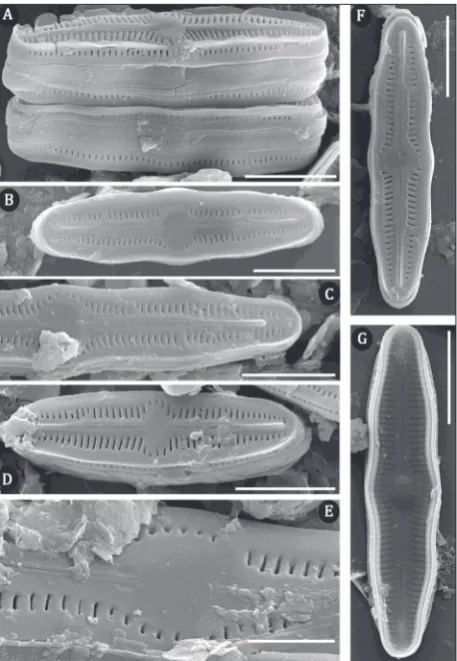
Humidophila brekkaensoides (Bock) Lowe,
Ko-ciolek, J.R.Johansen, Van de Vijver, Lange-Bertalot & Kopalová (Fig. 1A-W; Fig. 2.A-G)
Basionym: Navicula brekkaensoides Bock
Morphological characteristics (LM): The valves are linear, with protected and rounded ends. The margins are triundulate. Length is 11.6-24 µm, breadth is 3.6-5 µm. The axial area is linear, and narrow, with a large central area, that is round or elliptical. Striae radiate in the middle, becoming parallel at the ends, 27-31/10 µm (Fig. 1A-W).
Table 1. Species of genus Humidophila in caves of Serbia.
R HP D RC V SV L BG M S J C P PE RE RI ST PO RA
*Humidophila aerophila * * * * * * * * * * * * *
*Humidophila brekkaensoides * * * * * * *
*Humidophila comperei * * *
*Humidophila contemnata * * * * * * * *
Humidophila contenta * * * * * * * * * * * * * * * *
*Humidophila gallica * *
Humidophila paracontenta * * * * * * * * * * * * *
Humidophila perpusilla * * * * *
*Humidophila pyrenaica *
*Humidophila vidalii * * *
R – Ribnička, HP – Hadži Prodanova, D – Degurić, RC – Rćanska, V – Vernjikica, SV – Pećina kod Sove, L – Lazareva, BG – Bogovinska, M – Mandina, S –Samar, J – Jezava, C – Cerjanska, P – Prekonoška, PE – Petnička, RE – Resavska, RI – Risovača, ST – Stopić, PO – Potpeć, RA – Rajkova. A star before names presents species found for the first time in Serbia.
Fig. 1. Light microscopy (LM) micrographs.A-W– Humidophila
Morphological characteristics (SEM): The sternum is elevated relief-like, (Fig. 2B, C, D- F). Striae, com-posed of one transapically elongated areola, located in a shallow longitudinal depression (Fig. 2A-F). At the apices, striae terminate after the distal raphe endings (Fig. 2B-F). The external raphe branches are straight, with simple proximal endings (Fig. 2B-D, F, G). Distal raphe endings are elevated, terminating well before the ends (Fig. 2B-F). Internal proximal raphe endings are straight, and weakly T-shaped (Fig. 2G). Distal raphe endings are straight (Fig. 2G). On the girdle, three open copulae are visible, perforated by a single row of small, transapically elongated areola (Fig. 2A).
Distribution in Serbia: This species was found spo-radically at the cave entrance and was rare. However, inside Resavska Cave, the species was more abundant at both sampling points than in other caves (Table 1).
Humidophila comperei (Le Cohu & Van de Vijver)
Lowe, Kociolek, Johansen, Van de Vijver, Lange-Bertalot & Kopalová (Fig. 1X-AH, Fig. 3A-F)
Basionym: Diadesmis comperei Le Cohu & Van de Vijver
Morphological characteristics (LM): The valves are linear with broadly rounded ends. Length is 9.1-13.3 µm, breadth is 2.2-3.1 µm. The axial area is linear, and the central area is large. Rapha is straight, filiform. Striae are parallel, 31-36/10 µm (Fig. 1X-AH).
Morphological characteristics (SEM): The external raphe branches are straight. The central area is round and surrounded by areolae. Areolae are transapically elongated (Fig. 3A-E). Girdle is composed of several open, perforated bands (Fig. 3F).
Distribution in Serbia: This species was found at the entrances of three caves (Table 1).
Humidophila contemnata (E.Reichardt) Lowe,
Kociolek, Johansen, Van de Vijver, Lange-Bertalot & Kopalová (Fig. 1BL-BS; Fig. 6D, E)
Basionym: Diadesmis contemnata Reichardt Morphological characteristics (LM): The valves are linear to linear-elliptical, with broadly rounded ends. The margins are convex or slightly concave. Length is 6.2-10.4 µm, breadth is 2.7-4.2 µm. The axial area is
linear, the central area is small, rounded or elliptical, striae are parallel, 29-30/10µm (Fig. 1BL-BS).
Morphological characteristics (SEM): The axial area is broad (Fig. 6D, E). The external raphe branches are straight, with T-shaped distal raphe endings (Fig. 6D). The areolae are slightly transapically elongated (Fig. 6D, E).
Distribution in Serbia: H. contemnata was recorded in eight caves, primarly at the entrances. In addition, the species was found individually inside the Resavska and Rajkova caves, on the ground, and inside Lazar Cave, on the stone wall (Table 1).
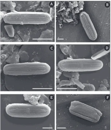
Humidophila contenta (Grunow) Lowe, Kociolek,
J.R. Johansen, Van de Vijver, Lange-Bertalot & Kopalová (Fig. 1AY-BK; Fig. 4A-F)
Basionym: Navicula contenta Grunow
Morphological characteristics (LM): The valves are linear, with broadly rounded ends and slightly concave margins. Length is 3.7-11.6 µm, breadth is 1.9-3.4 µm. Striae are parallel and interrupted in the center and difficult to resolve by LM (Fig. 1AY-BK).
Morphological characteristics (SEM): Central fascia is present (Fig. 4A-E). The axial area is wide and linear (Fig. 4A-E). The internal raphe branches are straight (Fig. 4F). The areolae are slightly transapi-cally elongated, 40-50/10 µm (Fig. 4A-E).
Distribution in Serbia: This species is found in most caves, except the Rćanska, Petnička, and Pećina kod Sove caves. It was found at different sampling sites (outside and inside of caves, on wet and dry walls, as well as on the ground and near artificial light) (Table 1). H. contenta was previously recorded in different lakes in Sara Mountain [40-42], Lepenac spring [43], Temska River [44], Zapadna Morava River [45], Dojk-inci River [46], Sava Lake [47] and the Velika Morava and Sava rivers [48].
Humidophila gallica (W.Smith) Lowe, Kociolek,
Q. You, Q. Wang & Stepanek (Fig. 1, CU-DB)
Basionym: Diadesmis gallica W. Smith
Morphological characteristics (LM): The valves are linear-elliptical, with broadly rounded ends. Length is 7.7-9.3 µm, breadth is 2.7-3.5 µm. Marginal spines are well developed, whereas valve interiors are not clearly visible in LM. Striae are not visible in LM (Fig. 1CU-DB).
Distribution in Serbia: This species was recorded on wet soils inside two caves (Table 1).
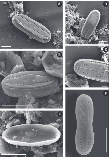
Humidophila paracontenta (Lange-Bertalot &
Werum) Lowe, Kociolek, Johansen, Van de Vijver, Lange-Bertalot & Kopalová 2014: 358 (Fig. 1BT-CH; Fig. 5, A-F)
Basionym: Didaesmis paracontenta Lange-Bertalot & Werum
Morphological characteristics (LM): The valves are parallel with broadly rounded sub-capitate apices. Length is 7.7-11.4 µm, and breadth is 2-3.4 µm. The
Fig. 3. SEM micrographs of H. comperei. Scale bar is 5 µm in A, C, D, and 2 µm in B, E, F.
axial area is narrow, and linear, whereas the central area is broad and rounded. Striae are fine, parallel, difficult to resolve by LM (Fig. 1, BT-CH).
Morphological characteristics (SEM): Central fascia is present (Fig. 5A-D), and sometimes surrounded by areolae (Fig. 5C). The external raphe branches are straight (Fig. 5A-D), sometimes with T-shaped distal raphe endings (Fig. 5D). The internal raphe branches are straight (Fig. 5E, F). The areolae are transapically elongated, approximately 40/10 µm (Fig. 5A-D).
Distribution in Serbia: This species was recorded in 13 caves (at entrances and on walls and soil inside caves) (Table 1). In addition, H. paracontenta was previously recorded in the Rasina River and Jasika and Konjski streams [49].
Humidophila perpusilla (Grunow) Lowe,
Koci-olek, J.R. Johansen, Van de Vijver, Lange-Bertalot & Kopalová (Fig. 1AS-AX; Fig. 6B, C)
Basionym: Navicula perpusilla (Grunow) D.G.Mann Morphological characteristics (LM): The valves are elliptic-lanceolate to linear-lanceolate, with rostrate to broadly rounded ends. Length is 9.2-14 µm, and breadth is 3.8-5.2 µm. The axial area is wide with a large central area. The raphe appears filiform, and linear. The striae are radiate, 25-30/10 µm (Fig. 1, AS-AX).
Morphological characteristics (SEM): The external and internal raphe branches are straight (Fig. 6B, C). The areolae are transapically elongated (Fig. 6B).
Distribution in Serbia: This species was found in five caves, mainly at entrances to caves. Among the lampenflora community, this species was only found in Resavska Cave, on the wet walls and soils (Table 1).
H. perpusilla has previously been recorded in different lakes of the Shara Mountain [40-42] and the Dojkinci River [46].
Fig. 5. SEM micrographs of H. paracontenta. Scale bar is 5 µm in
Humidophila pyrenaica (Lange-Bertalot & Werum) Lowe, Kociolek, Johansen, Van de Vijver, Lange-Bertalot & Kopalová (Fig. 1CI, CJ; Fig. 6A)
Basionym: Diadesmis pyrenaica Lange-Bertalot & Werum
Morphological characteristics (LM): The valves are linear with broadly rounded ends. Length is 9.1-10µm, and breadth is 2.8-3.1µm. The raphe is straight, and filiform, and the axial area is broad, with a slightly wide central area. The striae are parallel and difficult to resolve by LM (Fig. 1CI, CJ).
Morphological characteristics (SEM): The external raphe branches are straight. The central area is round and surrounded by areolae. Areolae are transapically elongated, approximately 35/10 µm (Fig. 6A).
Distribution in Serbia: H. pyrenaica was found in samples taken from Samar Cave and was registered in only one season (May) (Table 1).
Humidophila vidalii (Van de Vijver, Ledeganck
& Beyens) Lowe et al. (Fig. 1,CK-CT)
Basionym: Diadesmis vidalii B. Van de Vijver & P.Ledeganck
Morphological characteristics (LM): The valves are linear, with broadly rounded ends. Length is 5.8-10.4 µm and breadth is 2.6-3.7 µm. The axial area is wide, linear, and the central area is absent. The raphae are straight, and filiform. Striae are not visible in LM (Fig. 1CK-CT).
Distribution in Serbia: This species was found in three caves. This species was most abundant at the entrance of the Resavska Cave, where samples were taken from the walls, but it was also found sporadi-cally inside the Lazareva and Podpeć caves (Table 1).
DISCUSSION
The genus Humidophila contains well-known diatoms that are usually found on wet rocks, walls, wet soils, moss vegetation and in cave habitats [39, 50]. The genus includes diverse species, which have been described in Europe [38, 50], China [30], Hawaii [21] and the Antarctic region [22]. The taxa in this genus possess a small, linear to lanceolate-elliptic valve and broadly
rounded ends. Most of the species were originally classified as belonging to the genus Diadesmis [51], subgenus Paradiadesmis (Lange-Bertalot & Le Cohu), but after detailed morphological examinations, they were transferred into the genus Humidophila [21].
Phototrophic microorganisms are expected to be found on the walls and rocks at entrances of caves because they can receive sufficient natural light, wa-ter (from precipitation, dew or the nearest river) and nutrients in these locations [1, 52]. They can also be carried inside the cave by air, animals, water and hu-mans, and in the presence of artificial light, they can colonize different substrates such as walls, ceilings, and cave jewelry [2, 4]. Our results show ten species recorded in 19 caves in Serbia. Some of these species, such as H. contenta, H. paracontenta and H. perpusilla, have previously been documented in Serbia in different habitats, whereas the other species were recorded in the Serbia for the first time. H. contenta was previously recorded in different lakes ofthe Shara Mountain [40-42], Lepenac spring [43], Temska River [44], Zapadna Morava River [45], Dojkinci River [46], Sava Lake [47], Velika Morava and Sava rivers [48] with a large water-temperature range [39]. In addition to the caves,
H. paracontenta has been found in Rasina River and Jasika and Konjski streams [49], whereas H. perpusilla
habitat of this species and it may not be fully adapted to these conditions. H. gallica likely entered the caves via air, animals, or tourists, like H. aerophila, which was detected at the entrances of caves and in only one other location inside Podpeć Cave. This species is typically aerophytic and likely entered the cave via air flow and found suitable conditions for development due to the presence of artificial light. However, only one H. aerophila individual was recorded, so that it is difficult to determine whether it is capable of coloniz-ing the substrate in higher numbers. H. comperei was recorded at the entrances of three caves (Vernjikica, Samar, Prekonoška). According to Cohu and Van de Vijver [53], this species was found at Île de la Posses-sion (Crozet Archipelago) on dry soil, whereas our results show that it can be found on cave walls where the RH is high (Samar Cave, found in March, May and December). This is a rarely described species that most likely has a wide range of habitats. H. pyrenaica
was only detected at the entrance of Samar Cave in May, although samples were taken four times during a year. Unlike the inside of caves, where environmental parameters are generally stable (temperature (T) and relative humidity (RH)), these parameters are much more variable at the entrances to caves. Generally, T and RH vary, not only by season, but also between the day and night [1], which can result in changes in the phototrophic communities. According to Asencio and Aboal [54], during a two-year study, T and RH varied significantly during the day and throughout the year, T values ranged from 1.6 °C to 39.1 °C, whereas RH varied up to 77 %. The type of substrate and its heat capacity have also been suggested to affect the composition of phototrophic communities [1]. Inside caves, phototrophic communities will grow near natural light, unless artificial lights have been installed in the caves, allowing phototrophic communities to develop deeper within the cave environment [55,56].
The dimensions and descriptions of the recorded species in this study correspond with the descriptions from the identification keys, except for the rarely found species H. brekkaensoides, which was first described by Bock [57], who discovered it in a thin biofilm layer on rocks in the Alps. According to Bock’s findings [57], H. brekkaensoides, (Navicula brekkaensoides) can not be found on limestone because it prefers non-calcareous substrates. However, this species appears to occur in a wider spectrum of habitats, as the results of Buczkó [20]
and this study have shown that H. brekkaensoides can be found on limestone substrates. In addition, Reichardt [37] recorded this taxon in the Hochlantsch Mountain in Austria, from a slightly shaded limestone depression, and therefore whether this species prefers calcareous or non-calcareous substrates remains unclear as further details about the habitats are missing. Unlike cave en-trances where H. brekkaensoides was sporadically found, the population of this species was more abundant inside the cave near artificial light, which is likely the result of more stable environmental conditions (especially T and RH) and a tolerance for low nutrient input [52]. Furthermore, the dimensions of the valves reported by Bock [57] are different from those observed in our study. According to Bock [57], valve length is 17-23 µm and the breadth is 4-5 µm, whereas in our population, the length is 11.6-24 µm and the breadth is 3.6-5 µm. Compared with the description by Chattova et al. [39], the dimensions of H. vidalii were also different in our study. Chattova et al. reported a length of 7-17.5 µm and a breadth of 2-3.5 µm, whereas in our population the length is 5.8-10.4 µm and the breadth is 2.6-3.7 µm.
Data regarding the morphological and physiological characteristics of diatoms found in caves remain poorly described. According to Falasco [52], some species can exhibit morphological modifications when they grow in different environmental conditions. Gener-ally, diatoms from caves display smaller sizes and high resistance to desiccation, which are likely adaptations to life in extreme habitats with low nutrient input [52]. Because caves are described as oligotrophic habitats, with low nutrient input, low T, high RH, and low light [59] compared with the outside environment, species in caves compete for the same resources, which could affect the valve size.
Data regarding diatom diversity in caves remain scarce in the literature. Although these species are known to science, they have been identified for the first time in the caves of Serbia, and it is likely that the list of species present in these environments is incomplete. Moreover, details regarding morphological and physiological characteristics of these diatoms are still lacking, necessitating further research to obtain a better understanding of these communities.
CONCLUSIONS
These results describe species of the genus Humidophila
recorded in the caves of Serbia. Of the ten species identified; seven were found for the first time in Serbia, whereas the others have been registered in different habitats in Serbia (lakes, rivers, and streams). They were found at the entrance and inside tourist caves, near artificial light. The study includes a description of the rare species H. brekkaensoides, with a different range of valve dimensions than in the first reported description. Because the caves of the Balkan Peninsula have not been sufficiently explored in terms of their phototrophic microorganisms, it is necessary to con-ducted further research to obtain more information regarding this and other diatom species.
Funding: This research was supported by the Ministry of Science and Technological Development, Republic of Serbia, Project No. 176018, Project No. 176020, Project No. 37009, and the Synthesis Project, which is financed by the European Community Research Infrastructure Action under the FP7 “Capacities” Program. Acknowledgements: The authors want to thank the cave manage-ment, guides, families, and friends for their support.
Author contributions: All authors have contributed sufficiently to the project to be included as coauthors. NN, SP, GSS designed the research and took the samples. NN made LM pictures, tables and wrote the main part of the manuscript. SP made LM pictures and participated in the writing. DV prepared the diatom samples, made the SEM photographs and figures. JK helped with determination of diatom taxa; the whole process was revised by JK and GSS who also reviewed the manuscript with many helpful suggestions. All authors have read and approved the manuscript.
Conflict of interest disclosure: The authors report no conflict of interest, financial or other, exists.
REFERENCES
1. Pentecost A,Whitton BA. Subaerial Cyanobacteria. In: Whit-ton BA, editor. Ecology of Cyanobacteria II: their diversity in space and time. London:Springer;2012. p. 291-315.
2. Mulec J, Kosi G. Lampenflora algae and methods of growth control. J. C. K. Studies. 2009;71(2):109-15.
3. Mulec J. Human impact on underground cultural and natural sites, biological parameters of monitoring and remediation actions for insensitive surfaces: Caves of Slovenian show caves. J Nat Conserv. 2014;22(2):132-41.
4. Borderie F, Tête N, Cailhol D, Alaoui-Sehmer L, Bousta F, Rieffel D, Aleya L,Alaoui Sossé B. Factors driving epilithic algal colonization in show caves and new insights into com-bating biofilm development with UV-C treatments. Sci Tot Enviro. 2014;484: 43-52.
5. Popović S, Subakov Simić G, Stupar M, Unković N, Predojević D, Jovanović J. Ljaljević Grbić M. Cyanobacteria, algae and microfungi present in biofilm from Božana Cave (Serbia). Int J Speleol. 2015;44(2):141-9.
6. Jakovljević O, Popović S, Živić I, Stojanović K, Krizmanić J. Benthic diatoms of the Vrla River (Serbia) and their appli-cation in the water ecological status assessment. Oceanol Hydrobiol Stud. 2016;45(3):304-15.
7. Krizmanić J, Jakovljević O, Vidaković D, Jovanović J. First Record of The Genus Decussata (Patrick) Lange- Berta-lot (Bacillariophyta) In Serbia - Distribution of the Rare Species D. hexagona (Torka) Lange-Bertalot. Bot Serb. 2016;40(2):161-5.
8. Vidaković D, Radovanović S, Predojević D, Šovran S, Živić I, Stojanović K, Krizmanić J. Uncertainty of using habitat fidelity in biomonitoring based on benthic diatoms - the Raška River case study. Biologia. 2018;73(I.9): 831-9. 9. Ćirić M, Nikolić N, Krizmanić J, Gavrilović B, Pantelić A,
Petrović MV. Diatom diversity and ecological status of Lasovacka and Lenovacka streams near Zajecar: consid-eration of WFD implementation in Serbia. Arch Biol Sci. 2018;70:(4):691-8.
10. Albertano P. Epilithic algal communities in hypogean envi-ronments. G Bot Ital. 1993;127:386-92.
12. Poulíčková A, Hájková P, Křenková P, Hájek M. Distribution of diatoms and bryophytes on linear transects through spring fens. Nova Hedwig. 2004;78:411-24.
13. Poulíčková A, Hašler P. Aerophytic diatoms from caves in central Morovia (Czech Republic). Preslia. 2007;79:185-204. 14. Smith T, Olson R. A taxonomic survey of Lamp flora
(algae and cyanobacteria) in electrically lit passages within Mammoth Cave National Park, Kentucky. Int J Speleol. 2007;36:105-14.
15. Mulec J, Kosi G, Vrhošek D. Characterization of cave aero-phytic algal communities and effects of irradiance levels in production of pigments. J CavesKarst Stud. 2008;70:3-12. 16. Mulec J, Oarga-Mulec A, Šturm S, Tomazin R, Matos T.
Spacio-Temporal Distribution and Tourist Impact on Air-borne Bacteria in a Cave (Škocjan Caves, Slovenia). Diversity. 2017;9:28.
17. Llop E, Alvaro I, Hernández-Mariné M, Sammut S, Gómez-Bolea A. Colonization of Maltese catacombs by phototrophic biofilms. How much does light matter? IJHDE. 2012;1:289-94. 18. Mulec J. Lampenflora. In: White WB, Culver DC, edi-tors. Encyclopedia of Caves. Amsterdam:Academic Press; 2012.p.451-6.
19. Abdullin SR. Cyanobacterial-algal cenoses of the Shulgan-Tash cave, Southern Urals. Russ J Ecol. 2009;40:301-3. 20. Buczkó K. Diadesmis brekkaensoides (W.Bock) Moser,
Lange-Bertalot et Metzeltin: A new aerophytic diatom for the Hungarian flora. Studia Bot. Hung. 2003;34:5-10. 21. Lowe RL, Kociolek JP, Johansen JR, Van de Vijver B,
Lange-Bertalot H, Kopalová K. Humidophila gen. nov., a new genus for a clade of diatoms (Bacillariophyta) formerly within the genus Diadesmis: species from Hawai’i, including one new species. DiatomRes.2014;29(4):351-60.
22. Kopalová K, Kociolek PJ, Lowe RL, Zidarova R, Van Der Vijver B. Five new species of the genus Humidophila (Bacil-lariophyta) from the Maritime Antarctic Region. Diatom Res. 2015;30(2):117-31.
23. Miscoe HL, Johansen RJ, Kociolek JP, Lowe LR. The dia-tom flora and cyanobacteria on Kauai, Hawaii. Bibl Phycol. 2016;120: 3-74.
24. Garbacki N, Ector L, Kostikov I, Hoffmann L. Contribution à l’étude de la flore des grottes de Belgique. Belg J. Bot. 1999; 132:43-76. French.
25. Czerwik-Marcinkowska J, Mrozińska T. Algae and cyanobac-teria in caves of the Polish Jura. Pol Bot J. 2011;56:203-43. 26. Germain H. Les lieux de développement et de
multiplica-tion des diatomées d’eau douce. Contribumultiplica-tion à l’écologie des diatomées. Travaux du Laboratoire de Botanique de l’Université Catholique d’Angers N° 6. Nantes: Bulletin des Sciences Naturelles de l’Ouest. 1936; 200 p. French.
27. Taylor JC, Harding WR, Archibald CGM. An Illustrated Guide to Some Common Diatom Species from South Africa. Pretoria: Water Research Commission;2007. 178 p.
28. Lange-Bertalot H, Hofmann G, Werum M, Cantonati M. Freshwater Benthic Diatoms of Central Europe: Over 800 Common Species Used in Ecological Assessment. Engl. ed.with updated taxonomy and added species. Schmitten-Oberreifenberg: Koeltz Botanical Books; 2017. 942 p. 29. Clair SL, Rushforth SR.The diatoms of Timpanogos Cave
National Monument, Utah. AmJ Bot. 1976;63:49-59.
30. Lowe RL, Kociolek JP, You Q, Wang Q, Stepanek J. Diversity of the diatom genus Humidophila in karst areas of Guizhou, China. Phytotaxa. 2017;305(4):269-84.
31. Petrović J. Jame i pećine SR Srbije. Beograd: Vojnoizdavački zavod; 1976. 515 p. Serbian.
32. Đurović P. Speleološki atlas Srbije. Beograd: SANU; 1998. 290 p. Serbian.
33. Milanović S. Speleologija i speleoronjenje u hidrogeologiji karsta.Beograd: Rudarsko-geološki fakultet; 2012. 315p. Ser-bian.
34. Taylor JC, De Le Rey PA,Van Rensburg L. Recommendations for the collection, preparation and enumeration of diatoms from riverine habitats for water quality monitoring in South Africa. Afr J Aquat Sci. 2005; 30(1):65-75.
35. Krammer K, Lange-Bertalot H. Naviculaceae: neue und wenig bekannte Taxa, neue Kombinationen und Synonyme sowie Bemerkungen zu einigen Gattungen. 1st ed. Berlint: J. Cramer ; 1985. 230 p. German.
36. Krammer K,Lange-Bertalot H. Bacillariophyceae. 1. Teil: Naviculaceae. In: Ettl H, Gerloff J, Heynig H, Mollenhauer D, editors. Süßwasserflora von Mitteleuropa 2/1, Jena: G. Fischer Verlag; 1986. 876 p . German.
37. Reichardt E. Eine bemerkenswerte Diatomeenassoziation in einem Quellhabitat im Grazer Bergland, Österreich. Ein Beitrag zur Kenntnis seltener und wenig bekannter Diato-meen. Iconographia Diatomologica. 2004;13:419-79. German. 38. Werum M, Lange-Bertalot H. Diatoms in springs, from Cen-tral Europe and elsewhere under the influence of hydrogeol-ogy and anthropogenic impacts. Iconografia Diatomologica 13. Rugell: A.R.G. Gantner Verlag KG; 2004. 417 p. German.. 39. Chattova. B, Lebouvier M, Van De Vijver B. Morphological and taxonomical analyisis of the terrestrial diatom genus Humi-dophila (Bacillariophita) on Ile Amsterdam and Ile Saint-Paul (Southern Indian Ocean). Phytotaxa. 2018;336:28-42. 40. Urošević V. Phytoplankton and microbenthos in glacial lakes
Shara Mountain[dissertation]. [Priština]:Faculty of Natural Sciences and Mathematics,University of Priština; 1987. 281 p. 41. Urošević V.Alge visokoplaninskih jezera sirinićke strane Šar-planine. Priština: University of Pristina; 1994. 95p. Serbian. 42. Urošević V. The Yugoslav part of Sar-Planina Mt.Lake
Eco-system’s Periphyton Algae. University. Thought, Nat.Sci. 1998;5(2): 43-57.
43. Urošević V, Savić A. Algae of the Lepenac Springs on the Šar-Planina Mt.- University. Thought, Nat. Sci. 1996;3(1):23-32. 44. Andrejić JZ, Krizmanić J, Cvijan M. Diatom species composi-tion of the Nišava River and its tributaries Jerma and Temska rivers (Southern Serbia). Arch Biol Sci. 2012;64:1127-40. 45. Krizmanić J. Floristic, taxonomic and ecological studies of
Serbian diatoms with raphae (Bacillariophyceae, Bacillario-phycideae, Bacillariophyta) [dissertation]. [Belgrade]: Faculty of Biology, University of Belgrade; 2009. 595 p.
46. Krizmanić J, Ilić M, Vidaković D, Subakov-Simić G, Petrović J, Cvetanović K. Diatoms of the Dojkinci River (Stara Planina Nature Park, Serbia). Acta Bot Croat. 2015;74(2):317-31. 47. Trbojević I. Analysis of periphyton developed on artificial
substrates in the Sava lake and the Vrutci lake[dissertation]. [Belgrade]:Faculty of Biology, University of Belgrade; 2018. 275 p. 48. Vasiljević B, Simić S, Paunović M, Zuliani T, Krizmanić J,
diatom-based assessments of the ecological status of large riv-ers ‒ the Sava River Case Study. Sci Total Enviro. 2017;605-606:874-83.
49. Vidaković D, Jakovljević O, Predojević D, Radovanović S, SubakovSimić G, Lazović V, Krizmanić J. An updated list of Serbian diatom flora - new recorded taxa. Arch Biol Sci. 2018;70(2):259-75.
50. Kövèr C, Korponai J, Harangi S,Buczko K. A new Euro-pean record of Diadesmis fukushimae and its transference to Humidophila genus (Bacillariophyta). Acta Bot Croat. 2015;74(2):245-52.
51. Kützing FT. Die Kieselschaligen Bacillarien oder Diatomeen. Nordhausen: zu finden bei W. Köhne. 1844. 152 p. German. 52. Falasco E, Ector L, Isaia M, Wetzel CE, Hoffmann L, Bona F.
Diatom flora in subterranean ecosystems: a review. Int J Spel. 2014;43(3):231-51.
53. Cohu RL, Van de Vijver B. Le genre Diadesmis (Bacillari-ophyta) dans les archipels de Crozet et de Kerguelen avec la description de cinq espèces nouvelles. Ann. Limnol. 2002;38(2):119-32.
54. Asencio AD, Aboal M. Algae from La Serreta cave (Murcia, SE Spain) and their environmental conditions. Arch Hydro-biol Suppl Algol Stud. 2000;(96): 59-78.
55. Roldán M, Clavero E, Canals A, Gómez-Bolea A, Ariño X, Hernández- Mariné M. Distribution of phototrophic biofilms in cavities (Garraf, Spain). Nova Hedwig. 2004;(78):329-51. 56. Roldan M, Oliva F, Gonzalez del Valle MA, Saiz-Jimenez C,
Hernandez- Marine M. Does green light influence the fluo-rescence properties and structure of phototrophic biofilms? Appl Environ Microbiol. 2006;(72): 3026-31.
57. Bock W. Diatomeen extrem trockener Standorte. Nova Hed-wigia. 1963;5:199-254.
58. Mazina SE, Maximov VN. Photosynthetic organism commu-nities of the Akhshtyrskaya excursion cave. Moscow Univ Biol Sci Bull. 2011;(66):37-41.
59. Giordano M, Mobili F, Pezzoni V, Hein MK, Davis JS. Pho-tosynthesis in the caves of Frasassi (Italy). Phycologia. 2000;39:384-9.
Supplementary Material