Identification and Characterization of MCM3 as a Kelch-like
ECH-associated Protein 1 (KEAP1) Substrate
*
□SReceived for publication, April 8, 2016, and in revised form, September 12, 2016 Published, JBC Papers in Press, September 12, 2016, DOI 10.1074/jbc.M116.729418
Kathleen M. Mulvaney‡§, Jacob P. Matson¶, Priscila F. Siesser§, Tigist Y. Tamir§储, Dennis Goldfarb§**, Timothy M. Jacobs‡‡, Erica W. Cloer‡§, Joseph S. Harrison§¶, Cyrus Vaziri§ §§, Jeanette G. Cook§¶1, and Michael B. Major‡§储**2
From the Departments of‡Cell Biology and Physiology,¶Biochemistry and Biophysics,储Pharmacology,**Computer Science, and §§Pathology,§Lineberger Comprehensive Cancer Center, and‡‡Eshelman School of Pharmacy, University of North Carolina, Chapel Hill, North Carolina 27599
KEAP1 is a substrate adaptor protein for a CUL3-based E3 ubiquitin ligase. Ubiquitylation and degradation of the antioxi-dant transcription factor NRF2 is considered the primary func-tion of KEAP1; however, few other KEAP1 substrates have been identified. Because KEAP1 is altered in a number of human pathologies and has been proposed as a potential therapeutic target therein, we sought to better understand KEAP1 through systematic identification of its substrates. Toward this goal, we combined parallel affinity capture proteomics and candidate-based approaches. Substrate-trapping proteomics yielded NRF2 and the related transcription factor NRF1 as KEAP1 substrates. Our targeted investigation of KEAP1-interacting proteins revealed MCM3, an essential subunit of the replicative DNA helicase, as a new substrate. We show that MCM3 is ubiquity-lated by the KEAP1-CUL3-RBX1 complex in cells andin vitro. Using ubiquitin remnant profiling, we identify the sites of KEAP1-dependent ubiquitylation in MCM3, and these sites are on predicted exposed surfaces of the MCM2–7 complex. Unex-pectedly, we determined that KEAP1 does not regulate total MCM3 protein stability or subcellular localization. Our analysis of a KEAP1 targeting motif in MCM3 suggests that MCM3 is a point of direct contact between KEAP1 and the MCM hexamer. Moreover, KEAP1 associates with chromatin in a cell cycle-de-pendent fashion with kinetics similar to the MCM2–7 complex. KEAP1 is thus poised to affect MCM2–7 dynamics or function rather than MCM3 abundance. Together, these data establish new functions for KEAP1 within the nucleus and identify MCM3 as a novel substrate of the KEAP1-CUL3-RBX1 E3 ligase.
Kelch-like ECH-associated protein 1 (KEAP1)3is a substrate
adaptor protein for a Cullin3 (CUL3)-RBX1 E3 ubiquitin ligase complex (1–3). Recent studies have described the molecular architecture and mechanism for the KEAP1-CUL3-RBX1 ubiq-uitylation machine (4, 5). The most well studied and established substrate of the KEAP1 complex is the NFE2L2 transcription factor (henceforth referred to as NRF2) (1–3, 6, 7). A homodimer of KEAP1 tethered to CUL3 via its amino-terminal bric-a-brac/tramtrack/broad complex (BTB) domains binds to a single molecule of NRF2, and the C-terminal kelch domains of a KEAP1 homodimer bind to a high-affinity motif (ETGE) and a lower-affinity motif (DLG) within the NRF2 protein (2, 8, 9). Under homeostatic conditions, ubiquitylated NRF2 is rap-idly degraded by the proteasome, having a half-life of less than 30 min (10). KEAP1 acts as a sensor of cellular reduction-oxi-dation (redox) state through its 27 cysteine residues (6, 11, 12). The reactive cysteine residues within KEAP1 can be modified by reactive oxygen species (ROS), which is thought to trigger a conformational change in the KEAP1 complex (3, 6, 11). As a result, NRF2 is no longer efficiently degraded and thus accumu-lates, translocates to the nucleus, and promotes the transcrip-tion of antioxidant and cytoprotective genes (10, 13, 14). Spe-cifically, nuclear NRF2 forms heterodimers with small Maf proteins, and together they bind to the antioxidant response elements within the promoter region of NRF2 target genes, which include free radical scavengers, glutathione synthesis genes, and xenobiotic efflux proteins (7, 10, 15, 16). The up-reg-ulation of NRF2 target genes mitigates oxidative stress and con-fers resistance to a number of toxins, including chemothera-peutics (7, 13, 17, 18). The KEAP1-NRF2 signaling pathway serves as the primary defense of the cell against oxidative stress (13, 14).
Although NRF2 degradation has long been thought to be the primary function of KEAP1, we have shown that KEAP1 asso-ciates with a number of interesting and diverse proteins, sug-gesting previously unknown roles for KEAP1 (19, 20). In sup-port of this concept, three substrates have recently been *This work was supported by a grant from the Sidney Kimmel Cancer
Foun-dation (to M. B. M.), by American Cancer Society Grant RSG-14-068-01-TBE (to M. B. M.), by National Science Foundation Graduate Research Fellow-ship DGE-1144081 (to J. P. M.), and by NIGMS, National Institutes of Health Grant R01GM102413 (to J. G. C.). The authors declare that they have no conflicts of interest with the contents of this article. The content is solely the responsibility of the authors and does not necessarily represent the official views of the National Institutes of Health.
□S
This article containssupplemental Tables S1–S3.
The mass spectrometry proteomics data have been deposited in the Proteome-Xchange Consortium via the PRIDE partner repository with the dataset identi-fier PXD003929.
1To whom correspondence may be addressed: Dept. of Biochemistry and
Biophysics, 120 Mason Farm Rd., Campus Box 7260, Chapel Hill, NC 27599-7260. Tel.: 919-843-3867; E-mail: [email protected].
2To whom correspondence may be addressed: Dept. of Cell Biology and
Physiology, Lineberger Comprehensive Cancer Center, University of North Carolina, 450 West Dr., Lineberger Bldg., CB#7295, Chapel Hill, NC 27599-7295. Tel.: 919-259-2695; E-mail: [email protected].
3The abbreviations used are: KEAP, kelch-like ECH-associated protein; redox,
reduction-oxidation; ROS, reactive oxygen species; PAC, parallel affinity capture; SBP, streptavidin binding peptide; IP, immunoprecipitation; PLA, proximity ligation assay; tBHQ,tert-butylhydroquinone; AP, affinity purifi-cation; MEF, mouse embryonic fibroblast; RIPA, radioimmune precipita-tion assay; VSV, vesicular stomatitis virus; FDR, false discovery rate; TRITC, tetramethylrhodamine isothiocyanate.
reported for the KEAP1 E3 ligase: IKBKB (21), PGAM5 (22), and PALB2 (23). All three substrates contain an ETGE or ESGE motif that is essential for their interactions with and ubiquity-lation by KEAP1. Although we have a strong understanding of the dynamics and regulation of NRF2 as a KEAP1 substrate, these other substrates are less well studied. IKBKB is reported to be a KEAP1 substrate targeted for autophagy-mediated deg-radation, PGAM5 is thought to be ubiquitylated and targeted to the proteasome, and KEAP1-mediated PALB2 ubiquitylation regulates its function by blocking its interaction with BRCA1 (21–24). Thus, the KEAP1-CUL3-RBX1 ligase is capable of ubiquitylating its substrates to regulate substrate stability through either proteasome-mediated or autophagy-mediated degradation or to regulate substrate function by directing pro-tein-protein interactions.
In addition to the vital role the pathway plays in normal phys-iology, perturbations in KEAP1-NRF2 signaling have been reported in a variety of diseases, including cancer and inflam-matory, cardiovascular, and neurodegenerative diseases (25– 34). Most notably, sequencing efforts have determined that ⬃30% of non-small cell lung cancer tumors harbor mutations in the KEAP1-NRF2 pathway, and 12–15% of non-small cell lung cancer patient tumors have mutations within KEAP1 (20, 35–38). The high mutation frequency suggests a role for KEAP1-NRF2 in cancer progression. KEAP1 loss is thought to promote tumorigenesis through hyperactivation of NRF2, although little is known about other effects of KEAP1 mutation or loss. A better understanding of KEAP1 substrates would enhance our understanding of both normal KEAP1 function and of KEAP1-mutant tumors. We sought to define new KEAP1 substrates and to determine the function of their ubiq-uitylation by KEAP1.
Here we identify a subunit of the replicative DNA helicase minichromosome maintenance 3 (MCM3) as a KEAP1 strate. We selected it from our set of potential KEAP1 sub-strates for further study based on its important role in cell cycle regulation. Interestingly, human MCM subunits do not undergo ubiquitin-mediated proteolysis during normal prolif-eration; instead, the chromatin loading of the MCM complex is tightly controlled during the cell cycle to ensure once-per-cell cycle genome duplication. MCM complexes are chromatin-loaded strictly during G1phase, activated in S phase, and progressively unloaded as DNA replication forks terminate (reviewed in Ref. 39 – 41). The MCM2–7 complex is exten-sively modified by posttranslational modifications (42– 48), and, in particular, recent studies linked polyubiquitylation of the MCM7 subunit to MCM unloading in both Saccharomy-ces cerevisiaeand Xenopus laevis (49 –52). In S. cerevisiae, the SCFDia2 ligase ubiquitylates MCM7, and in X. laevis,
MCM7 is ubiquitylated by an unidentified cullin family member (49, 50). Thus, interaction with and polyubiquityla-tion by KEAP1 represents a potentially novel form of MCM regulation. We suggest that our discovery of KEAP1-medi-ated MCM3 ubiquitylation establishes a physical link between a key player in the oxidative stress response and chromosome replication.
Results
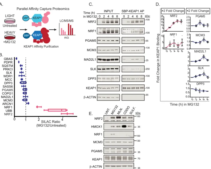
Identification of MCM3 as a KEAP1 Substrate—We used two complementary strategies to identify proteins ubiquitylated by the KEAP1-CUL3-RBX1 complex. First, we employed parallel affinity capture (PAC) mass spectrometry (Fig. 1A) (53). E3 ubiquitin ligases are processive in action: binding, ubiquitylat-ing and releasubiquitylat-ing substrates. As such, traditional purification of E3 ligases often fails to identify the transient interactions of co-complexed substrates. The PAC approach uses genetic or pharmacological tools to block substrate degradation, which results in stabilization of the E3-substrate interaction (53). HEK293T cells engineered for stable expression of KEAP1 fused with streptavidin binding peptide (SBP) and HA epitope were grown in stable isotope labeling with amino acids in cell culture (SILAC)-light medium or SILAC-heavy medium before addition of vehicle or MG132 proteasome inhibitor (Fig. 1A). Mass spectrometry analysis of streptavidin-purified protein complexes from these cells revealed SILAC ratios for KEAP1 and KEAP1-associated proteins (Fig. 1B and supplemental Table S1). As expected, NRF2 abundance increased within the KEAP1 complex following MG132 treatment (SILAC ratio, ⬃4). The NRF2-related transcription factor NFE2L1 (hence-forth referred to as NRF1) similarly increased within the KEAP1 complex following proteasome inhibition, suggesting that it is also a KEAP1 substrate (SILAC ratio,⬃2). NRF1 is an established KEAP1-associated protein but, surprisingly, has not been reported previously to be a KEAP1 substrate (54, 55). With the exception of NRF2 and NRF1, PAC-based analysis of the KEAP1 protein complex did not reveal new putative substrates. PGAM5 is ubiquitylated by KEAP1 and targeted for protea-some-dependent degradation (22). Unexpectedly, PGAM5 did not accumulate in cell lysates or on KEAP1 following protea-some inhibition. Additionally, other high-confidence KEAP1-interacting proteins that contain an E(T/S)GE motif also did not show increased binding to KEAP1 with proteasome inhibition.
decreased modestly within the KEAP1 complex during protea-some inhibition.
Although both NRF1 and NRF2 responded rapidly to proteasome inhibition, NRF2 is the only KEAP1 substrate that robustly accumulated in response to treatment with a ROS mimetic (sulforaphane) or KEAP1-CUL3 antagonist (MLN4924, bardoxolone methyl (CDDO-me)) (Fig. 1E). Col-lectively, these results suggest two distinct classes of putative KEAP1 substrates: NRF1 and NRF2, which are short-lived, stress-responsive proteins that are rapidly turned over by the proteasome, and a second, more stable and higher abundance class of KEAP1 substrates comprised of PGAM5, MCM3, SLK, and MAD2L1. We chose MCM3, a member of the essential DNA replicative helicase, for further study based on its impor-tant role in cell cycle regulation.
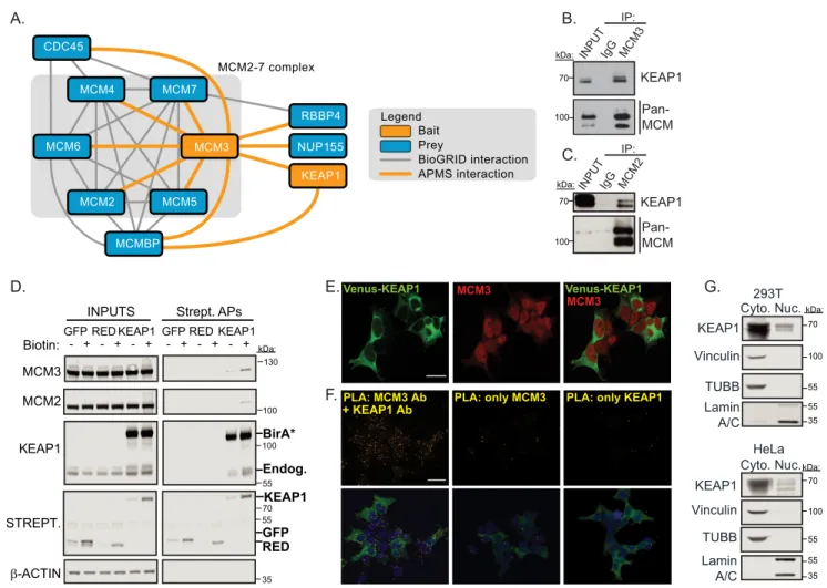
Biochemical Analysis of the KEAP1-MCM3 Complex— Hav-ing previously identified MCM3 as a high-confidence
KEAP1-interacting protein by affinity purification (AP)/MS (19, 20) and now as a putative substrate (Fig. 1, C andD), we sought to validate and determine the localization of this interaction. We first conducted a reciprocal MCM3 IP/MS experiment with ectopically expressed FLAG epitope-tagged MCM3 and detected endogenous KEAP1 in addition to all expected MCM3-associated proteins, largely those important for DNA replication (Fig. 2A). We also detected KEAP1 interaction with the MCM complex by endogenous co-immunoprecipitation using antibodies to MCM3 or MCM2 (another subunit of the MCM2–7 heterohexameric complex) (Fig. 2,BandC). Next, we expressed a KEAP1 fusion to a biotin ligase proximity detec-tor, BirA*, and tested MCM subunits forin vivobiotinylation. We detected biotin-stimulated modification of both endoge-nous MCM3 and MCM2 only in cells expressing the KEAP1-BirA* fusion, demonstrating its close proximity to the MCM hexamer (Fig. 2D). We next assessed where in the cell KEAP1 KEAP1 Affinity Purification
LIGHT
HEAVY Untreated
+MG132
KEAP1
KEAP1
m/z LC/MS/MS
A. C.
0 2 4
NRF2 UBB NRF1 ARCN1 MCM3 MAD2L1 COPG1 PGAM5 SASS6 DPP3 MCC WDR1 SLK PRKCI SQSTM PDPR GBAS
SILAC Ratio (MG132/Untreated) B.
Parallel Affinity Capture Proteomics
D.
F
o
ld
C
h
a
n
g
e
in KEAP1 Binding
Time (h) in MG132
NRF2
5 10 15 20 25
0.5 1.0 1.5 2.0 2.5
0.0 0.5 1.0
1.5 MCM3
DPP3
0 2 4 6 8
0.0 0.5 1.0 1.5
MAD2L1
0 1 2 3
SLK
0 1 2 3
PGAM5 >2 Fold Change <2 Fold Change
0 0.0
2.0 SBP-KEAP1 AP
INPUT Time (h)
in MG132: 0 2 4 6 8 0 2 4 6 8
NRF2
NRF1
PGAM5
MCM3
SLK MAD2L1
DPP3
KEAP1
β-ACTIN SBP
SBP
1
0 5
NRF1
0 2 4 6 8
10 100
35
130
25
250
100
55 100 100 kDa:
100
35 70 100
35 130 35
MCM3 NRF2
-ACTIN HMOX1
PGAM5
KEAP1
UNT. MG132 MLN. CDDO SULF.
NRF1 E.
β
kDa:
and MCM3 interact. Immunofluorescence analysis using antibodies against MCM3 in cells stably expressing VENUS-KEAP1 revealed that MCM3 was mainly nuclear, but a small fraction of MCM3 antibody reactivity diffusely localized to the cytosol, in contrast to VENUS-KEAP1, which was mainly cyto-plasmic with a small pool in the nucleus (Fig. 2E). To test in which compartment(s) KEAP1 and MCM3 associate, we per-formed anin situproximity ligation assay (PLA) using primary antibodies for KEAP1 and MCM3. Fig. 2Fshows representative images for this assay, demonstrating that KEAP1 and MCM3 are in close proximity to one another in both the nucleus and cytoplasm. Using subcellular fractionation followed by West-ern blotting, we observed that a small fraction of KEAP1 was indeed in the nucleus, in agreement with our microscopy anal-ysis and other reports that⬃5% of KEAP1 is nuclear (Fig. 2G) (56).
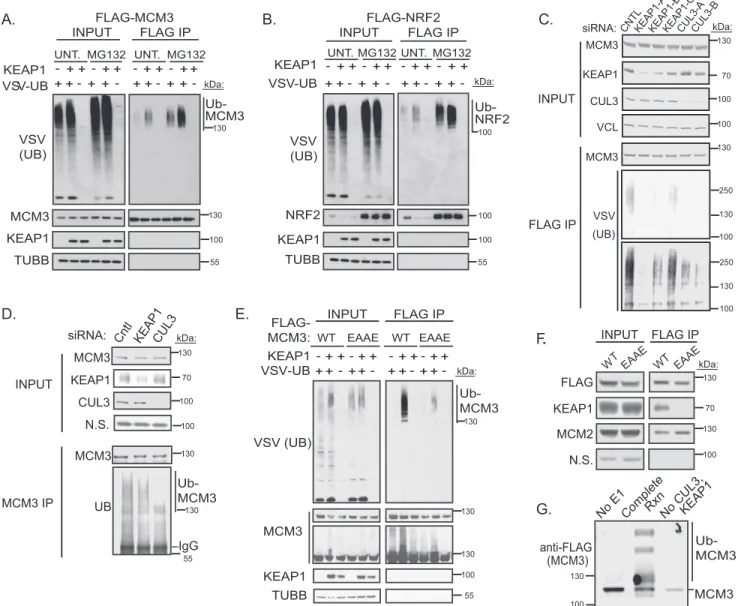
MCM3 Is a KEAP1 Substrate for Ubiquitylation in Vivo and in Vitro—Next we tested whether KEAP1 directly ubiquitylates MCM3. Under near-denaturing conditions, FLAG-MCM3 or
FLAG-NRF2 was immunoprecipitated from HEK293T cells expressing control GFP or KEAP1. Western blotting analysis showed strong induction of MCM3 ubiquitylation by KEAP1, similar to the positive control NRF2 (Fig. 3,AandB). Recipro-cally, siRNA-mediated silencing of KEAP1 suppressed ubiqui-tylation of FLAG-MCM3 (Fig. 3C). The degree of KEAP1 silencing by the three independent siRNAs strongly correlated with loss of MCM3 ubiquitylation. To evaluate the ubiquityla-tion of endogenous MCM3, we immunoprecipitated MCM3 under near-denaturing conditions from cells transfected with siRNAs targeting KEAP1 or CUL3. Both KEAP1 and CUL3 silencing suppressed ubiquitylation of endogenous MCM3 (Fig. 3D). These data demonstrate that the KEAP1-CUL3-RBX1 ligase is responsible for the majority of MCM3 ubiquitylation in proliferating cells. As expected, ubiquitylation of MCM3 by KEAP1 required MCM3-KEAP1 physical interaction. Specifi-cally, we mutated the ETGE motif within MCM3 to EAAE and found that this mutant was not ubiquitylated by KEAP1 and did not bind KEAP1 (Fig. 3,EandF). Together, these data suggest
E.Venus-KEAP1 Venus-KEAP1 G.
MCM3 MCM3
B.
INPUT
C.
Pan-MCM KEAP1
D.
INPUTS Strept. APs
KEAP1 MCM3
β-ACTIN
STREPT.
KEAP1
GFP RED
Biotin: - + - + - + - + - + - + GFP RED KEAP1 GFP RED KEAP1
MCM2
BirA*
Endog.
HeLa KEAP1
Vinculin
TUBB Lamin A/C
F.PLA: MCM3 Ab
+ KEAP1 Ab
PLA: only MCM3 PLA: only KEAP1
Cyto. Nuc. IgG MC
M3
KEAP1
Vinculin
TUBB
Lamin A/C
Pan-MCM KEAP1
293T Cyto. Nuc. INPUT
IP:
IgG MCM2 KEAP1
NUP155 MCM7
MCM3 CDC45
MCM4
MCMBP
MCM2 MCM5
RBBP4
MCM6
Bait Prey
APMS interaction BioGRID interaction Legend
MCM2-7 complex
A. IP:
100
55 100 kDa:
70
100 70
70
55 35
55 100 70
55 35 kDa:
kDa:
kDa: 130
100
55 kDa:
70 55
35 100
FIGURE 2.KEAP1 associates with MCM3 in the MCM2–7 complex in both the nucleus and cytoplasm.A, FLAG-KEAP1 and FLAG-MCM3 protein interaction networks were determined by FLAG IP/MS. Spotlite-scored high-confidence interactors are shown (supplemental Table S2).B, endogenous MCM3 IP was probed for KEAP1 and MCM proteins using an antibody to an epitope common to multiple MCM subunits.C, endogenous MCM2 IP was probed for KEAP1 and MCM proteins.D, HEK293T cells stably expressing BirA*-KEAP1 (a biotin ligase proximity detector) or cells stably expressing controls (BirA*-GFP or BirA*-HC Red (RED)) were subjected to streptavidin affinity purification and probed for the indicated proteins. Biotinylated proteins were detected using fluorescently labeled streptavidin (Strept).Endog, endogenous.E, HEK293T immunofluorescence of VENUS-KEAP1 and endogenous MCM3.Scale bar⫽20m.F, Duo-Link
that the KEAP1-CUL3 complex directly ubiquitylates MCM3. To confirm this, anin vitroubiquitylation assay was performed. The KEAP1-CUL3-RBX1 complex was sufficient to ubiquity-late MCM3 (Fig. 3G).
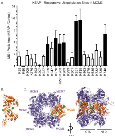
To identify the sites of ubiquitylation within MCM3, and specifically those that respond to KEAP1, we performed ubiq-uitin remnant profiling on immunopurified MCM3 complexes from control cells or cells overexpressing KEAP1. Specifically, tryptic peptides from FLAG-MCM3 complexes were subjected to ubiquitin remnant IP followed by LC/MS-MS. This method uses an antibody specific for the ubiquitin remnant left on the ubiquitylated lysine following tryptic digestion. The results
(Fig. 4A) further support our Western blotting data that KEAP1 indeed ubiquitylates MCM3. Using a 3-fold arbitrary threshold, six lysine residues were identified as responsive to KEAP1-de-pendent ubiquitylation: Lys-229, Lys-270, Lys-283, Lys-351, Lys-435, and Lys-748 (Fig. 4A). Of the ubiquitylated lysines mapped, Lys-435 showed the greatest -fold increase by KEAP1. This site was also found to be differentially ubiquitylated in an unbiased screen for cullin ring ligase substrates (57). To visual-ize these lysines on the structure of the MCM2–7 complex, we used protein structural modeling. Human MCM3 was threaded around a homologous archaeal MCM protein (58) and super-imposed over a published model of the yeast MCM2–7 hetero-KEAP1
VSV-UB
FLAG-MCM3
INPUT FLAG IP FLAG-NRF2 + + + -+ + + + -+ MG132
INPUT FLAG IP
+ + + -+ + + + -+ VSV (UB) NRF2 VSV (UB) MCM3 KEAP1 KEAP1 TUBB TUBB KEAP1 VSV-UB
UNT. UNT. MG132 UNT.MG132UNT. MG132
+ + + -+ + + + -+ + + + -+ + + + -+
Ub-MCM3 Ub-NRF2
A. C.
E. INPUT MCM3 IP Cntl KEAP1 siRNA: CUL3 MCM3 UB KEAP1 CUL3 N.S. MCM3 IgG F
.
D. Ub-MCM3INPUT FLAG IP
KEAP1 VSV-UB + + + -+ + + + -+ + + + -+ + + + -+ WT EAAE MCM3 KEAP1 TUBB VSV (UB) Ub-MCM3
No E1CompleteRxnNo CUL3, KEAP1
anti-FLAG (MCM3) WT EAAE Ub-MCM3 MCM3 G. FLAG KEAP1 MCM2
INPUT FLAG IP
WT EAAE
N.S.
WT EAAE
B. FLAG-MCM3: 55 100 100 130 55 100 130 100 130 70 100 100 130 130 55 kDa: kDa: kDa: kDa: kDa: 130 130 130 100 55 130 70 130 100 100 130 KEAP1 MCM3 CUL3 VCL VSV (UB) MCM3 INPUT FLAG IP
CNTLKEAP1-AKEAP1-BKEAP1-CCUL3-A CUL3-B siRNA: 250 100 130 130 70 100 100 130 250 100 130 kDa:
hexamer (59). The lysines observed to be most ubiquitylated in response to ectopic KEAP1 were found to be on predicted exposed surfaces of the C-terminal domain in MCM3 (Fig. 4,B andC).
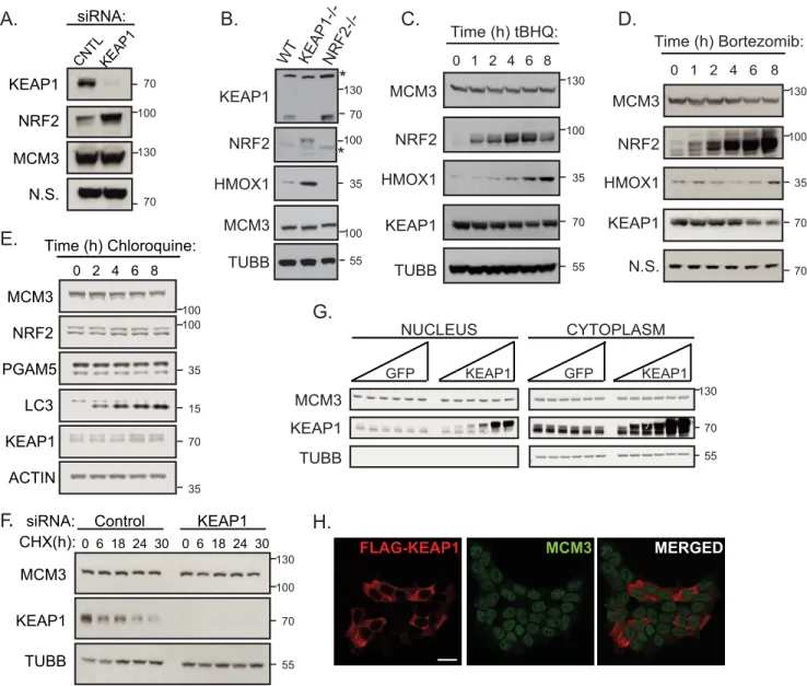
KEAP1 Does Not Regulate MCM3 Levels, Subcellular Local-ization, or MCM2–7 Complex Formation—After identifying MCM3 as a novel KEAP1 substrate, we sought to determine the function of this ubiquitylation. First, we tested whether KEAP1 targets MCM3 for proteasome-mediated degradation as it does its well known substrate NRF2. KEAP1 manipulation did not affect steady-state levels of total cellular MCM3. Specifically, KEAP1 knockdown, deletion, overexpression, or chemical antagonism caused no changes in total MCM3 protein levels, whereas all of these perturbations affected NRF2 levels (Fig. 5, A–D). Attempts to determine whether KEAP1 loss affects MCM3 half-life were hampered by the extremely long MCM3 half-life. A very long (30-h) chase with the protein synthesis inhibitor cycloheximide did not yield an appreciable change in MCM3 levels (Fig. 5F), in agreement with a report that showed that the MCM complex had a half-life of⬃24 hin vivo(60).
Treatment with the proteasome inhibitor bortezomib also did not stabilize MCM3 over the course of 8 h, in agreement with KEAP1-CUL3-RBX1 not targeting MCM3 for proteasome-me-diated degradation (Fig. 5D). Similarly, treatment with the lys-osomal inhibitor chloroquine did not stabilize MCM3 over an 8-h time course, supporting the conclusion that the KEAP1-CUL3-RBX1 ligase is not targeting MCM3 for lysosome-medi-ated degradation (Fig. 5E).
Next we tested whether KEAP1 could be ubiquitylating MCM3 to affect its subcellular localization. Using immunoflu-orescence in HEK293T cells transiently transfected with KEAP1, we found no difference in the localization of endoge-nous MCM3, which remains largely diffuse in the nucleus (Fig. 5H, compare cells expressing KEAP1 in red to those not expressing). We also expressed increasing amounts of exoge-nous KEAP1 and assayed the amounts of MCM3 in the nuclear and cytoplasmic compartments and found no difference in the amount of MCM3 in either compartment, suggesting that KEAP1 does not regulate total MCM3 subcellular localization (Fig. 5G).
The ability of MCM3 to associate with the other members of the MCM2–7 heterohexamer was evaluated by immunopre-cipitating either WT FLAG-MCM3 or the KEAP1-deficient binding mutant (FLAG-MCM3 EAAE) and probing with anti-MCM2. A comparable amount of MCM2 associated with both forms of MCM3, suggesting that KEAP1 binding and ubiquity-lation do not regulate MCM3 incorporation into the MCM2–7 hexamer (Fig. 3F). Furthermore, MCM3 was associated with the other MCM proteins at similar levels in the presence or absence of KEAP1 siRNA knockdown (data not shown).
KEAP1-dependent Ubiquitylation Is Not Responsive to Treatment with DNA-damaging Agents, ROS Mimetics, or Autophagy—As DNA damage by etoposide was recently reported to lead to increased phosphorylation and ubiquityla-tion of a number of sites in subunits of the MCM2–7 complex (61), including three ubiquitylation sites within MCM3, we examined whether KEAP1 could be one of the E3 ligases that ubiquitylate MCM3 in response to DNA damage. We found, however, that overnight treatment with a panel of DNA-dam-aging agents (etoposide, gemcitabine, and 4-Nitroquinoline 1-oxide (4NQO)) did not strongly affect the MCM3-KEAP1 interaction (Fig. 6A) or KEAP1-dependent ubiquitylation of MCM3 but did activate phospho-Chk1, a marker of DNA dam-age (Fig. 6B). These data suggest that KEAP1 is not the ligase modifying MCM3 in response to etoposide-mediated DNA damage.
The ability of KEAP1 to act an as efficient substrate adaptor for NRF2, its well known substrate, relies on the redox state of the cell because, during oxidative stress, KEAP1 undergoes electrophilic attack by ROS and is placed in a conformation no longer favorable to target NRF2 (3, 6, 11, 12, 62– 64). To test whether MCM3 is also a ROS-dependent substrate of KEAP1, we employed surrogate compounds (sulforaphane andtert -bu-tylhydroquinone (tBHQ)) that mimic ROS by attacking the reactive cysteines within KEAP1. These drugs are widely used as NRF2 agonists, although whether these inhibit NRF2 ubiq-uitylation or block release of ubiquitylated NRF2 is debated. Here we find that treatment with these compounds stabilizes
K3
5
K
80
K1
0
9
K
176
K1
9
3
K2
07
K2
27
K2
29
K2
47
K2
7
0
K
27
0
;K2
8
3
K2
8
3
K2
93
K3
01
K3
5
1
K4
3
5
K4
63
K5
79
K6
35
K7
32
K7
35
K7
48
1 2 4 8 1 6 32
MS
1
Pe
ak
A
rea (
K
E
A
P1
/Co
n
tr
o
l)
A.
B. C.
MCM3 MCM7
MCM5 MCM2
MCM6 MCM4
CTD NTD
3 MCM7 M
MCM5 MCM2
CM6 MCM4
MC MCM3
MCM7
CM5
90o
KEAP1-Responsive Ubiquitylation Sites in MCM3
FIGURE 4.Mapping the KEAP1-dependent ubiquitylation sites in MCM3. A, HEK293T cells were transfected with plasmids encoding FLAG-MCM3 with or without the SBPHA-KEAP1 plasmid, and a near-denaturing FLAG IP was performed as in Fig. 3. A tryptic digest and ubiquitin remnant IP were then performed, followed by LC/MS-MS on the resultant peptides. Ubiquitylated peptides of MCM3 detected are plotted as mean⫾S.E. MS1 peak areas of three biological replicate experiments.Black columnsare lysine residues that increased beyond an arbitrary threshold of 3-fold increase in the presence of SBPHA-KEAP1 (supplemental Table S3).B, protein structural modeling of human MCM3 (Uniprot code P25205-1) threaded around an archaeal MCM structure (PDB code 3F9V). The KEAP1-modified lysines detected inAare shown asgreen spheres.C, protein structural modeling of human MCM3 from
NRF2 but does not ablate KEAP1-dependent ubiquitylation of either NRF2 or MCM3 (Fig. 6,CandD). Together, these data suggest that the ubiquitylation of MCM3, like NRF2, is not inhibited by sulforaphane or tBHQ.
As MCM3, along with other important DNA replication fac-tors, was recently reported to undergo autophagy-mediated degradation, we tested whether KEAP1 could be ubiquitylating MCM3 to target it for lysosomal degradation (65). We found that treatment with a lysosome-mediated degradation inhibitor (chloroquine, 100Mfor 8 h) or an autophagy activator
(rapa-mycin, 10Mfor 8 h) did not affect KEAP1-dependent
ubiqui-tylation of MCM3 but did augment LC3 I/II conversion, a marker of autophagy (Fig. 6E). These data suggest that KEAP1 is not targeting MCM3 for autophagy-mediated degradation.
KEAP1 Associates with Chromatin in a Cell Cycle-dependent Fashion—Our detection of KEAP1-MCM3 association in the nuclei of actively proliferating cells suggested that KEAP1 may associate with MCM3 during a normal cell cycle. MCM3 is chromatin-loaded as part of the MCM2–7 complex during G1
phase and unloaded as DNA replication completes throughout S phase. To examine whether KEAP1 associates with chroma-tin in a cell cycle-dependent fashion, HeLa cells were
synchro-B.
D.
E.
KEAP1
NRF2
MCM3
N.S.
KEAP1 CNTL KEAP CNTL
siRNA:
A.
C.
KEAP1
NRF2
HMOX1
MCM3
TUBB
*
*
WT KEAP1-/- NRF2-/-70
100
35
100
55
G.
H.
MCM3
KEAP1
Control
TUBB
KEAP1 CHX(h): 0 6 18 24 30 0 6 18 24 30
siRNA:
/
FLAG-KEAP1 MCM3 MERGED
NRF2 MCM3
KEAP1
TUBB
Time (h) tBHQ:
0 1 2 4 6 8
HMOX1
NRF2
KEAP1
N.S.
1 2 4 6 Time (h) Bortezomib:
MCM3
0 8
HMOX1
MCM3
KEAP1
TUBB
NUCLEUS CYTOPLASM
KEAP1
KEAP1 GFP
GFP
100 70
130
70
100 130
70 35
55 70
70 35 100 130
55 70 100 130
130
70
55 Time (h) Chloroquine:
0 2 4 6 8 MCM3
NRF2
ACTIN LC3 PGAM5
KEAP1 70
100 100
F
.
35 35
15
130
nized, and lysates were collected during G1and early S phase.
The lysates were fractionated into chromatin and soluble frac-tions and immunoblotted for KEAP1, MCM3, and fraction-ation and loading controls. Strikingly, we found that KEAP1 loaded onto chromatin during G1phase as MCM3 did (Fig. 7A,
loading is seen at the 4- to 10-h time points, chromatin frac-tion). To investigate when KEAP1 also unloads from the chro-matin, HeLa cells were synchronized in early S phase, and lysates were collected from S to M phase. These lysates were fractionated into chromatin and whole cell fractions and probed for KEAP1, MCM3, and fractionation and loading
con-trols. KEAP1 unloaded in G2phase, similar to MCM3, but the
unloading of KEAP1 was slightly behind that of MCM3 (Fig. 7B, unloading is seen at the 6- to 10-h time points, chromatin frac-tion). Thus, KEAP1 associates with DNA in a cell cycle-depen-dent fashion, and KEAP1 is at the right place at the right time during the cell cycle to regulate the MCM complex.
In an asynchronous population, KEAP1 bound MCM3 pre-dominantly in the soluble fraction with a weaker association on chromatin (Fig. 7C). This observation is consistent with the fact that most of the cellular MCM is soluble, and only a fraction is chromatin-loaded. To test where in the cell this
KEAP1-depen-A. B. C.
D.
KEAP1
NRF2
MCM3 β-ACTIN P-CHK1
INPUT Strept. AP
CntrltBHQSulf.Etop.Gemcit.4NQO CntrltBHQSulf.Etop.Gemcit.4NQO
DNA Damage DNA Damage
INPUT FLAG IP GFP KEAP1
Unt. ET OP
.
GEM.Unt. ET OP
.
GEM. GFP KEAP1
KEAP1 VSV (UB)
MCM3
β-ACTIN P-CHK1
Unt. ET OP
.
GEM.Unt. ET OP
.
GEM. KEAP1:
INPUT FLAG IP NRF2
- + + +
NRF2 VSV (UB)
KEAP1
TUBB
- + + +
KEAP1:
MCM3
MCM3 VSV (UB)
KEAP1
NRF2
TUBB
- + + + - + + + Unt. Unt. tBHQSULF
MCM3
Unt. Unt. tBHQSULF
Unt. Unt. tBHQSULF Unt. Unt. tBHQSUL
F
INPUT FLAG IP
35 70 130 100 100
55 100
35 70 130 250
25
55 100
35 70 130 250
25
100 100
100 55 35
70 100 130 130
55 100
35 70 130 250
25
100 100 130
100
55
INPUT FLAG IP
KEAP1
LC3 MCM3 VSV (UB)
+KEAP1 +KEAP1
Unt. CHQ RA P
Unt. CHQRA P E.
Unt. CHQ RA P
Unt. CHQ RA P
dent ubiquitylation occurs, cells expressing FLAG-MCM3 and either GFP or KEAP1 were fractionated into chromatin and soluble fractions, subjected to FLAG (MCM3) IPs in both frac-tions, and probed by Western blotting for ubiquitin on MCM3. Most ubiquitylation of MCM3 by KEAP1 was seen in the solu-ble fraction. Ubiquitylation of MCM3 in the chromatin-bound fraction was detectable, although only during proteasome inhi-bition (Fig. 7D). Therefore, KEAP1-dependent ubiquitylation of MCM3 occurs in both the chromatin and soluble fractions.
Discussion
Through its ubiquitin ligase activity, KEAP1 serves as a sen-sor and molecular switch for the cellular response to oxidative stress. Here we provide data that support the emerging concept of NRF2-independent functions for KEAP1. To date, in addi-tion to NRF2 signaling and the coordinated antioxidant response, KEAP1 has been shown to regulate NF-B signaling through its degradation of IKBKB to target the mitochondrial membrane phosphatase PGAM5 for proteasome-mediated degradation and to regulate DNA break repair through
degra-dation-independent ubiquitylation of PALB2 (21–23). Our analyses here establish a fourth NRF2-independent function for KEAP1 and raise the additional possibility that KEAP1 regu-lates cell cycle progression and/or genome stability through ubiquitylation of MCM3.
Here we focused on a biochemical assessment of KEAP1 sub-strates in general and on defining MCM3 as a KEAP1 substrate in particular. Our future studies will delve into the role of KEAP1 in MCM3 function or dynamics. Our present data sug-gest that KEAP1 interacts with MCM3 when it is in the MCM2–7 hexameric complex. We detected KEAP1 in MCM2 immunoprecipitates, and, further, we observed biotinylation of MCM2 by the BirA*-tagged KEAP1. Together, these data sug-gest that KEAP1 interacts with the full MCM2–7 hexamer (e.g. Fig. 2C). Moreover, we detected KEAP1-mediated MCM3 ubiquitylation in both the soluble and chromatin-bound frac-tions (Fig. 7D). The association of KEAP1 with chromatin dur-ing S phase in a pattern that closely follows MCM loaddur-ing and unloading is also consistent with KEAP1 associating with the MCM complex rather than monomeric MCM3 because only
CHROMATIN SOLUBLE
INPUT FLAG IP
MG132:
VSV (UB)
MCM3
HIST3 HA (KEAP1) BTUBB
D.
GFP KEAP1GFP KEAP 1 - - + +
INPUT FLAG IP
Ub-MCM3 A.
I.
C.
KEAP1
MCM3
HIST3
TUBB Time (h) post release:
Chromatin Fraction
C 0 1 2 3 4 5 6 7 8 9 10
Soluble Fraction
n.s
G1
M M G1
C 0 1 2 3 4 5 6 7 8 9 10
KEAP1
MCM3
TUBB Time (h) post release:
Chromatin Fraction
0 1 2 3 4 5 6 7 8 9 10
Soluble Fraction
B.
INPUTS Strept. AP
GFP KEAP1 Sol. Chrom. Sol. Chrom.
MCM3
TUBB
HIST3
GFPKEAP1GFPKEAP1
S G2/M S G2/M
STREPT.
GFP KEAP1GFP KEAP1 GFP KEAP1GFP KEAP1
GFP KEAP1GFP KEAP1
- - + + - - + +
- - + +
GFPKEAP GFP 1
KEAP1
S S
0 1 2 3 4 5 6 7 8 9 10
130
55 70
15 130
55 130
55 15 70
130
130 70
55 100
130
70 100
55
35
130
100
55
15
FIGURE 7.KEAP1 associates with chromatin.A, HeLa cells were transfected with control or KEAP1 siRNA and synchronized by double thymidine-nocodazole block. Lysates were collected during G1and S phases, separated into soluble and chromatin fractions, and probed for KEAP1, MCM3, and loading and
fractionation controls (C).B, HeLa cells were transfected as inA, synchronized by double thymidine block, collected during S and G2phases, fractionated, and
the MCM complex is loaded for replication and not individual subunits (66). We thus favor the notion that KEAP1 impacts the replication function of the MCM complex, although we cannot yet rule out KEAP1 involvement in a novel non-replication role for MCM3, as has been reported for other individual MCM subunits (67, 68).
The high-affinity KEAP1-targeting motif (ETGE) is uniquely found in MCM3 and not in the other five MCM subunits, sug-gesting that MCM3 is at least one direct point of contact between the MCM complex and KEAP1. MCM4 contains the lower-affinity DLG binding motif for KEAP1, raising the possi-bility for a second point of contact. All six subunits of the human MCM2–7 heterohexamer have been reported to be ubiquitylated in cells (69). It is thus possible that KEAP1 ubiq-uitylates additional MCM proteins. Based on theS. cerevisiae andDrosophila melanogasterMCM complexes, we presume that human MCM3 is adjacent to both MCM5 and MCM7 in the hexamer (70, 71). MCM5 constitutes one side of the MCM2/5 “gate” where the MCM ring opens to allow double-stranded DNA to pass during MCM loading in G1phase (72,
73). KEAP1 could modulate MCM loading by regulating con-formational changes at the MCM2/5 interface. MCM3 is the subunit that directly contacts the helicase activator complex GINS, which only associates with MCM during helicase activa-tion and fork progression (71). KEAP1-mediated MCM3 ubiq-uitylation could impact helicase activation either globally or at a subset of origins. Interestingly, polyubiquitylation of the MCM7 subunit in bothS. cerevisiaeandX. laevisis associated with replication termination and MCM unloading (49, 50, 52) but not changes in MCM7 stability. The KEAP1 interaction with MCM3 could also impact MCM unloading. KEAP1 could thus link ROS sensing to the control of MCM chromatin load-ing, to activation of MCM-dependent DNA unwindload-ing, or to MCM unloading during S phase as a means to preserve DNA integrity and genome stability.
Human MCM complexes undergo cell cycle-dependent phosphorylation and sumoylation (46), and it is certainly pos-sible that additional E3 ubiquitin ligases participate with KEAP1 in MCM control. Nonetheless, KEAP1 is clearly the major MCM3 E3 ubiquitin ligase in actively proliferating cells (Fig. 3). Our data so far suggest that KEAP1 binds and ubiqui-tylates only a subset of the total MCM3 molecules and likely regulates their activity through altering MCM2–7 protein-pro-tein interactions or helicase activity. Future work will explore not only the molecular consequences of KEAP1-mediated MCM ubiquitylation but also under what cellular circum-stances KEAP1 may be stimulated to ubiquitylate MCM3. Our discovery that KEAP1 associates with chromatin during S phase may reflect a novel nuclear role for KEAP1 in monitoring replication fork progression and perhaps coordinating origin firing or replisome activity with cellular redox state. If so, then the KEAP1-MCM interaction represents a novel, nuclear role for KEAP1 outside of NRF2 regulation, emphasizing the breadth of KEAP1-regulated cellular events.
We have previously defined a static KEAP1 protein interac-tion network and demonstrated that it is enriched for proteins containing the KEAP1 binding motif, E(T/S)GE (19, 20). Although the ETGE motif in NRF2 has been established as a
KEAP1 degron therein, and several ETGE-containing proteins have been shown to activate NRF2 through competitive bind-ing to KEAP1 (74, 75), the function of the interactions between KEAP1 and these other E(T/S)GE-containing proteins remained largely unknown. In this study, we have combined PAC proteomics to capture substrates and annotated them against what we identified in our prior studies to be high-con-fidence KEAP1 interactors. Our data suggest that, in addition to MCM3, which we validated as a KEAP1-CUL3-RBX1 substrate here, we may have also discovered NRF1 as a novel KEAP1 substrate. NRF1 has been shown previously to bind KEAP1, but KEAP1 was shown not to be responsible for targeting NRF1 to the endoplasmic reticulum membrane or regulating an artifi-cial reporter of NRF1-driven transcription (54, 55). To the best of our knowledge, our study is the first report that NRF1 is a putative KEAP1 substrate for proteasome-mediated degrada-tion. We observe NRF1 accumulating with proteasome inhibi-tion in both whole cell lysates and in the KEAP1 complex by both AP/MS and AP-Western blotting. In-cell and in vitro ubiquitylation assays will be important to confirm the substrate status of NRF1. Our findings neither confirm nor rule out whether MAD2L1, SLK, DPP3, TSC22D4, FAM117B, or other ETGE-containing KEAP1 interactors arebona fidesubstrates. Because we and others have now demonstrated that KEAP1-CUL3-RBX1 substrates may not be turned over by the protea-some (23), the substrate status of each of these interactors will need to be evaluated individually.
substrate interface of KEAP1. Furthermore, this work provides a previously unappreciated link between KEAP1, genome sta-bility, and cell cycle progression.
Experimental Procedures
Tissue Culture, Treatments, Transfections, and Small Inter-fering RNAs—HEK293T, HDF-Tert, and HeLa cells were obtained from the American Type Culture Collection. The cell lines were passaged for no more than 3 continuous months after resuscitation. HDF-Tert, HEK293T, and HeLa cells were grown in DMEM supplemented with 10% FBS and 1% penicil-lin/streptomycin in a 37 °C humidified incubator with 5% CO2.
Mouse embryonic fibroblasts (MEFs) were cultured in Iscove’s modified Dulbecco’s medium supplemented with 10% FBS and 1% penicillin/streptomycin. The KEAP1 and NRF2 knockout MEFs were kindly provided by Thomas Kensler and Nobunao Wakabayshi. Drugs used for cell treatments were acquired as follows: MG132 (Calbiochem), bortezomib (SelleckChem), tert-butylhydroquinone (Sigma), sulforaphane (Sigma), etopo-side (Sigma), chloroquine (Sigma), and gemcitabine (Sigma). For transient transfections, cDNA expression constructs were transfected in HEK293T cells using Lipofectamine 2000 (Life Technologies) for 24 h before harvest. Transfection of siRNA (20 nM) in HEK293T cells was performed with Lipofectamine
RNAiMAX (Life Technologies). Transfection of siRNA in HeLa cells was done with either Dharmafect (50 nM) or
Lipo-fectamine RNAiMAX (20 nM). siRNA sequences were as fol-lows: control, CGUACGCGGAAUACUUCGATT; KEAP1-A, GGGCGUGGCUGUCCUCAAU; KEAP1-B, CAUGUGAU-UUAUUCUUGGAUACCUG; KEAP1-C, UGGCUGUCCU-CAAUCGUCUCCUUUA; CUL3-A, GGUCUCCUGAAUAC-CUCUCAUUAUU; and CUL3-B, GAAUGUGGAUGUCAG-UUCACGUCAA.
Immunoprecipitations, Affinity Pulldowns, and Western Blotting—These experiments were performed as described pre-viously (74) with minor modifications. Briefly, for streptavidin and FLAG affinity and immune purification, cells were lysed in 0.1% Nonidet P-40 lysis buffer (10% glycerol, 50 mMTris-HCl,
150 mMNaCl, 2 mMEDTA, 0.1% Nonidet P-40 supplemented
with protease inhibitor mixture (Thermo Scientific) and phos-phatase inhibitor (Thermo Scientific), 10 mMN
-ethylmaleim-ide, and 250 units Benzonase (Sigma)) and then passed through a 26.5-gauge needle three times. The cell lysates were cleared by centrifugation and incubated with streptavidin resin (GE Healthcare) or FLAG resin (Sigma) before washing with lysis buffer and eluting with NuPAGE 4⫻SDS loading buffer (Life Technologies). For siRNA knockdown, HEK293T cells were transiently transfected and lysed in RIPA buffer (1% Nonidet P-40, 0.1% SDS, 0.25% sodium deoxycholate, 150 mMNaCl, 10%
glycerol, 25 mMTris, and 2 mMEDTA supplemented with
pro-tease inhibitor mixture, phosphatase inhibitor, andN -ethylma-leimide) 60 –72 h post-transfection. For BirA* affinity purifica-tion, HEK293T cells were pretreated with 50Mbiotin for 2– 4
h and lysed in supplemented RIPA buffer, and cleared lysates were subjected to streptavidin AP as above and eluted with a 1:1:1:1 mixture of 1 MDTT, 4⫻ SDS loading buffer, 50M
biotin, and RIPA buffer. For endogenous IP (Fig. 2,BandC), HEK293T cells were lysed in co-IP buffer (50 mMHEPES (pH
7.2), 33 mMpotassium acetate, 1 mMMgCl2, 1 mMATP, 0.1%
Nonidet P-40, 5 mMCaCl2, and 10% glycerol with protease and
phosphatase inhibitors), then treated with 10 units of S7 micro-coccal nuclease (Roche), sonicated, and cleared by centrifuga-tion. Samples were rotated with MCM2 antibody or control rabbit IgG, followed by rotation with protein A-Sepharose (Roche), and then washed three times with co-IP buffer and eluted with 2⫻ SDS loading buffer, 5% 2-mercaptoethanol (Sigma), and boiling for 10 min. Lysates were resolved on 4 –12% SDS-PAGE gradient gels (Invitrogen), transferred to nitrocellulose or PVDF membranes, and probed using the fol-lowing antibodies: MCM3 (Bethyl, A300-192A), anti-KEAP1 polyclonal (ProteinTech, Chicago IL), anti-FLAG M2 monoclonal (Sigma), HA monoclonal (Roche), anti-MAD2L1 (Bethyl, Montgomery TX, A300-301A), anti-SLK (Bethyl, A300-499A), anti--actin polyclonal (Sigma, A2066), anti--tubulin monoclonal (Sigma, T7816), anti-DPP3 poly-clonal (Abcam, Cambridge, MA, 97437), anti-GFP (Abcam, ab290), anti-NRF2 H300 polyclonal (Santa Cruz Biotechnol-ogy, Santa Cruz, CA), anti-PGAM5 polyclonal (Abcam, 126534), anti-NRF1 polyclonal (Santa Cruz Biotechnology, D5B10), anti-MCM2 polyclonal (Bethyl, A300-191A), anti-Pan MCM (a kind gift from D. MacAlpine, Duke University), and anti-VSV polyclonal (Bethyl, A190-131A). Biotinylated pro-teins associated with BirA* AP blots were detected using fluo-rescently labeled streptavidin (IRDye 680CW-LI-COR). Pro-tein quantification was performed in the LI-COR imaging suite (Image Studio Lite), where all blots were determined to be in the linear range by the software.
Cell Fractionations and Cell Synchronization—HeLa cells were synchronized with a double thymidine block in early S phase by treatment with 2 mM thymidine (Sigma) for 18 h,
washout for 9 h, and readdition of 2 mMthymidine for an
addi-tional 17 h. HeLa cells were synchronized in M phase by double thymidine/nocodazole block: first, the double thymidine block (as above, with 24 h of 2 mMthymidine, 6 h of release, and
readdition of thymidine for 18 h), and then cells were released into fresh DMEM containing 100 ng/ml nocodazole for 8 –12 h. In each case, cells were washed twice with warm medium and released into fresh DMEM. Chromatin fractionation was per-formed by gentle lysis with CSK buffer (0.5% Triton X-100, 300 mMsucrose, 10 mMPIPES, 100 mMNaCl, and 2 mMEDTA)
supplemented with protease and phosphatase inhibitors for 20 min on ice. Lysates were centrifuged at 900⫻gfor 5 min at 4 °C to pellet the nuclei. Soluble fractions were transferred to new tubes. The nuclei were resuspended in CSK buffer containing 10 units DNase (RQ1, Promega) for 10 min at room tempera-ture and pelleted at 900⫻gfor 5 min at 4 °C. The remaining nuclei were washed once with CSK buffer, and the first DNase digest and the wash were pooled. Nuclear proteins solubilized by DNase digest were the chromatin fraction.
clones and gateway-cloned into pHAGE-CMV-FLAG-DEST. The MCM3 EAAE alanine mutant was created using PCR-based mutagenesis (Q5 site-directed mutagenesis kit, New Eng-land Biolabs) and sequence-verified before use.
Cell-based Ubiquitylation Experiments—These experiments were performed as reported previously (20) with few modifica-tions. Briefly, HEK293T cells were transfected with VSV-UB, FLAG-NRF2, or FLAG MCM3 and either SBPHA-KEAP1 or SBP-GFP as a control so that each condition received the same mass of DNA. The cells were lysed under near-denaturing con-ditions in 1% SDS lysis buffer (1% SDS, 150 mMNaCl, and 2 mM
EDTA) and boiled at 90 °C for 10 min. SDS lysis buffer was diluted 1:10 in cold 0.5% Nonidet P-40 lysis buffer supple-mented with protease, phosphatase, and deubiquitylase inhib-itors, and the lysates were cleared by centrifugation. FLAG IPs were carried out by incubating lysates with FLAG resin for 1– 4 h at 4 °C, followed by washing three times in lysis buffer and eluting with 4⫻SDS loading buffer and DTT.
In Vitro Ubiquitylation Experiments—Forin vitro ubiquity-lation studies, SBPHA-KEAP1 was generated using a TNT assay
(Promega), and MCM3 was purified from HEK293T cells stably expressing FLAG-MCM3 using 1% Triton-X lysis buffer (as described above for immunoprecipitations but with 500 mM
NaCl). For thein vitroubiquitylation assay, KEAP1 was mixed with recombinant human E1 (Ube1, Boston Biochem), UbcH5B (E2, Boston Biochem), CUL3-RBX1 (co-expressed in Esche-richia colias GST-Rbx1 and His-CUL3 fusions and purified sequentially over HiTRAP nickel-nitrilotriacetic acid (GE Healthcare) and glutathione-Sepharose (GE Healthcare) and proteolytically cleaved off the resin using thrombin (Sigma), followed by size exclusion chromatography over a Superdex-200 16/60 column (GE Healthcare)), ubiquitin (Boston Biochem), and FLAG-tagged MCM3 in buffer containing 25 mMHEPES (pH 8.0), 5 mMMgCl2, 2 mMDTT, and 4 mMATP.
Ubiquitylation was carried out for 20 min at 30 °C, and the products were analyzed by Western blotting with anti-FLAG antibody.
Protein Structural Modeling—The crystal structure of an archaeal MCM fromSulfolobus solfataricus(PDB code 4FDG) was identified as a template for predicting the atomic structure of human MCM3 by HHpred (PMID 15531603), and the ho-mology model was generated using MODELLER PMID 25199792). In the structural model, 94 residues at the C termi-nus as well as the residues between 510 –562 were omitted because of poor homology to the archaeal MCM. The MCM3 model was superimposed onto a published model of the MCM2–7 heterohexamer from yeast (59) using PyMOL (The PyMOL Molecular Graphics System, version 1.3, Schrödinger, LLC). PyMOL was used to prepare the images used in Fig. 4,B andC.
Affinity Purification and Mass Spectrometry—These experi-ments were performed as described previously (19, 74, 76) with minor modifications. Briefly, HEK293T cells (3–5 ⫻ 15-cm plates) stably expressing either FLAG-MCM3 or SBPHA-KEAP1 were lysed in 0.1% Nonidet P-40 lysis buffer for FLAG IP or streptavidin AP, respectively. Cell lysates were incubated, rotating with FLAG or streptavidin resin for 1 h at 4 °C, and then washed three times with lysis buffer. The precipitated
pro-teins were then trypsinized (Promega) on beads at 37 °C over-night (12–18 h) using the FASP protein digestion kit (Protein Discovery). For the KEAP1 substrate-trapping experiment (Fig. 1,AandB), SBPHA-KEAP1-expressing HEK293T cells were grown in SILAC media (light, K0R0; heavy, K6R10) for at least 10
cell divisions prior to harvesting for lysis. Tryptic peptides were cleaned up using a C18 spin column (Thermo Scientific) and then separated by reverse-phase HPLC using a nano-Aquity ultra performance liquid chromatography (UPLC) sys-tem (Waters Corp.). Chromatographic separation and mass spectrometry analysis of peptides from FLAG-MCM3 experi-ments (Fig. 2A) were performed using the same methods as in our previous work (76). Peptides from SBPHA-KEAP1 and ubiquitin remnant experiments were first trapped in a 2-cm trapping column (Acclaim威PepMap 100, C18 beads of 3.0-m particle size, 100-Å pore size) and a 25-cm EASY-spray analyt-ical column (75-m inner diameter, C18 beads of 2.0-m par-ticle size, 100-Å pore size) at 35 °C. The flow rate was 250 nl/min over a gradient of 1% buffer B (0.1% formic acid in ace-tonitrile) to 30% buffer B in 180 min, and an in-line Orbitrap Elite mass spectrometer (Thermo Scientific) performed mass spectral analysis. The ion source was operated at 2.4 –2.8 kV with the ion transfer tube temperature set at 300 °C. A full MS scan (300 –2000m/z) was acquired in Orbitrap with a 120,000 resolution setting, and data-dependent MS2 spectra were acquired in the linear ion trap by collision-induced dissociation using the 15 most intense ions. Precursor ions were selected based on charge states (3⫹2) and intensity thresholds (above
1e5) from the full scan; dynamic exclusion (one repeat during 30 s, a 60-s exclusion time window) was also used. The polysi-loxane lock mass of 445.120030 was used throughout spectral acquisition.
Protein Identification, Filtering, and Bioinformatics—For the MCM3 protein interaction network (Fig. 2A), the raw mass spectrometry data were searched with MaxQuant (1.5.2.6) along with an internal lab FLAG affinity purification mass spec-trometry (APMS) dataset of an additional 17 baits and 35 exper-iments. The search parameters were as follows: specific tryptic digestion, up to two missed cleavages, a static carbamidomethyl cysteine modification, variable protein N-terminal acetylation, and variable methionine oxidation using the human Uni-ProtKB/Swiss-Prot sequence database (release 2013_07). Pro-teins were then filtered for a 1% protein level false discovery rate (FDR). Filtering of false interactions was then accomplished using Spotlite (19), with a 10% FDR for the entire dataset, including the additional baits. These results were then imported into Cytoscape v3.2.1 for network visualization. The Spotlite results are provided insupplemental Table S2. For the SBPHA-KEAP1 SILAC experiments, the additional MaxQuant parameters were a variable Gly-Gly modification on lysines, and Lys-6 and Arg-10 heavy SILAC labels. The Maxquant results are provided insupplemental Table S1.
For ubiquitin remnant profiling, raw files were searched using SorcererTM-SEQUEST威 (build 5.0.1, Sage N Research)
within PeptideProphet, semitryptic digestion, a static carbam-idomethyl cysteine modification (57.021465), and variable methionine oxidation (⫹15.99492), ubiquitylated lysine (114.042931), and serine, threonine and tyrosine (STY) phos-phorylation (79.966331). A 1% peptide-level FDR was deter-mined by PeptideProphet. The mass spectrometry proteomics data have been deposited in the ProteomeXchange Consortium via the PRIDE (77) partner repository with the dataset identifier PXD003929.
Ubiquitin Remnant Profiling—For mapping the ubiquity-lated lysines within MCM3, immunoprecipitations for FLAG-MCM3 were done under near-denaturing conditions in the presence or absence of SBPHA-KEAP1. Then, the proteins were subject to tryptic digest and the peptides were flowed over beads conjugated to the ubiquitin remnant specific antibody (Cell Signaling) for 4 h at 4°C in IAP buffer supplied by the manufacturer. Beads were washed twice with IAP buffer and once with PBS. Peptides were eluted with 0.15% TFA for 5 min at room temperature and dried at room temperature by speed vacuuming. Peptides were shot in the mass spectrometer as described above. Ubiquitylated peptides identified by MS/MS were then quantified in Skyline v3.5. For each peptide, the MS1 intensity of its extracted ion chromatogram was integrated for each of 3 label-free replicate experiments (3 runs with KEAP1, 3 runs without KEAP1). Any peptide identified by MS/MS or which was aligned bym/z, retention time, and had an isotope dot product⬎.8 was included in the analysis. The mean total area of each MS1 belonging to a ubiquitylated MCM3 peptide was taken from the 3 replicate experiments and a ratio was created for the⫹KEAP1/-KEAP1 conditions. For any peptide found to be true in one condition, but that was missing in the other, the background at the same mass to charge window was quantified to calculate a-fold change. The complete results are provided inTable S3. The mass spectrometry proteomics data have been deposited to the ProteomeXchange Consortium via the PRIDE (77) partner repository with the dataset identifier PXD003929.
Immunostaining—These experiments were performed as reported previously with minor modifications (76). To deter-mine the subcellular distribution of KEAP1 and MCM3 pro-teins, HEK293T cells stably expressing VENUS-KEAP1 were plated on 10g/ml fibronectin-coated coverslips in DMEM supplemented with 10% fetal bovine serum and subjected to staining. Cells were fixed in 4% paraformaldehyde in cytoskel-etal buffer (5 mMPIPES (pH 6), 137 mMNaCl, 5 mMKCl, 1.1 mM
Na2HPO4, 0.4 mMKH2PO4, 0.4 mMMgCl2, 0.4 mMNaHCO3, 2
mMEGTA, and 50 mMglucose) for 15 min and permeabilized with 0.1% Triton X-100/PBS solution for 5 min. After blocking with 1% BSA/PBS, cells were incubated overnight with a rabbit polyclonal antibody against MCM3 (Bethyl, 1:250 dilution), fol-lowed by incubation with TRITC-conjugated donkey second-ary antibody (1:300 dilution) against rabbit IgG. Coverslips were mounted to slides using the Prolong Gold antifade reagent (Invitrogen/Molecular Probes), and images were acquired using a Zeiss LSM710 confocal laser-scanning microscope equipped with⫻40/1.3 Oil Plan Neo and⫻63/1.4 Oil Plan Apo objective lenses.
Proximity Ligation Assays—For detection of KEAP1 and MCM3 interactions, proximity ligation was performed with the Duolink II proximity ligation assay kit according to the protocol of the manufacturer (Sigma, DUO092101). Briefly, HEK293T cells stably expressing VENUS-KEAP1 were plated, fixed, and permeabilized as described above. Next, cells were incubated in blocking solution in a humidified chamber at 37 °C for 30 min, followed by overnight co-incubation with rabbit polyclonal antibodies against MCM3 (Bethyl, 1:250) and mouse monoclo-nal antibody against KEAP1 (Origene, 1:100) in antibody dilu-ent solution at 4 °C. Cells were washed in buffer A and incu-bated with PLA probes (PLA probe minus and plus in antibody diluent, 1:5 dilution) for 1 h at 37 °C, washed in buffer A, and incubated with ligation solution (1:5 dilution of ligation buffer and 1:40 dilution of ligase in pure water) for 30 min at 37 °C. After ligation, cell were washed in buffer A and subjected to amplification with Detection Reagents Red from (1:5 dilution amplification stock and 1:80 dilution of polymerase in water) for 100 min at 37 °C. Amplified samples were washed in buffer B and then mounted with Duolink II mounting medium with DAPI. Confocal Z-stack images were acquired using a Zeiss LSM710 spectral confocal laser-scanning microscope equipped with a⫻63/1.42 Oil PlanApo objective lens. ImageJ software was used to process the images to a two-dimensional illustra-tion of all dots in each cell.
Author Contributions—K. M. M. designed and performed the
majority of the experiments, analyzed and interpreted the experi-mental results, and provided the manuscript draft. J. P. M. per-formed cell synchronization and endogenous co-immunoprecipita-tion experiments and edited the manuscript. P. F. S. performed immunofluorescence and PLA experiments and edited the manu-script. T. Y. T. and D. G. ran the LC/MS-MS instrument for the MS experiments. D. G. also contributed to MS data analysis and made the protein interaction networks. T. Y. T. also performed a subset of the KEAP1 affinity purifications and edited the manuscript. J. S. H. and E. W. C. generated proteins for thein vitroubiquitylation assays and edited the manuscript. T. M. J. made the PyMOL models and edited the manuscript. C. V. conceived several experiments, advised on experimental design, and provided data interpretation. M. B. M. and J. G. C. edited the manuscript, analyzed and interpreted data, and advised on project maturation. All authors have reviewed the results and approved the final version of the manuscript.
Acknowledgments—We acknowledge the University of North Caro-lina R. L. Juliano Structural Bioinformatics Core Facility, particu-larly Brenda Temple, for structural modeling of human MCM3 and Christopher Brandl from the University of Western Ontario, who pro-vided PyMOL builds of the yeast MCM complex. We thank David MacAlpine of Duke University for the gift of the pan-MCM antibody and members of the Major, Cook, Vaziri, and Emanuele laboratories for technical support, protocols, reagents, and thoughtful discussion related to this project. Additionally, we thank Michael Emanuele for critical reading of this manuscript.
References
2. Furukawa, M., and Xiong, Y. (2005) BTB protein Keap1 targets antioxi-dant transcription factor Nrf2 for ubiquitination by the Cullin 3-Roc1 ligase.Mol. Cell. Biol.25,162–171
3. Zhang, D. D., Lo, S. C., Cross, J. V., Templeton, D. J., and Hannink, M. (2004) Keap1 is a redox-regulated substrate adaptor protein for a Cul3-dependent ubiquitin ligase complex.Mol. Cell. Biol.24,10941–10953 4. Baird, L., and Dinkova-Kostova, A. T. (2013) Diffusion dynamics of the
Keap1-Cullin3 interaction in single live cells.Biochem. Biophys. Res. Com-mun.433,58 – 65
5. Jiang, Z. Y., Chu, H. X., Xi, M. Y., Yang, T. T., Jia, J. M., Huang, J. J., Guo, X. K., Zhang, X. J., You, Q. D., and Sun, H. P. (2013) Insight into the intermolecular recognition mechanism between Keap1 and IKK com-bining homology modelling, protein-protein docking, molecular dynam-ics simulations and virtual alanine mutation.PLoS ONE8,e75076 6. Zhang, D. D., and Hannink, M. (2003) Distinct cysteine residues in Keap1
are required for Keap1-dependent ubiquitination of Nrf2 and for stabili-zation of Nrf2 by chemopreventive agents and oxidative stress.Mol. Cell. Biol.23,8137– 8151
7. Nguyen, T., Sherratt, P. J., Huang, H. C., Yang, C. S., and Pickett, C. B. (2003) Increased protein stability as a mechanism that enhances Nrf2-mediated transcriptional activation of the antioxidant response element: degradation of Nrf2 by the 26S proteasome. J. Biol. Chem. 278, 4536 – 4541
8. Tong, K. I., Katoh, Y., Kusunoki, H., Itoh, K., Tanaka, T., and Yamamoto, M. (2006) Keap1 recruits Neh2 through binding to ETGE and DLG motifs: characterization of the two-site molecular recognition model.Mol. Cell. Biol.26,2887–2900
9. Ogura, T., Tong, K. I., Mio, K., Maruyama, Y., Kurokawa, H., Sato, C., and Yamamoto, M. (2010) Keap1 is a forked-stem dimer structure with two large spheres enclosing the intervening, double glycine repeat, and C-ter-minal domains.Proc. Natl. Acad. Sci. U.S.A.107,2842–2847
10. McMahon, M., Itoh, K., Yamamoto, M., and Hayes, J. D. (2003) Keap1-de-pendent proteasomal degradation of transcription factor Nrf2 contributes to the negative regulation of antioxidant response element-driven gene expression.J. Biol. Chem.278,21592–21600
11. Dinkova-Kostova, A. T., Holtzclaw, W. D., Cole, R. N., Itoh, K., Waka-bayashi, N., Katoh, Y., Yamamoto, M., and Talalay, P. (2002) Direct evi-dence that sulfhydryl groups of Keap1 are the sensors regulating induction of phase 2 enzymes that protect against carcinogens and oxidants.Proc. Natl. Acad. Sci. U.S.A.99,11908 –11913
12. Yamamoto, T., Suzuki, T., Kobayashi, A., Wakabayashi, J., Maher, J., Mo-tohashi, H., and Yamamoto, M. (2008) Physiological significance of reac-tive cysteine residues of Keap1 in determining Nrf2 activity.Mol. Cell. Biol.28,2758 –2770
13. Kensler, T. W., Wakabayashi, N., and Biswal, S. (2007) Cell survival re-sponses to environmental stresses via the Keap1-Nrf2-ARE pathway.
Annu. Rev. Pharmacol. Toxicol.47,89 –116
14. Wakabayashi, N., Dinkova-Kostova, A. T., Holtzclaw, W. D., Kang, M. I., Kobayashi, A., Yamamoto, M., Kensler, T. W., and Talalay, P. (2004) Pro-tection against electrophile and oxidant stress by induction of the phase 2 response: fate of cysteines of the Keap1 sensor modified by inducers.Proc. Natl. Acad. Sci. U.S.A.101,2040 –2045
15. Itoh, K., Wakabayashi, N., Katoh, Y., Ishii, T., Igarashi, K., Engel, J. D., and Yamamoto, M. (1999) Keap1 represses nuclear activation of antioxidant responsive elements by Nrf2 through binding to the amino-terminal Neh2 domain.Genes Dev.13,76 – 86
16. Nguyen, T., Nioi, P., and Pickett, C. B. (2009) The Nrf2-antioxidant re-sponse element signaling pathway and its activation by oxidative stress.
J. Biol. Chem.284,13291–13295
17. Nguyen, T., Yang, C. S., and Pickett, C. B. (2004) The pathways and mo-lecular mechanisms regulating Nrf2 activation in response to chemical stress.Free Radic. Biol. Med.37,433– 441
18. Wang, X. J., Sun, Z., Villeneuve, N. F., Zhang, S., Zhao, F., Li, Y., Chen, W., Yi, X., Zheng, W., Wondrak, G. T., Wong, P. K., and Zhang, D. D. (2008) Nrf2 enhances resistance of cancer cells to chemotherapeutic drugs, the dark side of Nrf2.Carcinogenesis29,1235–1243
19. Goldfarb, D., Hast, B. E., Wang, W., and Major, M. B. (2014) Spotlite: web application and augmented algorithms for predicting co-complexed
pro-teins from affinity purification-mass spectrometry data.J. Proteome Res.
13,5944 –5955
20. Hast, B. E., Cloer, E. W., Goldfarb, D., Li, H., Siesser, P. F., Yan, F., Walter, V., Zheng, N., Hayes, D. N., and Major, M. B. (2014) Cancer-derived mu-tations in KEAP1 impair NRF2 degradation but not ubiquitination. Can-cer Res.74,808 – 817
21. Lee, D. F., Kuo, H. P., Liu, M., Chou, C. K., Xia, W., Du, Y., Shen, J., Chen, C. T., Huo, L., Hsu, M. C., Li, C. W., Ding, Q., Liao, T. L., Lai, C. C., Lin, A. C.,et al.(2009) KEAP1 E3 ligase-mediated downregulation of NF-B signaling by targeting IKK.Mol. Cell36,131–140
22. Lo, S. C., and Hannink, M. (2006) PGAM5, a Bcl-XL-interacting protein, is a novel substrate for the redox-regulated Keap1-dependent ubiquitin li-gase complex.J. Biol. Chem.281,37893–37903
23. Orthwein, A., Noordermeer, S. M., Wilson, M. D., Landry, S., Enchev, R. I., Sherker, A., Munro, M., Pinder, J., Salsman, J., Dellaire, G., Xia, B., Peter, M., and Durocher, D. (2015) A mechanism for the suppression of homo-logous recombination in G1cells.Nature528,422– 426
24. Kim, J. E., You, D. J., Lee, C., Ahn, C., Seong, J. Y., and Hwang, J. I. (2010) Suppression of NF-B signaling by KEAP1 regulation of IKKactivity through autophagic degradation and inhibition of phosphorylation.Cell Signal.22,1645–1654
25. Barone, M. C., Sykiotis, G. P., and Bohmann, D. (2011) Genetic activation of Nrf2 signaling is sufficient to ameliorate neurodegenerative phenotypes in aDrosophilamodel of Parkinson’s disease.Dis. Model Mech.4,701–707 26. Kim, H. J., and Vaziri, N. D. (2010) Contribution of impaired Nrf2-Keap1 pathway to oxidative stress and inflammation in chronic renal failure.
Am. J. Physiol. Renal Physiol.298,F662– 671
27. Kim, J. H., Choi, Y. K., Lee, K. S., Cho, D. H., Baek, Y. Y., Lee, D. K., Ha, K. S., Choe, J., Won, M. H., Jeoung, D., Lee, H., Kwon, Y. G., and Kim, Y. M. (2012) Functional dissection of Nrf2-dependent phase II genes in vascular inflammation and endotoxic injury using Keap1 siRNA.Free Radic. Biol. Med.53,629 – 640
28. Kim, M. S., Lee, W. S., and Jin, W. (2016) TrkB promotes breast cancer metastasis via suppression of Runx3 and Keap1 expression.Mol. Cells39, 258 –265
29. Konstantinopoulos, P. A., Spentzos, D., Fountzilas, E., Francoeur, N., San-isetty, S., Grammatikos, A. P., Hecht, J. L., and Cannistra, S. A. (2011) Keap1 mutations and Nrf2 pathway activation in epithelial ovarian cancer.
Cancer Res.71,5081–5089
30. Mann, G. E., Bonacasa, B., Ishii, T., and Siow, R. C. (2009) Targeting the redox sensitive Nrf2-Keap1 defense pathway in cardiovascular disease: protection afforded by dietary isoflavones.Curr. Opin. Pharmacol.9,139 –145 31. Ohta, T., Iijima, K., Miyamoto, M., Nakahara, I., Tanaka, H., Ohtsuji, M.,
Suzuki, T., Kobayashi, A., Yokota, J., Sakiyama, T., Shibata, T., Yamamoto, M., and Hirohashi, S. (2008) Loss of Keap1 function activates Nrf2 and provides advantages for lung cancer cell growth.Cancer Res.68,1303–1309 32. Singh, S., Vrishni, S., Singh, B. K., Rahman, I., and Kakkar, P. (2010)
Nrf2-ARE stress response mechanism: a control point in oxidative stress-me-diated dysfunctions and chronic inflammatory diseases.Free Radic. Res.
44,1267–1288
33. Sykiotis, G. P., and Bohmann, D. (2008) Keap1/Nrf2 signaling regulates oxidative stress tolerance and lifespan inDrosophila.Dev. Cell14,76 – 85 34. Sykiotis, G. P., and Bohmann, D. (2010) Stress-activated cap’n’collar
tran-scription factors in aging and human disease.Sci. Signal.3,re3 35. Cancer Genome Atlas Research Network (2014) Comprehensive
molec-ular profiling of lung adenocarcinoma.Nature511,543–550
36. Cerami, E., Gao, J., Dogrusoz, U., Gross, B. E., Sumer, S. O., Aksoy, B. A., Jacobsen, A., Byrne, C. J., Heuer, M. L., Larsson, E., Antipin, Y., Reva, B., Goldberg, A. P., Sander, C., and Schultz, N. (2012) The cBio cancer genomics portal: an open platform for exploring multidimensional cancer genomics data.Cancer Discov.2,401– 404
37. Gao, J., Aksoy, B. A., Dogrusoz, U., Dresdner, G., Gross, B., Sumer, S. O., Sun, Y., Jacobsen, A., Sinha, R., Larsson, E., Cerami, E., Sander, C., and Schultz, N. (2013) Integrative analysis of complex cancer genomics and clinical profiles using the cBioPortal.Sci. Signal.6,pl1