https://openaccess.leidenuniv.nl
License: Article 25fa pilot End User Agreement
This publication is distributed under the terms of Article 25fa of the Dutch Copyright Act (Auteurswet)
with explicit consent by the author. Dutch law entitles the maker of a short scientific work funded either
wholly or partially by Dutch public funds to make that work publicly available for no consideration
following a reasonable period of time after the work was first published, provided that clear reference is
made to the source of the first publication of the work.
This publication is distributed under The Association of Universities in the Netherlands (VSNU) ‘Article
25fa implementation’ pilot project. In this pilot research outputs of researchers employed by Dutch
Universities that comply with the legal requirements of Article 25fa of the Dutch Copyright Act are
distributed online and free of cost or other barriers in institutional repositories. Research outputs are
distributed six months after their first online publication in the original published version and with proper
attribution to the source of the original publication.
You are permitted to download and use the publication for personal purposes. All rights remain with the
author(s) and/or copyrights owner(s) of this work. Any use of the publication other than authorised under
this licence or copyright law is prohibited.
If you believe that digital publication of certain material infringes any of your rights or (privacy) interests,
please let the Library know, stating your reasons. In case of a legitimate complaint, the Library will make
the material inaccessible and/or remove it from the website. Please contact the Library through email:
OpenAccess@library.leidenuniv.nl
Article details
Gerlach D., Guo Y., De Castro C., Kim S.H., Schlatterer K., Xu F.F., Pereira C., Seeberger P.H.,
Ali S., Codee J.D.C., Sirisarn W., Schulte B., Wolz C., Larsen J., Molinaro A., Lee B.L., Xia G.,
Stehle T. & Peschel A. (2018), Methicillin-resistant Staphylococcus aureus alters cell wall
glycosylation to evade immunity, Nature 563: 705-709.
Letter
https://doi.org/10.1038/s41586-018-0730-xMethicillin-resistant Staphylococcus aureus alters
cell wall glycosylation to evade immunity
David Gerlach1,2,13, Yinglan Guo3,13, Cristina De Castro4, Sun-Hwa Kim5, Katja Schlatterer1,2, Fei-Fei Xu6, Claney Pereira6, Peter H. Seeberger6, Sara Ali7, Jeroen Codée7, Wanchat Sirisarn8, Berit Schulte2,9, Christiane Wolz2,9, Jesper Larsen10, Antonio Molinaro11, Bok Luel Lee5, Guoqing Xia8, thilo Stehle3,12,14* & Andreas Peschel1,2,14*
Methicillin-resistant Staphylococcus aureus (MRSA) is a frequent cause of difficult-to-treat, often fatal infections in humans1,2. Most humans have antibodies against S. aureus, but these are highly variable and often not protective in immunocompromised patients3. Previous vaccine development programs have not been successful4. A large percentage of human antibodies against S. aureus target wall teichoic acid (WTA), a ribitol-phosphate (RboP) surface polymer modified with N-acetylglucosamine (GlcNAc)5,6. It is currently unknown whether the immune evasion capacities of MRSA are due to variation of dominant surface epitopes such as those associated with WTA. Here we show that a considerable proportion of the prominent healthcare-associated and livestock-associated MRSA clones CC5 and CC398, respectively, contain prophages that encode an alternative WTA glycosyltransferase. This enzyme, TarP, transfers GlcNAc to a different hydroxyl group of the WTA RboP than the standard enzyme TarS7, with important consequences for immune recognition. TarP-glycosylated WTA elicits 7.5–40-fold lower levels of immunoglobulin G in mice than TarS-modified WTA. Consistent with this, human sera contained only low levels of antibodies against TarP-modified WTA. Notably, mice immunized with TarS-modified WTA were not protected against infection with tarP-expressing MRSA, indicating that TarP is crucial for the capacity of S. aureus to evade host defences. High-resolution structural analyses of TarP bound to WTA components and uridine diphosphate GlcNAc (UDP-GlcNAc) explain the mechanism of altered RboP glycosylation and form a template for targeted inhibition of TarP. Our study reveals an immune evasion strategy of S. aureus based on averting the immunogenicity of its dominant glycoantigen WTA. These results will help with the identification of invariant S. aureus vaccine antigens and may enable the development of TarP inhibitors as a new strategy for rendering MRSA susceptible to human host defences.
Novel prevention and treatment strategies against major antibiotic- resistant pathogens such as MRSA are urgently needed but are not within reach because some of the most critical virulence strategies of these pathogens are not understood8. The pathogenic potential of prominent healthcare-associated (HA)-MRSA and recently emerged livestock-associated (LA)-MRSA strains is thought to rely on par-ticularly effective immune evasion strategies, whereas community- associated (CA)-MRSA strains often produce more aggressive toxins1,2. Most humans have high overall levels of antibodies against S. aureus as a consequence of preceding infections, but antibody titres differ strongly for specific antigens and are often not protective in immunocompro-mised patients, for reasons that are not clear3. A large percentage of human antibodies against S. aureus is directed against WTA5,9,10,
which is largely invariant. However, some S. aureus lineages produce altered WTA, which modulates, for instance, phage susceptibility7,11.
To investigate whether some prevalent S. aureus lineages use additional WTA-targeted strategies to increase their fitness and pathogenicity, we screened S. aureus genomes for potential additional paralogues of WTA biosynthesis genes. We found three S. aureus prophages that encoded a protein, TarP, that has 27% identity to the WTA-β-GlcNAc
1Interfaculty Institute of Microbiology and Infection Medicine, Infection Biology, University of Tübingen, Tübingen, Germany. 2German Centre for Infection Research (DZIF), Partner Site Tübingen, Tübingen, Germany. 3Interfaculty Institute of Biochemistry, University of Tübingen, Tübingen, Germany. 4Department of Agricultural Sciences, University of Naples, Naples, Italy. 5National Research Laboratory of Defense Proteins, College of Pharmacy, Pusan National University, Pusan, South Korea. 6Max-Planck-Institute for Colloids and Interfaces, Potsdam, Germany. 7Leiden Institute of Chemistry, Leiden University, Leiden, The Netherlands. 8Lydia Becker Institute of Immunology and Inflammation, Division of Infection, Immunity and Respiratory Medicine, Faculty of Biology, Medicine and Health, University of Manchester, Manchester Academic Health Science Centre, Manchester, UK. 9Interfaculty Institute of Microbiology and Infection Medicine, Medical Microbiology, University of Tübingen, Tübingen, Germany. 10Bacteria, Parasites and Fungi, Statens Serum Institut, Copenhagen, Denmark. 11Department of Chemical Sciences, University of Naples, Naples, Italy. 12Vanderbilt University School of Medicine, Nashville, TN, USA. 13These authors contributed equally: David Gerlach, Yinglan Guo. 14These authors jointly supervised this work: Thilo Stehle, Andreas Peschel. *e-mail: thilo.stehle@uni-tuebingen.de; andreas.peschel@uni-tuebingen.de
a
b
RN4220 ΔtarM/S
pRB474
pRB474_tarS
pRB474_tarP
Φ11 Φ52a Φ80
ΦtarP-Sa3int
ΦtarP-Sa1int
ΦtarP-Sa9int
sak chp scn sep
c
ΔtarS ΔtarP ΔtarP
ΔtarS
ΔtarP ΔtarS
c-tarS
ΔtarP ΔtarS
c-tarP Wild type
SaPI transfer (T
rU/PFU
)
P < 0.0001
tarP int
tarP int
tarP int
10–3 10–2
10–1 P = 0.0031
NO
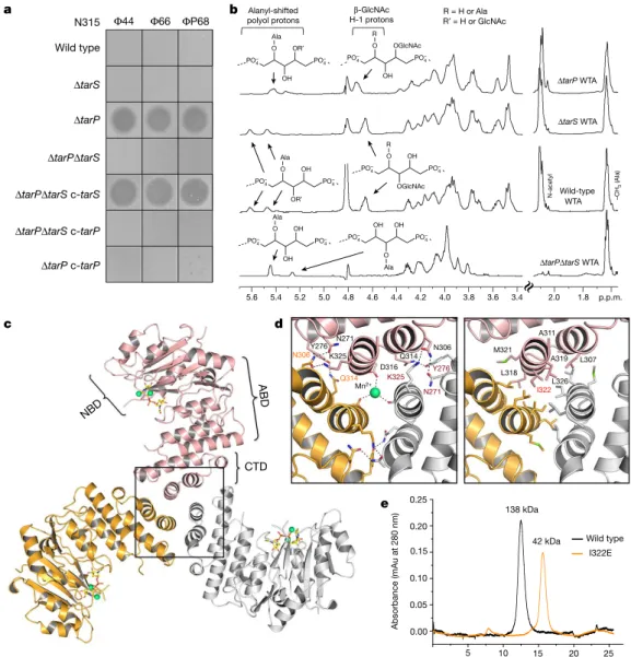
Fig. 1 | The phage-encoded TarP can replace the housekeeping WTA
β-GlcNAc transferase TarS. a, TarP is encoded next to different integrase
types (int gene) in prophages ϕtarP-Sa3int (with immune evasion
cluster scn, chp, sak, sep), found in HA-MRSA, and ϕtarP-Sa1int and
ϕtarP-Sa9int, identified in LA-MRSA. TarP variants in ϕtarP-Sa1int
and ϕtarP-Sa9int differed from TarP in ϕtarP-Sa3int in one amino acid
each (I8M and D296N, respectively). Both residues are distant from the
catalytic centre. b, Complementation of S. aureus RN4420 ΔtarM/S with
either tarS or tarP restores susceptibility to infection by WTA
GlcNAc-binding siphophages, as indicated by plaque formation on bacterial lawns.
Data shown are representative of three independent experiments. c, tarP
expression reduces siphophage Φ11-mediated transfer of SaPIbov in N315.
Values indicate the ratio of transduction units (TrU) to plaque-forming
units (PFU) given as mean ± s.d. of three independent experiments.
Statistical significances when compared to wild type were calculated by one-way ANOVA with Dunnett’s post-test (two-sided) and significant
P values (P≤ 0.05) are indicated. NO (none obtained) indicates no
obtained transductants.
2 9 N O V e M B e r 2 0 1 8 | V O L 5 6 3 | N A t U r e | 7 0 5
transferase TarS7 (Fig. 1a). tarP was found exclusively in isolates of the prominent HA-MRSA CC512, on a prophage that also encoded the
scn, chp and sak immune evasion genes13, and on two other prophages in the emerging LA-MRSAs CC39814 and CC515. All tarP-harbouring genomes also contained tarS.
When tarP from CC5 HA-MRSA strain N315 was expressed in a WTA glycosylation-deficient mutant of laboratory strain RN42207, it restored WTA glycosylation (Extended Data Fig. 1a) and suscep-tibility to siphophages, which need RboP WTA GlcNAc as a binding motif16 (Fig. 1b). The presence of β-GlcNAc on WTA is essential for full β-lactam resistance in MRSA strains7. When tarP was expressed in a WTA glycosylation-deficient mutant of CA-MRSA strain MW2 (CC1), it restored full oxacillin resistance (Extended Data Fig. 1b), confirming that tarP can replace tarS in several key interactions.
The expression of TarP led to susceptibility to siphophages, albeit to a lower extent than TarS (Extended Data Fig. 1c), although TarP did not
incorporate less GlcNAc into WTA than TarS (Extended Data Fig. 1d, Supplementary Table 3). Similarly, the siphophage-mediated horizontal transfer of an S. aureus pathogenicity island was reduced about tenfold in S. aureus N315 expressing tarP, compared to the same strain expressing only tarS (Fig. 1c), suggesting that TarP and TarS glycosylate WTA differently. Notably, N315 was resistant to podophages, but inac-tivation of tarP (but not of tarS) rendered it susceptible to podophages (Fig. 2a). We analysed the overall effect of tarP on podophage suscep-tibility patterns in 90 clinical CC5 and CC398 isolates and found that none of the tarP-containing strains, but all of the tarP-lacking strains, were susceptible to podophages (Extended Data Table 1). Thus, TarP causes podophage resistance and TarP-mediated modification of WTA is distinct from that mediated by TarS. Nuclear magnetic resonance (NMR) analyses revealed that both TarP and TarS add GlcNAc to WTA in the β-configuration. However, the attachment site in RboP differs: TarS glycosylates the C4 position17 whereas TarP attaches GlcNAc N315
ΔtarP
Wild type
ΔtarPΔtarS
ΔtarPΔtarS c-tarS
ΔtarPΔtarS c-tarP
ΔtarP c-tarP
Φ44 Φ66 ΦP68
ΔtarS
CTD
a b
N-acetyl
ΔtarPΔtarS WTA Wild-type
ΔtarS WTA
ΔtarP WTA
WTA –C
H3
(Ala
)
R = H or Ala R′ = H or GlcNAc
β-GlcNAc H-1 protons Alanyl-shifted
polyol protons
5.6 4.4 2.0
c d
e NBD
AB D
Elution volume (ml)
5 10 15 20 25
0.00 0.05 0.10 0.15 0.20
Absorbance (mAu at 280 nm
) Wild type I322E 138 kDa 42 kDa Y276 N271 N306 K325 Q314 D316
Mn2+ K325
Y276 N271
Q314 N306 M321
L318 A311 A319 L326 L307 I322 0.25 O OH OR′
PO4–
PO4–
Ala
O
OH OGlcNAc
PO4–
PO4–
R
O
OR′
OH
PO4–
PO4–
Ala O
OGlcNAc OH
PO4–
PO4–
R
O
OH OH
PO4–
PO4–
Ala
OH
O OH
PO4–
PO4–
Ala
4.6 4.8 5.0 5.2
5.4 4.2 4.0 3.8 3.6 3.4 1.8 p.p.m.
Fig. 2 | TarP protects N315 from podophage infection by alternative glycosylation of WTA at RboP C3. a, Expression of tarP renders N315 resistant to podophages. Representative data from three independent
experiments are shown. b, 1H NMR spectra reveal different ribitol
hydroxyl glycosylation of N315 WTA by TarS (C4) or TarP (C3). The RboP units with attached GlcNAc are depicted above the corresponding proton resonances. Representative data from three experiments are shown. In-depth description of the structural motifs identified in the spectra is given in the Supplementary Information. c, Crystal structure of TarP homotrimer (pink, orange, grey) bound to UDP-GlcNAc (yellow) and two
Mn2+ ions (lime green). The nucleotide-binding domain (NBD),
acceptor-binding domain (ABD), and C-terminal trimerization domain (CTD) of the pink monomer are labelled. d, Views into the trimer interface (boxed in c). Left, polar interactions. Hydrogen bonds and salt bridges are shown
as black dashed lines. The Mn2+ is 2.1 Å from each Asp316 carboxylate.
Right, hydrophobic interactions, with the mutated residue Ile322 highlighted in red. e, Size-exclusion chromatography elution profiles. Based on calibration of the column, the TarP wild-type and I322E mutant
proteins have estimated molecular weights of 138 kDa (n= 8) and 42 kDa
(n= 3), respectively, in agreement with the calculated molecular weights of
to C3 (Fig. 2b, Extended Data Fig. 2, Supplementary Table 2). This difference may be crucial for impairing phage infection. Moreover, NMR analysis revealed that TarP is dominant over TarS because in N315, which bears both genes, GlcNAc was almost exclusively attached to RboP C3 (Fig. 2b).
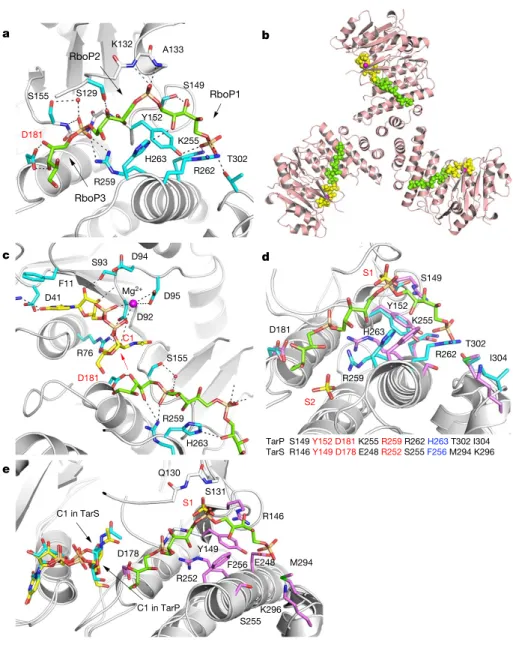
We solved the TarP structure at high resolution to elucidate how TarP generates a different glycosylation product from TarS. Like TarS18, TarP forms stable homotrimers, but it uses a different trimerization strategy because it lacks the C-terminal trimerization domain found in TarS (Fig. 2c, Extended Data Fig. 3). Instead, hydrophobic and polar inter-actions of a small helical C-terminal domain generate the TarP trimer (Fig. 2d, e). WTA polymers comprising three or six RboP repeating units (3RboP or 6RboP-(CH2)6NH2, respectively) were synthesized and used for soaking TarP crystals (Supplementary Information Fig. 2, 3), yielding the first protein structure visualizing the binding of a WTA-based polymer (Fig. 3, Extended Data Fig. 4). In the ternary complex TarP–UDP-GlcNAc–3RboP, the distance between the C3-hydroxyl of the third unit of 3RboP (RboP3) and the anomeric C1 of GlcNAc is 4.2 Å. Furthermore, at 3.1 Å, Asp181 is well within hydrogen bond-ing distance of the C3-hydroxyl of RboP3. The observed distances and geometry nicely explain the unusual glycosylation of WTA at the C3-hydroxyl. We propose that TarP uses a direct SN2-like glycosyltrans-ferase reaction, as discussed for other inverting GT-A fold enzymes19,20. In this mechanism, Asp181 would act as the catalytic base, deprotonat-ing the C3-hydroxyl on RboP3 and enabldeprotonat-ing a nucleophilic attack on
the GlcNAc C1, thus yielding a β-O-GlcNAcylated polyRboP (Fig. 3c). Mutagenesis of Asp181 to alanine rendered TarP inactive, supporting this putative mechanism (Extended Data Table 2).
The ternary structure of TarP–UDP-GlcNAc–3RboP allows us to predict how polyRboP binds to the homologous TarS enzyme. Three residues that are critical for binding and catalysis (including Asp181) are identical in TarP and TarS, while five other residues differ18 (Fig. 3d). Lys255 and Arg262, for instance, which interact electrostat-ically with a WTA phosphate group in TarP, are replaced with Glu248 and Ser255, respectively, in TarS, which may lead to reduced affinity for WTA and might explain why TarP is dominant over TarS in vivo. On the basis of the location of UDP-GlcNAc, the identical Tyr149, Asp178 and Arg252 side chains, the conserved aromatic side chain of Phe256, and a site that contains a bound sulfate ion from the crystallization solution (S1) and probably binds phosphate in TarS (Fig. 3e), the polyR-boP chain would be shifted to the upper right, and the relative position of RboP units in the binding site would be altered in TarS. Such an altered binding mode would move the C4-hydroxyl of the target RboP towards C1 of GlcNAc, allowing TarS to glycosylate at the C4 position.
S. aureus WTA is a dominant antigen for adaptive immune responses5,9. The observation that the position of GlcNAc on RboP had a profound impact on binding by podophage receptors raises the question of whether human antibodies also discriminate between the two isomeric polymers and whether MRSA clones use TarP to subvert immune recognition. We analysed several human antibody Y149
TarP S149 Y152D181 K255 R259 R262 H263 T302 I304 TarS R146 Y149D178 E248 R252 S255 F256 M294 K296
S1 D178 F256 R252 H263 R146 E248 M294 Q130 S131 K296 S255 S155 S129 Y152 S149 D181 C1 S1 S2 D181 R259 S149 Y152 K255 R262 T302
C1 in TarS
C1 in TarP
a b d c e I304 RboP1 F11 D41 S93 D94 D95 D92 R76 S155 R259 H263 D181 Mg2+ RboP2 RboP3 K132 A133 K255 T302 R262 H263 R259
Fig. 3 | Interactions of TarP with UDP-GlcNAc and d-ribitol-5-phosphate trimer (3RboP), and comparison of polyRboP binding sites of TarP and TarS. a, 3RboP
binding site in the TarP–3RboP complex, with key amino acids shown (cyan). Asp181 is highlighted in red. The ribitol of 3RboP is coloured green and d-ribitol-5-phosphate units 1, 2 and 3 (RboP1, RboP2, and RboP3) are labelled. Hydrogen bonds and salt bridges are shown as black dashed lines. b, Ternary complex of TarP with UDP-GlcNAc and 3RboP.
UDP-GlcNAc, Mg2+ and 3RboP are shown as
full-atom models coloured yellow, magenta, and green, respectively. c, View into the active site of TarP. C1 of UDP-GlcNAc and Asp181 are highlighted with red labels. The arrow indicates how the C3-hydroxyl in RboP3 could nucleophilically attack GlcNAc C1.
d, Comparison of the polyRboP-binding site
of TarP with the corresponding region in TarS. Residues of TarP and 3RboP are coloured as in
a. TarS residues are coloured violet and the two
sulfates are labelled S1 and S2. Only residues of TarP are labelled, for clarity. Key TarP and TarS residues lining the polyRboP-binding site are shown at the bottom, with three identical (red) and one conserved amino acids (blue).
e, Superposition of UDP-GlcNAc-bound TarS
with the ternary TarP–UDP-GlcNAc–3RboP complex. UDP-GlcNAc and 3RboP bound to TarP are coloured as in b, whereas UDP-GlcNAc bound to TarS is coloured in cyan. Only the TarS residues are shown (coloured as in d), for clarity. The arrows indicate the C1 positions of UDP-GlcNAc in TarP and TarS.
2 9 N O V e M B e r 2 0 1 8 | V O L 5 6 3 | N A t U r e | 7 0 7
preparations for their capacity to opsonize a panel of N315 strains with or without tarP and/or tarS. The mutant lacking any WTA glycosylation bound the lowest amount of IgG compared to WTA glycosylation-positive strains (Fig. 4a), demonstrating that glyco-sylated WTA is a prominent S. aureus antigen in humans. Exclusive expression of tarS led to strongly increased IgG binding compared to the glycosylation-deficient mutant, indicating that β-GlcNAc on RboP C4 is an important epitope for human anti-S. aureus anti-bodies. By contrast, expression of tarP in the presence or absence of tarS led to only slightly increased IgG binding compared to the glycosylation-deficient mutant. The capacity of TarP to impair the deposition of IgG on S. aureus differed with individual serum donors and reached average levels in pooled serum preparations (Fig. 4a). When tarP was deleted in three further CC5 isolates, they showed similarly increased capacities to bind human serum antibodies com-pared to the wild-type strains (Extended Data Fig. 1e). Additionally,
tarP deletion led to a substantially increased capacity of human neutrophils to phagocytose opsonized S. aureus (Fig. 4b, Extended Data Fig. 1g). Thus, only a small percentage of S. aureus-specific antibodies can bind WTA with β-GlcNAc on RboP C3, and tarP- expressing S. aureus are less likely to be detected and eliminated by human phagocytes.
We purified N315 WTA that had been glycosylated by TarS or TarP and used it to immunize mice. Antibodies binding to regular (TarS-modified) WTA increased continuously over three weeks after vaccination (Fig. 4c). By contrast, no or only very low amounts of IgG directed against TarP-glycosylated WTA emerged, indicating that WTA modified at RboP C3 is much less immunogenic than WTA modified at RboP C4. This experiment was repeated three times with three different WTA preparations and yielded broadly similar data.
Vaccination with S. aureus WTA bearing GlcNAc at RboP C4 protects mice against infection by CA-MRSA strains USA300 (CC8) or USA400 (CC1), which both lack tarP5,21. Remarkably, vaccination with regular (TarS-modified) or TarP-modified WTA did not lead to any notable protection against subsequent infection with tarP-expressing N315 compared to mock vaccination, despite the robust antibody response against regular WTA (Fig. 4d). Together, our results demon-strate that tarP protects S. aureus against adaptive host defences by allowing bacteria to evade recognition by preexisting anti-
S. aureus antibodies and by exploiting the poor immunogenicity of TarP-modified WTA.
It is possible that TarP-modified WTA mimics a currently unknown autoantigen and is therefore hardly immunogenic. On the other hand, regular S. aureus WTA can be ingested by antigen-presenting cells
50 100 150 0 0
IgG enriched for WTA Pooled IgG (Abcam) Pooled IgG (Athens R&T)
a 3,000 0 6,000 9,000 12,000 15,000
Wild typeΔ tarSΔtarP
ΔtarS Δtar P Phagocytotic ef ficiency (MFI) 0 0.15 ng μl–1 0.3 ng μl–1 b
Relative IgG deposition (%)
ΔtarSΔtarP ΔtarS
ΔtarP ΔtarSΔtarP
Δtar SΔtar
P c 0 0.5 1.0 1.5
d0 d7 d14d21d26
ΔtarP WTA
ΔtarS WTA
0.1 0.2 0 0.5 1.0
1.5 d0d21
1st experiment
2nd experiment
3rd experiment
Relative serum levels (absorbance at 450 nm)
d Alum ΔtarS + alum ΔtarP + alum 1 × 10²
1 × 10⁴ 1 × 10⁶ 1 × 10⁸
CFU per ml (kidney)
Wild type Wild type 0 50
100 150 200 0 50 100 150
50 100 150 Wild type
ΔtarS ΔtarP ΔtarSΔtarP ΔtarSΔtarP c-tarS ΔtarSΔtarP c-tarP
Wild type ΔtarS ΔtarP ΔtarSΔtarP ΔtarSΔtarP c-tarS ΔtarSΔtarP c-tarP
IgG (donor A) IgG (donor B) IgG (donor C)
50 100 0
P = 0.0149
2.0 d0 d21 0.3
0
d0 d7 d14 d21 d26 100 150 200
0
P < 0.0001
P < 0.0001
150 200
P < 0.0001
P < 0.0001
P = 0.0210
P < 0.0001
P = 0.0027
P = 0.0412 P = 0.0130
P < 0.0001
P = 0.0015
P < 0.0001
P = 0.0200
P = 0.0047
P = 0.0009
P = 0.0261
P = 0.0021
P = 0.0004 P = 0.0143
P = 0.0184
P P = 0.0143 = 0.0395
Wild type + alum
Fig. 4 | TarP attenuates immunogenicity of WTA. a, TarP expression
reduces deposition of IgG from human serum on N315 cells. The protein
A gene spa was deleted in all strains. Top, human IgG isolated from three
individual healthy donors (A, B, and C; n= 4); bottom, left, IgG from
human serum enriched for RN4220 WTA binding (n= 4); middle and
right, pooled human IgG from different suppliers (Abcam, n= 4; Athens
R&T, n= 6). Results were normalized against wild type and shown as
means with s.d. of n experiments. P values for comparison with wild type
were calculated by one-way ANOVA with Dunnett’s post-test (two-sided),
and P≤ 0.05 was considered significant. Significant P values are displayed.
b, TarP reduces neutrophil phagocytosis of N315 strains lacking protein
A, opsonized with indicated concentrations of IgG enriched for WTA binding. Values are depicted as mean fluorescence intensity (MFI). Means of two dependent replicates of a representative experiment are shown.
The other two representative experiments can be found in Extended Data Fig. 1g. c, TarP abrogates IgG response of mice towards WTA. For each
experiment, WTA from N315 ΔtarP or ΔtarS was isolated independently.
At least three mice per group were vaccinated and analysed for specific IgG at indicated time points after vaccination. Results are depicted as mean absorbance with s.d. Individual mice are indicated by colour. Increase in IgG levels was assessed by one-way ANOVA with Tukey’s
post-test (two-sided). Significant differences (P≤ 0.05) are indicated
with corresponding P values. d, Vaccination with WTA does not protect
mice against tarP-expressing N315, as shown for bacterial loads in kidney
and presented to T cells, in a largely unexplored way, thereby evoking specific immunoglobulins and immunological memory22,23. It is possi-ble that TarP-modified WTA is refractory to this process. Thus, TarS- and TarP-modified WTA could be helpful for decoding glycopolymer presentation pathways and for defining the most promising WTA epitopes for the development of protective vaccines against S. aureus.
Protection against S. aureus infections is urgently needed, in particu-lar for hospitalized and immunocompromised patients2,4. Antibodies can in principle protect against S. aureus, but their titres and specif-icities vary largely among humans and they are often not protective in immunocompromised patients3, probably in particular against
S. aureus clones that mask dominant epitopes, for instance using TarP. Unfortunately, all previous human vaccination attempts with protein or glycopolymer antigens have failed, for reasons that are unclear24. Our study identifies a new strategy used by pandemic MRSA clones to subvert antibody-mediated immunity, which should be considered in future vaccination approaches. S. aureus WTA with GlcNAc at RboP C3 has been reported as a type-336 antigen, but was not further explored25. We found that tarP is present in type-336 S. aureus (Extended Data Fig. 1f). However, TarP-modified WTA is a very poor antigen and vaccines directed against GlcNAc at WTA RboP C3 or C4 may fail against many of the pandemic MRSA clones. The structural character-ization of TarP will instruct the development of specific TarP inhibitors that could become important in combination with anti-WTA vaccines or antibiotic therapies. We found tarP-encoding prophages in 70–80% of south-west German HA-MRSA CC5 and 40% of Danish LA-MRSA CC398 isolates (Extended Data Table 1), pointing to a crucial role of
tarP in the fitness of these lineages and raising concerns of further dissemination by horizontal gene transfer. TarP is a new and probably crucial component of the S. aureus virulence factor arsenal26,27, high-lighting the important roles of adaptive immunity and its evasion in
S. aureus infections.
Online content
Any methods, additional references, Nature Research reporting summaries, source data, statements of data availability and associated accession codes are available at https://doi.org/10.1038/s41586-018-0730-x.
Received: 28 June 2018; Accepted: 18 October 2018; Published online 21 November 2018.
1. Tong, S. Y., Davis, J. S., Eichenberger, E., Holland, T. L. & Fowler, V. G. Jr. Staphylococcus aureus infections: epidemiology, pathophysiology, clinical manifestations, and management. Clin. Microbiol. Rev. 28, 603–661 (2015). 2. Lee, A. S. et al. Methicillin-resistant Staphylococcus aureus. Nat. Rev. Dis. Primers
4, 18033 (2018).
3. Stentzel, S. et al. Specific serum IgG at diagnosis of Staphylococcus aureus bloodstream invasion is correlated with disease progression. J. Proteomics 128, 1–7 (2015).
4. Missiakas, D. & Schneewind, O. Staphylococcus aureus vaccines: deviating from the carol. J. Exp. Med. 213, 1645–1653 (2016).
5. Lehar, S. M. et al. Novel antibody–antibiotic conjugate eliminates intracellular
S. aureus. Nature 527, 323–328 (2015).
6. Weidenmaier, C. & Peschel, A. Teichoic acids and related cell-wall glycopolymers in Gram-positive physiology and host interactions. Nat. Rev.
Microbiol. 6, 276–287 (2008).
7. Brown, S. et al. Methicillin resistance in Staphylococcus aureus requires glycosylated wall teichoic acids. Proc. Natl Acad. Sci. USA 109, 18909–18914 (2012).
8. Tacconelli, E. et al. Discovery, research, and development of new antibiotics: the WHO priority list of antibiotic-resistant bacteria and tuberculosis. Lancet Infect.
Dis. 18, 318–327 (2018).
9. Kurokawa, K. et al. Glycoepitopes of staphylococcal wall teichoic acid govern complement-mediated opsonophagocytosis via human serum antibody and mannose-binding lectin. J. Biol. Chem. 288, 30956–30968 (2013). 10. Lee, J. H. et al. Surface glycopolymers are crucial for in vitro anti-wall teichoic
acid IgG-mediated complement activation and opsonophagocytosis of
Staphylococcus aureus. Infect. Immun. 83, 4247–4255 (2015).
11. Winstel, V. et al. Wall teichoic acid structure governs horizontal gene transfer between major bacterial pathogens. Nat. Commun. 4, 2345 (2013). 12. Nübel, U. et al. Frequent emergence and limited geographic dispersal of
methicillin-resistant Staphylococcus aureus. Proc. Natl Acad. Sci. USA 105, 14130–14135 (2008).
13. McCarthy, A. J. & Lindsay, J. A. Staphylococcus aureus innate immune evasion is lineage-specific: a bioinfomatics study. Infect. Genet. Evol. 19, 7–14 (2013).
14. Bal, A. M. et al. Genomic insights into the emergence and spread of international clones of healthcare-, community- and livestock-associated meticillin-resistant Staphylococcus aureus: Blurring of the traditional definitions.
J. Glob. Antimicrob. Resist. 6, 95–101 (2016).
15. Hau, S. J., Bayles, D. O., Alt, D. P., Frana, T. S. & Nicholson, T. L. Draft genome sequences of 63 swine-associated methicillin-resistant Staphylococcus aureus sequence type 5 isolates from the United States. Genome Announc. 5, e01081-17 (2017).
16. Xia, G. et al. Wall teichoic acid-dependent adsorption of staphylococcal siphovirus and myovirus. J. Bacteriol. 193, 4006–4009 (2011).
17. Vinogradov, E., Sadovskaya, I., Li, J. & Jabbouri, S. Structural elucidation of the extracellular and cell-wall teichoic acids of Staphylococcus aureus MN8m, a biofilm forming strain. Carbohydr. Res. 341, 738–743 (2006).
18. Sobhanifar, S. et al. Structure and mechanism of Staphylococcus aureus TarS, the wall teichoic acid β-glycosyltransferase involved in methicillin resistance.
PLoS Pathog. 12, e1006067 (2016).
19. Lairson, L. L., Henrissat, B., Davies, G. J. & Withers, S. G. Glycosyltransferases: structures, functions, and mechanisms. Annu. Rev. Biochem. 77, 521–555 (2008).
20. Kozmon, S. & Tvaroska, I. Catalytic mechanism of glycosyltransferases: hybrid quantum mechanical/molecular mechanical study of the inverting
N-acetylglucosaminyltransferase I. J. Am. Chem. Soc. 128, 16921–16927
(2006).
21. Takahashi, K. et al. Intradermal immunization with wall teichoic acid (WTA) elicits and augments an anti-WTA IgG response that protects mice from methicillin-resistant Staphylococcus aureus infection independent of mannose-binding lectin status. PLoS One 8, e69739 (2013).
22. Weidenmaier, C., McLoughlin, R. M. & Lee, J. C. The zwitterionic cell wall teichoic acid of Staphylococcus aureus provokes skin abscesses in mice by a novel CD4+ T-cell-dependent mechanism. PLoS One 5, e13227 (2010).
23. Wanner, S. et al. Wall teichoic acids mediate increased virulence in
Staphylococcus aureus. Nat. Microbiol. 2, 16257 (2017).
24. Pozzi, C. et al. Vaccines for Staphylococcus aureus and target populations. Curr.
Top. Microbiol. Immunol. 409, 491–528 (2017).
25. Fattom, A., Sarwar, J., Kossaczka, Z., Taylor, K. & Ennifar, S. Method of protecting against staphylococcal infection. US Patent US20060228368A1 (2006). 26. Thammavongsa, V., Kim, H. K., Missiakas, D. & Schneewind, O. Staphylococcal
manipulation of host immune responses. Nat. Rev. Microbiol. 13, 529–543 (2015).
27. Spaan, A. N., Surewaard, B. G., Nijland, R. & van Strijp, J. A. Neutrophils versus
Staphylococcus aureus: a biological tug of war. Annu. Rev. Microbiol. 67, 629–650
(2013).
Acknowledgements We thank S. Popovich and P. Kühner for technical assistance; E. Weiß for help with phagocytosis experiments; R. Rosenstein and X. Li for discussions; B. Blaum and G. Zocher for assistance with NMR analysis and support for structure phasing and discussion; and the Swiss Lightsource beamline staff of the Paul Scherrer Institute for beam time and technical support. This work was financed by grants from the German Research Foundation to A.P. (TRR34, CRC766, TRR156, RTG1708), T.S. (TRR34, CRC766), C.W. (TRR34, CRC766, TRR156, RTG1708), and G.X. (CRC766); the German Center of Infection Research to A.P. (HAARBI); the Ministry of Science and Technology, Thailand Government to W.S.; the Korean Drug Development Foundation to S.-H.K. and B.L.L. (KDDF-201703-1); and the Max-Planck-Society to P.H.S.
Reviewer information Nature thanks M. Crispin, F. DeLeo, M. Gilmore and J. Zimmer for their contribution to the peer review of this work.
Author contributions D.G. characterized TarP in vivo and its genomic context, created mutants, designed experiments, purified WTA, and performed experiments with human IgGs. Y.G. designed experiments, purified proteins, crystallized proteins, solved the structures, and performed in vitro analysis of TarP. C.D.C. performed NMR experiments. C.D.C. and A.M. analysed the NMR data and wrote the NMR discussion. S.-H.K. performed and B.L.L. designed and interpreted mouse immunization and infection experiments. K.S. designed IgG deposition experiments. B.S. and C.W. collected and characterized CC5 MRSA strains. J.L. collected and characterized CC398 strains. J.L. and C.W. analysed
S. aureus genomes. F.-F.X, C.P., and P.H.S. designed and synthesized 3RboP.
S.A. and J.C. designed and synthesized 6RboP-(CH2)6NH2. W.S. performed MIC
experiments. G.X. identified tarP, and characterized and interpreted MIC data. D.G., Y.G., A.P., T.S., and G.X. designed the study, analysed results, and wrote the paper.
Competing interests The authors declare no competing interests.
Additional information
Extended data is available for this paper at https://doi.org/10.1038/s41586-018-0730-x.
Supplementary information is available for this paper at https://doi.org/ 10.1038/s41586-018-0730-x.
Reprints and permissions information is available at http://www.nature.com/ reprints.
Correspondence and requests for materials should be addressed to T.S. or A.P. Publisher’s note: Springer Nature remains neutral with regard to jurisdictional claims in published maps and institutional affiliations.
2 9 N O V e M B e r 2 0 1 8 | V O L 5 6 3 | N A t U r e | 7 0 9
MEthodS
No statistical methods were used to predetermine sample size. The experiments were not randomized. The investigators were not blinded to allocation during experiments and outcome assessment.
Bacterial strains and growth conditions. S. aureus strains N315, RN4220, and MW2 (wild type and mutants) were used for this study. Collections of CC5 isolates
of the Rhine-Hesse pulsed-field gel electrophoresis type28 and of the LA-MRSA
lineage CC398 from the Danish Statens Serum Institut29,30 were analysed for the
presence of tarP and for podophage susceptibility. Additionally, 48 spa-type t002
(ST5) and 16 spa-type t003 (ST225) isolates were obtained from the MRSA
collec-tion of the University Hospital Tübingen and analysed for tarP presence by PCR.
S. aureus strains were cultivated in tryptic soy broth (TSB) or basic medium (BM;
1% tryptone, 0.5% yeast extract, 0.5% NaCl, 0.1% glucose, 0.1% K2HPO4, w/v).
MICs of oxacillin were determined by microbroth dilution according to established
guidelines31.
Experiments with phages. tarP-encoding phages were identified in genome
sequences using the webtool Phaster32 in representative strains listed with
GenBank accession: ΦtarP-Sa3int with immune evasion cluster (IEC) in
CC5 (strain N315, BA000018.3), ΦtarP-Sa1int, found in LA-MRSA of CC5
(strain ISU935, CP017090), and ΦtarP-Sa9int found in CC398 (strain E154,
CP013218).
Phage susceptibility was determined using a soft-agar overlay method16. In brief,
10 µl phage lysate of 104–106 PFU was dropped onto soft agar containing 100
µl bacterial suspension (OD600 of 0.1). Plates were incubated at 37 °C overnight.
The efficiency of plating was determined as described33. Transfer of SaPIs was
determined according to previously described methods11. In brief, SaPI particle
lysates were generated from S. aureus strain JP1794, which encodes a SaPI with a
resistance marker for tetracycline34. PFU of SaPI lysate was determined on RN4220.
200 µl bacterial culture (OD600 of 0.5) was mixed with 100 µl of SaPI particle lysate
(SaPIbov1 (Φ11), 106 PFU/ml), incubated at 37 °C for 15 min. Appropriate
dilu-tions were plated on TSB plates containing 3 µg/ml of tetracycline, and CFU were
checked after overnight incubation.
WTA isolation and structure analysis. WTA from S. aureus was isolated and
purified according to previously described methods11. In brief, WTA was released
from purified peptidoglycan by treatment with 5% trichloroacetic acid and dialy-sed extensively against water using a Spectra/Por3 dialysis membrane (MWCO of 3.5 kDa; VWR International GmbH). Obtained soluble WTA was quantified by
determining the content of phosphate35 and GlcNAc36. For PAGE analysis of WTA,
samples (400 nmol of phosphate per lane) were applied to a 26% polyacrylamide
(Rotiphorese Gel 40 (19:1)) resolving gel and separated at 25 mA for 16 h37. The
gel was equilibrated in a solution of 40% ethanol and 5% acidic acid at room tem-perature for 1 h and the WTA ladders were visualized by incubation with alcian blue (0.005%) for several hours.
NMR spectroscopy experiments were carried out on a Bruker DRX-600 spectrometer equipped with a cryo-probe, at 288 K (WT-WTA, TarS-WTA, and TarP-WTA) or 298 K (double-mutant WTA lacking any glycosylation). Chemical
shifts of spectra recorded in D2O were calculated in p.p.m. relative to internal
acetone (2.225 and 31.45 p.p.m.). The spectral width was set to 10 p.p.m. and the frequency carrier placed at the residual HOD peak, suppressed by pre-saturation. Two-dimensional spectra (TOCSY, gHSQC, gHMBC, and HSQC-TOCSY) were measured using standard Bruker software. For all experiments, 512 FIDs of 2,048 complex data points were collected, 32 scans per FID were acquired for homo-nuclear spectra, and 20 or 100 ms of mixing time was used for TOCSY spectra.
Heteronuclear 1H-13C spectra were measured in the 1H-detected mode, gHSQC
spectrum was acquired with 40 scans per FID, the GARP sequence was used for
13C decoupling during acquisition; gHMBC scans doubled those of gHSQC
spec-trum. As for HSQC-TOCSY, the multiplicity editing during selection step version was used, scans tripled those of the HSQC spectrum and two experiments were acquired by setting the mixing time to 20 or 80 ms. During processing, each data
matrix was zero-filled in both dimensions to give a matrix of 4K × 2K points
and was resolution-enhanced in both dimensions by a cosine-bell function before Fourier transformation; data processing and analysis were performed with the Bruker Topspin 3 program.
Molecular biology. All primers used for PCR, cloning, and mutagenesis are
listed in Supplementary Table 1. tarP (UniProt A0A0H3JNB0, NCBI Gene ID
1260584) was amplified using genomic DNA of S. aureus N315 and inserted in
Eschericha coli/S. aureus shuttle vector pRB47438 at the BamHI and SacI sites, to
transform S. aureus, or into pQE80L at BamH1 and HindIII sites, to transform
E. coli BL21(DE3). A thrombin cleavage site was inserted between the His-tag and mature protein in pQE80L. Single mutations of TarP were introduced by
PCR-based site-directed mutagenesis39. The obtained amplicons were confirmed by
sequencing. For the construction of marker-less S. aureus deletion mutants of tarS
or tarP, the pIMAY shuttle vector was used40. The IgG-binding surface protein A
gene (spa) was deleted using the pKORI shuttle vector41. Protein A deletion had
no impact on phage siphophage or podophage susceptibility, indicating that it did not alter WTA amount or structure.
Protein expression, purification, and activity assay. E. coli BL21(DE3) were grown
in LB medium at 30 °C. Expression of tarP was induced with 1 mM IPTG at 22 °C at
an OD600 of 0.6. After 15 h, cells were harvested, washed with wash buffer (50 mM
Tris-HCl, pH 8.0, 1 mM EDTA), and lysed by sonication with lysis buffer (70 mM
NaH2PO4, pH 8.0, 1 M NaCl, 20% glycerol, 10 U/ml of benzonase nuclease).
After centrifugation (15,000g), the supernatant was filtered with a 0.45 µm filter,
loaded onto a His Trap FF column (GE Healthcare, 5 ml), and washed with buffer
A (50 mM NaH2PO4, pH 8.0, 1 M NaCl, 20% glycerol) supplemented with 45 mM
imidazole and buffer B (buffer A with 90 mM imidazole). Finally, the protein was eluted with buffer C (buffer A with 500 mM imidazole), and the fractions were pooled, and further purified by size-exclusion chromatography on a Superdex 200 10/30 column equilibrated with buffer D (20 mM MOPS, pH 7.6, 400 mM
LiCl, 10 mM MgCl2, 5 mM β-mercaptoethanol, 5% glycerol). The peak fractions
were pooled and concentrated to 1.4 mg/ml for crystallization. For selenomethio-nyl-form TarP production, bacteria were grown in a selenomethionine-containing medium (Molecular Dimension) and auto-induction was carried out. The protein was purified as described above. The activity of wild-type and mutated TarP, as well as donor substrate specificity of TarP were determined with the ADP Quest Assay
kit (DiscoverRx, Extended Data Tables 2, 3). The reaction volume was 20 µl with
1 mM UDP-GlcNAc, 1.5 mM purified WTA from RN4220 ΔtarM/S. The reaction
was started with protein and incubated at room temperature for 1 h. Released UDP, coupled into a fluorescence signal, was detected in a 384-well black assay plate with 530 nm excitation and 590 nm emission wavelengths using TECAN Infinite M200.
Crystallization and data collection. Crystals were obtained by vapour diffusion
at 20 °C. 1 µl protein solution was mixed with 1 µl reservoir solution containing
25% PEG 3350, 250 mM MgCl2, and 0.1 M sodium citrate, pH 5.7. The
selenome-thionyl-form protein was crystallized under the same conditions. For crystals of TarP with UDP-GlcNAc, 27 mM UDP-GlcNAc was introduced in the reservoir
solution containing 250 mM MgCl2 or 230 mM MnCl2. Crystals of TarP with Mg2+
were used for soaking of synthetic 3RboP (60 mM), 6RboP-(CH2)6NH2 (41 mM),
or UDP-GlcNAc (20 mM) combined with 3RboP (52 mM) for 5 min. For data collection the crystals were cryo-protected with 20% glycerol in reservoir solution and flash-frozen in liquid nitrogen. Diffraction data were collected at beamline X06DA of Swiss Light Source in Villigen, Switzerland, or at beamline BL14.1 at BESSY-II, Helmholtz Zentrum Berlin.
Phasing, model building, and refinement. For phase determination, two data sets
from a selenomethionine-containing TarP crystal were collected at wavelengths of 0.97941 Å (peak) and 0.97952 Å (inflection). The structure was solved by multi- wavelength anomalous dispersion (MAD) at 2.60 Å resolution. All data were
reduced using XDS/XSCALE software packages42. Initial phases were derived from
the substructure of 26 selenium atom sites per asymmetric unit with the program
suite SHELX C/D/E43. The heavy atom parameters were further refined and the
initial phases were improved by SHARP/autoSHARP44. The initial model was
generated with PHENIX45 and the final model was achieved by cycles of iterative
model modification using COOT46, and restrained refinement with REFMAC. TLS
was used in the later stages47,48. The four binary and one ternary complex structures
were solved by molecular replacement using PHASER49 and the unliganded TarP
structure was used as a search model. UDP-GlcNAc, 3RboP, Mg2+, or Mn2+ were
removed from the models to calculate the simulated annealing (mFo − DFc) omit
maps using PHENIX. The anomalous difference map of Mn2+ at 1.89259 Ǻ was
generated by FFT within CCP4, from which two Mn2+ in the active site and one
Mn2+ at the trimer interface were identified. The coordinate and parameter files
for 3RboP and 6RboP-(CH2)6NH2 were calculated using the PRODRG server50.
The structure figures were generated by PyMOL51 and the models were evaluated
using MolProbity52. Statistics for the data collection, phasing, and refinement are
reported in Extended Data Tables 4 and 5.
Synthesis of ribitol phosphate oligomers. Synthesis of 3RboP. Target compound
1, d-ribitol-5-phosphate trimer (3RboP), was prepared by the phosphoramidite
method53,54 (Supplementary Fig. 2). In brief, the primary alcohol of commercially
available compound 2 was converted into levulinoyl ester by using levulinic acid
and N,N'-dicyclohexylcarbodiimide (DCC), and the allyl group of 3 was removed
with tetrakis(triphenylphosphine)palladium to produce compound 4. The primary alcohol of 4 reacted with phosphine derivative 5 in the presence of
diisopropylam-monium tetrazolide55 to generate phosphoramidite 6. At the same time, compound 4
was coupled with dibenzyl N,N-diisopropylphosphoramidite 7, which was
cata-lysed by 1H-tetrazole, and the product was further oxidized by tert-butyl
hydrop-eroxide, yielding protected d-ribitol-5-phosphate 8. Cleavage of the levulinoyl ester of 8 with hydrazine hydrate resulted in benzyl protected d-ribitol-5-phosphate 9,
which was further coupled with phosphoramidite 6 and oxidized with tert-butyl
12. Subsequent removal of the levulinoyl group and hydrogenolysis of 13 to remove
all benzyl groups yielded 3RboP 1. All chemicals and experimental procedures as well as characterization of products can be found in the Supplementary Methods.
Synthesis of 6RboP-(CH2)6NH2. Aminohexyl d-ribitol-5-phosphate hexamer
(6RboP-(CH2)6NH2) was synthesized using a new method (Supplementary Fig. 3).
All chemicals (Acros, Biosolve, Sigma-Aldrich and TCI) for the synthesis were used as received and all reactions were performed under a protective argon atmosphere at room temperature, unless otherwise stated. Procedures for phosphoramidite coupling, oxidation, detritylation, global deprotection, TLC analysis and charac-terization of these compounds can be found in Supplementary Methods.
Human samples. Venous blood samples were obtained from male and female
healthy volunteers (20–50 years) with protocols approved by the Institutional Review Board for Human Subjects at the University of Tübingen (014/2014BO2 und 549/2018BO2). Informed written consent was obtained from all volunteers. Blood samples were used for purification of either serum IgGs or neutrophils as described below.
IgG from human plasma. IgG was purified from plasma of human donors using
the NAb Protein G Spin Kit (ThermoFisher), purity was checked by SDS PAGE, and protein concentration was determined using Bradford assay. Anti-WTA-IgG was
prepared as described9. To analyse the IgG-binding capacity of S. aureus cells,
expo-nentially growing bacterial cultures were adjusted to an OD600 of 0.5, diluted 1:10
in PBS, and 100 µl of diluted bacteria was mixed with 100 µl of IgG diluted in PBS
with 1% BSA. The concentration of IgG was 250 ng/ml for IgG enriched for WTA
binding, 10 µg/ml for IgG from pooled human serum (Athens R&T 16-16-090707,
Abcam ab98981), or 5 µg/ml for single-donor IgG preparations. A control without
IgG was included in all experiments for all mutants. Samples were incubated at 4 °C for 1 h, centrifuged, washed 2–3 times with PBS, and further incubated with
100 µl FITC-labelled anti-human IgG (Thermo Scientific, 62-8411, 1:100 in PBS
with 1% BSA, 62-8411) at 4 °C for 1 h. Bacteria were centrifuged, washed 2–3 times with PBS, and fixed with 2% paraformaldehyde (PFA). Surface-bound IgG was quantified by flow cytometry using a BD FACSCalibur. For all flow cytometry
experiments a mutant panel lacking spa, the gene for the IgG-binding protein A,
was used. The subsequent gating strategy is exemplified in Extended Data Fig. 5a.
IgG-mediated phagocytosis. Stationary-phase S. aureus cells were washed once
with PBS and labelled by incubation in PBS containing 10 µM carboxyfluorescein
succinimidyl ester (CFSE; OD600 of 1.7) at 37 °C for 1 h. The bacteria were washed
three times and resuspended in PBS. CFU were determined by plating on TSB plates and bacteria were heat-inactivated at 70 °C for 20 min. CFSE-labelled
S. aureus (1 × 10⁷ cells/ml) in PBS with 0.5% BSA were opsonized with
anti-WTA-IgG (0.15 or 0.3 ng/µl) at 4 °C for 40 min. Neutrophils from human donors, isolated
via Ficoll-Histopaque density gradient centrifugation56, were diluted to a
con-centration of 2.5 × 10⁶/ml in neutrophil medium (10% HSA, 2 mM l-glutamine,
2 mM sodium pyruvate, 10 mM HEPES). 200 µl neutrophil suspension was
incu-bated with 25 µl opsonized bacteria (final MOI 0.5) in a 96-well plate at 37 °C for
30 min, centrifuged (350g, 10 min), washed once with 200 µl PBS, and fixed with
2% PFA at room temperature for 15 min. Cells were washed twice with PBS and analysed by flow cytometry, whereby surface-bound and ingested bacteria were measured without discrimination. An example of the neutrophil gating strategy can be found in Extended Data Fig. 5b.
Mice. Six-week-old sex-matched wild-type C57BL/6J mice, purchased from
ORIENT BIO (Charles River Breeding Laboratories in Korea), were kept in micro-isolator cages in a pathogen-free animal facility. The conducted experi-ments were performed according to guidelines and approval (PNU-2017-1503) by the Pusan National University-Institutional Animal Care and Use Committee (PNU-IACUC). Sample size was chosen to obtain significant outcomes (alpha
error ≤ 5%), based on results from previous experiments21. Experiments were
performed in a non-blinded, non-randomized fashion.
Mouse vaccination and infection. 30 µg of purified WTA from S. aureus N315
wild-type or isogenic ΔtarP, or ΔtarS mutants was dissolved in 15 µl PBS and
mixed with the same volume of aluminium hydroxide gel adjuvant (Alhydrogelr 1.3%, 6.5 mg/ml, Brennatag). The mixtures were incubated at 37 °C with agitation for 1 h and injected three times at one-week intervals via mouse footpads. Seven days after the third injection, blood was obtained from the retro-orbital sinus
and centrifuged (9,000g) at 4 °C for 10 min. The supernatants were aliquoted
(50 µl) and stored at –80 °C for ELISA quantification of WTA-binding IgG as
described57. Sera were diluted 1:100 and tested by ELISA on 96-well plates coated
with 2.5 µg/ml sonicated WTA preparations (WTA from N315, ΔtarS or ΔtarP,
respectively).
To prepare an inoculum for infection, N315 wild-type bacteria were grown in
TBS at 37 °C with agitation (180 r.p.m.) until they reached an OD600 of 1.0. After
centrifugation (3,500g) at 4 °C for 10 min, bacteria adjusted to 5 × 10⁷ CFU in 50 µl
PBS containing 0.01% BSA were intravenously injected (n= 5 per group). Injected
bacterial numbers were verified by plating serial dilutions of the inoculum onto TSA plates. To determine residual bacterial dissemination to kidneys, challenged
mice were euthanized, and organs were extracted aseptically and homogenized in 1 ml of saline using a Polytron homogenizer (PT3100). The homogenates were serially diluted and plated on TSA to determine CFU counts. CFU were calculated per 1 ml of kidney.
Statistical analyses. Statistical analysis was performed by using GraphPad Prism
(GraphPad Software, Inc.). Statistically significant differences were calculated by
appropriate statistical methods as indicated. P values of ≤ 0.05 were considered
significant.
Reporting summary. Further information on experimental design is available in
the Nature Research Reporting Summary linked to this paper.
Data availability
All major data generated or analysed in this study are included in the article or its supplementary information files. The coordinates and structure factors were deposited in the Protein Data Bank under accession numbers 6H1J, 6H21, 6H2N, 6H4F, 6H4M and 6HNQ. Source data for experiments with animals (Fig. 4c, d) are provided. Additionally, a gel image of Extended Data Fig. 1f is supplied as Supplementary Fig. 1. All other data relating to this study are available from the corresponding authors on reasonable request.
28. Schulte, B., Bierbaum, G., Pohl, K., Goerke, C. & Wolz, C. Diversification of clonal complex 5 methicillin-resistant Staphylococcus aureus strains (Rhine-Hesse clone) within Germany. J. Clin. Microbiol. 51, 212–216 (2013).
29. Larsen, J. et al. Meticillin-resistant Staphylococcus aureus CC398 is an increasing cause of disease in people with no livestock contact in Denmark, 1999 to 2011. Euro Surveill. 20, 30021 (2015).
30. Sieber, R. N. et al. Drivers and dynamics of methicillin-resistant livestock-associated Staphylococcus aureus CC398 in pigs and humans in Denmark.
mBio 9, e02142-18 (2018).
31. European Committee for Antimicrobial Susceptibility Testing (EUCAST) of the European Society of Clinical Microbiology and Infectious Diseases (ESCMID). Determination of minimum inhibitory concentrations (MICs) of antibacterial agents by broth dilution. Clin. Microbiol. Infect. 9, ix–xv (2003).
32. Arndt, D. et al. PHASTER: a better, faster version of the PHAST phage search tool. Nucleic Acids Res. 44, W16–W21 (2016).
33. Winstel, V., Sanchez-Carballo, P., Holst, O., Xia, G. & Peschel, A. Biosynthesis of the unique wall teichoic acid of Staphylococcus aureus lineage ST395. MBio 5, e00869 (2014).
34. Tormo, M. A. et al. Staphylococcus aureus pathogenicity island DNA is packaged in particles composed of phage proteins. J. Bacteriol. 190, 2434–2440 (2008).
35. Chen, P. S., Toribara, T. Y. & Warner, H. Microdetermination of phosphorus. Anal.
Chem. 28, 1756–1758 (1956).
36. Smith, R. L. & Gilkerson, E. Quantitation of glycosaminoglycan hexosamine using 3-methyl-2-benzothiazolone hydrazone hydrochloride. Anal. Biochem. 98, 478–480 (1979).
37. Xia, G. et al. Glycosylation of wall teichoic acid in Staphylococcus aureus by TarM.
J. Biol. Chem. 285, 13405–13415 (2010).
38. Brückner, R. A series of shuttle vectors for Bacillus subtilis and Escherichia coli.
Gene 122, 187–192 (1992).
39. Liu, H. & Naismith, J. H. An efficient one-step site-directed deletion, insertion, single and multiple-site plasmid mutagenesis protocol. BMC Biotechnol. 8, 91 (2008).
40. Monk, I. R., Shah, I. M., Xu, M., Tan, M. W. & Foster, T. J. Transforming the untransformable: application of direct transformation to manipulate genetically
Staphylococcus aureus and Staphylococcus epidermidis. MBio 3, e00277-11
(2012).
41. Bae, T. & Schneewind, O. Allelic replacement in Staphylococcus aureus with inducible counter-selection. Plasmid 55, 58–63 (2006).
42. Kabsch, W. Xds. Acta Crystallogr. D Biol. Crystallogr. 66, 125–132 (2010). 43. Sheldrick, G. M. Experimental phasing with SHELXC/D/E: combining chain
tracing with density modification. Acta Crystallogr. D Biol. Crystallogr. 66, 479–485 (2010).
44. Vonrhein, C., Blanc, E., Roversi, P. & Bricogne, G. Automated structure solution with autoSHARP. Methods Mol. Biol. 364, 215–230 (2007).
45. Adams, P. D. et al. PHENIX: a comprehensive Python-based system for macromolecular structure solution. Acta Crystallogr. D Biol. Crystallogr. 66, 213–221 (2010).
46. Emsley, P., Lohkamp, B., Scott, W. G. & Cowtan, K. Features and development of Coot. Acta Crystallogr. D Biol. Crystallogr. 66, 486–501 (2010).
47. Murshudov, G. N. et al. REFMAC5 for the refinement of macromolecular crystal structures. Acta Crystallogr. D Biol. Crystallogr. 67, 355–367 (2011).
48. Murshudov, G. N., Vagin, A. A. & Dodson, E. J. Refinement of macromolecular structures by the maximum-likelihood method. Acta Crystallogr. D Biol.
Crystallogr. 53, 240–255 (1997).
49. McCoy, A. J. et al. Phaser crystallographic software. J. Appl. Crystallogr. 40,
658–674 (2007).
50. Schüttelkopf, A. W. & van Aalten, D. M. PRODRG: a tool for high-throughput crystallography of protein-ligand complexes. Acta Crystallogr. D Biol. Crystallogr. 60, 1355–1363 (2004).
51. Schrodinger, LLC. The PyMOL Molecular Graphics System, Version 1.8 (2015). 52. Chen, V. B. et al. MolProbity: all-atom structure validation for macromolecular
crystallography. Acta Crystallogr. D Biol. Crystallogr. 66, 12–21 (2010).
53. Beaucage, S. L. & Caruthers, M. H. Deoxynucleoside phosphoramidites—a new class of key intermediates for deoxypolynucleotide synthesis. Tetrahedr. Lett. 22, 1859–1862 (1981).
54. Elie, C. J. J. et al. Synthesis of fragments of the capsular polysaccharide of
Haemophilus influenzae type b: Part IIII-3. A solid-phase synthesis of a
spacer-containing ribosylribitol phosphate hexamer. Recl. Trav. Chim. Pays Bas 108, 219–223 (1989).
55. Dreef, C. E., Elie, C. J. J., Hoogerhout, P., van der Marel, G. A. & van Boom, J. H. Synthesis of 1-O-(1,2-di-O-palmitoyl-sn-glycero-3-phospho)-d-myo-inositol
4,5-bisphosphate: an analogue of naturally occurring (ptd)Ins(4,5)P2. Tetrahedr. Lett. 29, 6513–6515 (1988).
56. Dürr, M. C. et al. Neutrophil chemotaxis by pathogen-associated molecular patterns—formylated peptides are crucial but not the sole neutrophil attractants produced by Staphylococcus aureus. Cell. Microbiol. 8, 207–217 (2006).
Extended Data Fig. 1 | See next page for caption.
Extended Data Fig. 1 | Characterization of TarP, deposition of human IgGs, and presence of tarP in the producer of antigen 336. a, Analysis
of WTA by PAGE. WTA from RN4220 ΔtarM/S expressing either tarP
or tarS was compared with non-glycosylated WTA. Data shown are
representative of two experiments. b, MIC values of oxacillin against
MW2 wild type, tarS mutant, and tarP-complemented tarS mutant. Data
are medians of ten independent experiments. c, Efficiency of plating
(EOP) of phage Φ11 against tarS or tarP-expressing RN4420 ΔtarM/S.
Values of tarP relative to tarS expression are given as mean ± s.d. (n= 3).
Statistical significance was calculated by paired Student’s t-test
(two-sided) with significant P values (P≤ 0.05) indicated. d, The level of WTA
glycosylation catalysed by TarP or TarS was determined by analysing the GlcNAc and phosphate content of WTA isolated from a N315 strain panel. Depicted is the ratio of GlcNAc and phosphate as mean with s.d. of three technical replicates. The values are in good agreement with NMR data
(Supplementary Table 3). e, Relative deposition of IgG from intravenous immunoglobulins enriched for WTA binding on different CC5
wild-type and tarP mutant cells. Values are given as mean percentage ± s.d.
of four independent experiments. Statistical significance was calculated
by paired Student’s t-test (two-sided). P values ≤ 0.05 were considered
significant and are indicated. f, Presence of tarP and tarS in S. aureus
ATCC55804, expressing antigen 336, described as 3-O-GlcNAc-WTA25.
Extended Data Fig. 2 | NMR analysis of WTA from N315 mutant panel. All depicted experiments were repeated twice. y-axes and x-axes
show 13C and 1H chemical shifts, respectively. a–d, NMR spectra of
non-glycosylated WTA (ΔtarSΔtarP mutant). a, HSQC expansion of the
region containing the ribitol and glycerol protons shifted by acylation;
b, c, HSQC-TOCSY-20 and HSQC-TOCSY-80 spectra, respectively. d, HSQC area of the non-acylated ribitol and glycerol proton. e–h, NMR
spectra of TarS-WTA (ΔtarP mutant). e, HSQC expansion of the region
containing the ribitol and glycerol protons shifted by acylation.
f, g, HSQC-TOCSY-20 and HSQC-TOCSY-80, respectively. h, HSQC area
of the non-acylated ribitol and glycerol proton. i–o, NMR spectra of
TarP-WTA (ΔtarS mutant). i, HSQC expansion of the region containing the
ribitol and glycerol protons shifted by acylation. j, k, HSQC-TOCSY-20 and HSQC-TOCSY-80 spectra, respectively. l, HSQC area of the non-acylated ribitol and glycerol protons. m, Expansion of l with HSQC (black/grey) overlapped with HSQC-TOCSY-20 (cyan). n, Overlap of HSQC-TOCSY-20 (cyan) and HSQC-TOCSY-80 (black). o, HSQC (black) and HMBC (grey) detailing the GlcNAc signals. p, NOESY expansion
detailing the correlations of the β-GlcNAc anomeric protons: GlcNAc ‘b*’
differs from unit ‘b’, which has the same anomeric proton chemical shift, but is linked to a different ribitol unit. All densities are labelled with the letters used in Supplementary Table 2. The density marked with an asterisk in m is consistent with ribitol glycosylated at O-4.
Extended Data Fig. 3 | Secondary structure of a TarP monomer and interactions with UDP-GlcNAc. a, Cartoon representation of a TarP
monomer bound to UDP-GlcNAc (yellow) and Mn2+ (lime green). The
CTD is coloured red. b, Interactions of TarP with UDP-GlcNAc and Mn2+,
coloured as in a. Hydrogen bonds and salt bridges are shown as black dashed lines. c, Interactions of TarP with UDP-GlcNAc (yellow)
and Mg2+ (magenta). d, Simulated-annealing (mF
o − DFc) omit map of
UDP-GlcNAc (grey mesh, contoured at 2.0σ) and Mn2+ (magenta mesh, at
3.0σ) in the TarP–UDP-GlcNAc–Mn2+ complex structure. UDP-GlcNAc
and Mn2+ are coloured as in a. e, Simulated-annealing (mF
o − DFc) omit
map of UDP-GlcNAc (grey mesh, at 2.0σ) and Mg2+ (blue mesh, at 2.0σ)
in the TarP–UDP-GlcNAc–Mg2+ complex structure. UDP-GlcNAc and
Extended Data Fig. 4 | Simulated-annealing (mFo − DFc) omit maps
of 3RboP and UDP-GlcNAc, and characterization of TarP mutant proteins. a, Chemical structures of synthetic 3RboP and
6RboP-(CH2)6NH2. The unit numbers are indicated. b, Simulated-annealing
(mFo − DFc) omit map of 3RboP (lime green) in the binary structure
(magenta mesh, contoured at 2.0σ). c, Simulated-annealing (mFo − DFc)
omit map of UDP-GlcNAc (yellow), Mg2+ (magenta) and 3RboP (lime
green) in the ternary complex structure (red mesh, at 1.8σ, blue mesh,
at 2.0σ or magenta mesh, at 1.5σ). d, Circular dichroism spectra of
wild-type and mutant TarP proteins (for wild wild-type, R76A and D181A, n= 3;
for D92A, Y152A and R259A, n= 2). e, Size-exclusion chromatography
elution profiles of wild-type and mutant TarP proteins (for wild type,
n= 8; for R76A, D181A and R259A, n= 3; for D92A and Y152A, n= 2,
all with similar results). Mutant proteins D94A, E180A, D209A, K255A, R262A, and H263A showed similar circular dichroism spectra and size-exclusion chromatography elution profiles (data not shown).
Extended Data Fig. 5 | Gating strategy for flow cytometry experiments. a, Gating strategy for IgG deposition experiments. To distinguish bacteria
from background signals, pure PBS was measured. Left, bacterial gating occurred at the FSC/SCC density plot omitting PBS-derived signals. Bacterial aggregates of high SSC and FSC values were excluded from the gated population as well. Right, the mean fluorescence of the bacterial population (black) was determined and compared with non-IgG-treated bacteria (grey) to control for nonspecific binding of the secondary FITC-labelled antibody. Subsequently, mean fluorescence values of individual mutants were compared relatively to the corresponding wild-type strain.
b, Gating strategy for phagocytosis experiments. Neutrophils were
separated by Histopaque/Ficoll gradient and subsequent gating of neutrophils occurred at the FSC/SCC density plot upon size and complexity (left). Histopaque/Ficoll gradient isolations showed a
Extended data table 1 | tarP presence and podophage susceptibility of CC5 strains, comprising sequence type (St) 5 and 225, and CC398 isolates
tarP presence in three different S. aureus collections was determined by PCR using primer pair TarP_Ty_Fw/Rv. Phage susceptibility to podophages Φ44, Φ66, and ΦP68 was determined by soft-agar overlay. Plaque formation indicated susceptibility, absence of visible plaque formation indicated resistance. ND, not determined.
Extended data table 3 | donor substrate specificity of tarP
Extended data table 4 | Crystallographic data statistics for tarP and tarP–UdP-GlcNAc–Mg2+
Values in parentheses are for highest-resolution shell. Two data sets for TarP-SeMet were collected from the same single crystal. *I is the mean intensity, σ(I) is the standard deviation of reflection intensity I.
Extended data table 5 | Crystallographic data statistics for tarP–UdP-GlcNAc–Mn2+, tarP–3RboP, tarP–6RboP-(Ch
2)6Nh2 and
tarP–UdP-GlcNAc–3RboP
Values in parentheses are for highest-resolution shell.
*I is the mean intensity, σ(I) is the standard deviation of reflection intensity I. **r.m.s.d., root-mean-square deviation of bond length or bond angle.