TRPV4-MEDIATED MECHANOTRANSDUCTION IN ARTICULAR CARTILAGE FUNCTION AND DISEASE
Christopher Joseph O’Conor
A dissertation submitted to the faculty of the University of North Carolina at Chapel Hill in partial fulfillment of the requirements for the degree of Doctor of Philosophy in the
Department of Biomedical Engineering in the School of Medicine.
Chapel Hill 2014
© 2014
ABSTRACT
Christopher Joseph O’Conor: TRPV4-mediated mechanotransduction in articular cartilage function and disease.
(Under the direction of Farshid Guilak and Elizabeth Loboa)
Osteoarthritis (OA) is a debilitating and painful disease of synovial joints that affects an estimated 27 million people in the United States. Limitations to current treatments include the ineffectiveness of conservative treatments, as well as the risks, costs, and limited life-span of surgical interventions. The progressive joint degeneration of OA involves the modulation of cartilage extracellular matrix metabolism towards degeneration. Mechanical loading is known to play a central role in cartilage function, both in the normal and
pathological settings. This dissertation provides important insights into how mechanical factors the influence the function of articular chondrocytes in ways that are relevant to cartilage maintenance and disease.
ACKNOWLEDGEMENTS
I would like to acknowledge my family for their continued support throughout this long, rewarding, educational marathon. My advisor, Farshid Guilak, a limitless source of support and guidance, thank you for pushing me to be the best scientist and person I can be. The work of Holly Leddy, Amy McNulty, Natasha Case, and Bridgette Furman, and their willingness to serve as role models, mentors, and collaborators, provided a foundation on which to formulate and execute this work. I have also had the pleasure to work alongside both an outstanding Pratt Fellow in Halei Benefield, as well as an equally remarkable Howard Hughes Medical Research Fellow Hari Ramalingam, for whose contributions I am incredibly grateful. I also want to acknowledge Bob Nielsen, Steve Johnson, and Elaine Campbell, for their behind-the-scenes work that kept this project going.
TABLE OF CONTENTS
TABLE OF CONTENTS ... viii
LIST OF TABLES ... ix
LIST OF FIGURES ... ix
LIST OF ABBREVIATIONS ... xi
CHAPTER 1. INTRODUCTION ... 1
1.1. ARTICULAR CARTILAGE ... 1
1.2. OSTEOARTHRITIS ... 2
1.2.1. Epidemiology and Pathogenesis ... 2
1.2.2. Treatments ... 4
1.3. CARTILAGE MECHANOTRANSDUCTION ... 4
1.3.1. Osmo-mechanotransduction ... 4
1.3.2. Mechanically-influenced Osteoarthritis ... 6
1.4. TRANSIENT RECEPTOR POTENTIAL VANILLOID 4 ... 7
1.4.1. The TRPV4 Ion Channel ... 7
1.4.2. TRPV4 in Articular Chondrocytes ... 8
1.5. IN VITRO MODELS OF CARTILAGE MECHANOTRANSDUCTION ... 9
1.5.1. Biophysical signal generation during dynamic loadings ... 10
1.5.2. Candidate structures for biophysical signal transduction ... 15
CHAPTER 2: TRPV4-MEDIATED CHONDROCYTE MECHANOTRANSDUCTION ... 22
2.1. INTRODUCTION ... 22
2.2. MATERIALS AND METHODS ... 24
2.3. RESULTS ... 28
2.4. DISCUSSION ... 38
CHAPTER 3. TRPV4 IN OBESITY-ASSOCIATED OSTEOARTHRITIS ... 44
3.1. INTRODUCTION ... 44
3.2. MATERIALS AND METHODS ... 45
3.3. RESULTS ... 48
3.4. DISCUSSION ... 51
CHAPTER 4. THE ROLE OF TRPV4 IN A MODEL OF INSTABILITY-INDUCED OSTEOARTHRITIS ... 56
4.1. INTRODUCTION ... 56
4.2. MATERIALS AND METHODS ... 57
4.3. RESULTS ... 60
4.4. DISCUSSION ... 63
CHAPTER 5. CONCLUSIONS ... 65
5.1. TECHNIQUES AND FUTURE DIRECTIONS ... 65
APPENDIX A. ... 69
LIST OF TABLES
LIST OF FIGURES
Figure 2.1: Photograph of the custom loading bioreactor ... 26
Figure 2.2: TRPV4 function in agarose-embedded chondrocytes. ... 30
Figure 2.3: Loading without pre-culture ... 31
Figure 2.4: Gene expression with pre-culture ... 33
Figure 2.5: TRPV4-mediated biosynthesis with dynamic loading ... 34
Figure 2.6: TRPV4-dependent gene expression ... 35
Figure 2.7: Two weeks of daily dynamic and quasi-static osmotic loading ... 36
Figure 2.8: TRPV4 activation via daily osmotic loading or GSK101 ... 37
Figure 2.9: TRPV4 activation enhances matrix synthesis ... 38
Figure 3.1: Trpv4-/- mice exhibit increased adiposity in response to high-fat feeding ... 49
Figure 3.2. Trpv4-/- mice have more severe diet-induced osteoarthritis and altered chondrocyte histomorphology. ... 50
Figure 3.3: Adult stem cells from Trpv4-/- mice exhibit altered differentiation potential ... 51
Figure 4.1: Cartilage-specific TRPV4 knockout ... 60
Figure 4.2: TRPV4 cKO produces little changes in body composition ... 61
Figure 4.3: Osteoarthritis progression and synovial inflammation in TRPV4 cKO mice following DMM. ... 62
LIST OF ABBREVIATIONS OA Osteoarthritis
TRPV4 Transient Receptor Potential Vanilloid 4 ECM Extracellular matrix
PCM Pericellular matrix MMP Matrix metalloproteinase
ADAMTS5 a disintegrin and metalloproteinase and thrombospondin motif COMP Cartilage oligomeric protein
s-GAG Sulfated Glycosaminoglycan NOS2 Nitric oxide synthase 2 (inducible) microCT X-ray Microcomputed tomography TGFβ Transforming Growth factor beta
ACAN Aggrecan
COL2A1 Collagen type II, alpha 1 MSC Mesenchymal stem cell ASC Adipose-derived stem cell
Cre Cyclic recombinase
RT-PCR Real time polymerase chain reaction
EY Young’s modulus
CHAPTER 1. INTRODUCTION1
1.1 ARTICULAR CARTILAGE
Articular cartilage, the avascular connective tissue that covers diathrodial joints, provides a near frictionless surface to support and distribute mechanical loads generated during movement and locomotion.
A large fraction of articular cartilage is water and dissolved ions (70-80% of wet weight), followed by an extracellular matrix (ECM) component (20-30%), which gives cartilage its material and functional properties, and a sparse population of chondrocytes (~1-2%), the cells of articular cartilage [1]. The ECM of articular cartilage is primarily composed of type II collagen (12-18% wet weight) and proteoglycans (5-10% wet weight) [1-3]. Large fibers of type II collagen provide tensile strength, while proteoglycans, dense with fixed negative charges, attract counter ions and water that swell the tissue. Although chondrocytes exhibit little division or repopulation, healthy cartilage exhibits regular matrix turnover via a balance of anabolic and catabolic processes. For example, the half-life of matrix components range from ~11 days for proteoglycans [4] to ~100 years for collagens [5]. Importantly, the composition and structure of cartilage varies with depth, location in the joint, and distance from chondrocytes. In particular, the pericellular matrix (PCM) of articular cartilage, which surrounds the chondrocyte, is rich in type VI collagen, as well as proteoglycans such as
1 Portions of this chapter previously appeared as an article in Stem Cells Research & Therapy. The original
fibronectin, aggrecan, and decorin. The lower Young’s modulus and hydraulic permeability of the PCM relative to the ECM also leads to a unique biochemical and biophysical
environment, particularly during compressive loading. Specifically, the PCM modulates the stress-strain field and the flux of water and other molecules during compressive deformation [6].
Unlike most tissues that a) contain a dense cell population, b) maintain regular cell turnover, and c) receive nutrients and other reparative factors from a blood supply, fully matured articular cartilage is a) sparely populated with chondrocytes, b) undergoes limited cell proliferation and migration, and c) is avascular. As such, resident chondrocytes are chiefly responsible for regulating the maintenance and repair of articular cartilage, with the latter being remarkably limited.
1.2 OSTEOARTHRITIS
1.2.1 Epidemiology and Pathogenesis
Osteoarthritis (OA), the most common form of arthritis, is defined by the progressive and painful degeneration of the articular cartilage and surrounding joint tissues. OA is a very prevalent disease, affecting an estimated 27 million Americans, or more than 10% of the total United States adult population, and is the fourth most common cause of hospitalization [7]. Diagnosing OA can be difficult in the early stages of the disease and is often diagnosed after the disease has progressed. Clinical diagnosis of OA can be based solely on patient history and physical examination, but typically is used in combination with radiographic evidence to guide patient care. While OA is most commonly associated with aging, with a 10 fold
multi-factorial. Risk factors for OA, such as trauma, dysplasia, impingement, inflammatory and metabolic diseases, obesity, gender, occupation, and destabilization of the joint (typically due to an injury to the surrounding menisci, ligaments, and capsules), are all known to initiate and/or accelerate the progression of OA [9]. However, joint use is not uniformly a risk factor for OA. For example, moderate levels of daily physical exercise are protective against the development of cartilage defects [10]. Thus, joint loading is capable of playing both a chondroprotective and chondrodegenerative role.
Though the progression of OA can vary, the initial stages of OA include the enhanced proteolytic breakdown of cartilage ECM via matrix metalloproteinases (MMPs) and
members of the family of a disintegrin and metalloproteinase and thrombospondin motifs (ADAMTSs). This results in proteoglycan loss, increasing tissue water content and decreasing the osmolarity of the tissue, though the concentration of collagen typically remains constant. With further disease progression, fibrillation and erosion of the surface zone begins to occur, releasing cartilage fragments into the joint space. During this process, chondrocytes respond by enhancing both anabolic and catabolic ECM metabolism [11], though this response is typically insufficient to restore the tissues original properties. Chondrocyte morphology also changes during OA progression, with the appearance of cell hypertrophy in the middle and deep zones, as well as cell proliferation, resulting in
1.2.2 Treatments
The early treatments for OA are quite limited, and include pain relief in the form of analgesics and anti-inflammatory drugs. Low impact physical activity such as walking or swimming has also been shown to decrease pain, improve function, and improve the moods of symptomatic OA patients, as has self-management education [7]. Reducing comorbidities such as obesity also improves the outcomes of OA patients [12]. Patients with advanced OA can benefit from surgical intervention, often in the form of total joint arthroplasty, or joint replacement. OA is the most common indication for joint replacement, with almost one million knee and hip replacements performed in the US each year. While effective, these replacements have a limited effective lifespan necessitating complex revision surgery, and are costly, at $42.3 billion dollars per year [7]. Better, disease-modifying treatments at the earlier stages of OA to prevent the development and progression of OA have the potential to greatly improve patient morbidity and reduce the overall healthcare burden.
1.3 CARTILAGE MECHANOTRANSDUCTION 1.3.1 Osmo-mechanotransduction
Joint loading regulates cartilage ECM metabolism and tissue homeostasis by the transduction of compressive loading into intracellular signals within chondrocytes [13, 14]. However, the mechanism(s) by which these mechanical signals are transduced and
surrounding PCM and ~1/400th the stiffness of the ECM [6]. This variation results in strain amplification of the PCM and cell relative to the bulk tissue. The overall low permeability of cartilage also generates an initial hydrostatic pressurization of the tissue of up to 10 MPa [15]. As the tissue equilibrates, water is driven out of the tissue, causing fluid flow and shear stress fields within the tissue, while at the same time lowering the osmolarity of the tissue. Thus, the four main biophysical factors generated during cartilage loading are deformation, fluid shear stress, and hydrostatic and osmotic pressurization [16].
One particular mechanism of cartilage mechanotransduction, involving osmolarity, has been proposed: compression of the cartilage ECM during joint loading, causing
preferential exudation of water (followed by passive resorption upon unloading), leads to fluctuations in tissue osmolarity [13], causing changes in chondrocyte intracellular Ca2+ [17, 18] which in turn regulates the metabolic function in chondrocytes, including ECM gene expression and inflammatory cytokine production [19, 20]. There are numerous mechanisms by which intracellular calcium signaling may affect chondrocyte function. Ca2+ signaling is known to modulate many signaling pathways, including nuclear factor of activated T lymphocytes (NFAT), Protein kinase C, nuclear factor kappa-light-chain-enhancer of activated B cells (NF-κB), c-Jun N-terminal kinase 1 (JNK1), and cAMP response element-binding protein (CREB) pathways [21]. Downstream of these pathways may also be
pathways relevant to chondrocytes, such as Sox9 [22] and Smad signaling [23], inflammatory mediators, and ECM gene expression [22].
collagen type II, and cartilage oligomeric protein (COMP) expression [24-26] and sulfated glycosaminoglycan (s-GAG) biosynthesis, and decreased ADAMTS5, nitric oxide synthase 2 (NOS2) and MMP expression [27]. In contrast, non-physiologic loading, such as static and high magnitude loading, can produce deleterious effects on cartilage health, such as cell death, tissue swelling, and tissue catabolism. Mechanistic studies have implicated effectors, such as MMPs, aggrecanases, and cyclooxygenase expression in this response [28, 29]. 1.3.2 Mechanically-influenced Osteoarthritis
Though historically thought to be an unavoidable process associated with advanced age, OA is now recognized to be a product of a complex interplay between biochemical and biomechanical factors [30-32] that cause an imbalance in anabolic and catabolic signaling pathways in joint tissues [33, 34]. For example, obesity increases the propensity for OA development [35], and appears to relate to both the obesity-associated changes joint
progression.
1.4 TRANSIENT RECEPTOR POTENTIAL VANILLOID 4 1.4.1 The TRPV4 Ion Channel
Transient Receptor Potential (TRP) cation channels serve as polymodal sensors in
mammalian cells and come in six subtypes: TRPC (“canonical”), TRPV (“vanilloid”), TRPM
(melastatin”), TRPP (“polycystin”), TRPML (“mucolipin”), and TRPA (“ankyrin”). All TRP
gene products are intrinsic membrane proteins with six transmembrane domains, a cation
pore region between domain 5 and 6, and intracellular amino and carboxy termini involved in
regulating channel function and trafficking. Functional TRP channels consist of four similar,
and often homologous, TRP subunits. Importantly, TRP channels have been found to not
only function as biological sensors, but play important roles various homeostatic functions
across organ systems [42].
TRPV1-4 are non-selective, thermosensitive cation channels. TRPV4 was the first
osmotically activated channel described in vertebrates [43, 44]. Mammalian TRPV4 homologues share a high level of sequence similarity (∼95–98%). The full length TRPV4
protein consists of 871 amino acids comprising intracellular N- and C-termini and six
transmembrane spanning α-helical domains, as well as multiple intracellular ankyrin repeats
and a calmodulin binding domain [45]. TRPV4 also associates with cytoskeletal elements
can be activated and/or modulated by factors such as mechanical pressure, heat,
inflammatory cytokines and eicosanoids [49, 55-59]. In addition to physiologic stimuli, pharmacologic agonists/antagonists have been developed that specifically target TRPV4 activation, such as GSK1016790A (GlaxoSmithKline, Inc.), a TRPV4 agonist, and GSK205 (GlaxoSmithKline, Inc.), a selective inhibitor of TRPV4 [60].
1.4.2 TRPV4 in Articular Chondrocytes
TRPV4 plays a key role in controlling the response of chondrocytes to osmotic changes. Hypo-osmotic treatment activates TRPV4 in chondrocytes, allowing an influx of Ca2+ to occur, followed by the release of intracellular Ca2+ stores [60]. TRPV4-mediated Ca2+ influx upregulates Sox9 expression, a transcription factor which functions as a master
regulator of chondrogenesis in chondroprogenitor cell lines [22]. In the same study, Trpv4 gene expression was also found to correlate precisely with the aggrecan and collagen II gene expression profiles during chondrogenesis. Functional mutations of TRPV4 disrupt normal skeletal development, including endochondral ossification, as well as joint health in maturity [45, 61-63], signifying the importance of TRPV4-mediated signaling in the proper
development and function of the musculoskeletal system.
decreases in human osteoarthritic cartilage, also pointing to a potential involvement of TRPV4 in human OA development and progression [61].
1.5 IN VITRO MODELS OF CARTILAGE MECHANOTRANSDUCTION There is growing interest in understanding the mechanobiology of not only
chondrocytes, but multipotent stem cells as well, also capable of synthesizing cartilage-like tissues. These cells are abundant, expandable, available from various tissue depots including bone marrow, fat, and synovium [65-67], and provide a potential cell source for the
regeneration and replacement of damaged articular cartilage resulting from injury or diseases such as osteoarthritis [68-71]. MSCs and chondrocytes have also been found to respond similarly to mechanical stimulation [17], suggesting that a similar mechanism of
mechanotransduction may exist in these two cell populations. However, an effective cell-based tissue replacement requires a stably differentiated cell population capable of producing and maintaining a functional neo-tissue. There is great interest in leveraging native
mechanical and biophysical cues to enhance the current strategies for stem cell-based cartilage tissue repair.
Joint loading leads to complex cartilage tissue strains, including components of compression, tension, and shear, producing direct cellular and nuclear deformation [72]. In addition, indirect biophysical factors are also generated, as a result of the exudation of interstitial water and ions from cartilage, including streaming potentials, changes in local pH and osmolarity, and hydrostatic pressure [73]. Cartilage explant models have yielded
valuable insights into biological signaling mechanisms of cartilage mechanotransduction
site-to-site variability of the extracellular matrix, and a sparse, non-homogenous cell
population.
Cell-scaffold models, where enzymatically-isolated chondrocytes are embedded into
three-dimensional scaffolds (e.g. agarose, alginate, collagen), have been widely utilized to
study the nature of cartilage mechanotransduction. Cell-scaffold models typically utilize a
pool of cells from full-thickness tissue digests, although it is possible to separately isolate the
top, middle, and deep zones to study the nature of these distinct populations. Primary chondrocytes maintain a stable chondrogenic phenotype in 3D culture, and can produce an appreciable amount of functional matrix [75, 76] even in the absence of growth factors or serum [77, 78].While application of dynamic compression to isolated chondrocytes or mesenchymal stem cells seeded into hydrogels or polymeric scaffolds will recapitulate many of these biophysical changes that occur in native cartilage, it is important to understand that the amount of extracellular matrix relative to the original scaffold or hydrogel present within the constructs, as well as the mechanical properties of these scaffolds, will influence the range of biophysical stimuli generated by loading.
1.5.1 Biophysical signal generation during dynamic compression
Deformation. Mechanical loading of hydrogel scaffolds results in the transmission of strains to cells embedded within such constructs [76, 79]. The relationship between ECM and cell-level strains in agarose-laden chondrocytes is also comparable to that of in situ
culture. Deformation experiments can be done both in monolayer culture, most often by cyclic stretch on flexible tissue culture plates, as well in 3D agarose culture, most often using compressive loading. Cyclic stretching in monolayer is known to alter ion channel
expression, including “big” potassium channel (BK) [81], with TRPV4 expression not being altered in this setting. In chondrocyte-agarose cultures, a mechanical threshold for cell death has been observed, as well as the protective effects of the neo-pericellular matrix [82]. Specifically, chondrocyte death is a positively correlated with both magnitude and duration of straining. While 5 hours of 20-25% construct strain leads to over 10% cell death in 1 day old constructs, additional days abolished the strain-induced cell death.
shown that isolated chondrocytes are responsive to hydrostatic pressure. Acute application of static hydrostatic pressure at 5 MPa for 4 hours enhanced Col2α1 and aggrecan expression
by chondrocytes in agarose gels [89], while application of both dynamic and static
hydrostatic pressure at 10 MPa to scaffoldless chondrocyte constructs for 1 hour/day on days 10-14 of culture led to increased sGAG production and compressive stiffness at day 28 [90]. Applying dynamic hydrostatic pressurization (3-10 MPa, 1Hz) to human MSCs either seeded within scaffolds or in pellet culture in the presence of transforming growth factor beta (TGFβ) increased expression of cartilage extracellular matrix genes and enhanced
biochemical content compared to TGFβ alone [91-93]. In these studies, hydrostatic pressure was applied by directly pressurizing the culture medium for 1-4 hours/day beginning in the first week of culture, indicating that the MSC response to hydrostatic loading does not require a pre-culture period. Miyanishi et al. examined dose dependency of hydrostatic loading with TGFβ supplementation, and found that while 0.1 MPa was sufficient to increase Sox9 expression, upregulation of Col2α1 gene expression only occurred with loading at 10
MPa [94]. Hydrostatic pressure also transiently increased cartilage-associated genes in the absence of TGFβ [92, 95, 96]. Additional studies with rat MSCs cultured in alginate applied hydrostatic pressure following an initial 8-day pre-culture in chondrogenic medium including TGFβ. Dynamic hydrostatic pressure applied by pressurization of the gas phase above the culture medium for 7 days at 13-36 kPa and 0.25 Hz, parameters lower than in previous studies, increased expression of Col2α1 and aggrecan, as well as sGAG accumulation, both
TGFβ signaling only modestly reduced the upregulation of Col2α1 by loading and had no
influence on the upregulation of aggrecan (ACAN) by loading, suggesting the involvement of other signaling pathways in mediating the response to hydrostatic pressure [98].
Osmotic Pressure. Healthy articular cartilage has an interstitial osmolarity ranging from 350 to 450 mOsm, due to the high concentration of negatively charged proteoglycans in the tissue, which attracts counterions [13]. Extracellular matrix production by articular chondrocytes has been shown to be sensitive to the medium osmolarity. Culture for 48 hours in 550 mOsm medium increased s-GAG synthesis by chondrocytes in alginate beads relative to culture in 380mOsm medium, while culture in 270 mOsm medium decreased s-GAG synthesis [99]. Chondrocytes cultured in medium at 370 mOsm for 6 days exhibited the greatest s-GAG accumulation and s-GAG synthesis by chondrocytes in alginate compared to culture in medium with either higher or lower osmolarity [100]. Longer-term studies have indicated that neo-tissue formation by articular chondrocytes in hydrogel systems is
influenced by osmolarity of the culture medium, but the results have been contradictory [101, 102]. Freshly isolated chondrocytes in alginate accumulated less s-GAG at 270 mOsm
compared to osmolarities ranging from 380 - 550 mOsm [101], while culture-expanded chondrocytes produced neo-tissue with superior mechanical properties when cultured in agarose at 300 mOsm compared to 400 mOsm [102].
exposure [103]. However, the level of Col2α1 mRNA and its half-life were decreased by
exposure to hyperosmotic conditions. Hyperosmotic medium also increased phosphorylation of p38 MAP kinase, and induction of Sox9 mRNA by hyperosmotic treatment was disrupted in the presence of a pharmacologic inhibitor to p38 MAP kinase. A similar study in equine articular chondrocytes showed that hyperosmotic treatment had varying effects on Sox9 mRNA levels dependent on whether treatment was applied in a static or cyclic manner and whether chondrocytes were from normal or osteoarthritic cartilage [104].
In these studies with isolated chondrocytes, the osmolarity of the culture medium was kept constant. However, articular chondrocytes in situ are exposed to cyclic changes in osmolarity due to joint loading and unloading during normal daily activity. Compression of articular cartilage causes extrusion of water relative to solutes due to fixed charges on the sulfated GAG chains, which leads to an increase in tissue osmolarity. High frequency
Growth and chondrogenic differentiation of mesenchymal stem cells are also influenced by culture medium osmolarity. High osmolarity medium (485 mOsm) reduced proliferation of both rat mesenchymal stem cells (MSCs) and human adipose-derived stem cells (ASCs) [107, 108]. Increasing the osmolarity of chondrogenic differentiation medium containing TGFβ by 100 mOsm enhanced Sox9, Col2α1, and aggrecan expression, as well as
expression of the hypertrophic chondrocyte markers collagen type X and Runx2, in day 21 monolayer cultures of human MSCs [109].
1.5.2 Candidate structures for biophysical signal transduction.
The cell membrane. Ion channels expressed by chondrocytes include potassium channels, sodium channels, transient receptor potential (TRP) non-selective cation channels, and chloride channels [110]. Various ion channels in chondrocytes appear to be regulated by changes in osmolarity, as well as different forms of mechanical stimulation. Calcium
signaling has also been observed in chondrocytes in response to hydrostatic pressurization [111] and compressive loading [112]. Changes in intracellular calcium downstream of ATP secretion and binding to purinergic receptors has also been proposed as a mechanical signaling pathway in chondrocytes [113]. Calcium signaling has been linked to the
donors in a scaffoldless culture system at 4 weeks of culture, providing evidence that ion channel regulation can also influence functional properties of neo-cartilage [116].
The cytoskeleton. The cytoskeleton in articular chondrocytes is primarily composed of actin microfilaments, microtubules, and vimentin intermediate filaments [117]. Disruption of actin microfilaments with cytochalasin D decreases the viscoelastic mechanical properties of chondrocytes [118] and alters chondrocyte nuclear deformation in response to
compression of cartilage explants [72]. The actin cytoskeleton in articular chondrocytes has also been shown to undergo reorganization with osmotic stress and Ca2+ signaling [17, 19, 60], as well as compressive loading and hydrostatic pressure [119]. These studies suggest that the cytoskeleton is involved in the response of chondrocytes to mechanical loading, yet studies directly implicating the cytoskeleton in transducing mechanical load are lacking. Prior work has shown that integrins are involved in responses of chondrocyte-hydrogel constructs to dynamic compressive loading [120, 121]. Linkages between integrins and cytoskeletal components are thought to be integral to mechanotransduction in various cell types [122], but such linkages in chondrocytes have not been well-defined.
including TRPV4, are also found on primary cilia. Interestingly, chemical disruption of the primary cilia on articular chondrocytes blocked the increase in intracellular calcium caused by exposure to hypo-osmotic stress or a TRPV4 channel agonist [60]. Acute compressive loading of sternal chondrocyte-agarose disks has been shown to induce an increase in calcium signaling, upregulation of aggrecan expression, and higher s-GAG accumulation; these loading effects were absent in IFT88(orpk) mutant chondrocytes that lack primary cilia [129]. Together, these studies suggest that the primary cilium may contribute
TRPV4-mediated mechanical signal transduction in chondrocytes.
The nucleus. Deformation of the nucleus in chondrocytes may be important in
propagating the cellular response to biophysical stimuli [130]. The connections between the extracellular matrix, integrins, cytoskeleton, the linker of nucleoskeleton and cytoskeleton (LINC) complex, and nuclear lamina allow for direct transmission of biophysical forces from the cell exterior to the nucleus and potentially to subnuclear structures. The nucleus in
chondrocytes deforms in response to compression of articular cartilage explants [72] and chondrocyte/agarose constructs [131]. Application of osmotic stress to chondrocytes also influences nuclear volume and structure [132], with changes in the nucleus likely reflecting alterations in intracellular ionic and macromolecular concentrations [133].
1.6 ANIMAL MODELS OF OSTEOARTHRITIS
clinically, and allow for the longitudinal evaluation of changes in synovial and systemic
cytokine levels, behavioral and biomechanics testing, and joint microCT and joint histology.
Models that primarily examine the inflammatory factors in OA include intra-articular
injections of complete Freund’s adjuvant [134], as well as intra-articular fracture [135],
which induce high levels of inflammation. In these scenarios, the biomechanical factors are
mostly likely overshadowed by the intense and sustained inflammatory response.
The purest biomechanical model of OA is perhaps hind limb immobilization, which has been used to study the effects of physiologic joint loading on cartilage metabolism and protection of OA pathogenesis [136]. Similar to this is treadmill exercise, which has also been used to study the chondroprotective effects of running during OA development [137].
Obesity [138] and surgical joint destabilization [139] represent two models of
osteoarthritis that are both highly similar to human OA pathogenesis, and are known to
involve both mechanical and inflammatory factors. Like humans, induction of obesity in
mice by a high-fat diet markedly accelerates idiopathic OA progression and severity [138, 140]. A high fat diet in the setting of joint destabilization [141], as well as intra-articular fracture [142], also increases the severity of OA. This effect of obesity on OA progression appears to be due to multiple factors, including the specific dietary components, altered mechanical load due to weight changes, and the systemic level of inflammation that occurs with obesity.
reproducible [143]. In addition, this model has proven effective in identifying a number of
disease-modifying enzymes including ADAMTS5 [144], MMP-13 [145], and PAR-2 [146].
The study of individual genes in mouse models, classically performed by germline
gene knockout, is a powerful way to assess the role of specific proteins or pathways in
normal and pathological processes Advances in transgenic technology, specifically Cre/loxP
and FRT/FLP, have further provided the ability to create virtually any modification in the
genetic background, including gene insertion, point mutation, short and long deletions, and
inversions. In the Cre/loxP system, loxP sites, consisting of two 34 bp sequences, are inserted
via homologous recombination to flank the sequence of interest. This strain is then combined
with an animal expressing cyclic recombinase (Cre), which provides site-specific
recombination at the loxP sites. To reduce the time from design to execution of conditional
knockout work, there is an international mouse knockout consortium (IKCM) whose goal is
to compile an open library of conditional alleles for every gene (including microRNA genes)
of the mouse genome (http://www.knockoutmouse.org/). Further control over this system
can be achieved by driving Cre expression in a tissue-specific fashion. For example, by using
Col2α1 [147] or aggrecan [148] promoters to drive Cre expression, Cre expression, and thus
recombination, is limited to chondrocytes. Furthermore, genetic manipulation can also be
controlled temporally by making Cre expression inducible via either tamoxifen [149] or
doxycycline [150] to limiting any potential embryonic lethality, developmental effects, or
1.7 DISSERTATION SCOPE AND OBJECTIVE
This dissertation addresses the role of TRPV4 in chondrocyte mechanotransduction, at both the molecular and organ system level. A tissue-engineering approach is used in Chapter 2 to investigate how TRPV4 participates in chondrocyte mechanotransduction. Chapters 3 and 4 examine the in vivo role of TRPV4-mediated mechanotransduction during mechanically-influnced cartilage degeneration, by studying transgenic animal models of TRPV4 signaling in the settings of osteoarthritis pathogenesis.
Hypothesis #1: Chondrocytes respond to compressive loading via activation of TRPV4. Specific Aim 1: To study the role of TRPV4-mediated signaling on chondrocyte biosynthesis and the resulting ECM properties in response to dynamic compressive loading. My goal was (a) to determine the extent that TRPV4 activation is responsible for the known metabolic effects of cyclic compressive loading on matrix metabolism in chondrocyte-laden agarose constructs, (b) to compare mechanical loading the effects of loading to pharmacologic and osmotic TRPV4 activation, and (c) to investigate potential pathways downstream of TRPV4-mediate Ca2+ signaling that may be involved in the mechanical regulation of chondrocyte matrix biosynthesis.
Hypothesis #2: Loss of TRPV4 signaling exacerbates acute cartilage degeneration secondary to high-fat diet-induced obesity via altered stem cell differentiation potential.
diet-induced obesity and (b) the development of obesity-associated OA, and (b) to relate these findings to the role of TRPV4 signaling on stem cell differentiation potential.
Hypothesis #3: Cartilage-specific deletion of TRPV4 in adult mice will prevent acute cartilage degeneration secondary to joint destabilization by inhibiting aberrant TRPV4-mediated mechanotransduction.
CHAPTER 2. TRPV4-MEDIATED CHONDROCYTE
MECHANOTRANSDUCTION2
2.1 INTRODUCTION
Articular cartilage is the dense connective tissue that lines the surfaces of diarthrodial joints and provides a low friction surface for joint loading and articulation. The extracellular matrix (ECM) of articular cartilage is primarily comprised of proteoglycans and type II collagen, in addition to a sparse population of chondrocytes responsible for synthesizing and maintaining this tissue. The mechanical environment of articular cartilage plays an important role in regulating the development and maintenance of the tissue. For example, dynamic compressive loading of cartilage supports ECM biosynthesis [151], while abnormal loading, such as disuse, static loading, or altered joint biomechanics can disrupt ECM homeostasis [74, 152] and lead to osteoarthritis (OA) [32], a degenerative joint disease characterized by an imbalance of chondrocyte anabolic and catabolic activities. Most of the hypotheses on the etiology of OA involve biomechanical loading as a factor [32, 153]. As such, understanding chondrocyte mechanotransduction, i.e. how chondrocytes sense and respond to their physical environment is vital to understanding how OA develops and progresses, and may lead to new treatments for this disease.
2
Chondrocyte mechanotransduction appears to involve the integration and transduction of multiple biophysical signals that arise from joint loading, including direct matrix, cellular, and nuclear strain, hydrostatic pressurization, fluid shear, and changes in tissue osmolarity [154]. Ion channels, integrin signaling, and the primary cilia have all been implicated in transducing the external biophysical environment of chondrocytes into electrical and/or chemical intracellular signaling [60, 127, 155]. Specifically, intracellular Ca2+ signaling has emerged as a common regulatory mechanism for controlling gene and protein expression [156-158].
The Transient Receptor Potential Vanilloid 4 (TRPV4) channel is a multi-modally activated, Ca2+-preferred membrane ion channel widely implicated in transducing external environmental cues into specific metabolic responses via the generation of intracellular Ca2+ ([Ca2+]i) transients [45, 159]. Human TRPV4 mutations that alter channel function are known to disrupt normal skeletal development and joint health [45, 61-63], and similarly, targeted deletion of TRPV4 in mice leads to loss of chondrocyte osmo-mechanotransduction and subsequently, severe joint degeneration [64]. TRPV4-mediated Ca2+ signaling has also been shown to enhance chondrogenic gene expression in chondroprogenitor cell lines [22], as well as increase matrix synthesis in chondrocyte-based self-assembled constructs [160]. However, the precise role of TRPV4 in transducing and regulating chondrocyte metabolic activity in response to mechanical loading is unclear.
(GSK101). The TRPV4 antagonist GSK205 was used to examine the role of this channel in regulating the response of chondrocytes to mechanical loading, while the GSK101 and osmotic loading were used to evaluate the effects of TRPV4 activation in the absence of mechanical loading.
2.2 MATERIALS AND METHODS
Cell Culture. All cell culture was performed in standard feed media (DMEM-HG, 10% FBS, 0.1 mM NEAA, 15 mM HEPES, 40 mg/mL ι-proline, 1x antibiotics/antimycotics, and fresh 50 mg/mL vitamin C) unless otherwise noted, at 37°C and 5% CO2. Media
osmolarity was measured and adjusted as detailed below. To modulate TRPV4 channel activity, media was supplemented with 10 mM GSK205, a TRPV4 selective antagonist [60], or 1 nM GSK101 (Sigma-Aldrich, St. Louis, MO), a TRPV4 selective agonist, with controls receiving the same amount of vehicle (0.1-0.2% DMSO) as the experimental groups. To limit exposure to these TRPV4 modulators, every treatment with GSK205 and GSK101 was followed by a wash in fresh feed media, aspiration and a new feed of fresh media.
Osmotic Conditioning. Media osmolarity was measured with a freezing point
media every 5 minutes (0.00167Hz) for 1 hour/day, immediately following which all inserts were returned to wells of fresh 400 mOsm media (n=7-9). This loading regime was carried out 5 days/week for 2 weeks. During loading, control disks were transferred between identical wells of 400 mOsm media, while quasi-static loaded disks (+200 static and -200 static) were transferred between identical wells of 600 or 200 mOsm media and dynamically loaded disks (+200 dynamic and -200 dynamic) were transferred between wells of 400 mOsm media and either 600 or 200 mOsm media.
Cell Isolation and Construct Formation. Full-thickness porcine articular
chondrocytes were enzymatically isolated from the femurs and ulnas of skeletally immature pigs (~30kg) as described previously [60] and frozen in freezing media (10% DMEM-HG, 10% DMSO, 80% FBS). Thawed and pooled cell suspensions from at least five donors were washed once with PBS, suspended in feed media and mixed 1:1 with 4% molten type VII agarose (Sigma-Aldrich) to form chondrocyte-laden disks (2% agarose, 4x2.25mm, 10-20M cells/mL). All constructs were allowed three days to equilibrate before the start of each study (Day 0). Media was changed for five consecutive days a week, except for the constructs used for Ca2+ imaging and during pre-culture periods, in which media was changed every 2-3 days.
the Ca2+ indicator dyes, and a positive signal was defined as an increase in the [Ca2+]i that was >3 SDs over the average response to control. The number of peaks in signaling cells was also measured using a custom written Matlab program (MathWorks, Natick, MA).
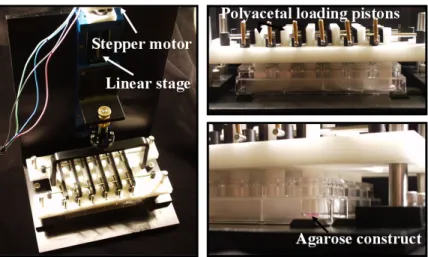
Custom Loading Bioreactor. Dynamic compressive loading was performed as previously described (Figure 2.1) [161]. Briefly, a closed-loop displacement controlled bioreactor with 24 individual polyacetal pistons connected to a linear stage driven by a stepper motor was used to deliver a 10% peak-to-peak sinusoidal strain (7% offset) at 1Hz for 3 hours/day. Unloaded control constructs were handled and cultured alongside loaded constructs.
Figure 2.1: Photograph of the custom loading bioreactor. A closed-loop computer controlled system controlling a linear motor to apply precise dynamic compressive loads to
agarose constructs.
chloroform extraction and clean up on RNeasy Mini columns. Following RNA quantification (ND-1000, Fisher Scientific, Waltham, MA), cDNA was synthesized (Superscript VILO Express Supermix, Life Technologies, Carlsbad, CA), and amplification was performed with a OneStepPlus real-time PCR system, using intron spanning primers and Power SYBR Reaction Mix (Life Technologies). Primer sequences were designed using PrimerBlast (NCBI) (Table 1). Relative expression levels were quantified using the delta delta Ct method and normalized to 18S.
Biochemical Assays. Constructs intended for biochemical analysis were digested in a 125 µg/mL papain solution (Sigma-Aldrich), 100 mM phosphate buffer, 10 mM cysteine, and 10 mM EDTA, pH 6.3) for 16 h at 65°C. The temperature was raised to 70°C for 10 minutes to melt the agarose, followed by vortexing. Total sulfated glycosaminoglycan (s-GAG) content was quantified with the dimethylmethylene blue assay [162], using bovine chondroitin sulfate as the standard. Total collagen content was quantified using the
orthohydroxyproline assay [163], using 0.134 as the ratio of orthohydroxyproline to collagen. Total DNA content was quantified using the PicoGreen fluorescent double-stranded DNA assay (Life Technologies).
(ab97021), treated with HRP streptavidin and AEC Red Single (Histostain-Plus BS: Life Technologies), counterstained with hemotoxylin, and mounted with Clearmount (Life Technologies). Sections from porcine osteochondral plugs were used as positive controls to confirm staining specificity.
Mechanical Testing. Mechanical testing was performed in unconfined compression using the Enduratec ELF system (Bose, Framingham, MA) at room temperature in PBS. Disks were equilibrated in creep (~0.01N tare load for 300s) and then compressed at 1 µm/s to 10% stress, at which point the strain was held and disks were allowed to stress relax to equilibrium (1200s). The equilibrium force was determined by the average force over the last 10 seconds, and the equilibrium compressive Young’s modulus (EY) was calculated by normalizing this equilibrium force to the original disk cross-sectional area (measured by digital calipers) and dividing this by the strain. The dynamic moduli were obtained by superimposing a 2% peak-to-peak sinusoidal displacement strain on the 10% strain at frequencies of 0.1, 0.5 and 1.0Hz, with five cycles imposed at each frequency. The dynamic moduli (G*) were determined by dividing the average amplitude of the resulting stress wave by the applied strain.
Statistical Analysis. Data were analyzed using multiple-factor ANOVA, followed by a Fisher’s post-hoc test when appropriate. Nominal data were compared between treatment groups using the chi-square test.
2.3 RESULTS
TRPV4 Channel Function in Agarose-Embedded Chondrocytes.
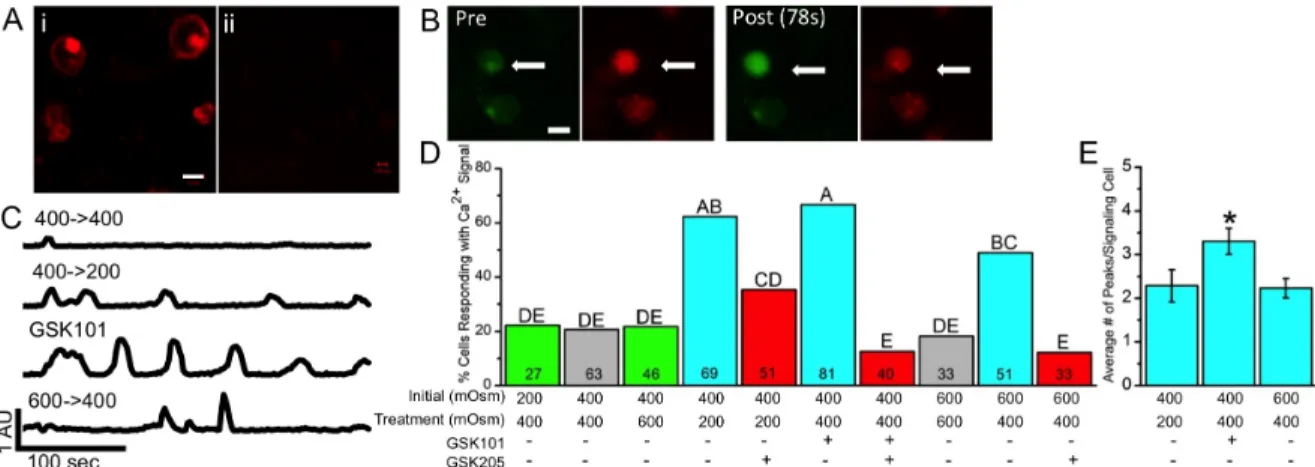
membrane of chondrocytes three days after casting in agarose (Figure 2.2A). Using fluorescence ratio imaging, Ca2+ concentrations in individual agarose-embedded
chondrocytes were measured in response to increases or decreases in osmolarity and TRPV4 activation. A significant increase in Ca2+ signaling was observed with both hypo-osmotic treatments (400→200 mOsm and 600→400 mOsm, p<0.001 and p<0.01 respectively), as well as GSK101 treatment (p<0.001), as compared to their respective iso-osmotic controls (Figure 2.2B,C), while the percentage of cells exhibiting a [Ca2+]i signal was the same between the two iso-osmotic controls (400→400 mOsm and 600→600 mOsm) (Figure 2.2D). GSK101 produced higher percent cell signaling than 600→400 mOsm (p=0.04), but not the 400→200 mOsm group (p=0.58); the 400→200 mOsm group was also not different from the 600→400 mOsm group (p=0.15). While both hypo-osmotic and GSK101 treatments caused more cells to signal, GSK101 treatment generated more [Ca2+]i transients in signaling cells than the two hypo-osmotic treatments (Figure 2.2E, p<0.05). Pre-incubation with the TRPV4 inhibitor GSK205 inhibited the effect of hypotonic loading (600→400 mOsm and 400→200 mOsm), as well as that of GSK101 treatment, and returned the percent of cells signaling back to control levels (400→200 mOsm+GSK205 vs 400→400 mOsm: p=0.08; 600→400 mOsm+GSK205 vs 600→600 mOsm: p=0.49; GSK101+GSK205 vs 400→400 mOsm: p=0.29). Neither hyper-osmotic loading condition tested (200→400 mOsm,
Figure 2.2: TRPV4 function in agarose-embedded chondrocytes. (A) Positive labeling of TRPV4 in agarose-embedded chondrocytes (i) and no labeling in the no primary control (ii),
scale bar=10µm. (B) Confocal images of a chondrocyte signaling in response to GSK101
(arrow), indicated by the increased ratio of green:red fluorescence post-GSK101 incubation, scale bar=15µm. (C) Representative Ca2+ traces for conditions that demonstrate significant
[Ca2+]i signaling as compared to the iso-osmotic control. (D) Percentage of cells responding to the below osmotic and chemical conditions. Data not sharing a common superscript letter indicate a significant difference (p<0.05). Bars do not have error bars because the percent responding metric does not have an error associated with it. Numbers inside the bars are the
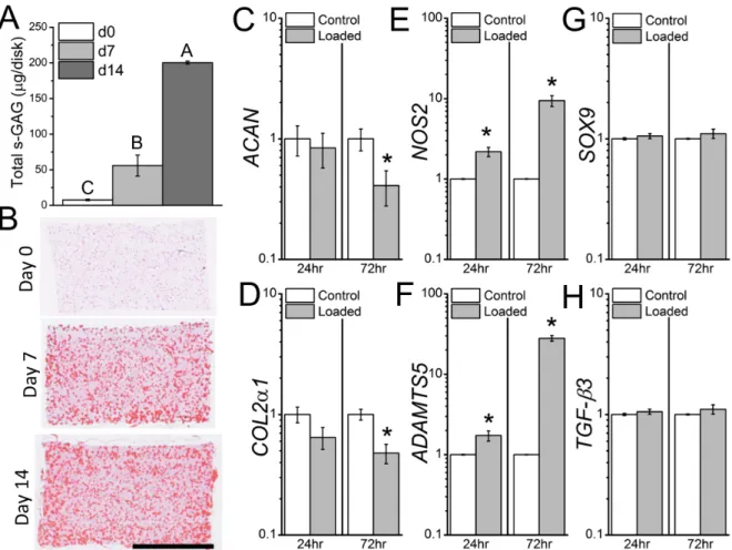
Figure 2.3: Loading without pre-culture. Without pre-culture, and the associated elaboration of a pericellular matrix, mechanical loading suppresses matrix and enhances catabolic gene expression. (A) s-GAG content increases dramatically with pre-culture of the
chondrocyte-laden agarose constructs. (B) Safranin-O staining shows pericellular localization of proteoglycans. (C-H) Gene-expression in 3-day pre-cultured (Day 0) chondrocyte-laden
agarose constructs 24hrs and 72hrs following mechanical loading. Mechanical loading suppressed ACAN (C) and COL2α1 (D) gene expression 72hrs following loading, and enhanced (E) NOS2 and (F) ADAMTS5 gene expression 24hrs and 72hrs following loading,
while not affecting (G) SOX9 or (H) TGF-β3 gene expression. Data not sharing a common superscript letter indicate a significant difference (p<0.05), * greater than Control (p<0.05),
n=3-4, mean ± SEM. Scale bar = 2mm.
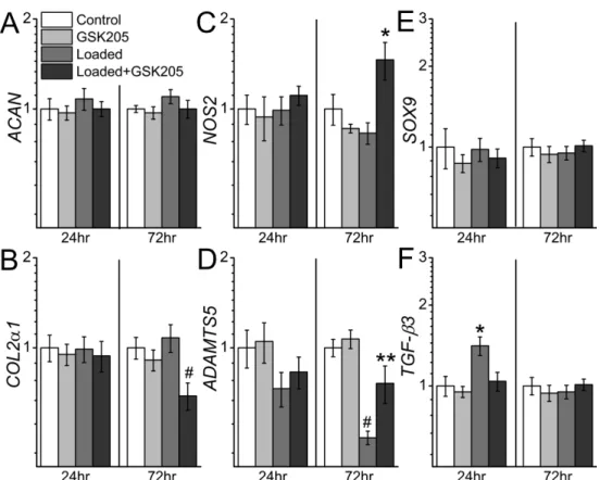
However, 72hrs later loading+GSK205 caused a decrease in COL2α1 (p<0.01) and an increase in NOS2 (p=0.02) gene expression compared to control, an effect that was not present with loading alone. In addition, ADAMTS5 expression was decreased with loading 72hrs after loading (p<0.001) and this effect was partially attenuated by GSK205 (Loaded vs Loaded+GSK205: p<0.01). Loading and GSK205 treatment had no effect on SOX9 gene expression, however TGF-β3 expression was significantly increased at 24hr (p<0.01) with loading and this effect was fully blocked by GSK205 (Control vs Loaded+GSK205: p=0.78). This effect was also transient, as TGF-β3 expression in the loaded group returned to control levels by 72hrs. The anabolic and anti-inflammatory effect of mechanical loading on
chondrocyte gene expression was also dependent on the two-week pre-culture period; when chondrocyte-laden constructs were loaded on Day 0 (three days after casting), ACAN and COL2α1 gene expression was suppressed and inflammatory genes ADAMTS5 and NOS2
Figure 2.4: Gene expression with pre-culture. TRPV4-dependent gene-expression in two-week pre-cultured chondrocyte-laden agarose constructs 24hrs and 72hrs after mechanical
loading. Mechanical loading enhanced TGF-β3 (F) and suppressed ADAMTS5 gene expression (D) in a TRPV4-dependant manner. Inhibition of TRPV4 during mechanical loading also enhanced NOS2 (C) and suppressed COL2α1 (B) gene expression. * greater than all other groups (p<0.05), # smaller than all other groups (p<0.05), ** less than Control
and GSK205, greater than Loaded+GSK205 (p<0.05), n=4-6, mean ± SEM. TRPV4 Inhibition During Dynamic Loading Inhibits Mechanically-Regulated Enhancement of Matrix Accumulation and Functional Properties. To determine if the enhancement of construct biochemical and functional properties in response to dynamic loading was also TRPV4-dependent, pre-cultured constructs underwent daily dynamic
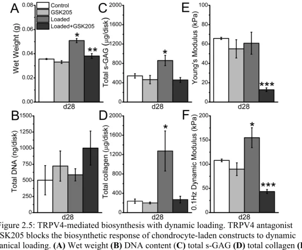
compressive loading for four weeks (3 hours/day, 5 days/week), in the presence or absence of GSK205. Daily transient exposure to GSK205 had no effect on construct wet weight,
biochemical content or functional properties after four weeks of culture (Figure 2.5A).
response (Figure 2.5A). Loaded disks also exhibited an increase in both total sulfated
glycosaminoglycan (s-GAG) content (p=0.01) and total collagen content (p<0.01), while the presence of GSK205 prevented these increases (Figure 2.5C,D). Loaded disks also exhibited an increased dynamic modulus over free-swelling controls (p=0.02), while loaded disks exposed to GSK205 had a significantly lower dynamic modulus than all other groups (Figure 2.5F). In addition, although dynamic loading did not significantly affect the equilibrium Young’s modulus of the constructs, the addition of GSK205 during loading significantly decreased this property (Figure 2.4E, p<0.001).
Figure 2.5: TRPV4-mediated biosynthesis with dynamic loading. TRPV4 antagonist GSK205 blocks the biosynthetic response of chondrocyte-laden constructs to dynamic mechanical loading. (A) Wet weight (B) DNA content (C) total s-GAG (D) total collagen (E)
Young’s modulus and (F) dynamic modulus of constructs following four weeks of mechanical loading. * greater than all other groups (p<0.05), ** greater than GSK205
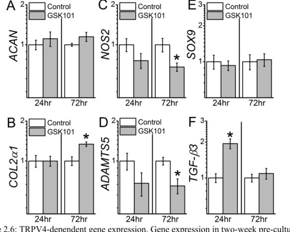
Direct Activation of TRPV4 Potently Drives Gene Expression, Biochemical, and Functional Changes Analogous To Dynamic Loading. To measure the direct effects of TRPV4 channel activation on chondrocyte gene expression, chondrocyte-laden constructs were transiently exposed to TRPV4 agonist GSK1016790A (GSK101). Similar to the effects of dynamic loading, NOS2 (p<0.05) and ADAMTS5 (p<0.01) gene expression were reduced 72hrs after treatment with GSK101 (Figure 2.6). TGF-β3 gene expression was elevated 24hrs post-treatment (p<0.001), while GSK101 had no effect on ACAN (24hrs: p=0.61; 72hrs: p=0.18) and SOX9 gene expression (24hrs: p=0.50; 72hrs: p=0.94). COL2α1 (72hrs) gene expression was also enhanced with GSK101 treatment (p=0.001).
Figure 2.6: TRPV4-dependent gene expression. Gene expression in two-week pre-cultured chondrocyte-laden agarose constructs 24hrs and 72hrs following TRPV4 agonist GSK101 treatment. GSK101 enhanced (B) COL2α1 and (F) TGF-β3 and decreased (C) NOS2 (D) and
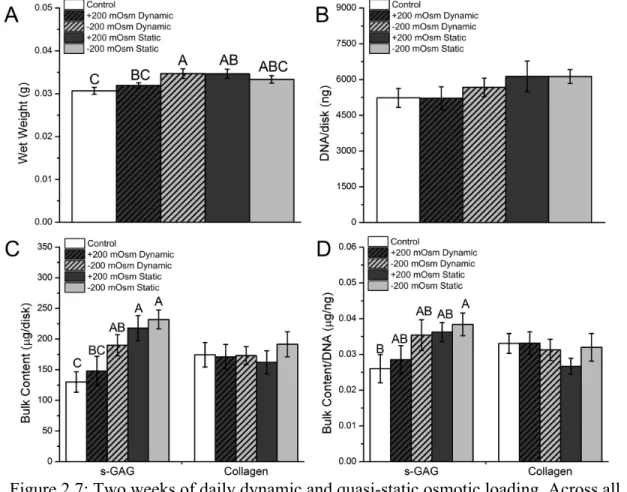
In addition to direct activation of TRPV4 using GSK101, we also examined the effects of osmotic loading as an alternative approach for activating TRPV4 and enhancing ECM production. We compared four different osmotic loading regimes and found that after two weeks, both a +200 mOsm and -200 mOsm quasi-static stimulus enhanced total s-GAG accumulation (Figure 2.7).
Figure 2.7: Two weeks of daily dynamic and quasi-static osmotic loading. Across all regimes, daily osmotic loading did not affect total DNA content (B) (p=0.415) or total collagen content (C) after two weeks. Hyper- and hypotonic quasi-static loading, as well as
dynamic hypotonic loading, increased total s-GAG content (C) compared to the control constructs, while only quasi-static hypotonic significantly increased s-GAG/DNA (D). Data
not sharing a common superscript letter indicate a significant difference (p<0.05), n=8-9, mean ± SEM.
in wet weight and DNA per disk, as well as total s-GAG and collagen accumulation. When normalized to DNA content, s-GAG/DNA increased significantly with GSK101 treatment (p=0.001), as did collagen/DNA (p=0.05). Agonist treatment also enhanced both the Young’s (p=0.02) and dynamic modulus (p<0.001). Histologic staining for s-GAG and type II
collagen further indicated an enhanced accumulation of both ECM components with GSK101 (Figure 2.9).
Figure 2.8: TRPV4 activation enhances matrix synthesis. TRPV4 activation enhances extracellular matrix accumulation. (A) Wet weight (B) DNA content (C) total GAG (D)
s-GAG/DNA (E) total collagen (F) total collagen/DNA (G) Young’s Modulus and (H) Dynamic Modulus of constructs following two and four weeks of stimulation. Data not sharing a common superscript letter indicate a significant difference (p<0.05), n=6-8, mean ±
Figure 2.9: TRPV4 activation enhances matrix accumulation and distribution. Gross morphology, Safranin-O, and Collagen type II IHC after four weeks of culture, scale bar =
2mm.
2.4 DISCUSSION
Our results confirm the hypothesis that TRPV4 channel activation plays a critical role in the mechanoregulation of chondrocyte physiology and matrix metabolism in response to dynamic compressive loading. This study revealed a TRPV4-dependent response of
chondrocytes to mechanical stimulation, involving transcriptional induction of anabolic growth factor gene expression and inhibition of pro-inflammatory mediators. This response was functionally validated in long-term culture, where inhibition of TRPV4 prevented mechanically-induced chondrocyte ECM biosynthesis and matrix accumulation. The role of TRPV4 channel activation on chondrocyte physiology was further confirmed using direct chemical activation of TRPV4, which produced a similarly potent anabolic response as mechanical loading.
mechanical loading that has been observed in a large number of previous studies. For example, compressive dynamic loading typically does not confer an anabolic response in chondrocyte-laden constructs for at least two weeks [165], while the postponement of
compressive loading for this same period of time is explicitly beneficial [27, 166]. Moreover, while compressive loading of intact cartilage explants can stimulate proteoglycan synthesis immediately [151, 167], the response of chondrocytes embedded in agarose is enhanced with additional weeks of pre-culture [168]. Cell-matrix interactions in the pericellular region are believed to play a critical role in transducing mechanical signals [169-171]. Several studies have highlighted the association of pre-culture with accumulation of a proteoglycan-rich pericellular matrix [6, 27, 164, 172], which can contribute to strain shielding of the chondrocytes [6, 164] as well as the conversion of mechanical loading to changes in interstitial osmolarity [106, 173]. The results of the present study not only reproduced this dependence of the anabolic effect of mechanical loading on pre-culture, but also suggest specific mechanisms involving the conversion of mechanical to osmotic stress, as well as the transduction of osmotic stress to an intracellular response via TRPV4 channel activation.
compression is removed. In addition, GSK101 elicited significantly more [Ca2+]i transients during the imaging period than the hypo-osmotic loading conditions (600→400 and
400→200mOsm), an indication that pharmacologic targeting of TRPV4 may provide a more potent method of activating TRPV4 than a single hypo-osmotic stimulus of physiologic magnitude.
[Ca2+]i signaling is believed to be one of the earliest events in the response of chondrocytes to mechanical stimulation [112, 174, 175]. Previous studies have observed [Ca2+]i signaling in response to mechanical loading [176, 177]; interestingly, these studies detected [Ca2+]i signaling in chondrocyte-laden constructs without substantial pre-culture (<72hrs), although chondrocytes synthesize small amounts of pericellular proteoglycans even within 2 days of culture in agarose [169] that could contribute to mechanically-induced [Ca2+]i signaling. Thus, the effects of loading with and without pre-culture, observed in this study as well as in previous studies, may be due differences in the characteristics of the [Ca2+]i signal with pre-culture, or perhaps other environmental and cellular factors that change over time [165, 168, 169, 178]. Though chondrocyte [Ca2+]i signaling exhibits a high sensitivity to variations in osmolarity or volume [17, 18, 179, 180], there is little or no sensitivity to physiologic levels of other physical stimuli, such as direct membrane stretch, suggesting that membrane connections to the pericellular matrix and/or intracellular
We observed transcriptional control of ADAMTS5, NOS2, COL2α1, and TGF-β3 in response to mechanical loading or GSK101-mediated TRPV4 channel activation.
Furthermore, inhibition of TRPV4 during loading prevented mechanoregulation of these genes. ADAMTS5 is a previously identified mechanoresponsive gene in loaded chondrocyte constructs [27] and has also been implicated in mechanically-induced models of
osteoarthritis [185]. Interestingly, disruption of primary cilia, a structure also required for TRPV4-mediated transduction of osmotic swelling [60], has been shown to increase
ADAMTS5 gene expression [186]. Mechanical loading of chondrocytes in agarose can reduce nitric oxide production [187], and our results suggest that mechanical activation of TRPV4 contributes to the suppression of NOS2 expression. GSK101 enhanced COL2α1 expression, and although COL2a1 did not increase with loading, inhibiting TRPV4 during loading did reduce its expression. Thus, TRPV4-mediated Ca2+ appears to support ECM gene expression, particularly under dynamic mechanical loading. TRPV4-mediated enhancement of COL2α1 gene expression has also been observed in the ATDC5 cell line [22]. However, unlike the response of ATDC5 chondroprogenitors, we did not concurrently observe transcriptional enhancement of chondrocyte SOX9 with TRPV4 activation, suggesting that fully
potential mechanism for the biophysical to biochemical transduction of mechanical loading in articular cartilage.
Inhibition of TRPV4 with GSK205 blocked the compositional and functional enhancement of mechanically-loaded chondrocyte-laden constructs, further supporting the role of TRPV4-mediated mechanotransduction in regulating chondrocyte matrix metabolism. Functional testing of the mechanically loaded constructs revealed that the mechanically loaded disks with GSK205 had functional properties below that of the unloaded controls. Blocking TRPV4 during loading may have allowed for mechanical fatigue of the agarose in the setting of inhibited ECM production (e.g., a decrease in COL2α1 expression) or perhaps increased catabolic or disrupted matrix organization processes (e.g., an increase in NOS2 and ADAMTS5 expression). GSK101 treatment also produced increases in construct matrix accumulation and functional properties, which again could be due to both increased
production and/or suppressed catabolism and matrix organization. Future transcriptome-wide analyses [191, 192] may be useful in identifying the pathways that drive these loading-induced changes.
Activation of TRPV4 with GSK101 potently enhanced matrix accumulation and functional properties of the chondrocyte-agarose constructs, even in the absence of a pre-culture period. In addition, matrix deposition in GSK101-treated constructs appeared more uniform throughout the construct depth than in the control and osmotically-treated groups (Figure 2.9). In this regard, the use of a small molecule chemical agonist to accelerate tissue formation may have significant advantages over the use of growth factors or direct
lead to nonuniform tissue deposition. It will also be important to determine if targeting TRPV4 can also enhance the chondrogenesis and maturation of human stem cell-based tissue engineered cartilage.
CHAPTER 3. TRPV4 IN OBESITY-ASSOCIATED OSTEOARTHRITIS3
3.1 INTRODUCTION
Obesity is one of the most significant and modifiable risk factors for osteoarthritis (OA) [195]. However, local biomechanical factors associated with changes in the onset and progression of knee OA in the obese population [35, 196] cannot explain the relationship between obesity and OA in non-load bearing joints [197]. Obesity and related metabolic syndromes are associated with chronic low-grade inflammation and systemic tissue damage [198]. There is further evidence to suggest that these systemic metabolic factors participate in the development of OA in both weight bearing and non-weight bearing joints [138];
however, the mechanisms by which these systemic factors alter the course of OA remain unclear [199].
The transient receptor potential vanilloid 4 (TRPV4) ion channel is a Ca2+-preferred cation channel, originally characterized as a transducer of osmotic stress [43, 44]. TRPV4-mediated Ca2+ signaling in response to osmotic fluctuations in the cartilage is one potential mechanism by which chondrocytes sense and respond to joint loading [60]. TRPV4 signaling plays a crucial role in skeletal development [200, 201]. Furthermore, genetically-encoded deletion of TRPV4 in mice leads to accelerated joint degeneration with aging [64].
3
Interestingly, chondrocyte TRPV4 is also known to interact with pro-inflammatory mediators and cytokines to mediate catabolic signaling and nociception [202, 203].
We hypothesized that the absence of TRPV4-mediated signaling in the presence of the catabolic, biomechanical and inflammatory factors of obesity would accelerate OA progression in the high-fat diet model of OA. To examine the link between the observed phenotype of Trpv4−/− mice and function of TRPV4 at the cellular level, we measured the effects of TRPV4 deficiency on the intrinsic capabilities of bone marrow-derived stem cells (MSCs) and adipose-derived stem cells (ASCs), isolated from Trpv4−/− and Trpv4+/+ mice, to differentiate towards the adipogenic, osteogenic, and chondrogenic lineages.
3.2 MATERIALS AND METHODS
Animal handling. All animal care and experimental procedures were conducted
under a protocol approved by the Duke University Institutional Animal Care and Use Committee. 10 week old male pan-Trpv4 knockout (Trpv4-/-) mice and wild-type C57BL/6 (Trpv4+/+) mice were placed on either a high-fat diet (60% kcal; #12492) or control diet (10% kcal; #12540B) (Research Diets, Inc., New Brunswick, NJ) (n=36). Animals were maintained on their respective diets for 22 weeks, during which time, spontaneous
locomotor activity was measured by an automated photobeam apparatus, with light-phase and dark-phase locomotor activity collected with the VersaMax program (Omnitech Digiscan, AccuScan Instruments, Inc., Columbus, OH) [138].
Body composition. Immediately following sacrifice, total body fat of each mouse
Histological evaluation of OA. After DXA scanning, hind limbs were formalin
fixed and paraffin embedded. 6µm coronal sections of the knee joint were stained with Hematoxylin, Safranin-O and Fast Green. Sections from the tibiofemoral cartilage-cartilage contact region in the medial and lateral condyles were scored for degenerative changes using a modified Mankin system by three blinded graders, evaluating for cartilage structural damage, proteoglycan loss, tidemark duplication, subchondral bone thickening,
fibrocartilage, chondrocyte hypertrophy, and chondrocyte cloning [204]. Average scores were tabulated for each of the four compartments of the joint and combined to one total joint score. Subcutaneous fat tissue from 10-week old Trpv4-/- and Trpv4+/+ mice was also fixed, paraffin embedded, and 6µm sections were stained with Hematoxylin and Eosin.
Stem cell isolation, purification and expansion. Bone marrow derived stem cells
(MSC) and subcutaneous adipose-derived stem cells (ASC) were isolated from the femurs and tibias (MSC isolation), and the inguinal fat pad (ASC isolation) of 3-5 Trpv4-/- and Trpv4+/+ mice (8-10 weeks old), using a previously described method involving
fluorescence-activated cell sorting (FACS) to obtain cells with specific cell markers (MSC:
CD45-/TER-119-/Sca-1+/PDGFRα+; ASC: CD45-/TER-119-/CD31-/Sca-1+/CD34+), and expanded to P3 as described in [205, 206].
Tridifferentiation. At passage 3, MSCs and ASCs were induced toward
(33µM biotin, 17µM pantothenate, 1µM bovine insulin, 1µM dexamethasone (all from Sigma-Aldrich, St. Louis, MO)), with the addition of 250µM isobutylmethylxanthine (IBMX) and 2µM rosiglitazone (Avandia™, GlaxoSmithKline, Middlesex, UK) for the first three days only)). Adipogenic potential was assayed by Oil-Red-O stain release. For
osteogenesis, cells were plated at 10,000/cm2 on standard tissue culture plastic for three days in osteogenic control media (DMEM-HG (Invitrogen), 10% FBS and 1% P/S/F), at which time, experimental wells were changed to osteogenic control media plus osteogenic factors (10mM b-glycerophosphate (Sigma), 250µM ascorbate-2-phosphate (Sigma), 2.5µM retinoic acid (Sigma), and 50ng/mL hBMP-2 (R&D systems, Minneapolis, MN)).
Osteogenic potential was assayed by Alizarin stain release. For chondrogenic
differentiation, 250,000 MSCs and ASCs were pelleted in 15mL polypropylene tubes by centrifugation at 300g for 5 minutes. Cells were fed a chondrogenic control medium of DMEM-HG (Invitrogen), 1% ITS+ (BD biosciences, San Jose, CA), 50 µg ascorbate-2-phosphate (Sigma), 40 µg/mL proline (Sigma), with ASC pellet media being supplemented with 10% FBS. On day three, experimental pellets were changed to chondrogenic control media plus 10ng/mL human transforming growth factor-_3 (hTGF-b3) and 500ng/mL human bone morphogenetic protein-6 (hBMP-6, R&D). After 28 days, pellets were processed for Alcian Blue/Nuclear Fast Red staining or biochemical analysis for double stranded DNA with the PicoGreen assay and glycosaminoglycan content with the 1,9 dimethylmethylene blue (DMB) assay.
Statistical analysis. Normality was tested, and data were log-transformed before
post-hoc analysis using α=0.05. Significant differences were reported at the 95% CI unless otherwise noted. Data are presented as mean±SEM.
3.3 RESULTS
Trpv4−/− mice are more susceptible to diet-induced obesity than Trpv4+/+ mice.
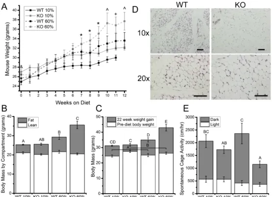
Trpv4−/− mice weighed significantly more than Trpv4+/+ mice at 10 weeks of age (Figure 3.1A,B). After being fed a high-fat diet, Trpv4−/− mice gained significantly more weight than Trpv4+/+ mice (Figure3.1A,B). DXA measurements after high-fat feeding revealed that the differences in body mass with genotype and diet were due to body fat, with Trpv4−/− mice gaining significantly more body fat than Trpv4+/+ mice following high-fat feeding (Figure 3.1C). To further examine the obese phenotype of Trpv4−/− mice, histological sections were taken of the inguinal fat pad of 10-week-old normally fed mice and showed that even prior to high-fat feeding, Trpv4−/− mice may possess larger adipocytes than Trpv4+/+ controls (Figure 3.1D).
High-fat fed Trpv4−/− mice have lower cage activity than high-fat fed Trpv4+/+
mice. To further investigate the relationship between Trpv4 deficiency and increased weight
Figure 3.1: Trpv4-/- mice exhibit increased adiposity in response to high-fat feeding. (A) High-fat fed mice weighed significantly more than control diet mice by Week 7. By Week 10, KO 60% mice weigh significantly more than all other groups. (B) WT 60% mice gained
(insignificantly) more weight after 32 weeks compared to WT 10% mice (p=0.0837), whereas KO 60% mice gained more weight than all other groups. (C) Post-diet, mice did not
differ in the amount of lean body mass. WT 60% mice had significantly more body fat than WT 10% mice, but KO 60% had the more body fat than all other groups. (D) Histological sections of subcutaneous adipose tissue from 10 week old mice, where KO adipocytes appear
larger than WT adipocytes, Scale bar = 100 µm. Data are shown as mean ± SEM. & Trpv4 KO > WT (p<0.05), * 60% diet > 10% diet (p<0.05), ^ 60% KO > all other groups (p<0.05),
+ 60% KO > 10% WT (p<0.05). Data not sharing a common superscript letter indicate a significant difference (p<0.05).
Trpv4 deficiency increases knee OA following high-fat feeding. A modified
Mankin score was tabulated and analyzed that combined the score for cartilage structural degeneration and proteoglycan loss as recommended in reference. Neither Trpv4−/− nor