THE EFFECT OF SINTERING PROCESS ON ZIRCONIA’S OPTICAL AND PHYSICAL PROPERTIES
Lida Swann
A thesis submitted to the faculty of the University of North Carolina at Chapel Hill in partial fulfillment of the requirements for the degree of Master of Science in the
School of Dentistry (Department of Prosthodontics).
Chapel Hill 2014
© 2014 Lida Swann
ABSTRACT
Lida Swann: The effect of sintering process on zirconia’s optical and physical properties
(Under the direction of Lyndon F. Cooper)
Zirconia restorations present an alternative dental restorative option with an average flexural strength of 1000 MPA. One of the challenges of this material is the decreased translucency when compared with less strong ceramic materials like feldespatic porcelain or lithium disilicate. A potential solution to this challenge is to the increase of sintering temperature and or increase holding times.
The present study evaluated the effect of different sintering temperatures and holding times on contrast ratio as a measure of translucency, and evaluated if any temperatures or holding times could have a negative effect on materials strength. We also looked at the effect of cyclic loading vs conventional load to failure testing.
Contrast ration was measured on a spectophotometer for all specimens. 60 specimens (5 per group) were subjected to mechanical cyclic loading under water at a load of 100N for 100,000 cycles at a frequency of 1.5Hz.
After fatigue loading samples and control group were tested on an Instron machine until catastrophic failure occurred.
ACKNOWLEDGEMENTS
To my mentor and program director Dr. Lyndon Cooper, for all your support and sharing your skills, knowledge and experience with me.
To my committee Dr. Terry Donovan and Dr. Gustavo Mendonca for all your help with this research project.
To Ivoclar Vivadent, for providing the materials and equipment for the execution of this research project and to Dr. Thomas Hill and Omar Nihlawi for sharing their research experience and their technical support.
TABLE OF CONTENTS
LIST OF FIGURES ... vii
Introduction ... 1
Table 1. Ceramic restorations in dentistry ... 2
1.1 Biocompatibility ... 8
1.2 Strength ... 10
1.3 Wear ... 15
1.4 Accuracy ... 17
1.5 Esthetics ... 19
2.The Effect of Sintering Process on Zirconia’s Optical and Physical Properties. .... 23
Introduction ... 23
2.1 Materials and Methods ... 25
2.2 Statistics ... 32
2.3 Results ... 33
2.3.1 Model for Strength ... 33
2.3.2 Model for Translucency ... 37
2.4 Discussion ... 39
LIST OF TABLES
Table 1- Ceramic restorations in dentistry ... 2
Table 2- Translucency- contrast ratio of dental ceramics ... 21
Table 3- Sintering temperatures ... 28
Table 4- Test fatigued group ... 28
Table 5- Control group ... 29
Table 6- Results for holding time ... 34
Table 7- Results for temperature ... 35
Table 8- Translucency results for temperature ... 38
LIST OF FIGURES
Figure 1- Transformation toughening of zirconia ... 13
Figure 2- Wear of zirconia vs enamel ... 16
Figure 3- Disks design file ... 26
Figure 4- Milled zirconia disk ... 26
Figure 5- Diagram of research sequence ... 27
Figure 6- SD Mechatronik chewing simulator CS-4 ... 30
Figure 7- Samples measurement ... 30
Figure 8 - Instron 33R4204 ... 31
Figure 9- Biaxial strength test for control group ... 36
Introduction
Biomaterials in dentistry must address several requirements that include biocompatibility, strength related to intended purpose and esthetics. The history of dental prostheses reflects a progression from function to esthetics with gold restorations being largely replaced by porcelain fused to metal restorations during a period from the 1970’s to 1990’s. The introduction of various all-ceramic restorations beginning in the 1980’s initiated a continuous transition from metal-based ceramics to different multilayered and monolithic all ceramic restorations.
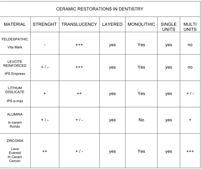
The central issue for all ceramic restorations has been the balancing of esthetics (color and translucency) with strength or function. Different materials have been utilized (Table 1) and their esthetic value traditionally has been inversely related to strength. The basis for this clinical paradox is the use of glass phase ceramics to impart translucency to dental ceramics and the use of relatively opaque crystalline ceramics to achieve strength. Today, lithium disilicate restorations (e.g., IPS e-max Ivoclar Vivadent (175 Pineview Drive, Amherst, NY 14228 USA) exemplifies a glass phase ceramic with remarkable translucency and color
Table 1- Ceramic restorations in dentistry
CERAMIC RESTORATIONS IN DENTISTRY
MATERIAL STRENGHT TRANSLUCENCY LAYERED MONOLITHIC SINGLE UNITS
MULTI UNITS
FELDESPATHIC
Vita Mark - +++ yes Yes yes no
LEUCITE REINFORCED
IPS Empress
+ / - +++ yes Yes yes no
LITHIUM DISILICATE
IPS e-max
+ ++ yes Yes yes + / -
ALUMINA
In-ceram Rondo
+ / - + / - yes No yes +
ZIRCONIA
Lava Everest In Ceram
Cercon
++ + / - yes Yes yes +++
ceramic restorations is attributable to several factors including the relative cost of gold alloys, the integration of Zirconia materials into the CAD CAM workflow, and the esthetic value of ‘white’ dental materials. Suggested by this migration of clinical preferences from metal ceramics to all ceramic materials is the satisfactory performance of the all-ceramic material.
The past decade of clinical research has provided some insight regarding the performance of zirconia prostheses. A systematic review by Raigrodski looked at the survival and complications of zirconia FDP. He reported survival rates that ranged from 73.9% to 100% within 12 studies. Five studies reported 100% survival rates during the observations period. One study reported 73.9% survival of frameworks and the rest (6 studies) had survival rates ranging between 88.2% and 96.6%. The common complication reported was chipping and it was suggested that with the development of new layering porcelains better clinical properties would be expected. [2]
posterior restorations, and, or second molar abutments. Larsson’s systematic review in 2014 [5], suggested that the success rate of tooth-supported and implant-supported zirconia-based crowns is adequate, similar, and comparable to that of conventional porcelain-fused-to-metal crowns. A recent laboratory study utilized indentation to induce chipping of monolithic zirconia and lithium disilicate materials. The results confirm that ceramic veneered-zirconia displayed high chipping and monolithic lithium disilicate resisted this chipping, monolithic zirconia was most resistant to this induced chipping behavior [6].
When considering the outcome of zirconia single unit full coverage restorations, less information is available. The earliest clinical efforts of zirconia-based restorations involved the creation of a milled zirconia coping supporting a compatible ceramic veneer. Most recently, a 5 – year cohort study revealed that less than 60% of the crowns demonstrated success at 5 years and 11 of 47 crowns at 5 years required replacement due to chipping. This 2014 paper advised that new materials should be more carefully evaluated before introduction to clinical use. [7]
Previous outcomes for zirconia restorations including crowns have been considered in several systematic reviews. A 5-year retrospective study of survival of zirconia single crowns fitted in a private clinical setting by Anders showed promising
results for zirconia single crowns. Most crowns (78%) were placed on premolars
and molars. Among the 143 crowns that were followed for 5 years, 88% did not have
any complications. The reported complications were: extraction of abutment tooth (7;
3%), loss of retention (15; 7%), need of endodontic treatment (9; 4%) and porcelain
Jung’s systematic review focused on single crowns. The five to ten year
results showed high survival rates for tooth and implant supported single crowns,
but technical, biological and aesthetic complications were common. It is possible to
suggest that many of the complications were mostly related to restoring implants compared to tooth supported restorations [9].
As revealed in the aforementioned reviews concerning zirconia restoration performance, one of the early and prominent observations made regarding the clinical performance of zirconia-based all ceramic restorations was chipping of the veneering porcelain from the zirconia frameworks. While many different investigators have suggested fundamental reasons for this phenomenon (reviewed below), the clinical response to chipping is a concern for layered zirconia restorations.
When used as a framework, zirconia has an inherent basic esthetic value, due to the fact that it is white and can be alternatively colored to mimic surrounding dentin. Further it can be provided with high opacity to cover discolored teeth and implant components [10]. This can be advantageous to the technician who is trying to conceal a dark underlying tooth structure, a metal post, or the remainder of amalgam restorations left after initial preparation.
revealed that chipping within the veneering ceramic or at the framework/veneer interface frustrates higher clinical success and survival of these restorations. Veneer chipping, not framework fracture, appears to be the weak link in zirconia-based restorations.
For traditional porcelain fused to metal restorations, layering porcelains possess a lower coefficient of thermal expansion (CTE) than the metallic substrate. In this manner the veneering porcelain gains strength by compressive loading. Unfortunately, the simple mis-matching of CTEs doesn't work effectively for zirconia-layered restorations. This results in the creation of stress fields throughout the restoration leading to cracking and delamination during the cooling phase of the veneering process. [11]. Fortunately, specific ceramic veneering materials have been developed for application to Zirconia frameworks. Given that veneering materials and framework zirconia CTEs are today well matched, it is unlikely that CTE represents the root cause of veneered zirconia framework chipping.
rate of change in temperature may, in addition to differences in CTE, influence the strength of the veneer / framework bond. For example, Belli et al (2013) demonstrated by chewing simulation and compressive loading that attempts to minimize the thermal residual stresses within the veneer (closer CTEs) and application of slow-cooling cycles (< 30oC/minute) delayed the experimental failure of zirconia-veneer crowns. This was more important where larger differences in CTE were displayed. In an interesting and related paper Belli, the fractographic analysis of zirconia –veneered crowns demonstrated failures occurred solely within the veneering porcelains [12]. Others have also reported that a longer the cooling rate after firing will affect negatively bond strength of ceramic veneered restorations [13,14]. The number of firing cycles has also been considered to affect the bond strength, and it has been reported that between 3 and 5 firing cycles would be recommended to obtain a better bonding [15,16]. On the other hand one report argues than more than six firings will reduce bond strength [17].
The surface finish of a zirconia framework could also affect the core- veneer bond strength. Roughening of zirconia by the means of air abrasion could potentially improve bond strength but in the same time it could also make the core more susceptible to fractures. 50 um alumina oxide particles create less severe damage than when 120 particles are used [19].
Finally, the application of wet thick layers of porcelain onto a dried zirconia facilitates the t-m conversion and can create residual stress [20]. This too may result from water-mediated crystalline transformation of the zirconia framework. Contemporary workflow for veneered zirconia restorations include very long drying periods to assure firing occurs in the absence of water. It may be concluded that the process of veneering application has an important role in is the bond between the zirconia surface and the veneering porcelain.
In three different reports, using different systems (lava, DC-Zircon) , zirconia frameworks that were veneered with layering porcelain all developed cracking or crazing over two years of observation, ranging from 80% to 50% loss of material [21,22,23]. These studies may indicate that the observed porcelain fractures were material-system specific. It also indicates that factors such as type of zirconia framework thickness, and framework design, may very well be a cause for ceramic fracture.
1.1 Biocompatibility
the ball heads for total hip replacements [25]. Zirconia is still used in this application and other medical prosthetics to this day. Implied was acceptable biocompatibility. Clark showed that zirconia was found to be better than other ceramic biomaterials in use circa 1990, because it possessed higher strength and hardness [4].
confirmed these results with an in vivo study where y-TZP accumulated fewer bacteria than Ti in terms of total numbers of bacteria and presence of potential pathogens such as rods [36]. It may be concluded that zirconia materials offer advantages of biocompatibility for use as endosseous biomaterials and oral biomaterials due, both to its remarkable strength and durability as well as the surface properties of the material.
1.2 Strength
Introduction of zirconia-based ceramics as a restorative dental material has generated much interest in the dental profession. The mechanical properties of zirconia are the highest ever reported for any ceramic used prosthetic dentistry. The strength of zirconia has allowed the incorporation of high-strength all ceramics, into its use for posterior FDP [37]. High-strength, coupled with the possible high aesthetics that zirconia offers, allows the material to become a highly valuable option in our prosthetic armamentarium.
Zirconium dioxide (ZrO2, zirconia) has a unique crystallographic property that greatly improves its strength and toughness. Zirconia crystals can have a monoclinic (M), tetragonal (T) or cubic structure depending on temperature. Figure 1
A crystal structure is the spacing of the atoms of zirconium and oxygen and produces a resulting volume. At high temperatures, zirconia has a cubic structure. As the temperature is lowered, the atoms rearrange themselves and the structure becomes tetragonal. Further cooling results in additional rearrangement into a monoclinic structure. The transformation from tetragonal to monoclinic is
accompanied by a volume change. The volume change accompanying the
tetragonal to monoclinic transformation is what makes zirconia stronger and tougher than aluminum oxide and therefore, unique as a dental structural material for multiple unit posterior bridges.
Figure 1- Transformation toughening of zirconia
Biomedical grade zirconia usually contains 3 to 5 mol% yttria (Y2 O3) as a stabilizer (3Y-TZP). The mechanical properties of 3Y-TZP strongly depend on its grain size. Above a critical grain size, 3Y-TZP is less stable and more susceptible to
spontaneous t → m transformation whereas smaller grain sizes (<1 um) are
associated with a lower transformation rate [41]. Moreover, below a certain grain size (∼0.2 um), the transformation is not possible, leading to reduced fracture
toughness [42]. Consequently, the sintering conditions have a strong impact on both stability and mechanical properties of the final product as they dictate the grain size Higher sintering temperatures and longer sintering times lead to larger grain sizes [43,44]
compressive strength stresses, but at the same time, can also alter the phase integrity of the material and increase the susceptibility of aging [45]. When considering bulk zirconia as required for an anatomical, monolithic prosthesis, laboratory manipulation and clinical alterations required following sintering may influence the strength of the material. For example, air abrasion is utilized as a cleaning step during veneering and cementation procedures. Air abrasion was shown to increase the flexural strength of zirconia by T-M phase transformation that places the surface ceramic in compression. Machining with fine diamond instruments (<40 um) provided a similar effect on the compression surface zone, where as large grit instruments (> 125 um) created flaws beyond the depth of the surface zone, thereby weakening the structure [46]. It should be acknowledged that beyond the physical damage, heat induced by grinding might be of the magnitude to induce low temperature degradation of the material that leads to weakness.
Other factor to consider for the long-term success of zirconia-based restorations in multiunit FPD's is the size of the interproximal connectors. The minimal interproximal connector surface should be at least 6.25mm. Also, the height of the terminal abutments, is fundamental to achieving proper interproximal connectors, and should be evaluated with utmost care [37].
To circumvent the need for veneering altogether, another option is to develop fracture resistant, partially translucent monolithic ceramics. Monolithic all ceramic restorations are becoming more accepted due to higher strength, by avoiding weak veneer-core interfaces. All ceramic restorations, such as, IPS e-max (lithium disilicate), Wieland (zirconia), 3M lava Plus (zirconia), offer several all ceramic monolithic, restorative options, that have acceptable aesthetics, and eliminate the need for veneering ceramics altogether. [41]
1.3 Wear
During the early 2000’s zirconia was perceived as a material that would cause high wear to the opposing dentition. Today this perception is being challenged [49] and multiple studies have shown how zirconia can be a material gentle to opposing dentition, in comparison to glass ceramics that are layered on PFM restorations. This low wear property can be attributed to zirconia’s microstructure, and it’s small grain size, that allows for a mirror polished surface to be created, that is kind to opposing enamel surfaces. [50-51]
was more than polished zirconia, it was still less than commonly used porcelains for PFM restorations [53]. See figure 2
Figure 2- Wear of zirconia vs enamel
1.4 Accuracy
Zirconia is currently utilized in construction of dental prostheses by CAD-CAM milling of partially sintered materials. These materials shrink in dimension with sintering. When utilized as a framework with veneered ceramic, further distortion may occur. However, when compared to the internal fit and marginal adaptation of metal ceramic crowns, there was no difference in the fit of the different restorations. However, prior to veneering, metal copings displayed better internal fit [55]. When extended to larger restorations, the ceramic veneering of CAD/CAM frameworks did lead to distortion as measured by higher strain development using strain gauges. Non-veneered anatomic zirconia restorations showed little distortion and significantly less strain upon measurement [56].
is not clear what the influence of other milling parameters (rate, speed, tool path) will impose on the ultimate integrity of the surface layer [57].
With regards to marginal integrity several studies have been done and showed that with the aid of new milling systems and in some instances digital
impression systems. Seelbach looked at the marginal adaptation of alumina and
zirconia restorations various CAD CAM Systems including Lava COS, CEREC and
iTero, conventional impressions were used as well. The overall marginal fit for the
groups was 44+/- 26. He reported that within the limitations of his in-vitro study the
marginal adaptation of this all-ceramic crowns is acceptable and comparable to the
ones made with conventional methods. [58]
A systematic review described the marginal discrepancies for zirconia FDP.
The occurrences of marginal gaps for six studies were reported as follows. Marginal
integrity was clinically unacceptable for 16.7% [59], marginal integrity was
considered a success [60], marginal discrepancies were detected in 11.5 %[61],
visible evidence of ditching along the margins 5-26% [62], marginal gaps were
evident in 58.7 [63] , and visible evidence and or catch of the explorer was present in
one case [2].
report of the marginal fit of a 14 - unit zirconia FDP framework produced using Zeno CAD CAM on master dies was reported. The mean marginal gaps were again small (25 +/-29 um), but larger than that measured for single unit crowns placed on the retainers. The quality of fit was location dependent, suggesting that large frameworks are subject to complex changes. It should be further noted that these frameworks were not veneered [66].
1.5 Esthetics
The difficulty in reproducibly achieving good esthetics with PFM restorations and the desire for metal free solutions has led to the increased use of zirconia. The unique optical properties of zirconia require new and different understanding of how the materials are managed [67].
Translucency and color are important and often inseparable variables for dental restorations. Translucency may be an innate optical property of the zirconia material related to its crystalline structure. Colorants may be infused within the partially sintered zirconia and incorporated during sintering. Hjerppe examined the influence of shading on zirconia disks [68]. Differences in biaxial strength were measured among the samples (885 MPa – 1007MPa) compared to the control group (1132 MPa).
boundaries between the various phases and is influenced by the crystals’ particle size, shape, volume concentrations, and relative refractive indices. Physical properties for these types of materials are optimized by the use of bonding techniques, but still it is recommended the use of these of materials to be limited to the esthetic zone.
Table 2- Translucency- contrast ratio of dental ceramics
Regarding the present knowledge pertaining to zirconia – based dental prostheses, the following conclusions are suggested:
2) Layered zirconia restorations display chipping of the veneer ceramic material, but framework fracture is rare.
3) Anatomic, monolithic zirconia restorations may offer promises of improved physical strength and absence of chipping, but the translucency of the bulk zirconia may challenge esthetics.
Monolithic zirconia restorations were recently introduced to address the strength and chipping complications reported. The concept of avoiding veneering porcelains is directed at elimination of chipping. However, elimination of the veneering porcelains may reduce the technician and clinician ability to impart natural appearances attributable to translucency. It is the aim of this project to examine the relationship of translucency and strength of zirconia as a function of sintering conditions.
2.The Effect of Sintering Process on Zirconia’s Optical and Physical Properties.
Introduction
One of the emerging challenges inherent to the adoption of new CAD-CAM procedures for fabricating dental prosthesis is identifying the physical limitations presented. Anusavice (2012) described many of the limitations in dental materials testing in prediction of clinical outcomes [72]. For the emergent use of Zirconia restorations that are presently supported by work-flow (digital), cost (versus gold) and esthetics (all-ceramic) issues, a complex set of concerns must be addressed. For example, if the strength of zirconia is sufficient for fabrication of single as well as multiunit restorations, can it be utilized in a highly esthetic manner? Although Zirconia is white, it is relatively opaque. Clinical strategies to utilize this promising dental material in an effective, efficient and esthetic manner require careful assessment.
Zirconia is strong and tough. Its strength is based on its crystalline structure. In its tetragonal form, stresses lead to local transformation to monoclinic form that resists or stops crack propagation [73]. This can be measured in bulk and observed by fractography. Zirconia is white. This appeals to dentists and patients alike.
approach, milling zirconia from the sintered state has been largely discounted in recent years.
One approach to utilizing zirconia for reproducibly esthetic and lasting restorations is by CAD CAM generation of a framework that is veneered with a compatible veneering ceramic. The other approach involves the generation of a fully anatomical prosthesis or ‘monolithic’ zirconia prosthesis that is devoid of veneering ceramic. Modification of the zirconia’s white color occurs by infusion of colorants prior to sintering.
Ceramic veneered zirconia prostheses require the careful design and manufacture of a framework that is produced by CAD CAM procedures from bulk zirconia. Recent reports suggest that proper contour and design of the framework be employed to assure ceramic veneer integrity [74]. Other studies confirm that milled zirconia frameworks fit with acceptable fidelity [75]. Beautiful ceramics can be applied to these frameworks and remarkable prostheses can be made. However, when considering the growing data set regarding the clinical outcomes for this approach to using zirconia for clinical dentistry, chipping of the framework (often an irreversible complication) occurs with relatively high frequency [76]
a monolithic prosthesis is a complex procedure requiring the use of multiple colors and a precise drying and sintering procedure. When completed, a monolithic zirconia prosthesis lacks a ceramic veneer that is susceptible to chipping. However, compared to veneered prostheses, relative opacity may be noted [79].
Monolithic zirconia restorations were recently introduced to address the strength and chipping complications reported. The concept of avoiding veneering porcelains is directed at elimination of chipping. However, elimination of the veneering porcelains may reduce the technician and clinician ability to impart natural appearances attributable to translucency. It is the aim of this project to examine the relationship of translucency and strength of zirconia as a function of sintering conditions.
2.1 Materials and Methods
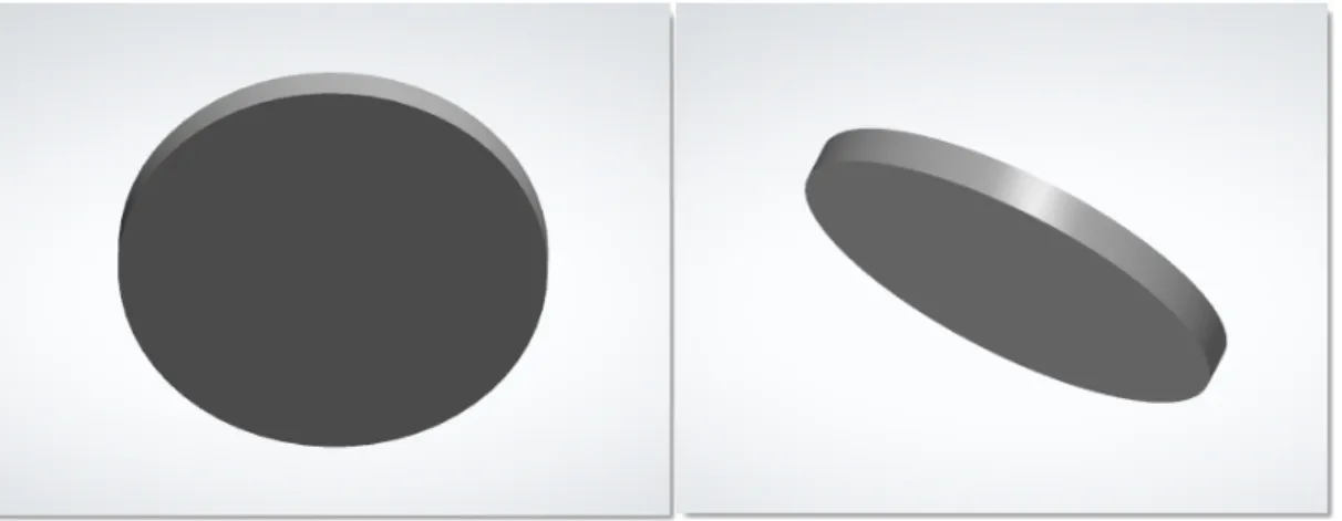
Figure 3- Disks design file
Figure 4- Milled zirconia disk
Figure 5- Diagram of research sequence
All disks were sintered in a four stage protocol using Origin® DuoTron™ Pro
Furnace, B&D Dental Technologies 2371 S. Presidents Dr., Ste. E ,West Valley City, UT, and were sintered according to manufacturer’s recommendations in regards to Stages 1,2 and 4 (Closing, heat ramp and Cooling). Stage 3 (holding temperature) was the variable considered in this study as the holding temperature and times were modified to explore what combination would give the best outcome as far as optical and physical properties.
Disks were divided into 12 groups and each groups was assigned a holding (sintering) temperature and holding time according to table 1.
CAD
DESIGN
SPECTO-‐ PHOTOMETRY
THERMO-‐ CYCLING & FATIGUE
STRENGHT BIAXIAL TEST SINTER
Table 3- Sintering temperatures Max
temperature
Holding time 1450 1500 1550 1600
1H 10 10 10 10
2H 10 10 10 10
3H 10 10 10 10
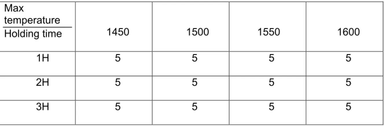
All disks were surfaced after sintering using table top polisher Ecomet 3 (Buehler 41 Waukegan Road Lake Bluff, Illinois), using water at 100 RPM. Buelher Carbimet disks were used first grit 320/P400 followed by 600/P1200. Each of the 12 groups was later subdivided into 2 subgroups representing fatigued and non-fatigued (control) samples (n=5).
Table 4- Test fatigued group Total specimens 60
Max
temperature
Holding time 1450 1500 1550 1600
1H 5 5 5 5
2H 5 5 5 5
Table 5- Control group Total specimens 60
Max
temperature
Holding time 1450 1500 1550 1600
1H 5 5 5 5
2H 5 5 5 5
3H 5 5 5 5
Using a calibrated spectrophotometer (UltraScan VISDelta TRAC with
EasyMatch QC, Calibration date 11-6-13, Hunter Associates Laboratory Inc.
Reston VA samples were evaluated for contrast ratio(CR) as a measure of translucency. Contrast Ratio is the ratio between the reflectance of a specimen over a black background to that over a white background of a known reflectance [80, 81]. The CR values are calculated according to the equation CR = Yb/Yw, in which Yb represents the spectral reflectance of light of the specimen over a black background and Yw over a white background. The CR value of a totally transparent material is 0, while the value of a totally opaque material is 1.
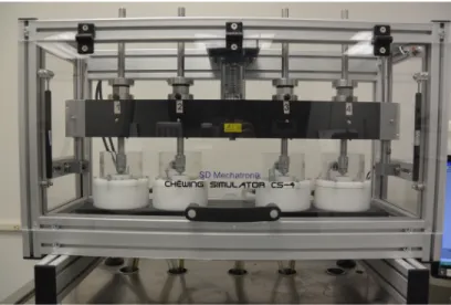
Specimens were later fatigued and thermo cycled using SD Mechatronik
chewing simulator CS-4 (SD MECHATRONIK GMBH Miesbacher Straße 34
D-83620 Feldkirchen-Westerham GERMANY). Samples where mounted in a
(TC: 6000 58/558; 2 min each cycle) and mechanical loading (100 N; 1.6 Hz 100.000 cycles) was performed with parameters based on previous reports.
Figure 6- SD Mechatronik chewing simulator CS-4
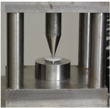
Specimens in group A (fatigued) and group B (control), were measured for strength with a biaxial strength test (piston on 3 balls) as described in the ISO standard 6872 for dental ceramics [82]. Each disk was measured with digital caliper and values were taken into account up to 2 decimal points. Average disk thickness of 1.20 was observed for the specimens.
Tension-compression test machine Instron 33R4204 ,Instron Worldwide Headquarters 825 University Ave. Norwood, MA. with a crosshead speed of 0.15
mm/min was used (Calibration day 04/10/13). To support the specimen 3 steel balls with a diameter of (3mm) were positioned in a support circle with (16mm) diameter The disk - shaped specimens were positioned concentrically on these supports and load was applied centrally with a stainless steel stylus 1.4 mm in diameter at the tip. The load at the point of fracture was recorded and biaxial strength for each specimen recorded and the mean and standard deviations were calculated. Figure 8
2.2 Statistics
2.3 Results
2.3.1 Model for Strength
The results obtained from biaxial loading of samples revealed only modest differences among samples created using all conditions. Generally, a trend for greater strength with higher temperatures and longer holding times was observed (Table 4 and 5). More specific observations are revealed by the statistical evaluations.
The model can be summarized in the following formula,
Strength= group+temperature+holdtime
The Type 3 results for this model are,
Source DF Type III SS Mean Square F Value Pr > F Group 1 0.0424 0.0424 0.00 0.9988 Temp 3 143180.9626 47726.9875 2.68 0.0506 Holdtime 2 112575.1159 56287.5580 3.16 0.0464
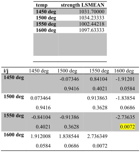
different (smaller) than that with 1600 degree temperature (p=0.0072). But the differences between other pairs (i.e. 1450 vs 1500, 1450 vs 1550, 1450 vs 1600, 1500 vs 1550 and 1500 vs 1600) are not statistically different.
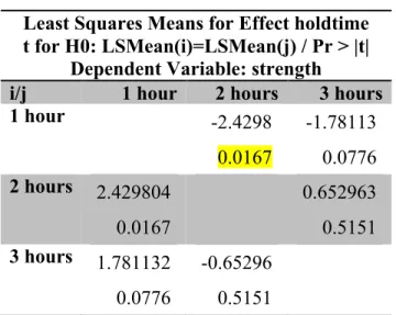
Table 6- Results for holding time
holdtime strength LSMEAN 1 hour 999.30664 2 hours 1072.35000 3 hours 1052.85000
Least Squares Means for Effect holdtime t for H0: LSMean(i)=LSMean(j) / Pr > |t|
Dependent Variable: strength
i/j 1 hour 2 hours 3 hours 1 hour -2.4298 0.0167 -1.78113 0.0776 2 hours 2.429804
0.0167
0.652963 0.5151 3 hours 1.781132
Table 7- Results for temperature
temp strength LSMEAN 1450 deg 1031.70000 1500 deg 1034.23333 1550 deg 1002.44218 1600 deg 1097.63333
i/j 1450 deg 1500 deg 1550 deg 1600 deg 1450 deg -0.07346 0.9416 0.84104 0.4021 -1.91201 0.0584 1500 deg 0.073464
0.9416 0.913863 0.3628 -1.83854 0.0686 1550 deg -0.84104
0.4021 -0.91386 0.3628 -2.73635 0.0072 1600 deg 1.912008
The following graphics summarizes the above results:
Figure 10- Biaxial strength test for fatigued group
2.3.2 Model for Translucency
The model can be summarized in the following formula,
𝑡𝑟𝑎𝑛𝑠𝑙𝑢𝑐𝑒𝑛𝑐𝑦 =𝑡𝑒𝑚𝑝𝑒𝑟𝑎𝑡𝑢𝑟𝑒+ℎ𝑜𝑙𝑑𝑡𝑖𝑚𝑒
The Type 3 results for this model are,
Source DF Type III SS Mean Square F Value Pr > F Temp 3 0.00156971 0.00052324 7.02 0.0005 Holdtime 2 0.00004025 0.00002012 0.27 0.7644
Average translucency was not statistically significantly different among the 3 holding times (P=0.76) but was statistically significantly different for the 4 temperatures after adjusting for holding time. The average contrast ratio (i.e. translucency) between 1450 and 1550 degrees , 1450 degree and 1600 degrees, and 1500 and 1600 degrees are significantly different. The average contrast ratios between the other pairs of temperatures (1450 vs 1500; 1500 vs 1550; and 1550 vs 1600) are not statistically significantly different.
Least Squares Means for Effect temp t for H0: LSMean(i)=LSMean(j) / Pr > |t|
Dependent Variable: trans
i/j 1450 deg 1500 deg 1550 deg 1600 deg 1450 deg 0.871674 0.3872 2.492523 0.0158 4.247859 <.0001 1500 deg -0.87167
0.3872 1.620849 0.1109 3.376185 0.0014 1550 deg -2.49252
0.0158 -1.62085 0.1109 1.755336 0.0849 1600 deg -4.24786
<.0001 -3.37619 0.0014 -1.75534 0.0849
2.4 Discussion
In this investigation, the influence of firing time and temperature on zirconia’s physical properties of strength and translucency were examined. Using disks and a biaxial model of strength testing, the current results confirm the relatively high strength of zirconia that approximates 1,000 MPa. This is a central advantage of zirconia among other dental ceramics available today. For example the reported strength of other materials are: Lithium disilicate 400 MPa, Leucite reinforced ceramics (Empress) 140 MPa.
disk shaped specimen is supported below by 3 ball bearings distributed o a circular pattern. The load is applied from above by use of a piston concentric with the support ball bearings.
The 3 - point flexural strength test that has been commonly used to test dental ceramics presents a problem since it can be sensitive to flaws along the specimen edges [82]. Biaxial flexure described in ISO 6872 is a reliable method of choice, since the effect of possible flaws in the edges is eliminated by applying the load in the central area [82]. Several studies have used biaxial flexural strength as a
method to predict performance of all-ceramic materials [83-85]. For example,
Pittayachawan in 2007 tested the biaxial strength of lava and reported Biaxial
flexural strength around 1100 MPa. [86]
This study employed a thermocycling regimen in the evaluation of fatigue. This may be of particular relevance to zirconia testing. Low-temperature degradation (LTD) has been associated with several 3Y-TZP-based biomaterials, but it is difficult to simulate in the laboratory. Thermocycling has been described as an effective way to simulate the oral environment and evaluate the effect of (LDT). Currently, Y2O3
(yttria) or CeO2 (ceria) are being incorporated to zirconia. These stabilizers improve the retention of the tetragonal structure at room temperature. Perdigao in 2012 evaluated the effect of thermocycling in zirconia with yttria and Ceria as stabilizers and reported lower monoclinic fraction in the group that contained Ceria as stabilizer. The group that contained yttria reflected greater susceptibility to LTD.
light adsorbed from the sample to calculate translucency. There exist other methods of measuring translucency that include spectroradiometry. These instruments measure different parameters of irradiance and radiance. When Lim et al compared the measures of translucency using both spectroradiometry and spectrophotometry; values were higher for the spectroradiometry measures. The measures were highly correlated. A previous study measured translucency of various ceramics and the CR results were between 0.82 and 0.89 for zirconia samples such as Vita Y Z zirconia and Lava, and for more translucent materials such as Empress values range from 0.69 to 0.77. [70].
among all groups. Further studies may be performed with an immersed sample or highly polished sample to reduce the influence of scatter and edge-effect. The opportunity to use glazed samples was avoided in response to the report of Heffernan (2002) that revealed modestly higher translucency for glazed materials. In this regard, the clinical use of polished zirconia may be preferred due to favored wear versus natural teeth or prostheses. [87,88]
A clinically relevant zirconia substrate dimension was studied. Its use in
anatomic monolithic prostheses often requires 16 – 20 mm2 connector dimensions
minimally. Implant supported prostheses with anterior tooth display will have incisor thicknesses approaching or exceeding anatomic limits. Here, samples were 1.2 mm thick. Among the many variables affecting translucency, thickness reduces translucency of dental porcelains [87-88].
Sintering temperatures are one variable that influences the strength of zirconia. Previous investigations demonstrated zirconia demonstrated highest strengths with sintering temperatures of 1400 C and 1550 C. Sintering temperatures of 1650 C and above lead to changes in the microstructure that decreased the materials strength. One of the limitations described in this study was the fact that they only used one type of zirconia and those results might not be applicable for all brands. [70].
were used. It may be suggested that clinical manipulations of zirconia be conducted in calibrated ovens. Further, under no circumstances should firing be conducted using reduced holding times.
The hypothesis that elevated sintering temperatures will increase the translucency of the resulting zirconia was not supported by the spectrophotometric analyses. Globally, translucency values of 0.78 to 0.82 were observed. These values reflect previous measures of zirconia and confirm that zirconia is less translucent than lithium disilicate materials with translucency values measured to be approximately 0.7 [71]. We speculate that further polishing, glazing or wet measurement of these samples would reduce the reflectance component of the measurement and improve translucency. Whether or not highly polished or glazed materials that are represented clinically are more or less translucent than the samples measure here cannot be determined. However, the issue of reflectance would affect all samples similarly and mask translucency with equal magnitude. Although there is a trend toward greater translucency with higher sintering temperatures and times, it is unclear how this may translate into the clinical environment.
respectively. No significant difference was found in the opacity of glazed and non-glazed specimens of IPS Empress 2, In-Ceram Alumina, In- Ceram Zirconia, and metal-ceramic. Glazing cycles decreased the opacity for all veneered materials, except for In-Ceram Zirconia and metal-ceramic specimens. We could possibly suggest that the effect of glazing for translucency measures is material dependent.
Clinical performance of contemporary all ceramics systems depend on a variety of factors that finally would determine how a material would perform in the oral environment. Prior to clinical manipulation evaluation of material properties in vitro might help to predict its clinical performance. The opportunity to compare the measured translucency with previous reports is limited. Existing studies have used very different samples of different thickness and made of different zirconia substrates. This work, however, demonstrates the modest sensitivity of zirconia translucency to holding temperatures and times.
One potential clinical implication that may be suggested from this investigation is that while ‘fine-tuning’ of the firing cycle can provide minor enhancement of zirconia translucency without deleterious effects on strength, other approaches to creating highly translucent restorations using monolithic zirconia are needed.
2.5 Conclusions
Within the limitations of these experiments, the follow observations were made:
1) Increasing temperature from 1450 – 1600oC led to minor increase in biaxial
2) Increasing the holding times from 1 to 2 to 3 hours at temperatures ranging
from1450 – 1600oC led to increased flexural strength when holding times
exceed 1 hour.
REFERENCES
1 Piconi C , G Maccauro Zirconia as ceramic biomaterial Biomaterials 20 (1999) 1—25
2 Raigrodski, Ariel J., et al. "Survival and complications of zirconia-based fixed dental prostheses: a systematic review." The Journal of prosthetic dentistry 107.3 (2012): 170-177
3 Schley, Jaana‐Sophia, et al. "Survival probability of zirconia‐based fixed dental prostheses up to 5 yr: a systematic review of the literature." European journal of oral sciences 118.5 (2010): 443-450.
4 Sailer, Irena, et al. "A systematic review of the survival and complication rates of all‐ceramic and metal–ceramic reconstructions after an observation period of at least 3 years. Part II: fixed dental prostheses." Clinical Oral Implants Research 18.s3 (2007): 86-96.
5 Larsson, Christel, and Ann Wennerberg. "The clinical success of zirconia-based crowns: a systematic review." International Journal of Prosthodontics 27.1 (2014).
6 Zhang, Yu, et al. "Edge chipping and flexural resistance of monolithic ceramics." Dental Materials 29.12 (2013): 1201-1208.
7 Spies, Benedikt Christopher, Susanne Stampf, and Ralf‐Joachim Kohal. "Evaluation of Zirconia‐Based All‐Ceramic Single Crowns and Fixed Dental Prosthesis on Zirconia Implants: 5‐Year Results of a Prospective Cohort Study." Clinical implant dentistry and related research (2014).
8 Örtorp, Anders, Maria Lind Kihl, and Gunnar E. Carlsson. "A 5-year retrospective study of survival of zirconia single crowns fitted in a private clinical setting." Journal of dentistry 40.6 (2012): 527-530.
9 E Jung, Ronald, et al. "Systematic review of the survival rate and the incidence of biological, technical, and aesthetic complications of single crowns on implants reported in longitudinal studies with a mean follow‐up of 5 years." Clinical oral implants research 23.s6 (2012): 2-21.
10 McLaren EA, Giordano RA. Zirconia-based ceramics: material properties, esthetics and layering techniques of a new veneering porcelain, VM9. Quintessence of Dental Technology 2005;28:99–112.
15 J.R. Queiroz, P. Benetti, M. Massi, L.N. Junior, A. Della BonaEffect of multiple firing and silica deposition on the zirconia-porcelain interface bond strength
16 F.Z. Trindade, M. Amaral, R.M. Melo, M.A. Bottino, L.F. Valandro Zirconia-porcelain bonding: effect of multiple firings on microtensile bond strength J Adhes Dent, 15 (2013) [in press]
17 Queiroz JR, Benetti P, Massi M, Junior LN, Della Bona A. Effect of multiple firing and silica deposition on the zirconia-porcelain interface bond strength. Dent Mater 2012;28:763–
18 Mainjot, Amélie K., et al. "Influence of zirconia framework thickness on residual stress profile in veneering ceramic: measurement by hole-drilling." Dental Materials 28.4 (2012): 378-384.
19 Wang CAD CAM, Aboushelib MN, Feilzer AJ (2008). Strength influencing variables on CAD/CAM zirconia frameworks. Dent Mater 24:633-638.
20 Tholey MJ, Swain MV, Thiel N (2009). SEM observations of porcelain CAD CAM-TZP interface. Dent Mater 25:857-862.
21. Belli, Renan, et al. "Thermal-induced residual stresses affect the lifetime of zirconia–veneer crowns." Dental Materials 29.2 (2013): 181-190.
22 Komine F, Saito A, Kobayashi K, Koizuka M, Koizumi CAD CAM, Matsumura CAD CAM. Effect of cooling rate on shear bond strength of veneering porcelain to a zircnoia ceramic material. J Oral Sci 2010;52:647–52.
23 Gostemeyer G, Jendras M, Dittmer MP, Bach FW, Stiesch M, Kohorst P. Influence of cooling rate on zircinia/veneer interfacial adhesion. Acta Biomater 2010;6:4532–8.
24 Helmer JD, Driskell TD. Research on bioceramics. Symp. on Use of Ceramics as Surgical Implants. South Carolina (USA): Clemson University, 1969.
25 Christel P, Meunier A, Dorlot J-M et al. Biomechanical compatibility and design of ceramic implants for orthopaedic surgery. Bioceramics: material charateristics versus in vivo be- havior. Ann NY Acad Sci 1988;523:234—56.
26 ISO TC 150/SC 1. Implants for surgery—ceramic materials based on yttria-stabilized tetragonal zirconia (CAD CAM-TZP). ISO/DIS 13356, 1995.
27 Christel P, Meunier A, Heller M, Torre J-P, Cales B, Peille CN. Mechanical properties and short-term in vivo evaluation of yttrium-oxide-partially-stabilized zirconia. J Biomed Mater Res 1989;23:45—61.
29 Hayashi, K., et al. "Re-evaluation of the biocompatibility of bioinert ceramics i> in vivo</i>." Biomaterials 13.4 (1992): 195-200.
30 Özkurt, Zeynep, and Ender Kazazoglu. "Zirconia dental implants: a literature review." Journal of Oral Implantology 37.3 (2011): 367-376.
31 Neunzehn, Jörg, Beate Lüttenberg, and Hans-Peter Wiesmann. "Investigation of biomaterials by human epithelial gingiva cells: an in vitro study." Head & face medicine 8.1 (2012): 35.
32 Bachhav, Vinay Chila, and Meena Ajay Aras. "Zirconia-based fixed partial dentures: A clinical review." Quintessence International 42.2 (2011).
33Nascimento, Cássio do, et al. "Bacterial adhesion on the titanium and zirconia abutment surfaces." Clinical oral implants research 25.3 (2014): 337-343.
34 Bremer, Felicia, et al. "In vivo biofilm formation on different dental ceramics." Quintessence International 42.7 (2011).
35 Scarano A, Piattelli M, Caputi S, Favero GA, Piattelli A. Bacterial adhesion on commercially pure titanium and zirconium oxide disks: an in vivo human study. Journal of Periodontology 2004;75:292–6.
36. Rimondini L, Cerroni L, Carrassi A, Torricelli P. Bacterial colonization of zirconia ceramic surfaces: an in vitro and in vivo study. International Journal of Oral Maxillofacial Implants2002;17:793–8.
37 Denry, Isabelle, and J. Robert Kelly. "State of the art of zirconia for dental applications." Dental materials 24.3 (2008): 299-307.
38 Cavalcanti, A. N., et al. "Y-TZP ceramics: key concepts for clinical application." Operative dentistry 34.3 (2009): 344-351.
39 Raigrodski AJ. Contemporary materials and technologies for all-ceramic fixed partial dentures: a review of the literature. Journal of Prosthetic Dentistry 2004;92:557–62.
40. Luthy, Filser F, Loeffel O, Schumacher M, Gauckler LJ, Hammerle CH. Strength and reliability of four-unit all- ceramic posterior bridges. Dental Materials 2005;21: 930–7.
41 Rekow, E. D., et al. "Performance of dental ceramics challenges for improvements."
43 Preis, Verena, et al. "In vitro failure and fracture resistance of veneered and full-contour zirconia restorations." Journal of dentistry 40.11 (2012): 921-928.
44 Pjetursson, Bjarni E., et al. "A systematic review of the survival and complication rates of all‐ceramic and metal–ceramic reconstructions after an observation period of at least 3 years. Part I: single crowns." Clinical Oral Implants Research 18.s3 (2007): 73-85.
45 Deville S, Chevalier J, Gremillard L. Influence of surface finish and residual stresses on
46 Kosmač, Tomaž, et al. "The effect of surface grinding and sandblasting on flexural strength and reliability of Y-TZP zirconia ceramic." Dental materials 15.6 (1999): 426-433.
47 Reich S, Petschelt A, Lohbauer U (2008). The effect of finish line prepara-tion and layer thickness on the failure load and fractography of ZrO2copings. J Prosthet Dent 99:369-376.
48 Clausen JO, Abou Tara M, Kern M (2010). Dynamic fatigue and fracture resistance of non-retentive all-ceramic full-coverage molar restorations. Influence of ceramic material and preparation design. Dent Mater 26:533-538.
49 Komine, Futoshi, Markus B. Blatz, and Hideo Matsumura. "Current status of zirconia-based fixed restorations." Journal of oral science 52.4 (2010).
50 Albashaireh ZSM, Ghazal M, Kern M. Two-body wear of differentceramic materials opposed to zirconia ceramic. J Prosthet Dent2010;104:105–13.
51 Sorensen JA, Sultan EA, Sorensen PN. Three-body wear of enamelagainst full crown ceramics. In: 89th IADR; 2011 [Abstr. No. 1652].
52 Jung, Yu-Seok, et al. "A study on the in-vitro wear of the natural tooth structure by opposing zirconia or dental porcelain." The journal of advanced prosthodontics 2.3 (2010): 111-115.
53 Janyavula, Sridhar, et al. "The wear of polished and glazed zirconia against enamel." The Journal of prosthetic dentistry 109.1 (2013): 22-29.
54 Stober, T., et al. "Enamel wear caused by monolithic zirconia crowns after 6 months of clinical use." Journal of oral rehabilitation (2014).
56 Karl, Matthias, et al. "Passivity of fit of CAD/CAM and copy-milled frameworks, veneered frameworks, and anatomically contoured, zirconia ceramic, implant-supported fixed prostheses." The Journal of prosthetic dentistry 107.4 (2012): 232-238.
57 Luthardt, R. G., et al. "Reliability and properties of ground Y-TZP-zirconia ceramics." Journal of dental research 81.7 (2002): 487-491.
58 Seelbach, Paul, Cora Brueckel, and Bernd Wöstmann. "Accuracy of digital and conventional impression techniques and workflow." Clinical oral investigations 17.7 (2013): 1759-1764.
59 Sailer I, Gottnerb J, Kanelb S, Hammerle CH. Randomized controlled clinical trial of zirconia-ceramic and metal-ceramic posterior fixed dental prostheses: a 3-year follow-up. Int J Prosthodont 2009; 22: 553–560.
60 Sailer I, Feher A, Filser F, Gauckler LJ, Luthy CAD CAM, Ham- merle CH. Five-year clinical results of zirconia frameworks for posterior fixed partial dentures. Int J Prosthodont 2007; 20: 383–388.
61 Schmitt J, Holst S, Wichmann M, Reich S, Gollner M, Hamel J. Zirconia posterior fixed partial dentures: a prospec- tive clinical 3-year follow-up. Int J Prosthodont 2009; 22: 597– 603.
62. Raigrodski AJ, Chiche GJ, Potiket N, Hochstedler JL, Mohamed SE, Billiot S, Mercante DE. The efficacy of posterior three-unit zirconium-oxide-based ceramic fixed partial dental prostheses: a prospective clinical pilot study. J Prosthet Dent 2006; 96: 237–244.
63 Molin MK, Karlsson SL. Five-year clinical prospective evaluation of zirconia-based denzir 3-unit FPDs. Int J Prosth- odont 2008; 21: 223–227.
64 Martínez-Rus, Francisco, et al. "Evaluation of the absolute marginal discrepancy of zirconia-based ceramic copings." The Journal of prosthetic dentistry 105.2 (2011): 108-114.
65 Att, Wael, et al. "Marginal adaptation of three different zirconium dioxide three-unit fixed dental prostheses." The Journal of prosthetic dentistry 101.4 (2009): 239-247.
67. Vichi, Alessandro, et al. "Color related to ceramic and zirconia restorations: a review." Dental materials 27.1 (2011): 97-108
68 Hjerppe, Jenni, et al. "Effect of shading the zirconia framework on biaxial strength and surface microhardness." Acta Odontologica 66.5 (2008): 262-267.
69 Spear, Frank, and Julie Holloway. "Which all-ceramic system is optimal for anterior esthetics?." The Journal of the American Dental Association 139.suppl 4 (2008): 19S-24S.
70 Spink, Lisa. "A Comparison of Absolute Translucency and Relative Translucency of Dental Ceramic
71 Heffernan, Michael J., et al. "Relative translucency of six all-ceramic systems. Part I: core materials." The Journal of prosthetic dentistry 88.1 (2002): 4-9.
72 Anusavice, Kenneth J. "Standardizing failure, success, and survival decisions in clinical studies of ceramic and metal–ceramic fixed dental prostheses." Dental Materials 28.1 (2012): 102-111.
73 Cavalcanti, A. N., et al. "Y-TZP ceramics: key concepts for clinical application." Operative dentistry 34.3 (2009): 344-351.
74 Broseghini, Cristiano, et al. "Aesthetic functional area protection concept for prevention of ceramic chipping with zirconia frameworks." International Journal of Prosthodontics 27.2 (2014).
75 Abduo, Jaafar, et al. "Fit of screw-retained fixed implant frameworks fabricated by different methods: a systematic review." International Journal of Prosthodontics 24.3 (2011).
76 Kimmich, Magdalena, and Christian FJ Stappert. "Intraoral treatment of veneering porcelain chipping of fixed dental restorations: a review and clinical application." Journal of the American Dental Association (JADA) 144.1 (2013).
77 Limmer, Bryan Michael. Complications and Patient Centered Outcomes with a Monolithic Zirconia Implant Supported Fixed Prosthesis. Diss. University of North Carolina, 2012.
78 Thalji, Ghadeer N., and Lyndon F. Cooper. "Implant‐Supported Fixed Dental Rehabilitation with Monolithic Zirconia: A Clinical Case Report." Journal of Esthetic and Restorative Dentistry (2014).
Journal of dentistry 35.11 (2007): 819-826.
80 Miyagawa, Y., J. M. Powers, and W. J. O'brien. "Optical properties of direct restorative materials." Journal of Dental Research 60.5 (1981): 890-894.
81 Yu, Bin, Jin-Soo Ahn, and Yong-Keun Lee. "Measurement of translucency of tooth enamel and dentin." Acta Odontologica 67.1 (2009): 57-64.
82 ISO Standard No. 6872, Dental ceramic, 2nd edn. Geneva: International Organization for Standardization; 1995. p. 1-9.
83.Guazzato M, Albakry M, Swain MV, Iron- side J. Mechanical properties of In-Ceram Alumina and In-In-Ceram Zirconia. Int J Prosthodont 2002;15:339-46.
84.Wagner WC, Chu TM. Biaxial flexural strength and indentation fracture tough- ness of three new dental core ceramics. J Prosthet Dent 1996;76:140-4.
85.Wen MY, Mueller HJ, Chai J, Wozniak WT. Comparative mechanical property charac- terization of 3 all-ceramic core materials. Int J Prosthodont 1999;12:534- 86 Pittayachawan, Piyapanna, et al. "The biaxial flexural strength and fatigue property of Lava™ Y-TZP dental ceramic." Dental Materials 23.8 (2007): 1018-1029
87 Heffernan, Michael J., et al. "Relative translucency of six all-ceramic systems. Part I: core materials." The Journal of prosthetic dentistry 88.1 (2002): 4-9.