BIOCHEMICAL ACTIVITIES AND GENETIC FUNCTIONS OF THE DROSOPHILA MELANOGASTER FANCM HELICASE IN DNA REPAIR
Noelle-Erin F. Romero
A dissertation submitted to the faculty of the University of North Carolina at Chapel Hill in partial fulfillment of the requirements for the degree of Doctor of Philosophy in the Curriculum of Genetics and Molecular Biology
Chapel Hill 2016
ABSTRACT
Noelle-Erin F. Romero: Biochemical activities and genetic functions of the Drosophila melanogaster Fancm helicase in DNA repair
(Under the direction of Steve Matson and Jeff Sekelsky)
The DNA damage response in eukaryotes involves multiple, complex, and often redundant pathways that respond to various types of DNA damage that affect one or both strands of DNA. One type of toxic DNA damage that can occur is a double-strand break (DSB). Repair of a DSB can lead to the formation of a recombination product known as a crossover (CO). Crossovers in mitotic cells can be deleterious and lead to chromosomal rearrangements or cell death. In order to limit crossing over during DSB repair, eukaryotes possess mechanisms to ensure crossovers do not occur. In this manner, several helicases function during repair of DSBs to promote accurate repair and prevent the formation of
crossovers through homologous recombination.
Among these helicases is the Fanconi anemia group M (FANCM) protein. FANCM is one of 17 Fanconi anemia (FA) proteins and is one of the most broadly? conserved FA proteins. FANCM and its orthologs, Mph1 and Fml1, are DNA junction-specific helicases/translocases that process homologous recombination (HR) intermediates. Additionally, FANCM has been implicated in a number of DNA metabolic processes including activation of the S-phase checkpoint, trasversal of interstrand crosslinks, recruitment of the proteins such as the FA core complex and Blm to sites of DNA damage, and prevention of mitotic crossovers during double-strand break repair.
Drosophila melanogaster Fancm. I show that purified Fancm is a 3ʹ to 5ʹ ATP-dependent helicase that can
ACKNOWLEDGEMENTS
This work would not have been completed without the support from my thesis advisors, Jeff Sekelsky and Steve Matson. Grampa Steve- Thanks for teaching me about biochemistry, buying me bagels, and letting me yell at you. Special thanks to members of the Matson and Sekelsky Labs, past and present, who’ve made my time at UNC fun and memorable. Thanks for laughing with me, laughing at me, and keeping science fun. Suzy Q- serenity now! JK Ho- thanks for not getting super mad at me when I fucked up suduko <3; sevens everywhere! Baby Mama- Sometimes you just need to vent and rant, thanks for letting me always bug you and distract you. TBags-I love you, but not as much as I love Susan. Seriously though, Tits out! Ldoc- thanks for bringing some color into the old white world of science.
Science is hard. Being a minority in science is harder. If someone’s not lifting you up, they’re dragging you down. I would have given up if not for all those who lifted me up: my peer support, my extended UNC family of IMSD and IME, and the wonderful mentors and role models I’ve had. Ashalla Freeman- Thank you for making me part of your family, supporting me, encouraging me, and being an amazing friend. Kathy Wood- I don’t know what I would do with you. Thanks for always fighting for us, lifting us up, and celebrating us. Kacey Hammel- Thanks for saving me, for believing in me, and seeing me through. Sibby Anderson-Thompkins- Thanks for keeping it real, giving me a chance, and supporting me. Lastly, my greatest support comes from my family. To my mom and sister, I love you.
TABLE OF CONTENTS
LIST OF FIGURES ...ix
LIST OF TABLES ... x
LIST OF ABBREVIATIONS ...xi
CHAPTER 1-INTRODUCTION ... 1
PRE-REPLICATIVE REPAIR ... 2
POST-REPLICATIVE REPAIR ... 4
DOUBLE-STRAND BREAK REPAIR VIA HOMOLOGOUS RECOMBINATION ... 5
FANCM AND HOMOLOGOUS RECOMBINATION ... 10
FIGURES ... 13
REFERENCES ... 17
CHAPTER 2- BIOCHEMICAL ANALYSIS OF THE ... 25
D. MELANOGASTER
FANCM ORTHOLOG ... 25
INTRODUCTION ... 25
MATERIALS AND METHODS ... 28
Expression and purification of
Drosophila
FANCM ... 28
DNA Substrates ... 29
ATPase assays ... 29
Helicase Assays ... 29
Fluorescence Anisotropy ... 30
RESULTS AND DISCUSSION... 30
Fancm is a ssDNA-dependent ATPase ... 30
FIGURES ... 36
REFERENCES ... 49
CHAPTER 3- GENETIC ANALYSIS OF
D. melanogaster Fancm
... 52
INTRODUCTION ... 52
MATERIALS AND METHODS ... 53
Drosophila stocks ... 53
Mitotic crossover assay... 54
DNA damage sensitivity assays ... 54
RESULTS AND DISCUSSION... 54
Helicase-dead and truncated Fancm are each able to prevent a subset of mitotic crossovers ... 54
Separation of function in Fancm’s roles in the response to DNA damage ... 57
FIGURES ... 61
REFERENCES ... 68
CHAPTER 4 Concluding remarks ... 72
Functions of fancm in SDSA ... 74
Protein-protein interactions ... 76
Checkpoint signaling ... 80
Posttranslational modifications ... 81
Role for Fancm during DNA replication ... 82
SUMMARY ... 84
FIGURES ... 85
LIST OF FIGURES
Figure 1.1- Pre and Post replication repair ... 13
Figure 1.2- Model for DSB repair Via Homologous Recombination ... 14
Figure 1.3 - The Fanconi anemia family repair network ... 15
Figure 1.4 - Model for ICL removal ... 16
Figure 2.1 - Purification of Fancm∆ and Fancm∆KM ... 37
Figure 2.2 - Schematic of Fancm ... 38
Figure 2.3 - ATP hydrolysis by Fancm as a function of temperature ... 39
Figure 2.4 - ATP hydrolysis by Fancm as a function of time ... 40
Figure 2.5 - ATP hydrolysis by Fancm as a function of NaCl concentration ... 41
Figure 2.6 - ATP hydrolysis by Fancm ... 42
Figure 2.7 - Fancm binding of DNA substrates as measured by fluorescence anisotropy ... 43
Figure 2.8 - Fancm unwinds duplex DNA in an ATP dependent manner ... 44
Figure 2.9 - Unwinding of partial duplex DNA substrates by Fancm ... 45
Figure 2.10 - Unwinding of bubble-like structure by Fancm ... 46
Figure 2.11 - Unwinding of D-loop intermediate substrates by Fancm ... 47
Figure 2.12 - Timecourse unwinding of partial duplex DNA substrates by Fancm ... 48
Figure 3.1 - Schematic of Fancm ... 62
Figure 3.2 - Fancm transgenic flies ... 63
Figure 3.3 - Spontaneous mitotic crossover rates ... 64
Figure 3.4 - Sensitivity of Fancm to HN2 ... 65
Figure 3.6 - Sensitivity of Fancm to MMS ... 67
Figure 4.1 - COBALT alignment and predicted phosphorylation site ... 86
LIST OF TABLES
LIST OF ABBREVIATIONS AMP-PNP Adenosine-5ʹ-(β,γ-imido) triphosphate
AP Apurinic Site
ATP Adenosine-5ʹ- triphosphate
BSA Bovine serum albumin
bp Base pair
CO Crossover
D-loop Displacement loop
dHJ Double Holliday junction
DNA Deoxyribonucleic acid
dsDNA Double-stranded deoxyribonucleic acid
DTT Dithiothreitol
DSB Double-strand break
DSBR Double-strand break repair
EDTA Ethylenediamine tertaacetic acid
FA Fanconi anemia
FL Transgene of full length Fancm
FLKM Transgene of full length Fancm with a mutation in the Walker A motif
HhH Helix-hairpin-helix
HJ Holliday junction
HR Homologous Recombination
ICL Interstrand Crosslink
IR Ionizing Radiation
LB Luria-Bertani broth
MBP Maltose binding protein
MMR Mismatch repair
MMS Methyl methanesulfonate
NCO Noncrossover
NER Nucleotide excision repair
NHEJ Non-homologous end joining
nt Nucleotide
NTP Nucleotide triphosphate
OD Optical density
OH Overhang
PCR Polymerase chain reaction
SF Superfamily
ssDNA Single-stranded deoxyribonucleic acid
TBE Tris base, boric acid, and EDTA
TE Tris-HCl (pH8.0) EDTA
TLS Translesion synthesis
Tris Tris [hydroxymethyl] aminomethane
tr Transgene of truncated Fancm
trKM Transgene of truncated Fancm with a mutation in the Walker A motif
UTR Untranslated region
γ-[32P]ATP Adenosine-5ʹ- triphosphate 32P labeled on the phosphate
Δ Truncation of 840 amino acids
CHAPTER 1-INTRODUCTION
Maintaining the structural integrity of DNA is essential to the health and vitality of a cell and organism as it serves as a permanent copy of the cell’s genome. Changes in its structure and/or sequence can have severe consequences as it can cause alterations in other cell components, such as proteins and structural RNAs. It is therefore critical that the integrity of DNA be maintained. While the duplex structure of DNA makes it a particularly stable repository of genetic information (1, 2), there are many times in which DNA molecules are more susceptible to damage. This includes periods of time in which the separation of the duplex DNA is necessary, such as replication during cell division (mitosis and meiosis), as well as during transcription of DNA into RNA. During these times the DNA can be altered by the proteins that act upon it or damaged by various factors.
For instance, the incorporation of incorrect bases or deletion of bases during DNA replication can have a profound effect on an organism. Polymerases, proteins responsible for replication, can encounter regions of highly repetitive DNA where the (3, 4) polymerase becomes susceptible to slippage or
(depurination) that occurs from the cleavage of the bond between the base and the deoxyribose, or through deamination of adenine, cytosine, or guanine (5–7). Exogenous exposure to environmental agents, such as toxins, UV radiation, and pollutants can cause breakage of DNA or toxic lesions. Covalent modifications to the sugar phosphate backbone (backbone modifications) or the nitrogenous bases of DNA (base modifications) can distort or break the helix. The type and frequency of the modification or lesion can vary depending on both the lesion type and the modifying agent. Induced or exogenous damage includes the formation of interstrand crosslinks (ICLs) generated by
chemotherapeutic agents, and pyrimidine dimers, in which two adjacent pyrimidines are joined by a ring-like structure, as seen in the case of damage induced by UV radiation. Alkylation, or the addition of a methyl or ethyl group to various positions on the DNA base, is another form of induced damage(5, 8, 9). Modifications, such as oxidative base modifications, can be numerous and can distort the helix.
A number of repair processes exist in order to ensure that detrimental modifications made to DNA are removed to ensure cell vitality and that any damage that occurs to the DNA is corrected efficiently and effectively. Repairing of DNA damage is essential as damage to DNA can block replication or
transcription and can result in a high frequency of mutations, which can be detrimental to cell reproduction and viability (4, 5, 10). To maintain the integrity of their genomes, cells have therefore evolved a robust set of mechanisms to repair damaged DNA. These myriad DNA repair mechanisms can be classified as either pre-replicative repair or post-replicative repair depending on where in the cell cycle they take place (Figure 1.1). Pre-replicative repair includes reversal of chemical damage or modification (direct reversal) or removal of the damaged base and synthesis of new DNA (excision repair) (9, 11). These systems act to correct DNA damage before replication allowing DNA synthesis to proceed using an undamaged DNA strand as a template. If these systems fail to remove the damage, alternative mechanisms for dealing with damaged DNA are employed post-replication.
PRE-REPLICATIVE REPAIR
results in the restoration of the original base in an unaltered state without synthesis or remodeling of the DNA. This method of repair is generally viewed as a highly efficient way of dealing with specific types of DNA damage that occur with a high frequency. For instance, UV light is a major source of DNA damage and can induce 6-4 photoproducts and pyrimidine dimers. The formation of pyrimidine dimers distorts the helical structure of the DNA and blocks transcription or replication past the site of damage. The direct reversal of this dimer occurs through a process known as photoreactivation. This process utilizes energy from visible light to break the ring structure that binds the pyrimidines together (12, 13). Although UV irradiation is the cause of almost all skin cancer in humans, photoreactivation is not a repair mechanism found in humans, although a variety of prokaryotic and eukaryotic cells employ photoreactivation as a means of repair (5, 9, 10).
When direct reversal of the DNA lesion is not possible, excision of the damaged base is
employed. Unlike direct reversal of DNA damage, excision of damage is a more general means to repair a broad range of alterations to DNA. Various types of excision repair mechanisms exist and are highly important DNA repair strategies in prokaryotic and eukaryotic cells. Excision repair involves the removal of the damage and synthesis of new DNA to fill the resulting gap. Types of excision repair include nucleotide-excision repair (NER), base excision repair (BER), and mismatch repair (MMR).
Similar to MMR, NER also involves the excision of DNA damage and restoration of the original sequence. In E. coli, NER is carried out by the UvrABC complex. UvrA recognizes damaged DNA, recruits UvrB and C, which are then responsible for cleaving on the 3ʹ and 5ʹ side of damage. Using mammalian cell lines and the identified genes that are involved in NER, the NER pathway in humans has been elucidated. Similar to the pathway in E. coli, XPA recognizes damaged DNA and recruits the heterodimer XPF/ERCC1 and XPG to the repair complex. XPG and XPF/ERCC1 are endonucleases that cleave DNA on the 3′ and 5′ sides of the damage. XPA also recruits XPB and XPD, which acts as a helicase to unwind the damaged DNA that was excised by XPF/ERCC1. The resulting gap is filled in by DNA polymerase and sealed by DNA ligase (18, 22).
POST-REPLICATIVE REPAIR
If damage persists post-replicative repair mechanisms are used to restore the integrity of the DNA. Pyrimidine dimers left unrepaired, ICLs, and many other types of lesions cannot be copied by DNA polymerases and block movement of the replication fork. One mechanism used to overcome these blockages is recombinational repair. Recombinational repair utilizes the undamaged homologous template to synthesize new, undamaged DNA (22–24). The damaged portion (e.g. a pyrimidine dimer or crosslink), can then subsequently be removed by one of the excision repair mechanisms. During the repair process of lesions that affect both strands, a combination of repair methods may be utilized. Repair of ICLs, explored later in this chapter, uses both excision and recombinational repair processes to restore DNA to its native state. Recombinational repair can also be used when damage occurs to the DNA phosphodiester sugar backbone. Strand modifications include single and double strand breaks, which can result from environmental and metabolic sources (10, 25–28).
operates during all phases of the cell cycle and is considered the predominant pathway in mammalian cells for DSB repair while HR is restricted to late-S and G2 phases. NHEJ is considered error-prone and eliminates DSBs through direct ligation of the broken ends (32). HR-directed repair is largely an error-free mechanism as it utilizes the undamaged sister chromatid, or homologous chromosome, as a template (31, 33). The work in this thesis focuses on DSB repair via HR and will not focus on NHEJ as the primary mode for repair of DSBs in Drosophila melanogaster is HR.
DOUBLE-STRAND BREAK REPAIR VIA HOMOLOGOUS RECOMBINATION
The fact that identical information is held on the sister chromatid, and a duplicate copy is available in the homologous chromosome, makes repair via homologous recombination an essential pathway to ensure accurate repair of broken DNA. DSBs, which can be induced by radiation and select toxins, are repaired via recombinational mechanisms in which an intact DNA molecule is used. Our current knowledge regarding the mechanism of homologous repair derives largely from studies done in yeast. The process of double strand break repair via homologous recombination was first proposed by Robin Holliday. Holliday and colleagues postulated that, during meiosis, DNA repair gives rise to crossovers (COs), and gene conversions (30, 34). Current models for repair via homologous recombination are based on the model of double-strand break repair (DSBR) originally outlined by Szostak (34). Key to this model are several essential steps (Figure 1.2):
evidence exists to support the direct assumption that double-strand break model of meiotic recombination accurately reflects mitotic recombination, core features, such as those outlined above, are believed to also hold true in mitotic DSB repair (38, 39).
While the essential steps of recombination are maintained between both processes, one of the fundamental differences between meiotic and mitotic recombination is regulation and promotion of recombination products. In meiotic dividing cells, recombination products (COs) are actively promoted as they contribute to genetic diversity and ensure proper segregation of chromosomes during cell division (38). Meiotic recombination is indispensable for accurate chromosome segregation and is promoted by generation of a DSB by a meiosis-specific nuclease. The subsequent crossover formation occur at various frequencies depending on the organism, but generally require at least one CO per chromosome, and help facilitate proper chromosomal alignment and subsequent segregation (40). Disruption of this process can lead to a number of aberrations and deleterious effects, such as genomic deletions and improper segregation of chromosomes (nondisjunction) which can lead to aneuploidy (39).
While homologous recombination through programed DSBs is essential in meiotic cells to ensure proper chromosomal segregation and promote genetic diversity, HR in mitotic cells is used to repair spontaneous and induced DSB damage. As mentioned above, repair of ICLs and other base
modifications utilize specialized nucleases to generate nicks in the DNA backbone to facilitate removal of the offending lesion. Excision of the damaged base can result in DSBs as nucleases used to excise the damage can nick both strands of the sugar phosphate backbone. The generation of a DSB can lead to the use of HR as a repair mechanism. However, the formation of COs during HR via DSB repair in mitotically dividing cells can be hazardous as they can result in loss of heterozygosity and gross
Repair of DSBs through HR requires multiple repair and recombination proteins. Processing of DSBs involves resection in which the 5ʹ ends on either side of the break are trimmed back to create 3ʹ single-stranded DNA overhangs. This process involves specialized proteins and protein complexes, namely Mre11-Rad50-Nbs1, and Exo1 exonuclease (27, 43). The single-stranded DNA tails are then coated by the single-stranded DNA binding protein RPA to remove secondary structures. RPA is subsequently displaced by Rad51 with the help of Rad51 mediator proteins (44) generating a single-stranded DNA tail coated with Rad51. This Rad51 nucleoprotein filament executes homology-mediated search and invasion of the homologous template. In humans, this template is the sister chromatid, but in some cases and some species, like Drosophila, this template can be the homologous chromosome. Following invasion, DNA synthesis is carried out by a DNA polymerase (31) (Figure 1.2).
Following invasion and subsequent DNA synthesis, one of two steps can occur: SDSA or HJ formation. If a Holliday junction is formed, cleavage by structure-specific endonucleases, such as
the energy of nucleoside triphosphate hydrolysis to transiently convert duplex DNA to single-stranded DNA. One family of conserved DNA helicases/translocases, whose members are involved in HR mediated repair, are relatives of archaeal Hef (Helicase-associated endonuclease for fork-structured DNA) (47–50). The Hef protein from Pyrococcus furiosus contains a conserved DEAD-box helicase motif toward the N-terminus and an endonuclease reminiscent of the nucleases ERCC4 endonuclease in its C-terminus (51). Hef processes DNA intermediates that are generated during HR, such as forks and four-way junctions (HJs) (52) either through its helicase activity or nuclease activity. Hef functions as a
homodimer in cleaving DNA forks and processing Holliday junctions into splayed arms, indicating roles for this protein during DNA replication and repair (50, 52). These helicases are members of the SF2 helicase superfamily. Relatives of Hef are found as orthologs of the human Fanconi anemia group M (FANCM) protein.
FANCM was first characterized as the yeast gene mph1 from Saccharomyces cerevisiae during a mutator screen to genetically characterizes genes of unknown function (53). This initial screen showed that deletion of MPH1 led to an increased spontaneous mutation rate. The impact of this protein was not fully examined until both the human ortholog, FANCM, and archael Hef were identified and the
biochemical activity was examined (50, 54, 55). FANCM was identified as a 250 kDa component of the FA core complex and identified in an FA patient who carried a bi-allelic mutation. This identification of FANCM led to its classification as a new FA complementation group (55, 56).
There are over 17 FA genes and associated genes classified as FA family members. Mutations in any of the FA complement group of genes are associated with the same disorder, Fanconi anemia. Fanconi anemia is a hereditary disorder characterized by an increased incidence of cancer,
consisting of 8 different FA proteins, has also been found to interact with and direct proteins involved in HR, such as the BLM complex and BRCA1/2, to sites of DNA damage (60–63). Although still under investigation, the primary function for FA pathway is thought to be in repairing ICL damage (64, 65). However, the FA proteins are thought to function at various points during the repair of ICLs and can be separated into three groups: The FA core complex, the FANCD2/FANCI (ID) complex, and downstream targets (66). The downstream FA proteins are made up of FANCD1 (BRACA2), FANCJ (BRIP1/BACH1), FACNP (SLX4), and FANCN (PALB2) all of which are associated with recombinational repair (25, 59, 61) (Figure 1.3).
Regulation of the FA pathway is dependent on the ubiquitylation of the ID complex. Upon ubiquitination, the ID complex localizes to chromatin during S phase and in response to DNA damage induced by mitomycin C (MMC), ionizing radiation, and UV exposure (67–69). Monoubiquitylation, and subsequent activation and regulation of the downstream components, occurs through the function of the FA core complex. The FA core complex consists of 8 FA proteins (FANC- A, -B, -C, -E, -F, -G, -L, and -M) as well as FA associated proteins (FAAP24, FAAP100) (57, 70–74). All members of the FA core complex are required for the catalytic subunit, FANCL (75), to function as an E3 ubiquitin ligase (76–79). FANCM, unlike the rest of the core complex, is distinctly different in the involvement of ubiquitylation of the ID complex. Inactivation of FANCM results in an intact core complex and the partial ubiquitylation of FANCD2 (80), leading to the idea that FANCM is partially redundant with another protein, has functions outside of the FA pathway (81, 82), or act as a signaling protein that targets the core complex to DNA (70, 83, 84).
(22). The ICL is then untethered by an endonuclease. Recombinational repair can then be used to correct the resulting gap (Figure 1.4).
FANCM AND HOMOLOGOUS RECOMBINATION
Although many of these FA proteins are unique to mammals, and to some extent metazoans, one protein, FANCM, is a constant in eukaryotic organisms. D. melanogaster, for instance, lacks the full complement of FA proteins (54), yet the FA proteins that are present in D. melanogaster are important for repair of ICLs. FANCM therefore plays a key role in damage repair and has been implicated in
recombination. The S. cerevisiae FANCM ortholog, Mph1 (54, 66, 85, 86), has been shown to be involved in preventing crossovers (48), and mph1 mutants show hypersensitivity to DNA damaging agents such as ionizing radiation (IR) andmethyl methanesulfonate (MMS) (53). Biochemical studies using purified Mph1 show that it is a DNA helicase capable of unwinding Rad51-coated D-loops (48, 87), and that it can process DNA intermediates that form later in repair, including HJs (48, 87, 88). Unwinding of HJs and D-loops has also been observed using the S. pombe ortholog Fml1 (89). In contrast, no helicase unwinding activity has been detected for human FANCM (55, 90). Together, genetic and biochemical studies suggest roles for FANCM and its orthologs in HR that are dependent upon their ability to use ATP hydrolysis to unwind or remodel DNA structures so as to prevent COs (47, 48, 91–93).
FANCM’s proposed role outside of the FA response, and the biochemical evidence that yeast FANCM (Mph1) is involved in HR, suggests that the primary role of FANCM is in promoting CO avoidance by processing DNA intermediates that occur during DSB repair via HR (48, 87, 97). While this does not negate the role for FANCM in ICL repair, it indicates that FANCM may function during various repair pathways and with multiple repair proteins to coordinate repair events. Additional evidence for FANCM functioning in multiple repair pathways comes from structural studies. Various motifs and domains in FANCM have been suggested to have roles in recruiting additional repair proteins. For example, the C-terminus of human FANCM, like its Hef ancestor, has an ERCC4-like endonuclease domain. Although a critical lysine residue within the endonuclease motif found in ERCC4 domain of FANCM is mutated, and no nuclease activity has been detected (55, 70), this domain is involved in protein-protein interactions (84, 98, 99). This domain also houses tandem helix-hairpin-helix (HhH)2domains that promote both DNA binding and protein dimerization with a second (HhH)2 domain found in FAAP24.
While yeast Mph1 and Drosophila FANCM lack the ERCC4 domain, there are additional motifs that promote protein-protein interaction. Yeast Mph1 and human FANCM have several motifs in the C-terminus that facilitate interaction with chromatin, additional FA proteins, and repair complexes (60, 100, 101). In human FANCM, two specific motifs (MM1 and MM2) have been shown to allow for interaction with the FA complex and the Bloom syndrome helicase (BLM) complex, which is involved in DSB repair via HR (60). While these two motifs are not detected in yeastMph1 and Drosophila orthologs, there is still the potential for C-terminal interactions with other proteins involved in HR or DNA repair complexes.
truncations of Fancm in vivo and analyzed how these mutants respond to various DNA damaging agents and their function in CO prevention.
Here I show that purified Fancm can unwind duplex DNA in a 3ʹ to 5ʹ direction in an
FIGURES
REFERENCES
1. Ashton,N.W., Bolderson,E., Cubeddu,L., O’Byrne,K.J., Richard,D.J., Richard,D., Bolderson,E.,
Khanna,K., Broderick,S., Rehmet,K., et al. (2013) Human single-stranded DNA binding proteins are essential for maintaining genomic stability. BMC Mol. Biol., 14, 9.
2. Banfalvi,G. (1986) Structural organization of DNA. Biochem. Educ., 14, 50–59.
3. Gupta,R. and Brosh,R.M. (2007) DNA repair helicases as targets for anti-cancer therapy. Curr. Med. Chem., 14, 503–17.
4. Khan,I., Sommers,J.A. and Brosh,R.M. (2015) Close encounters for the first time: Helicase interactions with DNA damage. DNA Repair (Amst)., 33, 43–59.
5. Bauer,N.C., Corbett,A.H. and Doetsch,P.W. (2015) The current state of eukaryotic DNA base damage and repair. Nucleic Acids Res., 43, 10083–101.
6. Doetsch,P.W. and Cunningham,R.P. (1990) The enzymology of apurinic/apyrimidinic endonucleases. Mutat. Res. Repair, 236, 173–201.
7. De Bont,R. and Larebeke,N. van (2004) Endogenous DNA damage in humans: a review of quantitative data. Mutagenesis, 19, 169–185.
8. Branzei,D. and Foiani,M. (2008) Regulation of DNA repair throughout the cell cycle. Nat. Rev. Mol. Cell Biol., 9, 297–308.
9. HAMILTON,L. (1981) An excision—reinsertion step in pre-replication repair of ultraviolet-irradiated DNA, isolated from the nuclei of human placental cells. Biochem. Soc. Trans., 9, 224–225.
10. Jeggo,P.A., Pearl,L.H. and Carr,A.M. (2015) DNA repair, genome stability and cancer: a historical perspective. Nat. Rev. Cancer, 16, 35–42.
15. Modrich,P. and Lahue,R. (2003) Mismatch Repair in Replication Fidelity, Genetic Recombination, and Cancer Biology. http://dx.doi.org/10.1146/annurev.bi.65.070196.000533.
16. Krokan,H.E. and Bjoras,M. (2013) Base Excision Repair. Cold Spring Harb. Perspect. Biol., 5, a012583–a012583.
17. Scharer,O.D. (2013) Nucleotide Excision Repair in Eukaryotes. Cold Spring Harb. Perspect. Biol., 5, a012609–a012609.
18. Marteijn,J.A., Lans,H., Vermeulen,W. and Hoeijmakers,J.H.J. (2014) Understanding nucleotide excision repair and its roles in cancer and ageing. Nat. Rev. Mol. Cell Biol., 15, 465–481.
19. Kunkel,T.A. and Erie,D.A. (2005) DNA MISMATCH REPAIR*. http://dx.doi.org/10.1146/annurev.biochem.74.082803.133243.
20. Kunkel,T.A. (2004) DNA Replication Fidelity. J. Biol. Chem., 279, 16895–16898.
21. Schaaper,R.M. (1993) Base selection, proofreading, and mismatch repair during DNA replication in Escherichia coli. J. Biol. Chem., 268, 23762–5.
22. Muniandy,P.A., Liu,J., Majumdar,A., Liu,S. and Seidman,M.M. (2010) DNA interstrand crosslink repair in mammalian cells: step by step. Crit. Rev. Biochem. Mol. Biol., 45, 23–49.
23. Daley,J.M., Niu,H. and Sung,P. (2013) Roles of DNA helicases in the mediation and regulation of homologous recombination. Adv. Exp. Med. Biol., 767, 185–202.
24. San Filippo,J., Sung,P. and Klein,H. (2008) Mechanism of eukaryotic homologous recombination. Annu. Rev. Biochem., 77, 229–57.
25. Maher,R.L., Branagan,A.M. and Morrical,S.W. (2011) Coordination of DNA replication and
recombination activities in the maintenance of genome stability. J. Cell. Biochem., 112, 2672–82.
26. van Gent,D.C., Hoeijmakers,J.H. and Kanaar,R. (2001) Chromosomal stability and the DNA double-stranded break connection. Nat. Rev. Genet., 2, 196–206.
27. Shrivastav,M., De Haro,L.P. and Nickoloff,J. a (2008) Regulation of DNA double-strand break repair pathway choice. Cell Res., 18, 134–47.
28. Mehta,A. and Haber,J.E. (2014) Sources of DNA Double-Strand Breaks and Models of Recombinational DNA Repair. Cold Spring Harb. Perspect. Biol., 6, a016428–a016428.
30. Haber,J.E., Ira,G., Malkova,A. and Sugawara,N. (2004) Repairing a double-strand chromosome break by homologous recombination: revisiting Robin Holliday’s model. Philos. Trans. R. Soc. Lond. B. Biol. Sci., 359, 79–86.
31. Heyer,W.-D., Ehmsen,K.T. and Liu,J. (2010) Regulation of homologous recombination in eukaryotes. Annu. Rev. Genet., 44, 113–39.
32. Lieber,M.R. (2010) The mechanism of double-strand DNA break repair by the nonhomologous DNA end-joining pathway. Annu. Rev. Biochem., 79, 181–211.
33. Li,X. and Heyer,W.-D. (2008) Homologous recombination in DNA repair and DNA damage tolerance. Cell Res., 18, 99–113.
34. Szostak,J.W., Orr-Weaver,T.L., Rothstein,R.J. and Stahl,F.W. (1983) The double-strand-break repair model for recombination. Cell, 33, 25–35.
35. Cervantes,M.D., Farah,J.A. and Smith,G.R. (2000) Meiotic DNA Breaks Associated with Recombination in S. pombe. Mol. Cell, 5, 883–888.
36. Bell,L.R. and Byers,B. (1983) Homologous Association of Chromosomal DNA during Yeast Meiosis. Cold Spring Harb. Symp. Quant. Biol., 47, 829–840.
37. Schwacha,A. and Kleckner,N. (1994) Identification of joint molecules that form frequently between homologs but rarely between sister chromatids during yeast meiosis. Cell, 76, 51–63.
38. Kohl,K.P., Sekelsky,J., Adams,M.D., McVey,M., Sekelsky,J., Allers,T., Lichten,M., Andersen,S.L., Sekelsky,J., Argueso,J.L., et al. (2013) Meiotic and mitotic recombination in meiosis. Genetics, 194, 327–34.
39. Andersen,S.L. and Sekelsky,J. (2010) Meiotic versus mitotic recombination: two different routes for double-strand break repair: the different functions of meiotic versus mitotic DSB repair are reflected in different pathway usage and different outcomes. Bioessays, 32, 1058–66.
40. Kohl,K.P. and Sekelsky,J. (2013) Meiotic and Mitotic Recombination in Meiosis. Genetics, 194, 327–
44. Forget,A.L. and Kowalczykowski,S.C. (2010) Single-molecule imaging brings Rad51 nucleoprotein filaments into focus. Trends Cell Biol., 20, 269–76.
45. Mimitou,E.P. and Symington,L.S. (2009) Nucleases and helicases take center stage in homologous recombination. Trends Biochem. Sci., 34, 264–72.
46. McVey,M., Larocque,J.R., Adams,M.D. and Sekelsky,J.J. (2004) Formation of deletions during double-strand break repair in Drosophila DmBlm mutants occurs after strand invasion. Proc. Natl. Acad. Sci. U. S. A., 101, 15694–9.
47. Lorenz,A., Osman,F., Sun,W., Nandi,S., Steinacher,R. and Whitby,M.C. (2012) The fission yeast FANCM ortholog directs non-crossover recombination during meiosis. Science, 336, 1585–8.
48. Prakash,R., Satory,D., Dray,E., Papusha,A., Scheller,J., Kramer,W., Krejci,L., Klein,H., Haber,J.E., Sung,P., et al. (2009) Yeast Mphl helicase dissociates Rad51-made D-loops: Implications for crossover control in mitotic recombination. Genes Dev., 23, 67–79.
49. Zheng,X.-F., Prakash,R., Saro,D., Longerich,S., Niu,H. and Sung,P. (2011) Processing of DNA structures via DNA unwinding and branch migration by the S. cerevisiae Mph1 protein. DNA Repair (Amst)., 10, 1034–43.
50. Komori,K., Fujikane,R., Shinagawa,H. and Ishino,Y. (2002) Novel endonuclease in Archaea cleaving DNA with various branched structure. Genes Genet. Syst., 77, 227–41.
51. Nishino,T., Komori,K., Tsuchiya,D., Ishino,Y. and Morikawa,K. (2005) Crystal structure and functional implications of Pyrococcus furiosus hef helicase domain involved in branched DNA processing. Structure, 13, 143–53.
52. Komori,K., Hidaka,M., Horiuchi,T., Fujikane,R., Shinagawa,H. and Ishino,Y. (2004) Cooperation of the N-terminal Helicase and C-terminal endonuclease activities of Archaeal Hef protein in processing stalled replication forks. J. Biol. Chem., 279, 53175–85.
53. Scheller,J., Schürer,A., Rudolph,C., Hettwer,S. and Kramer,W. (2000) MPH1, a yeast gene encoding a DEAH protein, plays a role in protection of the genome from spontaneous and chemically induced damage. Genetics, 155, 1069–81.
54. Whitby,M.C. (2010) The FANCM family of DNA helicases/translocases. DNA Repair (Amst)., 9, 224– 36.
55. Meetei,A.R., Medhurst,A.L., Ling,C., Xue,Y., Singh,T.R., Bier,P., Steltenpool,J., Stone,S., Dokal,I., Mathew,C.G., et al. (2005) A human ortholog of archaeal DNA repair protein Hef is defective in Fanconi anemia complementation group M. Nat. Genet., 37, 958–63.
tumor-57. Xue,Y., Li,Y., Guo,R., Ling,C. and Wang,W. (2008) FANCM of the Fanconi anemia core complex is required for both monoubiquitination and DNA repair. Hum. Mol. Genet., 17, 1641–52.
58. Peng,M., Litman,R., Xie,J., Sharma,S., Brosh,R.M. and Cantor,S.B. (2007) The FANCJ/MutLalpha interaction is required for correction of the cross-link response in FA-J cells. EMBO J., 26, 3238–49.
59. Cantor,S.B. and Xie,J. (2010) Assessing the link between BACH1/FANCJ and MLH1 in DNA crosslink repair. Environ. Mol. Mutagen., 51, 500–7.
60. Deans,A.J. and West,S.C. (2009) FANCM connects the genome instability disorders Bloom’s Syndrome and Fanconi Anemia. Mol. Cell, 36, 943–53.
61. Wang,W. (2007) Emergence of a DNA-damage response network consisting of Fanconi anaemia and BRCA proteins. Nat. Rev. Genet., 8, 735–48.
62. D’Andrea,A.D. and Grompe,M. (2003) The Fanconi anaemia/BRCA pathway. Nat. Rev. Cancer, 3, 23–34.
63. Bridge,W.L., Vandenberg,C.J., Franklin,R.J. and Hiom,K. (2005) The BRIP1 helicase functions independently of BRCA1 in the Fanconi anemia pathway for DNA crosslink repair. Nat. Genet., 37, 953–7.
64. Clauson,C., Schärer,O.D. and Niedernhofer,L. (2013) Advances in understanding the complex mechanisms of DNA interstrand cross-link repair. Cold Spring Harb. Perspect. Biol., 5, a012732.
65. McHugh,P.J., Ward,T.A. and Chovanec,M. (2012) A prototypical Fanconi anemia pathway in lower eukaryotes? Cell Cycle, 11, 3739–44.
66. Kee,Y. and D’Andrea,A.D. (2010) Expanded roles of the Fanconi anemia pathway in preserving genomic stability. Genes Dev., 24, 1680–94.
67. Hussain,S. (2004) Direct interaction of FANCD2 with BRCA2 in DNA damage response pathways. Hum. Mol. Genet., 13, 1241–1248.
71. Castella,M., Jacquemont,C., Thompson,E.L., Yeo,J.E., Cheung,R.S., Huang,J.-W., Sobeck,A., Hendrickson,E.A. and Taniguchi,T. (2015) FANCI Regulates Recruitment of the FA Core Complex at Sites of DNA Damage Independently of FANCD2. PLoS Genet., 11, e1005563.
72. Kim,H. and D’Andrea,A.D. (2012) Regulation of DNA cross-link repair by the Fanconi anemia/BRCA pathway. Genes Dev., 26, 1393–408.
73. Dorsman,J.C., Levitus,M., Rockx,D., Rooimans,M.A., Oostra,A.B., Haitjema,A., Bakker,S.T., Steltenpool,J., Schuler,D., Mohan,S., et al. (2007) Identification of the Fanconi anemia complementation group I gene, FANCI. Cell. Oncol., 29, 211–8.
74. Ishiai,M., Kitao,H., Smogorzewska,A., Tomida,J., Kinomura,A., Uchida,E., Saberi,A., Kinoshita,E., Kinoshita-Kikuta,E., Koike,T., et al. (2008) FANCI phosphorylation functions as a molecular switch to turn on the Fanconi anemia pathway. Nat. Struct. Mol. Biol., 15, 1138–46.
75. Meetei,A.R., de Winter,J.P., Medhurst,A.L., Wallisch,M., Waisfisz,Q., van de Vrugt,H.J., Oostra,A.B., Yan,Z., Ling,C., Bishop,C.E., et al. (2003) A novel ubiquitin ligase is deficient in Fanconi anemia. Nat. Genet., 35, 165–70.
76. Alpi,A.F., Pace,P.E., Babu,M.M. and Patel,K.J. (2008) Mechanistic insight into site-restricted monoubiquitination of FANCD2 by Ube2t, FANCL, and FANCI. Mol. Cell, 32, 767–77.
77. Miles,J.A., Frost,M.G., Carroll,E., Rowe,M.L., Howard,M.J., Sidhu,A., Chaugule,V.K., Alpi,A.F. and Walden,H. (2015) The Fanconi Anemia DNA Repair Pathway Is Regulated by an Interaction between Ubiquitin and the E2-like Fold Domain of FANCL. J. Biol. Chem., 290, 20995–21006.
78. Rajendra,E., Oestergaard,V.H., Langevin,F., Wang,M., Dornan,G.L., Patel,K.J., Passmore,L.A., Ali,A.M., Pradhan,A., Singh,T.R., et al. (2014) The Genetic and Biochemical Basis of FANCD2 Monoubiquitination. Mol. Cell, 54, 858–869.
79. Alpi,A., Langevin,F., Mosedale,G., Machida,Y.J., Dutta,A. and Patel,K.J. (2007) UBE2T, the Fanconi anemia core complex, and FANCD2 are recruited independently to chromatin: a basis for the regulation of FANCD2 monoubiquitination. Mol. Cell. Biol., 27, 8421–30.
80. Kim,J.M., Kee,Y., Gurtan,A. and D’Andrea,A.D. (2008) Cell cycle-dependent chromatin loading of the Fanconi anemia core complex by FANCM/FAAP24. Blood, 111, 5215–22.
81. Collis,S.J., Ciccia,A., Deans,A.J., Horejsí,Z., Martin,J.S., Maslen,S.L., Skehel,J.M., Elledge,S.J., West,S.C. and Boulton,S.J. (2008) FANCM and FAAP24 function in ATR-mediated checkpoint signaling independently of the Fanconi anemia core complex. Mol. Cell, 32, 313–24.
84. Yang,H., Zhang,T., Tao,Y., Wang,F., Tong,L. and Ding,J. (2013) Structural insights into the functions of the FANCM-FAAP24 complex in DNA repair. Nucleic Acids Res., 41, 10573–83.
85. Kee,Y., Kim,J.M., D’Andrea,A.D. and D’Andrea,A. (2009) Regulated degradation of FANCM in the Fanconi anemia pathway during mitosis. Genes Dev., 23, 555–60.
86. Xue,X., Choi,K., Bonner,J.N., Szakal,B., Chen,Y.-H., Papusha,A., Saro,D., Niu,H., Ira,G., Branzei,D., et al. (2015) Selective modulation of the functions of a conserved DNA motor by a histone fold complex. Genes Dev., 29, 1000–5.
87. Prakash,R., Krejci,L., Van Komen,S., Anke Schürer,K., Kramer,W. and Sung,P. (2005)
Saccharomyces Cerevisiae MPH1 Gene, Required for Homologous Recombination-Mediated Mutation Avoidance, Encodes a 3′ to 5′ DNA Helicase. J. Biol. Chem., 280, 7854–7860.
88. Kang,Y.-H., Munashingha,P.R., Lee,C.-H., Nguyen,T.A. and Seo,Y.-S. (2012) Biochemical studies of the Saccharomyces cerevisiae Mph1 helicase on junction-containing DNA structures. Nucleic Acids Res., 40, 2089–106.
89. Sun,W., Nandi,S., Osman,F., Ahn,J.S., Jakovleska,J., Lorenz,A. and Whitby,M.C. (2008) The FANCM Ortholog Fml1 Promotes Recombination at Stalled Replication Forks and Limits Crossing Over during DNA Double-Strand Break Repair. Mol. Cell, 32, 118–128.
90. Gari,K., Décaillet,C., Delannoy,M., Wu,L. and Constantinou,A. (2008) Remodeling of DNA replication structures by the branch point translocase FANCM. Proc. Natl. Acad. Sci. U. S. A., 105, 16107–12.
91. Kuo,H.K., McMahan,S., Rota,C.M., Kohl,K.P. and Sekelsky,J. (2014) Drosophila FANCM helicase prevents spontaneous mitotic crossovers generated by the MUS81 and SLX1 nucleases. Genetics,
198, 935–45.
92. Mitchel,K., Lehner,K. and Jinks-Robertson,S. (2013) Heteroduplex DNA position defines the roles of the Sgs1, Srs2, and Mph1 helicases in promoting distinct recombination outcomes. PLoS Genet., 9, e1003340.
97. Nandi,S. and Whitby,M.C. (2012) The ATPase activity of Fml1 is essential for its roles in homologous recombination and DNA repair. Nucleic Acids Res., 40, 9584–95.
98. Wang,Y., Leung,J.W., Jiang,Y., Lowery,M.G., Do,H., Vasquez,K.M., Chen,J., Wang,W. and Li,L. (2013) FANCM and FAAP24 maintain genome stability via cooperative as well as unique functions. Mol. Cell, 49, 997–1009.
99. Huang,M., Kim,J.M., Shiotani,B., Yang,K., Zou,L. and D’Andrea,A.D. (2010) The FANCM/FAAP24 complex is required for the DNA interstrand crosslink-induced checkpoint response. Mol. Cell, 39, 259–68.
100. Chen,Y.H., Choi,K., Szakal,B., Arenz,J., Duan,X., Ye,H., Branzei,D. and Zhao,X. (2009) Interplay between the Smc5/6 complex and the Mph1 helicase in recombinational repair. Proc. Natl. Acad. Sci. U. S. A., 106, 21252–21257.
101. Vinciguerra,P. and D’Andrea,A.D. (2009) FANCM: A landing pad for the Fanconi Anemia and Bloom’s Syndrome complexes. Mol. Cell, 36, 916–7.
102. Räschle,M., Knipscheer,P., Knipsheer,P., Enoiu,M., Angelov,T., Sun,J., Griffith,J.D.,
Ellenberger,T.E., Schärer,O.D. and Walter,J.C. (2008) Mechanism of replication-coupled DNA interstrand crosslink repair. Cell, 134, 969–80.
103. McVey,M. (2010) Strategies for DNA interstrand crosslink repair: Insights from worms, flies, frogs, and slime molds. Environ. Mol. Mutagen., 10.1002/em.20551.
104. Long,D.T., Räschle,M., Joukov,V. and Walter,J.C. (2011) Mechanism of RAD51-dependent DNA interstrand cross-link repair. Science, 333, 84–7.
CHAPTER 2- BIOCHEMICAL ANALYSIS OF THE D. MELANOGASTER FANCM ORTHOLOG
INTRODUCTION
As discussed in Chapter 1, homologous recombination (HR) is critical in genome maintenance and is required for the accurate repair of DNA double-strand breaks (DSBs) as well as a variety of other lesions (1–3). When a DSB occurs, resection of the broken ends by an exonuclease generates a 3ʹ single-stranded DNA tail. Rad51 coated 3ʹ single-stranded DNA tail mediates strand pairing and invasion with the homologous chromosome. The process of strand invasion generates a displacement loop (D-loop). DNA synthesis occurs using the homologous template, allowing for accurate repair of the broken and resected end. The repair process can then diverge into a number of potential pathways resulting in different recombinational products: Noncrossovers (NCOs) with the conservation of the original parental DNA molecules, or the reciprocal exchange of DNA flanking the break site generating a crossover (CO) product (see Figure 1.2) (1, 4).
structural/functional analysis by Wigley, is used to classify helicases into one of six helicase superfamilies (SF) (6, 9). Helicases can be either be classified as toroidal, usually hexameric structures, or not. Toroidal enzymes, such as the MCM replicative helicases, belong to SF3 and SF6. SF1 and SF2 house non-ring forming enzymes and will be the focus of the helicases discussed here. Helicases belonging to SF1 and SF2 share a highly similar catalytic core yet perform distinct functions and interact with a broad class of substrates.
A distinct characteristic of SF1 and SF2 helicases is the conserved helicase core. This core contains several conserved sequence motifs (9). Some of the highest level of sequence conservation is within the motifs that coordinate binding and hydrolysis of the nucleoside triphosphate. This includes the Walker A motif, responsible for binding of the nucleoside triphosphate, and therefore essential for the hydrolysis of NTP. Mutations in helicases have been linked to multiple disease states such as cancer, developmental abnormalities, and degenerative diseases (10, 11). Various medical disorders result from defective helicases that impair DNA repair. The Bloom helicase (BLM), for instance, is a member of the RecQ SF2 helicase family. Mutations in BLM result in Bloom syndrome, a disorder characterized by short stature, developmental abnormalities, and a predisposition to cancer. BLM functions within a complex of TOP3A, RMI1, and RMI2 to migrate and dissolve D-loop intermediates that are formed during DSB repair. Mutations in BLM lead to an increased incidence of COs and, subsequently, gross chromosomal
rearrangements. The prevention of COs in mitotically dividing cells is essential to ensure genomic stability. Various proteins, including helicases, act to promote the formation of NCOs and prevent COs.
One such helicase involved in the promotion of NCOs is FANCM. FANCM is one member of a class of proteins that, when defective, is linked to the disease Fanconi Anemia (FA) (12). FA is
Although the primary repair response for which FANCM is known is in the repair of ICLs via recruitment the FA core components (13–15), there may be additional aspects of DNA repair in which it is involved. For instance, mutations in the Walker A box increase cell sensitivity to crosslinking agents but did not greatly affect the ubiquitination of FANCD2/I (13). This difference indicates that the ATPase activity of FANCM is not necessary for recruitment of additional components, but that the motor activity of the protein may be required during later steps of repair.
One possibility for the involvement of FANCM in repair of ICLs coordinating SDSA during the repair of DSBs generated when excising the ICL (Figure 1.4). During the repair of an ICL, a DSB may be generated (16, 17). As discussed earlier, the DSB repair pathway generates a D-loop which can be processed through SDSA, producing a noncrossover product, favored during mitotic recombination. The S. cerevisiae FANCM ortholog, Mph1 (18–21), has been shown to be involved in preventing crossovers
(22), and mph1 mutants show hypersensitivity to DNA damaging agents such as ionizing radiation (IR) and methyl methanesulfonate (MMS) (23). Biochemical studies using purified Mph1 show that it is a DNA helicase capable of unwinding Rad51-coated D-loops (22, 24), and that it can process DNA intermediates that form later in the repair pathway, including HJs (22, 24, 25). Unwinding of HJs and D-loops has also been observed using the S. pombe ortholog Fml1 (26). In contrast, no helicase unwinding activity has been detected for human FANCM (27, 28). Together, genetic and biochemical studies suggest roles for FANCM and its orthologs in HR that are dependent upon their ability to use ATP hydrolysis to unwind or remodel DNA structures so as to prevent CO products (22, 29–32).
MATERIALS AND METHODS
Expression and purification of Drosophila FANCM
Truncated FANCM, lacking 840 C-terminal residues (FANCMΔ), was cloned into pLIC-HisMBP using InFusion cloning (Clontech), with primers FAM1 and FAM2 (Table 2.1) and cDNA (DGRC). The K84M (FANCMΔKM) mutation was introduced into FANCMΔ using QuickChange Site-Directed
Mutagenesis kit (Agilent Technologies) with the pLIC-HisMBP-FANCMΔ construct as the template and the KMQC primer (Table 2.1). The protein expression plasmid was maintained in E. coli BL21DE3/pLysS and protein expression was induced by auto induction(33, 34). Briefly, bacterial cultures were grown in three liters of ZYM5052 autoinduction media (34) at 25°C for 24 hours. Cells were harvested by centrifugation, washed with 20 mL of STE buffer (10 mM Tris-HCl (pH 8.0), 1 mM EDTA, and 100 mM NaCl), harvested again by centrifugation and stored as a cell pellet at −80°C until use.
Drosophila FANCMΔ and FANCMΔKM were purified to near homogeneity (Figure 2.1) using Ni-NTA resin (Qiagen) and Amylose resin (New England Biolabs) to take advantage of the two affinity tags present on the fusion protein. Cells were lysed in buffer L (500 mM NaCl, 50 mM Tris-HCl (pH 7.0), 10% glycerol) with 100 mM PMSF, EDTA-free protease inhibitor cocktail, 0.1% triton X-100 and 1 mg/mL lysozyme by incubation at 4oC for 45 minutes and then sonicated to reduce viscosity in 10 second bursts. Cleared lysate was collected by centrifugation, incubated with 3 mL Ni-NTA resin, and 12 column
DNA Substrates
Synthetic oligonucleotides (Table 2.1) used for DNA substrate preparation were PAGE purified by the supplier (IDT). Radioactively labeled substrates were prepared by incubating 10 pmols
oligonucleotide with 3 μM [γ-32P]ATP and T4 polynucleotide kinase (New England Biolabs) at 37°C for 50 minutes followed by a 20 minute incubation at 70°C to inactivate the enzyme. Labeled oligonucleotide was then annealed to its complement oligonucleotide in a ratio of 1:1.3 labeled:unlabeled oligonucleotide for fork substrates or 1:1.3:1.3 labeled:unlabeled oligonucleotide for D-loop substrates. Annealing occurred in buffer A (50 mM NaCl, 10 mM Tris-HCl (pH 7.5), 1 mM MgCl2) by heating at 95°C for 5 minutes and slowly cooling to room temperature. Hybridized DNA substrates were separated from unannealed oligonucleotide and free [γ32P]ATP using a Sephadex G-50 column (Pharmacia).
ATPase assays
ATPase reactions were conducted using 212 nM of either FANCMΔ or FANCMΔKM. Reaction mixtures (20 µL) contained buffer C (25 mM Tris-HCl (pH7.5), 20 mM NaCl, 5 mM 2-mercaptoethanol, 10 μg/mL bovine serum albumin), M13mp18 ssDNA titrated from 0 to 120 nM (nucleotide phosphate) and 3 mM MgCl2. All reagents except ATP were mixed and allowed to incubate on ice. 3 mM ATP with trace amounts (~60 nCi/μL) of [γ-32P] ATP was added to initiate the reaction and incubation was at 37°C for 5 minutes. Aliquots (5 μL) were removed, and stop solution (5 μL) was added to a final concentration of 17 mM EDTA, 3.4 mM ATP, and 3.4 mM ADP. Of this mixture, 2 μL were spotted onto a cellulose matrix TLC-PET plate (Sigma) and developed in a 0.8 M LiCl/1M Formic acid solution. Plates were allowed to dry, exposed on a phosphor storage screen, and imaged using a Phosphorimager (Amersham
and 0.1% BPB.) All reactions were resolved on 7.5% non-denaturing polyacrylamide gels containing 0.5X TBE and 0.1% SDS, at room temperature for 2 hours at 180 v. Gels were transferred to Whatman paper, allowed to soak for 30 minutes in drying buffer (40% methanol, 10% acetic acid, 3% glygerol), and dried for 6 hours using a gel dryer. Dried gels were exposed on a phosphor storage screen and imaged using a Phosphorimager (Amersham Biosciences). All images were quantified using ImageQuant software.
Fluorescence Anisotropy
Reaction mixtures (50 μL) contained 10 nM fluorescently 5 labeled 6-FAM DNA substrate (Table 2.1), 25 mM Tris-HCl (pH7.5), 3 mM MgCl2, 20 mM NaCl, 5 mM βME and 10 μg/mL bovine serum albumin. The fluorescence anisotropy was measured as a function of Fancm concentration from 1 nM to 212 nM. Reactions were incubated at 25°C for 5 minutes.Fluorescence anisotropy was measured using a Jobin Yvon Horiba Fluorolog-3 fluorometer with a Wavelength Electronics temperature control box. Labeled dsDNA substrates were excited at 495 nm and emission was measured at 520 nm. Fluorescence anisotropy was calculated using the software provided by the instrument.
RESULTS AND DISCUSSION
Fancm is a ssDNA-dependent ATPase
Previous genetic studies of Drosophila Fancm indicated a modest role for Fancm in SDSA and in preventing mitotic crossovers (29). This study used a gap repair assay in which SDSA can be
distinguished from other types of repair, such as non-homologous end joining. In this assay a gap is generated by excision of a P element on the male X chromosome. This element carries an allele of white, apricot, that results in an orange eye color instead of the red wild-type color. Excision generates a 14 kb gap that is repaired using the sister chromatid with an intact P element as a template. Restoration of the white gene is the product if two-ended SDSA occurs. Aberrant SDSA disrupts the apricot allele and results in a white eye phenotype. In this way, involvement in SDSA can be measured. When Kuo et al. measured SDSA events (i.e. red eyes) Fancm mutants decreased SDSA by 50% compared to wild-type controls, indicating a reduced ability to complete repair by SDSA.
Human FANCM and its orthologs in yeast have been shown to dissociate D-loops (22, 26, 31, 35). To further understand the role(s) of Fancm in DNA repair, we investigated the biochemical properties of purified Fancm.
We were unable to express and purify full-length Fancm so we overexpressed a truncated form, FancmΔ, and a form of this truncated protein with a mutation in the Walker A motif, FancmΔKM, as His6x-MBP tagged proteins in E. coli and purified each to near homogeneity (Figure 2.1). The Walker A motif is a conserved motif characterizitic of SF2 helicases and binds the triphosphate tail of ATP and
consequently plays a role in ATP hydrolysis (36, 37). This truncation was generated to encompass the helicase domain and is based off of purified truncations of the fission yeast ortholog, Fml1 (26). The plasmid for construction was made so that expression of the protein would include the amino-terminal 649 amino acids of Fancm (Figure 2.2).
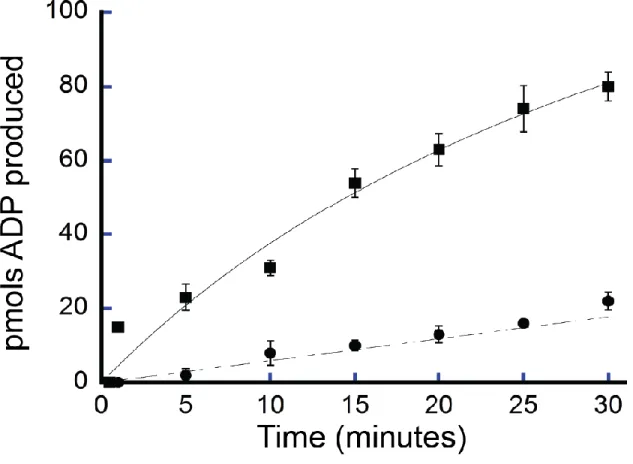
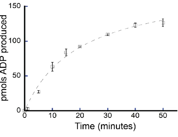
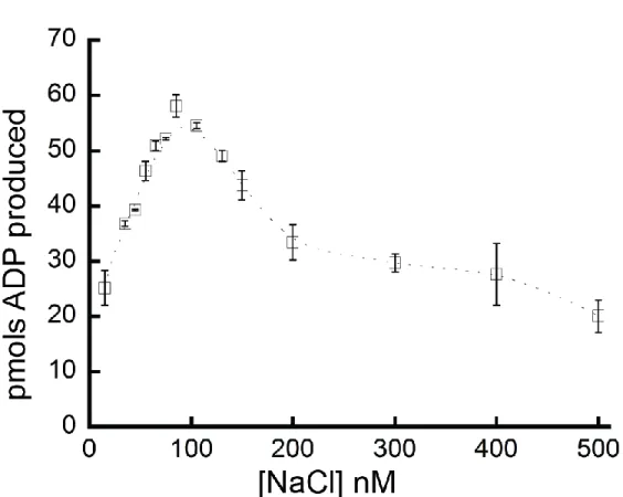
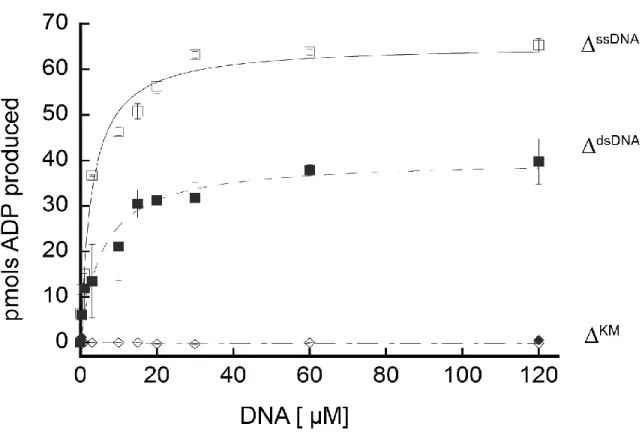
We confirmed the ATPase activity of purified FancmΔ and measured several biochemical parameters to characterize this activity. FancmΔ was found to have 5X greater ATPase activity at 37°C than 25°C (Figure 2.3). The ATPase activity also increased with time (Figure 2.4) and NaCl concentration (Figure 2.5), although ATPase activity declined at NaCl concentrations above 100 nM. For the purposes of this study, we chose conditions under which the ATPase activity was in a linear range. To this end, activity of Fancm was measured at 37°C for 5 minutes in 35 mM NaCl.
were a result of DNA binding (Figure 2.7 A). No significant differences in binding of Fancm to ssDNA as compared to dsDNA were detected.
Fancm is a 3ˈ to 5ˈ DNA helicase
To determine if Drosophila Fancm is active as a helicase, unwinding assays were performed using partial duplex DNA substrates under steady-state conditions. Purified protein was incubated with DNA substrate and the reaction was initiated by the addition of ATP. The wild-type (FancmΔ) helicase completely unwound a 15 bp partial duplex substrate with a 25 bp 3ˈ-ssDNA tail (15/40) (Figure 2.8 A, lane 3). There was no detectable unwinding of the substrate at an equal concentration of mutant protein FancmΔKM (Figure 2.8 A, lane 4). When the same reaction was conducted with a 15 bp partial duplex with 25 bp 5ˈ-ssDNA tail (-15/40), the wild-type helicase failed to unwind the substrate (Figure 2.8 B). This represents a directional bias for unwinding and classifies Fancm as a 3ˈ to 5ˈ helicase, consistent with previous work on the yeast ortholog Mph1 (24). In addition, these data support the conclusion that Fancm cannot unwind blunt-ended duplex DNA as no unwinding of the -15/40 substrate was detected even at longer incubation times.
As shown in Figure 2.8 C, no unwinding of the 15/40 substrate was detected when either ATP or MgCl2 were omitted from the reaction. Moreover, unwinding was undetectable when the non-hydrolyzable ATP analogue AMP-PNP was substituted for ATP. Taken together, these data indicate that unwinding by the Fancm helicase is dependent upon the ability of the protein to hydrolyze ATP and the FancmΔKM mutant is a ‘helicase-dead’ protein.
Fancm catalyzes limited unwinding reaction
(20/45). As seen with the 20/40 substrate, Fancm was only able to unwind 60% of the 20/45 substrate (Figure 2.9 A).
We also measured unwinding activity using two splayed-arm substrates, one with a 3ˈ-single stranded region of 25 bp, and one with a 3ˈ-singled stranded region of 20 bp; both substrates had a 15 bp duplex region. In each case the substrates were completely unwound, indicating that neither the length of the 3ˈ-tail nor the complexity of the substrate affects unwinding. An additional splayed arm substrate with a 25 bp duplex region and 25 nt 5ˈand 3ˈ-ssDNA arms was also tested (Figure 2.9 B), with no detectable unwinding. Although Fancm was able to unwind a 20 bp partial duplex, the increase from 20 bp to 25 bp reduced unwinding to undetectable levels under these conditions. It is possible that Fancm is able to unwind greater than 20 bp partial duplexes under different conditions as discussed later in this chapter.
Based on in vivo data (29), we hypothesized that Fancm may be involved in SDSA with a role in displacing D-loops. Previous studies have shown that the yeast ortholog, Mph1, can unwind the D-loop structures generated during recombination (22). To test the ability of Fancm to unwind complex DNA structures we constructed substrates resembling a recombination D-loop intermediate. We incubated Fancm with a 40 nt bubble-like structure with 25 bp of duplex on either end. As expected from previous studies, Fancm does not unwind the bubble (Figure 2.10), as the duplex region on either end of the bubble is longer than 20 bps.
We next tested if Fancm can unwind a D-loop by incubating Fancm with the bubble structure containing an ‘invading’ homologous strand in which the duplex region was limited to 15 bp. To determine whether the position of the invading strand had an effect on unwinding, the invading strand was
region to which the helicase can bind to initiate unwinding. The “middle” and “end” substrate both have regions that mimic the partial duplex with a ssDNA 3ʹ tail. However, the “front” position substrate does not have a partial duplex with a ssDNA 3ʹ tail, but instead has a 5ʹ ssDNA tail. As shown above, Fancm does not catalyze unwinding of a substrate with a 5ʹ -ssDNA tail (See Figure 2.8 B). However, in this more complex substrate there is an open ssDNA region on the opposite strand of the bubble. Fancm most likely unwinds enough of the duplex arm, generating a 3ʹ tail and thereby catalyzing reduced unwinding of the invading strand. When a 5ʹ-ssDNA tail was added to more closely mimic an “invading strand”, no difference in unwinding was detected (Figure 2.11 B).
To test if the initial rate of the reaction or the duration of the reaction affected unwinding, the rate of unwinding for each substrate was determined using 10 nM and 150 nM protein at various time points for the 15/40 and 20/40 DNA substrates (Figure 2.12 A and B). At 10 nm protein concentration (Figure 2.12 A), FancmΔ was able to fully unwind the 15/40 substrate over the course of the experiment. Under the same conditions FancmΔ was only able to partially unwind the 20/40 substrate. The same was observed for reactions using 150 nm protein (Figure 2.12 B). While the length of time did increase the amount of 20/40 substrate unwound -- 60% at 15 minutes to 78% at 40 minutes -- FancmΔ was unable to fully unwind the 20/40 substrate. There are many factors that might influence Fancm’s ability to catalyze unwinding of longer duplex regions. The structure of the protein, protein interactions, and even
posttranslational modifications could influence unwinding of DNA by Fancm.
with the same efficiency, then the inability to unwind greater than 20 bp of duplex DNA could reflect some mechanistic response of Fancm to limit unwinding.
The data presented here indicate that Fancm as a 3ˈto 5ˈ DNA helicase able to unwind up to 20 bp of partial duplex DNA substrates in an ATP-dependent manner. In addition, the enzyme is able to dissociate short duplex regions in more complex D-loop like structures.The failure of the protein to unwind longer duplex regions may be the result of in vitro conditions or lack of an important accessory protein. Efforts to detect unwinding of longer duplex regions under other conditions (e.g., different salt concentration) or in the presence of a ssDNA binding protein were unsuccessful.
Mph1 and Fml1 have both been shown to be active helicases unwinding up to 100 bp of duplex DNA (24). On the other hand, human FANCM has been shown to migrate D-loops and HJs, but no unwinding activity has been reported (24, 26–28, 35, 39). The data presented here suggest that Drosophila Fancm is similar to both the yeast and human orthologs but unique. Unlike the human
FIGURES Table 2.1-List of oligonucleotides used in this study.
NAME SEQUENCE
FAM1 AGTGGATAcCGGATCATGGATGTGAATTGGATGGACG
FAM2 GCTCGAATTCGGATCTCATCAGCTCATTTGGTAGGGTTTTATTC
KMQC GGGAATGACCTTCATCGCCGCGGTGGTTATG
40 GCTAGCAGTAGCCAGCATCGAACGTACGATCGGTAACGTA
45 GCTAGCAGTAGCCAGCATCGAACGTACGATCGGTAACGTAATGCA
15 CTGGCTACTGCTAGC
20 CGATGCTGGCTACTGCTAGC
-15 TACGTTACCGATCGT
SA25 GACGCTGCCGAATTCTGGCGTTAGGAGATACCGATAAGCTTCGGCTTAAA
SA25a ATCGATGTCTCTAGACAGCACGAGCCCTAACGCCAGAATTCGGCAGCGTC A1 CATTGCATATTTAAAACATGTTGGAAGGCTCGATGCATGCTGATAGCCTACTAGTGCTGC TGGCTTTCAAATGACCTCTTATCAAGTGAC A2 GTCACTTGATAAGAGGTCATTTGAATTCATGGCTTAGAGCTTAATTGCTGAATCTGGTGC TGGGATCCAACATGTTTTAAATATGCAATG
Front TCCCAGCACCAGATT Middle CAGCAATTAAGCTCT
End GCTCTAAGCCATGAA
SAL TTGATAAGAGGTCATCTGGCTACTGCTAGC
SAS TACGTTACCGATCGTTTGATAAGAGGTCAT
56F GACGCTGCCGAATTCTGGCGTTAGGAGATACCGATAAGCTTCGGCTTAA
DS TTAAGCCGAAGCTTATCGGTATCTCCTAACGCCAGAATTCGGCAGCGTC
3OH CCTAACGCCAGAATTCGGCAGCGT
.
A
Figure 2.12 - Timecourse unwinding of partial duplex DNA substrates by Fancm - Helicase reactions were performed as described under “Materials and Methods”. The indicated concentrations of Fancm were incubated with 0.1 nM of the indicated substrate for indicated time. Colored strand on each
REFERENCES
1. Heyer,W.-D., Ehmsen,K.T. and Liu,J. (2010) Regulation of homologous recombination in eukaryotes. Annu. Rev. Genet., 44, 113–39.
2. Kohl,K.P. and Sekelsky,J. (2013) Meiotic and Mitotic Recombination in Meiosis. Genetics, 194, 327–
334.
3. San Filippo,J., Sung,P. and Klein,H. (2008) Mechanism of eukaryotic homologous recombination. Annu. Rev. Biochem., 77, 229–57.
4. Li,X. and Heyer,W.-D. (2008) Homologous recombination in DNA repair and DNA damage tolerance. Cell Res., 18, 99–113.
5. Lorenz, a and Whitby,M.C. (2006) Crossover promotion and prevention. Biochem. Soc. Trans., 34, 537–41.
6. Singleton,M.R., Dillingham,M.S. and Wigley,D.B. (2007) Structure and mechanism of helicases and nucleic acid translocases. Annu. Rev. Biochem., 76, 23–50.
7. Lohman,T.M., Tomko,E.J. and Wu,C.G. (2008) Non-hexameric DNA helicases and translocases: mechanisms and regulation. Nat. Rev. Mol. Cell Biol., 9, 391–401.
8. Fairman-Williams,M.E., Guenther,U.-P. and Jankowsky,E. (2010) SF1 and SF2 helicases: family matters. Curr. Opin. Struct. Biol., 20, 313–324.
9. Gorbalenya,A.E. and Koonin,E. V. (1993) Helicases: amino acid sequence comparisons and structure-function relationships. Curr. Opin. Struct. Biol., 3, 419–429.
10. Brosh,R.M. (2013) DNA helicases involved in DNA repair and their roles in cancer. Nat. Rev. Cancer,
13, 542–558.
15. Castella,M., Jacquemont,C., Thompson,E.L., Yeo,J.E., Cheung,R.S., Huang,J.-W., Sobeck,A., Hendrickson,E.A. and Taniguchi,T. (2015) FANCI Regulates Recruitment of the FA Core Complex at Sites of DNA Damage Independently of FANCD2. PLoS Genet., 11, e1005563.
16. Muniandy,P.A., Liu,J., Majumdar,A., Liu,S. and Seidman,M.M. (2010) DNA interstrand crosslink repair in mammalian cells: step by step. Crit. Rev. Biochem. Mol. Biol., 45, 23–49.
17. Clauson,C., Schärer,O.D. and Niedernhofer,L. (2013) Advances in understanding the complex mechanisms of DNA interstrand cross-link repair. Cold Spring Harb. Perspect. Biol., 5, a012732.
18. Kee,Y., Kim,J.M., D’Andrea,A.D. and D’Andrea,A. (2009) Regulated degradation of FANCM in the Fanconi anemia pathway during mitosis. Genes Dev., 23, 555–60.
19. Kee,Y. and D’Andrea,A.D. (2010) Expanded roles of the Fanconi anemia pathway in preserving genomic stability. Genes Dev., 24, 1680–94.
20. Whitby,M.C. (2010) The FANCM family of DNA helicases/translocases. DNA Repair (Amst)., 9, 224–
36.
21. Xue,X., Choi,K., Bonner,J.N., Szakal,B., Chen,Y.-H., Papusha,A., Saro,D., Niu,H., Ira,G., Branzei,D., et al. (2015) Selective modulation of the functions of a conserved DNA motor by a histone fold complex. Genes Dev., 29, 1000–5.
22. Prakash,R., Satory,D., Dray,E., Papusha,A., Scheller,J., Kramer,W., Krejci,L., Klein,H., Haber,J.E., Sung,P., et al. (2009) Yeast Mphl helicase dissociates Rad51-made D-loops: Implications for crossover control in mitotic recombination. Genes Dev., 23, 67–79.
23. Scheller,J., Schürer,A., Rudolph,C., Hettwer,S. and Kramer,W. (2000) MPH1, a yeast gene encoding a DEAH protein, plays a role in protection of the genome from spontaneous and chemically induced damage. Genetics, 155, 1069–81.
24. Prakash,R., Krejci,L., Van Komen,S., Anke Schürer,K., Kramer,W. and Sung,P. (2005)
Saccharomyces Cerevisiae MPH1 Gene, Required for Homologous Recombination-Mediated Mutation Avoidance, Encodes a 3′ to 5′ DNA Helicase. J. Biol. Chem., 280, 7854–7860.
25. Kang,Y.-H., Munashingha,P.R., Lee,C.-H., Nguyen,T.A. and Seo,Y.-S. (2012) Biochemical studies of the Saccharomyces cerevisiae Mph1 helicase on junction-containing DNA structures. Nucleic Acids Res., 40, 2089–106.
26. Sun,W., Nandi,S., Osman,F., Ahn,J.S., Jakovleska,J., Lorenz,A. and Whitby,M.C. (2008) The FANCM Ortholog Fml1 Promotes Recombination at Stalled Replication Forks and Limits Crossing Over during DNA Double-Strand Break Repair. Mol. Cell, 32, 118–128.
28. Gari,K., Décaillet,C., Delannoy,M., Wu,L. and Constantinou,A. (2008) Remodeling of DNA replication structures by the branch point translocase FANCM. Proc. Natl. Acad. Sci. U. S. A., 105, 16107–12.
29. Kuo,H.K., McMahan,S., Rota,C.M., Kohl,K.P. and Sekelsky,J. (2014) Drosophila FANCM helicase prevents spontaneous mitotic crossovers generated by the MUS81 and SLX1 nucleases. Genetics,
198, 935–45.
30. Lorenz,A., Osman,F., Sun,W., Nandi,S., Steinacher,R. and Whitby,M.C. (2012) The fission yeast FANCM ortholog directs non-crossover recombination during meiosis. Science, 336, 1585–8.
31. Mitchel,K., Lehner,K. and Jinks-Robertson,S. (2013) Heteroduplex DNA position defines the roles of the Sgs1, Srs2, and Mph1 helicases in promoting distinct recombination outcomes. PLoS Genet., 9, e1003340.
32. Mazón,G. and Symington,L.S. (2013) Mph1 and Mus81-Mms4 prevent aberrant processing of mitotic recombination intermediates. Mol. Cell, 52, 63–74.
33. Studier,F.W. (2014) Stable expression clones and auto-induction for protein production in E. coli. Methods Mol. Biol., 1091, 17–32.
34. Studier,F.W. (2005) Protein production by auto-induction in high density shaking cultures. Protein Expr. Purif., 41, 207–34.
35. Gari,K., Décaillet,C., Stasiak,A.Z., Stasiak,A. and Constantinou,A. (2008) The Fanconi Anemia Protein FANCM Can Promote Branch Migration of Holliday Junctions and Replication Forks. Mol. Cell, 29, 141–148.
36. Walker,J.E., Saraste,M., Runswick,M.J. and Gay,N.J. (1982) Distantly related sequences in the alpha- and beta-subunits of ATP synthase, myosin, kinases and other ATP-requiring enzymes and a common nucleotide binding fold. EMBO J., 1, 945–51.
37. Koonin,E. V. (1993) A Superfamily of ATPases with Diverse Functions Containing Either Classical or Deviant ATP-binding Motif. J. Mol. Biol., 229, 1165–1174.