HARNESSING INTERSPECIES ANTAGONISM TO ENHANCE ANTIBIOTIC EFFICACY
Lauren Christine Radlinski
A dissertation submitted to the faculty at the University of North Carolina at Chapel Hill in partial fulfillment of the requirements for the degree of Doctor in Philosophy in the
Department of Microbiology and Immunology.
Chapel Hill 2020
© 2020
ABSTRACT
Lauren Christine Radlinski:
Harnessing interspecies antagonism to enhance antibiotic efficacy (Under the direction of Thomas H. Kawula and Brian P. Conlon)
Beyond genetically encoded mechanisms of resistance, the factors that contribute to antibiotic treatment failure within the host are poorly understood.
Traditional susceptibility assays fail to account for extrinsic determinants of antibiotic susceptibility present during infection and are therefore poor predictors of treatment outcome. To maximize the reach of current therapeutics, we must develop a more sophisticated understanding of antibiotic efficacy in the infection environment. Here we demonstrate that interspecies interactions between two important opportunistic
pathogens, Pseudomonas aeruginosa and Staphylococcus aureus, alters S. aureus response to antibiotics. We show that the P. aeruginosa-produced endopeptidase LasA potentiates lysis of S. aureus by vancomycin, rhamnolipids facilitate proton-motive force-independent aminoglycoside uptake, and that small molecule 4-hydroxy-2-heptylquinoline-N-oxide (HQNO) induces multidrug tolerance in S. aureus through respiratory inhibition and reduction of cellular ATP. We further demonstrate rhamnolipid-mediated potentiation of aminoglycoside uptake and killing of S. aureus restores
Furthermore, bacterial pathogens that replicate within the intracellular niche are protected from antibiotics that cannot penetrate the eukaryotic membrane. Identifying and disrupting the pathways used by these pathogens to modify the intracellular niche in order to survive is an alternative strategy for limiting bacterial proliferation. Here, we use Francisella tularensis as a model intracellular bacterial pathogento identify and describe the bacterial metabolic pathways and host-derived nutrients necessary for intracellular and in vivo growth. These findings reveal potential new therapeutic targets for disrupting bacterial nutrient acquisition that may be broadly applicable for treating other important intracellular pathogens.
Overall, the findings presented here suggest that antibiotic susceptibility is contingent on a multitude of factors including interspecies interaction and the physiological replicative niche. Further elucidation of key antibiotic susceptibility
ACKNOWLEDGEMENTS
First, I would like to acknowledge my advisors Thomas H. Kawula and Brian P. Conlon for their patient and enthusiastic support over the years. Thanks to Tom for guiding my development as an independent scientist during the first two tumultuous years of graduate school. Thanks to Brian for agreeing to take me on and giving me the opportunity to learn from him as he established his research program at UNC. Overall I am incredibly grateful that my graduate education allowed for the opportunity to learn from both of these amazing mentors and scientists. Next, thank you to the Kawula and Conlon lab members who have supported these projects through lively discussion and help at the bench. In particular, I want to acknowledge Sarah E. Rowe for her
contribution to Chapter 2 and Jason Brunton and Shaun Steele for their work on
TABLE OF CONTENTS
LIST OF TABLES ... x
LIST OF FIGURES ... xi
LIST OF ABBREVIATIONS ... xiv
CHAPTER 1 ANTIMICROBIAL STRATEGIES FOR THE COMPLEX INFECTION ENVIRONMENT ... 1
PART I. ANTICIPATING AND AVOIDING ANTIBIOTIC TREATMENT FAILURE ... 2
Antibiotic treatment failure: resistance vs. tolerance ... 2
Antibiotic tolerance is associated with ATP depletion ... 4
Other factors that contribute to antibiotic treatment failure ... 8
Antibiotic susceptibility assays are poor predictors of treatment outcome ... 11
Host-microbe interactions that influence antibiotic susceptibility ... 12
Interspecies interaction during polymicrobial infection alters antibiotic susceptibility ... 15
Physiological determinants of antibiotic susceptibility ... 17
Staphylococcus aureus adaptive metabolism contributes to its recalcitrance to antibiotic therapy ... 19
PART II. EXPLOITING SYNERGISTIC INTERACTIONS TO IMPROVE TREATMENT OUTCOME ... 21
Targeting antibiotic tolerant populations. ... 21
Combinational antibiotic therapy ... 23
Targeting and resuscitating dormant populations ... 26
PART III. IDENTIFYING THERAPEUTIC TARGETS FOR INTRACELLULAR PATHOGENS ... 28
The case for narrow-spectrum antibiotics ... 28
Disrupting niche modification to target recalcitrant pathogen populations ... 30
Targeting bacterial metabolism to inhibit proliferation ... 31
Francisella tularensis as a model for studying intracellular carbon catabolism ... 33
CHAPTER 2 PSEUDOMONAS AERUGINOSA EXOPRODUCTS DETERMINE ANTIBIOTIC EFFICACY AGAINST STAPHYLOCOCCUS AUREUS ... 37
IMPORTANCE ... 38
INTRODUCTION ... 39
RESULTS ... 41
Pseudomonas aeruginosa alters S. aureus susceptibility to antibiotic killing ... 41
Rhamnolipids increase tobramycin uptake and efficacy against S. aureus ... 43
P. aeruginosa induces multidrug tolerance in S. aureus through respiratory inhibition ... 46
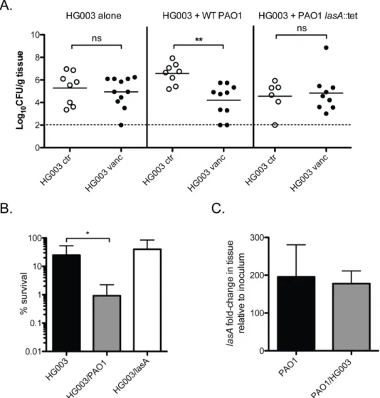
The LasA endopeptidase potentiates vancomycin bactericidal activity against S. aureus ... 48
P. aeruginosa potentiates vancomycin killing in a mouse model of P. aeruginosa/S. aureus co-infection ... 49
DISCUSSION ... 51
MATERIALS AND METHODS ... 55 CHAPTER 3 CHEMICAL INDUCTION OF AMINOGLYCOSIDE
INTRODUCTION ... 85
RESULTS ... 87
P. aeruginosa rhamnolipids potentiate aminoglycoside killing of S. aureus. ... 87
Rhamnolipid/tobramycin combinational therapy eradicates S. aureus persisters .. 89
Rhamnolipids repress the rise of tobramycin resistance, and re-sensitize resistant isolates to killing ... 91
Rhamnolipids sensitize other Gram-positive pathogens to aminoglycoside killing ... 93
Rhamnolipids induce distinct modifications to the S. aureus membrane to promote tobramycin uptake ... 93
DISCUSSION ... 96
MATERIALS AND METHODS ... 100
CHAPTER 4 DEFINING THE METABOLIC PATHWAYS AND HOST-DERIVED CARBON SUBSTRATES REQUIRED FOR FRANCISELLA TULARENSIS INTRACELLULAR GROWTH... 119
IMPORTANCE ... 120
INTRODUCTION ... 120
RESULTS ... 123
Gluconeogenesis, but not glycolysis, is essential for F. tularensis intracellular growth and virulence ... 123
F. tularensis possesses multiple pathways that supply gluconeogenic substrates to support intracellular growth ... 127
Amino acids feed the gluconeogenic pathway through the TCA cycle ... 129
Glycerol catabolism is required for F. tularensis in vivo growth ... 131
DISCUSSION ... 134
CHAPTER 5 SUMMARY OF RESULTS AND DISCUSSION ... 157 SUMMARY- CHAPTER 2: INTERSPECIES INTERACTION
DURING POLYMICROBIAL INFECTION ALTERS S. AUREUS
PHYSIOLOGY AND SUSCEPTIBILITY TO ANTIBIOTICS. ... 158 Interspecies interaction may play an underappreciated role in
dictating antibiotic treatment outcome. ... 160
SUMMARY- CHAPTER 3: RHAMNOLIPIDS INDUCE PMF-INDEPENDENT AMINOGLYCOSIDE UPTAKE TO RESTORE SENSITIVITY TO TOLERANT AND RESISTANT
S. AUREUS POPULATIONS. ... 164 Destabilizing membrane activity during aminoglycoside therapy
is a promising approach for targeting recalcitrant populations. ... 165 Identifying the underlying mechanism(s) of antibiotic tolerance
may reveal new paths to eradication. ... 167 SUMMARY- CHAPTER 4 FRANCISELLA TULARENSIS
UTILIZES NON-GLUCOSE CARBON SUBSTRATES TO
FUEL RAPID INTRACELLULAR PROLIFERATION. ... 171 Disrupting niche modification to target recalcitrant pathogen populations. ... 172
LIST OF TABLES
Table 2.1. LC-MS/MS quantification of HQNO production in P. aeruginosa strains ... 81 Table 2.2. Minimum inhibitory concentrations (MIC) of S. aureus HG003 ... 81 Table 2.3. Summary of P. aeruginosa isolate phenotypes and resulting... 82 Table 3.1. Tobramycin/rhamnolipid MIC values for other
Gram-positive and negative bacterial species ... 118 Table 3.2. Fractional inhibitory concentration (FICI1) of cell envelope
acting agents in combination with tobramycin against S. aureus HG003 ... 118 Table 4.1 Growth of Schu S4 and glpKA strains in broth, BMDMs or J774A.1 cells ... 156 Table 4.2. Summary table of Schu S4 WT and mutant strain growth
LIST OF FIGURES
Figure 1.1. Antibiotic tolerance, persistence, and resistance are
related but distinct phenomena that contribute to treatment failure... 35 Figure 1.2. Overview of extrinsic factors influencing antibiotic
susceptibility within the host ... 36 Figure 2.1. P. aeruginosa supernatant alters S. aureus antibiotic susceptibility ... 66 Figure 2.2. P. aeruginosa rhamnolipids potentiate aminoglycoside
uptake and cell death in S. aureus ... 67 Figure 2.3. Rhamnolipids do not cause cell death in S. aureus in
the absence of tobramycin ... 68 Figure 2.4. Exposure to P. aeruginosa supernatant alters methicillin
resistant S. aureus (MRSA) antibiotic susceptibility ... 69 Figure 2.5. Pseudomonas-produced toxins inhibit respiration in
S. aureus and induce antibiotic tolerance ... 70
Fig 2.6. P. aeruginosa secondary metabolites inhibit S. aureus aerobic respiration resulting in a drop in intracellular ATP and
protection from ciprofloxacin killing ... 71 Figure 2.7 P. aeruginosa supernatant inhibits S. aureus aerobic respiration ... 72 Figure 2.8. P. aeruginosa supernatant potentiates killing by
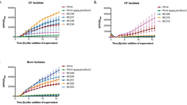
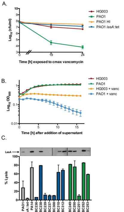
vancomycin via the LasA endopeptidase ... 73 Figure 2.9. P. aeruginosa endopeptidase LasA induces lysis in S. aureus ... 74 Figure 2.10. P. aeruginosa LasA potentiates vancomycin killing
of S. aureus during P. aeruginosa/S. aureus co-culture... 75 Figure 2.11. P. aeruginosa LasA lyses heat-killed S. aureus ... 76 Figure 2.12. P. aeruginosa potentiates vancomycin killing of
Figure 2.14. P. aeruginosa mediated alteration of S. aureus antibiotic susceptibility .... 79 Figure 2.15. Correlation analysis of P. aeruginosa exoproduct
production and impact on S. aureus antibiotic susceptibility ... 80 Figure 3.1. Rhamnolipids synergize with aminoglycosides against
tolerant S. aureus populations ... 108 Figure 3.2. Membrane-acting agents potentiate aminoglycoside killing ... 109 Figure 3.3. Sub-cytotoxic concentrations of rhamnolipids specifically
potentiate aminoglycoside killing of S. aureus ... 110 Figure 3.4. Rhamnolipid/aminoglycoside combinational therapy
targets S. aureus persisters ... 111 Figure 3.5. Reducing S. aureus translation with bacteriostatic
translation inhibitors slows the rate of RL/tobramycin killing ... 112 Figure 3.6. Rhamnolipids repress the rise of tobramycin resistance
and restore sensitivity to resistant isolates ... 113 Figure 3.7. Membrane targeting agents can potentiate aminoglycoside
killing without improving uptake ... 114 Figure 3.8. Rhamnolipids specifically induce PMF-independent
aminoglycoside uptake to resensitize tolerant S. aureus ... 115 Figure 3.9. Tobramycin and cell envelope-acting agent checkerboard assays ... 116 Figure 3.10. Rhamnolipids facilitate PMF-independent aminoglycoside uptake ... 117 Figure 4.1. An overview of Francisella tularensis subsp. tularensis
Schu S4 central carbon metabolism ... 146 Figure 4.2. F. tularensis GlpX is essential for replication on
gluconeogenic carbon substrates, within host macrophages,
and in a murine model of infection ... 147 Figure 4.3. Growth kinetics of F. tularensis ΔpfkA and ΔglpX mutants
during growth in CDM, CDM + glucose and CDM + glutamate ... 148 Figure 4.4. Growth of ΔpckA and ΔppdK in defined media, host cells
Figure 4.5. Intracellular growth characteristics of F. tularensis
Schu S4 and mutant strains in J774A.1 macrophage cells ... 150
Figure 4.6. GdhA fuels gluconeogenesis by shuttling carbon into the TCA cycle ... 151 Figure 4.7. GlpA, but not GlpK, is required for growth on glycerol-3P
and in BMDMs ... 152 Figure 4.8. Glycerol metabolism is essential for F. tularensis intracellular replication . 153 Figure 4.9. Active host cell lipolysis is required for efficient
F. tularensis intracellular replication ... 154
Figure 4.10. ATGL inhibition reduces F. tularensis growth within
LIST OF ABBREVIATIONS
ADEP acyldepsipeptide
AICAR 5-amnoimidazole-4-carboxamide ribonucleotide AME aminoglycoside-modifying enzyme
AMP adenosine monophosphate AMPK AMP-dependent protein kinase AMPs antimicrobial peptides
AsKO2 potassium arsenate
ATGL adipose triglyceride lipase ATP adenosine triphosphate AI-2 autoinducer-2
BAL bronchoalveolar lavage BHI brain heart infusion
BMDM bone marrow-derived macrophage BSA bovine serum albumin
Cam chloramphenicol
CCCP carbonyl cyanide 3-chlorophenylhydrazone CDM Chamberlain’s defined media
CEAAs cell envelope-acting agents
CF cystic fibrosis
CFU colony-forming units Cip ciprofloxacin
Da Dalton
DMEM Dulbecco’s modified Eagle medium Erm erythromycin
F6P fructose 6-phosphate FBP fructose 1,6-bisphosphate
FICI fractional inhibitory concentration index G3P glycerol-3 phosphate
Gent gentamicin
GFP green fluorescent protein GdhA glutamate dehydrogenase GlpA glycerol-3P dehydrogenase GlpK glycerol kinase
GlpX 1,6-bisphosphatase GML glycerol monolaurate HGT horizontal gene transfer
HI heat inactivated
HCN hydrogen cyanide
HQNO 2-heptyl-4-hydroxyquinoline N-oxide
Kan kanamycin
LB Luria-Bertani broth
MIC minimum inhibitory concentration MHB Mueller-Hinton broth
MMH modified Mueller-Hinton broth MOI multiplicity of infection
MRSA methicillin-sensitive Staphylococcus aureus MSSA methicillin-resistant Staphylococcus aureus NaCN sodium cyanide
NO nitric oxide
NO3 nitrate
O2 molecular oxygen
OD optical density
PBS phosphate-buffered saline
PckA phosphoenol pyruvate carboxykinase PCR polymerase chain reaction
PEP phosphoenol pyruvate PfkA phosphofructokinase
PPAR-γ peroxisome proliferator-activated receptor gamma PK/PD pharmacokinetic/pharmacodynamics
PMB polymyxin B
PMF proton motive force
PpdK pyruvate-phosphate dikinase
PYO pyocyanin
QS quorum sensing
RL rhamnolipid
RLU relative luminescence units RNS reactive nitrogen species ROS reactive oxygen species SCV small colony variant
SD standard deviation
TA toxin/antitoxin
Tob tobramycin
TSB tryptic soy broth
Vanc vancomycin
1Adapted and expanded from: Radlinski LC, Conlon BP. 2017. Antibiotic efficacy in the complex infection
environment. Current Opinion in Microbiology.
CHAPTER 1
ANTIMICROBIAL STRATEGIES FOR THE COMPLEX INFECTION ENVIRONMENT1
Since the discovery of penicillin in 1928, antibiotics have become an essential component of modern healthcare. They have made once life-threatening infections readily treatable, greatly prolonged the lives of immunocompromised individuals, and made possible the routine undertaking of invasive surgical procedures. Currently,
however, we are facing a growing crisis as resistance to antibiotics continues to spread, while the discovery of new antibiotics has stagnated[1,2]. For these reasons, it is more important than ever to use current antibiotics as effectively and appropriately as
possible, and to develop novel strategies to eradicate difficult-to-treat bacterial populations before they evolve resistance. As part of this effort, we must develop a more sophisticated understanding of antibiotic efficacy in the infection environment. This task involves identifying the extrinsic factors that directly potentiate or antagonize
antibiotic treatment failure occurs, and reveal opportunities for improving treatment outcome to promote patient health.
PART I. ANTICIPATING AND AVOIDING ANTIBIOTIC TREATMENT FAILURE
Antibiotic treatment failure: resistance vs. tolerance
Antibiotic treatment failure is most commonly associated with resistance. Antibiotic resistance occurs when a bacterium acquires a genetically heritable trait, typically through chromosomal mutation or horizontal gene transfer, that allows the organism to grow in increasing concentrations of an antibiotic. Resistance-conferring mutations include those resulting in target site alteration, induction of drug efflux,
metabolic bypass of the drug’s target, or direct enzymatic inactivation of the antibiotic[3]. An increase in resistance corresponds with an increase in the minimum inhibitory
concentration (MIC) of an antibiotic necessary to stop the growth of a bacterium. Resistance becomes life threatening when the MIC of a resistant isolate surpasses the maximum achievable concentration of antibiotic deliverable to the site of infection, as the drug will no longer inhibit growth of the pathogenic bacterial population within the host. Antibiotic resistance represents an increasingly urgent threat to public health as highlighted by a 2019 report by the Centers for Disease Control (CDC) that stated that more than 2.8 million cases of antibiotic resistant infections occur each year leading to over 35,000 deaths in the US alone[4].
Antibiotic tolerance is a phenotypic switch, usually to a metabolically quiescent state, that allows a subpopulation of bacteria to survive transient exposure to lethal antibiotic concentrations (Figure 1.1). As a tolerant population does not grow in the presence of antibiotic, this phenomenon is not associated in a change in MIC and thus tolerant bacterial populations often go undetected by in vitro clinical susceptibility assays[8]. Upon removal of the antibiotic, a tolerant population that has survived treatment will resume replication, and progeny of that population are equally susceptible to antibiotic killing relative to the parental cells. In the context of patient health, a tolerant bacterial population that has survived antibiotic therapy by entering a dormant state can
resuscitate and resume growth upon the cessation of treatment, thus contributing to chronic and relapsing disease[9–11].
Though antibiotic tolerance and resistance are distinct phenomena, the two are clinically and conceptually related. Poor adhesion to antibiotic therapy regimens has long been associated with the rise of antibiotic resistance, as intermittent antibiotic exposure selects for the outgrowth of resistant subpopulations. In a similar way, recent in vitro studies with Escherichia coli have demonstrated that intermittent exposure to ampicillin selects for bacterial populations that are highly tolerant to ampicillin killing[12]. These tolerant populations acquire mutations that prolong population lag time without changing the MIC. Though these mutations are genetically heritable, this is not
antibiotic resistance [13]. Further, Liu et al demonstrated that during the treatment of clinical isolates with combinational antibiotics, tolerance preceded the emergence of resistance[14]. Thus, within the host, incomplete clearance of a bacterial population during antibiotic therapy likely increases the frequency of antibiotic tolerance and drives the evolution of resistance.
Antibiotic tolerance was also recently implicated in the spread of antibiotic resistance mechanisms among bacterial species through horizontal gene transfer. The facultative intracellular enteric pathogen Salmonella enterica serovar Typhimurium (S. Typhimurium) colonizes both the lumen of the intestinal tract and within the cells of various host tissues. Luminal populations of S. Typhimurium are rapidly cleared by antibiotics[15]. However, tissue-associated, intracellular S. Typhimurium can tolerate antibiotic therapy for extended periods of time[16,17]. Following cessation of treatment, tolerant S. Typhimurium cells that have survived treatment resuscitate and migrate to the luminal space of the colon[18]. In vivo studies by Bakkeren et al. showed that these tissue-associated S. Typhimurium populations act as a bacterial reservoir for plasmids encoding clinically relevant mechanisms of resistance including β-lactamase activity[15]. After re-seeding the lumen of the gut, these cells can act as donors or recipients to facilitate the spread of resistance plasmids among various Enterobacteriaceae species, thus fostering the spread of antibiotic resistance among various members of the
microbiota.
Antibiotic tolerance is associated with ATP depletion
tolerance describes the capacity of a population to specifically survive antibiotic killing, persistence is defined only for bactericidal and not bacteriostatic antibiotics. Bactericidal antibiotics kill bacteria by corrupting active cellular processes[19]. Binding of
aminoglycosides to the 30S subunit of the ribosome, for instance, does not inhibit
protein synthesis, but instead facilitates mistranslation through incorporation of incorrect amino acids into the elongating peptide strand[20]. These misfolded proteins then proceed to wreak havoc on cell membrane permeability and other important functions, and eventually lead to cell death[19]. Similarly, quinolone antibiotics bind bacterial topoisomerase-DNA complexes to prevent strand rejoining following DNA cleavage. This essentially converts the topoisomerase into an endopeptidase that generates lethal double-stranded breaks in the bacterial chromosome[21]. Translation, DNA replication, and most other antibiotic targets are ATP-dependent processes, thus a reduction in intracellular ATP that occurs during metabolic dormancy is associated with a reduction in the number of active targets available for antibiotic action and an increase in antibiotic tolerance. Metabolic dormancy is associated with the induction of multidrug tolerance, suggesting that ATP depletion protects cells from multiple mechanisms of antibiotic killing.
In 1944 the microbiologist Joseph Bigger observed that a small sub-population of genetically susceptible Staphylococcus aureus cells survive intensive penicillin
survive antibiotic activity much better than the larger overall population (Figure 1.1) [23]. This phenomenon is readily observed in vitro and is characterized by a biphasic killing pattern where the bulk of the susceptible population succumbs to antibiotic killing at a much faster rate than the subpopulation of persister cells[8].
In all bacterial species tested, a small subset of cells (typically 0.001-1% of the population) stochastically enters an antibiotic-tolerant persister state, regardless of culture environment or growth phase[24,25]. To date, the precise mechanism(s) that facilitate the persister cell formation within an unstressed, exponentially growing culture are poorly understood. In S. aureus and E. coli, persister cells are associated with stochastic entrance into a stationary-phase-like state accompanied by a drop in
intracellular ATP concentration[26,27]. Indeed, a population-wide state of tolerance can be induced in S. aureus through exposure to arsenate, which depletes intracellular ATP through futile cycling of ADP-As[26,28]. These findings support a “low-energy”
hypothesis of persister cell formation, which proposes that ATP depletion is responsible for the induction of antibiotic tolerance. We recently demonstrated that within a growing culture, S. aureus cells with low expression of TCA cycle enzymes, and thus low levels of ATP generation, are tolerant to antibiotic killing[29]. This finding led to the hypothesis that stochastic fluctuation in TCA cycle enzymes may represent a prominent
mechanism of persister cell formation in S. aureus.
Regardless of the specific mechanism responsible for persister cell formation, the frequency of antibiotic tolerant cells within a population can be increased through
influence the metabolic state of the bacterial population. For instance, as cultures of S. aureus approach stationary phase, nutrients become scarce and population density increases exponentially. Subsequently, S. aureus entrance into stationary phase is associated with a drop in ATP and a population-wide state of tolerance[26]. Similarly, in E. coli biofilm, amino acid starvation precipitates a significant increase in tolerance to ofloxacin challenge[33]. In general, nutrient starvation and subsequent persister cell formation has been attributed as a primary contributing factor to the refractory nature of bacterial biofilms to antibiotic activity[10,33,34].
Though metabolic dormancy often induces antibiotic tolerance, single cell analysis of tolerant populations has led researchers to appreciate that tolerance does not always require ATP depletion. Recent work by Stapels et al. demonstrated that Salmonella Typhimurium persisters within macrophages maintain metabolic activity[35]. Non-growing, intracellular S. Typhimurium persisters are transcriptionally and
translationally active, and reprogram infected macrophages to drive M2 polarization and dampen the pro-inflammatory immune response [35]. Similarly, Pontes et al.
demonstrated that treating Salmonella cultures with the bacteriostatic antibiotic
Other factors that contribute to antibiotic treatment failure
Antibiotics must access a site of infection to kill or inhibit the growth of a
pathogen population. Pharmacokinetic/pharmacodynamics (PK/PD) studies determine the maximum serum concentration (Cmax) of an antibiotic achievable within the serum of the host. For simplicity, maximum serum concentrations are often substituted for
antibiotic Cmax concentrations at specific tissue or organ sites where most bacterial infections actually occur. Penetration of the antibiotic from serum to target tissue site depends on several factors including the molecular characteristics of the drug, the type of tissue infected, and the degree of inflammation[37]. Penetration occurs more rapidly in highly vascularized tissues, such as the liver. Poorly vascularized infection sites frequently reach lower antibiotic concentrations and are more difficult to target with antibiotics[38]. Osteomyelitis, for instance, is notoriously difficult to resolve through antibiotic therapy, and inadequate drug penetration due to low tissue vascularization may be partially responsible. A recent systematic review by Thabit et al. compiled data from a number of pharmacokinetic studies assessing the extent of antibiotic penetration into bone and joint tissues[39]. The range, peak (Cmax), or mean concentrations of over 30 different antibiotics were contrasted with MIC for the most common Gram-positive bacterial species associated with osteomyelitis. Quinolones, macrolides and linezolids penetrated bone and joint tissue well, meeting or exceeding the MIC90 values for most pathogens tested, however penicillins and cephalosporins poorly penetrated the
demonstrate in P. aeruginosa[41]. Thus, the factors that control drug penetration must be considered when selecting an appropriate therapeutic approach.
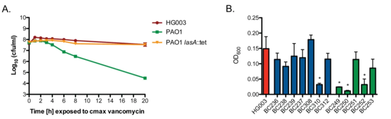
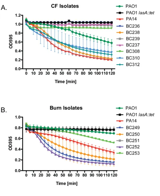
Furthermore, antibiotics often must access specific molecular target sites on the bacterium to facilitate death. Vancomycin is a frontline glycopeptide antibiotic used for the treatment of methicillin-resistant S. aureus (MRSA). Despite its widespread use, vancomycin treatment failure for endocarditis, bacterial pneumonia, or bacteremia range from 37-50%[42]. Vancomycin is hydrophilic molecule with a high molecular weight (over 1,400 Da) that readily binds plasma protein, resulting in poor tissue
penetration[37]. Upon reaching infected tissue, vancomycin must specifically bind to D-Ala-D-Ala residues of bacterial lipid II peptidoglycan precursor during cell wall
biosynthesis to elicit bactericidal activity[43]. Vancomycin also binds peptidoglycan of the mature cell wall, but this interaction does not kill the bacterium and instead
contributes to treatment failure as dense populations with excess non-lethal binding sites prevent vancomycin from accessing target lipid II[44]. For this reason, vancomycin fares poorly against stationary and biofilm associated S. aureus because these dense populations with thick cell walls contain many decoy D-Ala-D-Ala binding targets that limit vancomycin bactericidal activity[45,46]. Newly developed cell wall-acting antibiotics circumvent this obstacle by specifically binding lipid II at the S. aureus septum without binding mature peptidoglycan, and are thus much more effective against bacterial populations at high cell densities[47,48]
Similar to vancomycin, aminoglycoside failure is often attributed to the inability of these drugs to access bacterial ribosomal targets. Aminoglycoside antibiotics are
and charge, aminoglycosides do not readily penetrate eukaryotic membranes through passive diffusion[49]. Instead, eukaryotic uptake of aminoglycoside antibiotics likely results from active cellular mechanisms such as pinocytosis, and consequently
intracellular aminoglycoside accumulation is a slow process that typically takes 48-72 hours to reach detectable levels[49]. For this reason, prolonged aminoglycoside therapy is often necessary for resolving infections caused by intracellular pathogens.
maintain adequate energy levels for growth and replication through fermentative metabolism in the presence of sufficient glycolytic carbon substrates.
Antibiotic susceptibility assays are poor predictors of treatment outcome Current clinical antibiotic susceptibility testing consists primarily of in
vitro diagnostic assays (e.g. MIC assay) that measure the ability of an antibiotic to inhibit growth of a pure bacterial culture grown under artificial conditions. However, these assays do not assess the ability of a drug to eradicate an existing bacterial population, and fail to account for extrinsic determinants of antibiotic susceptibility present in the complex infection milieu. Further, while MIC assays are important for characterizing the resistance profiles of infectious isolates, they do not assess the capacity of those isolates to tolerate the presence of the antibiotic. Indeed, several studies have demonstrated poor correlation between clinical antibiotic
susceptibility testing and subsequent treatment outcome[54,55]. This poor correlation is particularly problematic in the case of deep-seated, chronic infections that fail to
respond to prolonged antibiotic therapy despite apparent drug susceptibility. This
suggests that environmental factors present within the host may influence a pathogen’s susceptibility to antibiotic killing.
efficacy in patients, reduce the duration of antibiotic therapy and decrease the risk of treatment failure, thereby minimizing the development and spread of antibiotic
resistance.
Host-microbe interactions that influence antibiotic susceptibility
Inhibition of bacterial growth by bacteriostatic antibiotics gives the host immune system a chance to contain and eliminate an infectious bacterial population. Similarly, while bactericidal antibiotics facilitate cell death, even powerful bactericidal agents fail to completely eradicate bacterial populations, as antibiotic tolerant persister cells can survive in the presence of the antibiotic for long periods of time[22,56]. Hence, both bacteriostatic and bactericidal antibiotics rely on cooperation with the immune system to fully clear an infection. In some cases, this cooperation may simply be additive, whereas an antibiotic inhibits growth or kills a portion of the population and the immune system then eliminates the survivors. On the other hand, specific host-bacterial interactions may specifically inhibit or potentiate antibiotic efficacy. Such antagonistic or synergistic interactions are only recently coming to light and their impact on in vivo efficacy is yet to be fully appreciated.
By comparing antibiotic efficacy in the presence or absence of host factors,
Sakoulas et al. observed that b-lactam antibiotics synergize with the host immune
considered b-lactam resistant by MIC testing, were sensitized to killing by various host
factors following b-lactam exposure[58]. Furthermore, in a murine model of intratracheal
infection, it was found that the macrolide antibiotic, azithromycin, synergizes with the host cathelicidin antimicrobial peptide, LL-37, resulting in bactericidal activity against Pseudomonas aeruginosa, Klebsiella pneumoniae, Acinetobacter baumannii and more recently Stenotrophomonas maltophilia, despite an apparent lack of susceptibility to azithromycin by MIC testing[59,60]. It is likely that other as yet unidentified interactions with host factors synergize with commonly used antibiotics to promote efficacy within a patient.
Interactions with the host may also be inhibitory to certain antibiotic activities. For instance, innate defenses can induce phenotypic resistance to the last-line antibiotic colistin in Enterobacter cloacae via activation of the histidine kinase PhoQ[61].
Importantly, in this study Band et al. demonstrate that an E. cloacae isolate described as colistin-susceptible via common clinical susceptibility testing can proliferate in the presence of colistin in vivo, leading to treatment failure and host death. Host-produced nitric oxide (NO) can inhibit PMF-dependent uptake of aminoglycoside antibiotics by inhibiting bacterial respiration and thus PMF generation in Salmonella, P. aeruginosa, and S. aureus [62], and DNA damage from exposure to reactive oxygen species (ROS) can induce persister cell formation in E. coli via the upregulation of toxin and drug efflux pump expression[63].
correlates with decreased antibiotic sensitivity[64–66]. Within this niche, bacteria are often physically protected from certain antibiotics, such as aminoglycosides, that
penetrate poorly into host cells[67]. However, poor drug penetrance cannot fully explain treatment failure in this environment, suggesting that other factors may contribute to the refractory nature of intracellular pathogens to antibiotic therapy[65,68,69]. Within the phagosome, bacteria face a variety of stressors including phagosome acidification, nutrient sequestration, and exposure to reactive oxygen and nitrogen species[70]. For Salmonella Typhimurium, vacuolar acidification and nutrient deprivation induces
antibiotic-tolerant persister cell formation through toxin-antitoxin module activation[17]. Similarly, nitrosative stress within the phagosome induces antibiotic tolerance of
internalized Mycobacterium tuberculosis[71].
These examples represent a microcosm of the many host-microbe interactions that influence antibiotic efficacy during infection. An improved understanding of how host factors mediate antibiotic susceptibility will improve our ability to predict antibiotic efficacy in vivo. Furthermore, consideration of these factors may lead to novel
antimicrobial strategies with enhanced activity within the complex host environment.
Interspecies interaction during polymicrobial infection alters antibiotic susceptibility
Rather than existing in isolation, invading microorganisms frequently encounter a complex polymicrobial community within the host, where interactions with the resident microbiota or co-infecting pathogens can directly influence the overall structure and dynamics of the community. Antibiotic susceptibility within this complex environment may vary dramatically from that of the same organism grown in pure culture[72]. An excellent example of community based antibiotic resistance can be seen in the deactivation of an antibiotic by a single bacterial species, extracellularly or
intracellularly, leading to de facto antibiotic resistance of the entire community[73,74]. In this case, antibiotic sensitive pathogens may elude antibiotic killing due to the activities of a co-existing organism[73]. As microbial expression of resistance factors such as antibiotic-modifying enzymes come with a fitness cost, during such instances of social “cheating” an antibiotic susceptible pathogen population can escape antibiotic action without the associated fitness or virulence cost[75,76].
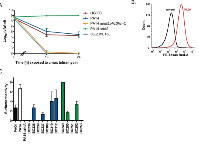
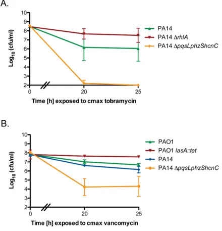
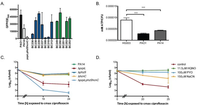
(HQNO) by P. aeruginosa elicits aminoglycoside tolerance in S. aureus by inhibiting the electron transport chain and depleting S. aureus cellular PMF, a necessary pre-requisite for aminoglycoside uptake[53]. P. aeruginosa-produced HQNO has also been shown to induce vancomycin tolerance in S. aureus by shifting S. aureus into a fermentative lifestyle[77]. As these pathogens frequently co-exist within the cystic fibrosis lung and in chronic wound infections, these interactions may represent an important determinant of antibiotic treatment outcome. Intraspecies quorum sensing (QS) has also been
associated with changes in the susceptibility of a population to antibiotic killing.
Production of the QS molecules CSP and acyl-homoserine lactone mediate multidrug-tolerant persister cell formation within populations of Streptococcus mutans and P. aeruginosa, respectively[78,79]. In an interesting example of interspecies crosstalk, indole production by the native commensal E. coli was demonstrated to induce antibiotic tolerance in pathogenic Salmonella enterica Typhimurium[80]. Similarly, interception of Haemophilus influenzae autoinducer-2 (AI-2) by Moraxella catarrhalis significantly increases M. catarrhalis tolerance to antibiotics through the induction of M. catarrhalis biofilm formation[81]. Indeed, biofilm-associated infections have long been associated with antibiotic treatment failure, and these infections are often polymicrobial in
nature[82]. Biofilm matrix production by one microbial species may induce antibiotic tolerance in another. In a recent example, it was demonstrated that C. albicans
extracellular matrix production during dual-species biofilm formation protects S. aureus from antibiotic killing in vivo[83].
instances where factors produced by one species can inadvertently influence the susceptibility of another to antimicrobial activities. Interspecies interactions can induce antibiotic resistance or tolerance, which may have a deleterious impact on antibiotic efficacy[72]. It is also likely that synergistic interactions occur that increase antibiotic efficacy, though only a few instances have been reported so far [84,85]. Identifying the determinants of antibiotic susceptibility in complex communities rather than relying on potentially misleading information garnered from monoculture susceptibility assays is essential for improving our ability to efficiently treat polymicrobial infection.
Physiological determinants of antibiotic susceptibility
Currently, antibiotic susceptibility is measured in nutrient rich media, under aerobic conditions, free of most stressors typically encountered during infection. However, the complex “macro-ecosystem” of a host is composed of a variety of physiologically distinct microenvironments subject to bacterial colonization. Nutrient availability and overall physiological states within these distinct niches can vary
drastically, and promote stark differences in bacterial metabolism. Even within the same spatial niche there often exists a significant degree of environmental heterogeneity, with aerobic, microaerophilic and anaerobic microniches in close proximity. Such is the case in late stage CF patients, where decreased mucociliary clearance promotes the
formation of mucus plugs within the alveoli of the lungs, creating anoxic
microenvironments within the aerobic lung[86]. Oxygen penetration is also often severely hampered in wound infections and abscesses[87,88]. Indeed, obligate
infections, implying that anoxic microenvironments exist within these infection sites[86,89]. Within the heterogeneous infection environment, facultative anaerobes such as S. aureus, E. coli or Streptococcus pneumoniae can colonize both aerobic and anaerobic niches to cause disease, and life within these niches requires specific
metabolic adaptation.
Physiologic heterogeneity in the infection environment may play a significant role in dictating antibiotic susceptibility. Indeed, certain antibiotic classes are active only against either aerobically or anaerobically growing bacteria. Metranidozole, for instance, is a prodrug that must be reduced by intracellular bacterial nitroreductases in order to exhibit bactericidal activity, which only occur in anaerobically growing bacteria[90]. Conversely, PMF-dependent uptake of aminoglycosides generally restricts their activity to aerobically respiring bacteria[50,91]. Active cellular respiration has also been linked to the lethality of other bactericidal antibiotics[92]. Respiration is a more efficient ATP generating process than fermentation, thus, actively respiring cells under oxygen rich conditions are expected to be higher in energy and more susceptible to antibiotic killing than cells in anoxic environments undergoing fermentation. In support of this
hypothesis, frequently acquired mutations that result in defective electron transport in S. aureus are commonly associated with persistent infection, as the SCVs that result are highly resistant to antibiotic killing activity[93]. SCVs are selected for by oxidative stress and low pH as well as through interaction with small molecules produced by P.
As bacteria compete both with other microorganisms and the host for nutrient sequestration during pathogenesis, nutrient availability undoubtedly plays a role in determining antibiotic susceptibility in vivo as well. Antibiotic tolerance increases
significantly during periods of nutrient limitation or diauxic carbon-source transition, and starving bacteria of specific nutrients during in vitro growthmarkedly increases antibiotic tolerance[30,96,97]. Biofilm-associated growth represents a major source of metabolic heterogeneity during infection, as nutrient and oxygen consumption by cells at the periphery of the biofilm coupled with limited nutrient diffusion can result in a starvation-induced state of dormancy for cells at the center of the biofilm that is associated with increased tolerance to antibiotic killing[98]. Supplying biofilms with limiting nutrients can restore bacterial susceptibility to antibiotic killing suggesting that starvation induced antibiotic tolerance may be responsible for the recalcitrance of biofilm infections to antimicrobial treatment[30,99,100].
Staphylococcus aureus adaptive metabolism contributes to its recalcitrance to antibiotic therapy
S. aureus is a major human pathogen responsible for numerous chronic and relapsing infections[101]. S. aureus stably colonizes the anterior nares and skin of approximately one-third of the human population[102]. Typically this co-habitation is harmless, however colonization of immunocompromised individuals or physical disruption of the epithelial barrier in a healthy host can lead to subsequent
necrotizing pneumonia and sepsis[104]. The rate of antibiotic treatment failure for these infections can reach 50%, and is often not associated with the emergence of antibiotic resistance[10,105–108]
As a facultative anaerobe, S. aureus can be found in a variety of physiologically distinct niches within a host. S. aureus encodes an extensive network of metabolic pathways that promote bacterial replication under a plethora of physiological
conditions[109]. Under aerobic conditions, S. aureus can catabolize a wide range of sugars and amino acids through glycolysis, acetogenesis, and TCA pathways to generate ATP and reducing equivalents to power cellular respiration[110]. In the absence of a terminal electron acceptor (O2, NO3-, etc.) or during respiration inhibition, S. aureus switches to fermentative lifestyle, typically fermenting glucose or other carbohydrates to a variety of fermentative end products including lactate, formate, ethanol, and potentially 2,3-butanediol[111–113]. This extensive metabolic network permits colonization of a wide range of niches within the complex host environment, and may explain the capacity of S. aureus to cause such a broad variety of infections[114]. Metabolic versatility makes systemic S. aureus infections difficult to resolve with
We propose that S. aureus represents an ideal model organism for studying how the extrinsic factors present during infection alter bacterial antibiotic susceptibility. This idea is explored further in Chapters 2 and 3. An improved understanding of how and why antibiotics fail to clear S. aureus populations within different niches of the host will not only aid in resolving recalcitrant S. aureus infection, but will also likely be broadly applicable for the resolution of other important pathogens responsible for chronic and relapsing infections.
PART II. EXPLOITING SYNERGISTIC INTERACTIONS TO IMPROVE TREATMENT OUTCOME
Targeting antibiotic tolerant populations.
Identifying the underlying causes of antibiotic treatment failure is a futile pursuit if we lack the therapeutic means to target these difficult-to-treat populations. How do we target dormant populations when most of our current antibiotic arsenal requires active bacterial targets to function? After discovering persisters, Bigger himself proposed that pulse-dosing cultures with antibiotics to allow persisters time to revive between
treatments and would eventually result in complete clearance of the population[22]. The plausibility of this hypothesis is supported by mathematical modeling and in vitro
treatment is challenging as it requires strict adherence to antibiotic regimens, often in the face of uncomfortable or even debilitating side-effects that reduce patient
compliance[120]. This is particularly apparent during Mycobacterium tuberculosis
treatment. M. tuberculosis is a slow-growing bacterial pathogen that replicates within the phagosome of alveolar macrophages[121]. Within this environment, host-mediated stressors including nutrient sequestration and nitrosative stress support suboptimal growth conditions and growth arrest of M. tuberculosis, rendering the pathogen tolerant to antibiotic killing [71]. Current guidelines for the treatment of drug-susceptible M. tuberculosis begins with a four-drug regimen of isoniazid, rifampin, pyrazinamide, and either ethambutanol or streptomycin that lasts for at least two months[122]. Not only is this intensive treatment regimen impractical for underdeveloped countries with poor access to healthcare, but harmful side effects make patient non-adherence common even in developed countries[123]. This has led to a rise in multi-drug resistant M. tuberculosis strains that necessitate even more extensive and crippling antibiotic regimens. Indeed, M. tuberculosis recently surpassed Human Immunodeficiency Virus (HIV) as the leading cause of death from infectious disease[124].
Clearly, extending antibiotic treatment duration is insufficient for combating treatment failure. Instead, researchers are pursing novel means for enhancing antibiotic efficacy against metabolically quiescent, tolerant populations that persist within infected hosts. These include combining antibiotics to target heterogeneous populations,
Combinational antibiotic therapy
Antibiotic synergy testing assessesthe efficacy of a combinational antibiotic therapy against a bacterial population in vitro. The effect of a two-compound
combinational therapy can be additive, where the cumulative antimicrobial effect is simply the sum of both individual therapies; synergistic, where the combinatorial activity of two compounds exceeds the sum of each compound alone; or antagonistic, where the effects of one compound decreases the antimicrobial effect of the other[125].
Over 50% of patients treated for septicemia and between 25-50% of patients with surgical site or pneumonia infections receive a combination of two or more antibiotics in an attempt to resolve the infection[126–128]. Prescription of multiple antibiotics with different spectra of activity may increase empiric coverage and efficacy of treatment, particularly when the susceptibility of infecting organism is unknown or there is
A second approach to combinational therapy focuses on antibiotic “sensitizers” or adjuvants. These are compounds that improve the efficacy of co-administered
antibiotics, usually without demonstrating antimicrobial activity on their own[133]. Typically, adjuvants function by reversing mechanisms of resistance in naturally
sensitive strains (e.g. combining an antibiotic with an antibiotic efflux pump inhibitor), or by sensitizing intrinsically resistant bacterial populations to killing (e.g. dispersion of antibiotic-tolerant biofilm)[133]. Clavulanic acid and amoxicillin for instance, make up a widely successful antibiotic cocktail that has been patented and commercialized as Augmentin®. Clavulanic acid inhibits the function of bacterial lactam-degrading lactamases during amoxicillin therapy, restoring the efficacy of amoxicillin against β-lactam-resistant populations[134]. One benefit of using antibiotic adjuvants is that a number of putative antimicrobial drugs that were shelved in the past for having low efficacy or a high intrinsic rate of resistance generation have been resurrected as promising adjuvant candidates. As researchers have only recently begun exploring potential antibiotic/adjuvant combinations, there may be a number of compounds with the capacity to significantly improve antibiotic lethality that have yet to be applied as such.
Disrupting cellular integrity to target antibiotic tolerant populations
attention to developing compounds that directly target bacterial cellular integrity, as this approach may permit eradication of dormant populations that are inherently tolerant to current therapeutics. Cell envelope integrity represents a promising but underdeveloped target for antibiotic action. Recent attention has focused on the applicability of using antimicrobial fatty acids, peptides, or other naturally occurring and synthetic compounds that physically disrupt the phospholipid bilayer as potential therapeutics, either alone in combination with a secondary antibiotic[59,135–137]. The primary draw of this approach is that membrane integrity is essential for the survival and virulence of all bacteria, regardless of metabolic state. Interaction with the membrane does not require ATP, thus tolerant persister populations are theoretically as susceptible to killing as metabolically active cells. Another attractive feature is that it is often more difficult for bacteria to evolve resistance to compounds that disrupt membrane integrity, as these compounds typically interact with multiple targets within the membrane. Indeed, results from in vitro studies show that de novo mutations that confer resistance to lipopeptides, antimicrobial peptides, and small molecules are exceedingly rare[138].
The primary drawback of targeting the membrane stability is that compounds that disrupt bacterial membranes may be cytotoxic to host cells for the same reason.
However, there are distinct physiological differences between bacterial and mammalian membrane composition, which may make it possible to specifically target bacterial membrane during treatment. Relative to mammalian membranes, bacteria lack
membrane is feasible and effective[138]. The antibiotic daptomycin, for instance, binds the surface of negatively charged bacterial membrane and oligomerizes to form pores and depolarize the membrane, leading to cell death[140,141]. As a membrane-acting agent, daptomycin often exhibits greater efficacy against non-growing S. aureus persister populations than traditional antibiotics[119]. Similarly, there are a number of membrane-acting compounds that have been recognized as safe by the United States Food and Drug Administration (FDA) for human consumption that exhibit antimicrobial activity[135,142,143]. Further, antimicrobial peptides and lipids constitute an important component of the human innate immune system, implying that these compounds may be safely administered to patients at concentrations that are bactericidal[144,145]. There may even be an opportunity to exploit synergistic interaction between naturally occurring antimicrobial lipids or peptides and antibiotics within the host to improve treatment outcome[59].
Targeting and resuscitating dormant populations
Others have pursued more creative ways to re-sensitize ATP-depleted populations to antibiotic killing by commandeering and manipulating normal cellular processes during treatment. For instance, Conlon et al. used the antibiotic
an exciting candidate for targeting dormant bacterial populations, as ADEP-treated cells essentially degrade themselves to death in an energy-independent manner. The
authors furtherdemonstrated that combining ADEP4 with rifampicin facilitated the
eradication of persister populations in vitro and in a deep-seated murine biofilm infection model[146].
Chemically resuscitating dormant cells prior to antibiotic exposure by providing nutrients may also improve persister eradication in vitro and in vivo. Eradication of E. coli and S. aureus persisters can be achieved through supplementation with glycolytic sugar molecules that enhance aminoglycoside uptake through PMF generation[149]. Similarly, supplementing cultures with glucose increases daptomycin efficacy against S. aureus persisters, and supplementation with nitrate or arginine potentiates tobramycin and ciprofloxacin killing of P. aeruginosa biofilm[99,150]. Though these in vitro studies support the idea of reviving antibiotic tolerant persisters to improve therapeutic
outcome, the clinical practicality and potential negative repercussions from providing pathogens with excess nutrients during treatment has yet to be evaluated.
The widespread onset of multidrug-resistant pathogenic strains, coupled with an evaporating pipeline of new antibiotics reaching market emphasizes the importance of maximizing the efficacy of current antibiotics. Identifying instances where combinational antibiotic therapy can improve the rate and capacity of an antibiotic to clear recalcitrant bacterial populations will reduce the duration of antibiotic tolerance and slow the rise of resistance. Importantly, however, synergistic interactions observed in vitro are not
investigation is necessary to identify new opportunities for exploiting pathogen
vulnerabilities during antibiotic therapy, as well as to assess the practicality and efficacy of implementing these therapies in patients.
PART III. IDENTIFYING THERAPEUTIC TARGETS FOR INTRACELLULAR PATHOGENS
The case for narrow-spectrum antibiotics
Broad-spectrum antibiotics act on both Gram-positive and negative bacterial species by targeting common cellular processes such as DNA replication
(fluoroquinolones), transcription (rifamycins), translation (aminoglycosides), and cell wall biosynthesis (β-lactams) that are essential for bacterial replication and survival. With the discovery of penicillin, broad-spectrum antibiotics were the first developed, and remain the most commonly applied antimicrobial strategies used today for resolving bacterial infection. These drugs allow clinicians to quickly treat patients when a bacterial infection is suspected but the pathogen is unknown, they can also be used prophylactically to prevent infection during invasive surgery and during labor[151], and can resolve polymicrobial infection when more than one pathogen is causing disease.
host commensal species that render a patient susceptible to subsequent infection. This is particularly apparent during nosocomial Clostridioides difficile infection, where
colonization follows antibiotic-mediated clearance of the host microbial flora[153]. Finally, while broad-spectrum antibiotics are effective at targeting extracellular bacterial pathogens, they are often ineffective against bacterial species that replicate within the cytoplasm or vacuolar space of host cells due to poor intracellular penetrance[67]. Many important antibiotic classes cannot enter the host cell cytosol and thus cannot gain access to the target bacterial population within[67]. Aminoglycosides, for instance, are the frontline therapy of choice for the facultative intracellular pathogen, Francisella tularensis, despite the fact that these antibiotics poorly penetrate the host cell[67]. As F. tularensis is among several bacterial species that can disseminate via cell-to-cell
transmission mechanisms without exposure to the extracellular space[154], extensive treatment periods that last several weeks are often necessary to treat this organism.
antimicrobial responses. Understanding and inhibiting these active microbial processes may prevent niche modification and restrict pathogen proliferation.
Disrupting niche modification to target recalcitrant pathogen populations
Preventing pathogens from cultivating an environment that supports replication within the host is a promising alternative to antibiotic therapy. During dysbiotic
Proteobacteria expansion, enteric pathogens actively modify their environment to establish a replicative niche[156]. The enteric pathogen Citrobacter rodentium, for instance, actively drives metabolic reprogramming of epithelial cells away from β-oxidation and towards aerobic glycolysis by triggering colonic crypt hyperplasia[157]. This change in host cell metabolism increases oxygenation at the mucosal surface and drives aerobic expansion of pathogenic Enterobacteriaceae[156]. Inhibiting colonic crypt hyperplasia during C. rodentium infection with the γ-secretase inhibitor, dibenzazepine, reduced the ability of C. rotentium to colonize this environment[157]. Similarly, treating mice with a PPAR-γ agonist (rosiglitazone) significantly reduced E. coli luminal
An ideal therapy for intracellular pathogens would prevent pathogen-mediated niche modification of the intracellular space. For professional phagocytic cells this would allow for the host to eliminate pathogens through the normal innate immune response (phagosome acidification, ROS generation, etc.). Within non-phagocytic cells, disrupting pathogen-mediated niche modification would likely reduce nutrient availability and slow bacterial proliferation. However, in most cases the means by which these organisms compete with the hosts own metabolic demands to derive metabolites from the
intracellular environment are unclear. An improved understanding of how intracellular bacterial pathogens modify the intracellular niche to obtain sufficient nutrients for
replication and dissemination is likely to reveal novel therapeutic avenues for combating these important pathogens. Furthermore, targeting bacterial growth by altering host cell metabolism may circumvent issues concerning drug penetration, as host-targeting therapeutics act on infected host cells and not the bacteria replicating within that environment.
Targeting bacterial metabolism to inhibit proliferation
the virulence of otherwise virulent bacterial species[163,164]. However, for most pathogens the metabolic pathways and host-derived nutrients necessary for in vivo growth are poorly defined.
Extracellular pathogens are subject to a constant flux of available nutrients within the host. By contrast, intracellular pathogens encounter more stable growth conditions within the cytoplasm or vacuolar space within the host cell. The relative simplicity of the intracellular environment has prompted significant strides in understanding the
metabolic requirements of intracellular pathogens[165]. Within this niche, bacteria have access only to nutrients they can scavenge from this compartment. Recent studies suggest that the intracellular environment is not simply an open buffet of freely available metabolites left over from host metabolic processes[165]. Instead, most intracellular nutrients are stored within complex structures and not immediately available to
intracellular pathogens[165]. To grow, intracellular bacteria must either harvest newly imported nutrients or direct the degradation of resident complex storage structures into their constituents (fatty acids, carbohydrates and amino acids). Successful intracellular pathogens have evolved the means for manipulating the intracellular environment to obtain sufficient carbon and trace elements necessary for replication[166–170]. As a reward, these pathogens are shielded from the innate immune system, competing microorganisms, and certain antibiotics that cannot access the intracellular space. Further, pathogens that can survive within motile macrophages and neutrophils may commandeer these cells as mode of protected dissemination through the
Francisella tularensis as a model for studying intracellular carbon catabolism F. tularensis is a Gram-negative, facultative intracellular bacterial pathogen and one of the most virulent organisms known. F. tularensis infects over 250 susceptible organisms, including humans[173]. Within these hosts, F. tularensis replicates within a variety of cell and tissue types including macrophages, epithelial cells, hepatocytes, neutrophils, fibroblasts and erythrocytes[174–177]. Following intracellular invasion, F. tularensis escapes the phagosome to replicate within the host cell cytosol[178]. A hallmark of F. tularensis pathogenesis is the bacterium’s ability to reach extreme
densities within this niche, often replicating 1,000-fold within 24 hours to fill 60% of host cytosolic volume with bacterial mass (unpublished data). This remarkable rate of growth demonstrates that F. tularensis is adept at harvesting and utilizing host cell nutrients in an environment that does not inherently contain sufficient free carbon to support the levels of replication observed. F. tularensis must actively modulate the host metabolic processes to amass sufficient carbon to support growth and dissemination. We
previously demonstrated that F. tularensis commandeers host cell autophagy to break down macromolecules and derive a source of free amino acids [179]. However, F. tularensis replicates to a considerable degree even in the absence of autophagy, demonstrating that this organism further exploits host metabolic processes to derive sufficient nutrients[179].
tularensis is adept at modulating host cell metabolism to fuel replication. Furthermore, as a facultative intracellular pathogen, F. tularensis contains a small and decaying genome that encodes a relatively simple set of carbon catabolic pathways that support intracellular replication[183]. For these reasons, we propose that F. tularensis is an excellent model for studying intracellular niche modification and carbon metabolism, and that doing so will reveal new insights into how we can improve antimicrobial targeting of recalcitrant intracellular pathogens. This topic is explored further in Chapter 4.
In all, though antibiotic susceptibility is traditionally examined in simple
homogenous conditions in vitro, more and more studies are revealing the dynamic and complex nature of antibiotic efficacy in the infection environment (Figure 1.2). The administration of antibiotics without consideration of these environmental factors may result in treatment failure, exacerbated disease progression, and the rise of resistant microorganisms. Moreover, we propose that pathogen sensitivity to antibiotic killing is contingent not only on genotype, but also the pathogen’s metabolic state, and on interactions that occur with the host and co-infecting microorganisms. Further
Figure 1.2. Overview of extrinsic factors influencing antibiotic susceptibility within the host. Environmental factors can antagonize or potentiate antibiotic efficacy killing of a pathogen. Antimicrobial peptides (AMPs) can synergize with antibiotics to increase killing of pathogens. Conversely, pathogen engulfment by phagocytic cells can inhibit antibiotic killing by preventing drug access to the pathogen or by directly
influencing pathogen metabolism and physiology through production of reactive oxygen or nitrogen species (ROS/RNS), vacuole acidification or nutrient sequestration. Inter- and intraspecies interactions can positively and negatively impact a pathogen’s susceptibility to antibiotic killing either through signaling processes or via direct
interaction, such is the case in polymicrobial biofilms. Finally, heterogeneity in oxygen or nutrient concentration within the infectious environment can influence bacterial
1 Radlinski LC, Rowe SE, Kartchner LB, Maile R, Cairns BA, Vitko NP, Gode CJ, Lachiewicz AM,
Wolfgang MC, Conlon BP. Pseudomonas aeruginosa exoproducts determine antibiotic efficacy against
Staphylococcus aureus. PLoS Biology. 2017 Nov 27;15(11):e2003981
CHAPTER 2
PSEUDOMONAS AERUGINOSA EXOPRODUCTS DETERMINE ANTIBIOTIC EFFICACY AGAINST STAPHYLOCOCCUS AUREUS1
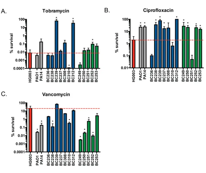
Chronic co-infections of Staphylococcus aureus and Pseudomonas aeruginosa frequently fail to respond to antibiotic treatment, leading to significant patient morbidity and mortality. Currently, the impact of interspecies interaction on S. aureus antibiotic susceptibility remains poorly understood. In this study, we utilize a panel of P.
aeruginosa burn wound and cystic fibrosis (CF) lung isolates to demonstrate that P. aeruginosa alters S. aureus susceptibility to bactericidal antibiotics in a variable, strain-dependent manner and further identify three instrain-dependent interactions responsible for antagonizing or potentiating antibiotic activity against S. aureus. We find that P. aeruginosa LasA endopeptidase potentiates lysis of S. aureus by vancomycin, rhamnolipids facilitate proton-motive force-independent tobramycin uptake, and 2-heptyl-4-hydroxyquinoline N-oxide (HQNO) induces multidrug tolerance in S. aureus through respiratory inhibition and reduction of cellular ATP. We find that the production of each of these factors varies between clinical isolates, and corresponds to the
These findings demonstrate that antibiotic susceptibility is complex and dependent not only upon the genotype of the pathogen being targeted, but also on interactions with other microorganisms in the infection environment. Consideration of these interactions will improve the treatment of polymicrobial infections.
IMPORTANCE
Accurate prediction of antimicrobial efficacy is essential for successful treatment of bacterial infection. Beyond genetically encoded mechanisms of resistance, the specific determinants of antibiotic susceptibility during infection remain poorly understood. Here we show that a single interspecies interaction between S.
aureus and P. aeruginosa can completely transform the antibiotic susceptibility profile of S. aureus. Through multiple distinct mechanisms, P. aeruginosa can antagonize or potentiate the efficacy of multiple classes of antibiotics against S. aureus. We identify the exoproducts responsible for altering S. aureus susceptibility to antibiotic killing, and furthermore demonstrate that these compounds are produced at varying levels in P. aeruginosa clinical isolates, with dramatic repercussions for S. aureus antibiotic
susceptibility. Finally, we use a mouse model of P. aeruginosa, S. aureus co-infectionto demonstrate that the presence of P. aeruginosa significantly alters the outcome of S. aureus antibiotic therapy in a host. These findings indicate that the efficacy of antibiotic treatment in polymicrobial infection is determined on the community level with