Original Research Article
Study on clinical profile and risk factors associated with pneumonia
Gangina Sriram, Akula Satyanarayana*
INTRODUCTION
Acute respiratory infections (ARI), particularly lower respiratory tract infections (LRTI), are the leading cause of death among children under five years of age and are estimated to be responsible for between 1.9 million and 2.2 million childhood deaths globally.1
Modernization, industrialization and urbanization are now posed with the problem of increase in ARI morbidity and mortality. It is clear that future health of children depends on preventing, diagnosing, treating and limiting ALRTI. The utility of simple clinical signs like rapid
breathing and chest in drawing to diagnose pneumonia in infants and young children has been well established. The use of these clinical signs in the early detection and treatment of children with pneumonia by primary health care workers forms the basis for the case management strategy formulated by the World health organization (WHO) to control mortality and morbidity.2
ARI can be preventable. However, socio environmental factors are acting as major obstacles in prevention of ARI. The epidemiological information regarding risk factors and management is scanty. A large gap exists in this knowledge about these factors, which needs to be ABSTRACT
Background: Modernization, industrialization and urbanization are now posed with the problem of increase in ARI morbidity and mortality. The epidemiological information regarding risk factors and management is scanty. A large gap exists in our knowledge about these factors, which needs to be fulfilled by systematic studies. The present study is designed to identify the risk factors of pneumonia in our area.
Methods: This was a prospective clinical study of pneumonia conducted on 94 children who were admitted to Paediatric ward in GSL Medical College General Hospital, Rajahmundry in study duration. Epidemiological factors affecting the same were studied and bronchoscopy was done whenever it was needed. A detailed history of the relevant symptoms, such as fever, cough, rapid breathing, refusal of feeds, noisy breathing, bluish discolouration etc., was collected.
Results: The most affected children belonged to the age group of 1 year to 3 years (64.9%). Bronchopneumonia (86.2%) was the most common clinical diagnosis made at admission. According to WHO ARI control programme, 28.7% had pneumonia, 54.3% had severe pneumonia and 17% very severe pneumonia. It was found that younger age group, malnutrition, kutcha house, crowding, poor sanitation facilities, cooking with fuel other than LPG (indoor pollution) and low socio economic status and high respiratory rate were significant risk factors for pneumonia in children.
Conclusions: ARI, especially pneumonia is one of the major causes of morbidity and mortality in children. Bronchopneumonia is the predominant form of presentation in infants and preschool children.
Keywords: Children, Malnutrition Pneumonia, Morbidity, Mortality, Risk factors
Department of Paediatrics, GSL Medical College & General Hospital, Rajahmundry, Andhra Pradesh, India
Received: 28 July 2019
Revised: 12 August 2019
Accepted: 16 August 2019
*Correspondence:
Dr. Akula Satyanarayana, E-mail: [email protected]
Copyright: © the author(s), publisher and licensee Medip Academy. This is an open-access article distributed under the terms of the Creative Commons Attribution Non-Commercial License, which permits unrestricted non-commercial use, distribution, and reproduction in any medium, provided the original work is properly cited.
fulfilled by systematic studies. The present study is designed to identify the risk factors of pneumonia in this area. Objective of this study is to assess the clinical profile and identification of risk factors of pneumonia
METHODS
The present prospective clinical study of pneumonia was conducted on 94 children who were admitted to Paediatric ward in GSL Medical College General Hospital, Rajahmundry from 2015 to 2017. Epidemiological factors affecting the same were studied and bronchoscopy was done whenever it was needed.
Inclusion criteria
Children in the age group of one year to five years with clinical features of pneumonia as per WHO ARI control programme were included.
Exclusion criteria
Children with congenital anomalies of heart and lungs, anatomical defects like cleft lip and cleft palate, immune compromised states like human immune deficiency virus infection (HIV) and children less than one year of age were excluded from the study.
Data collection procedure
A detailed history of the relevant symptoms, such as fever, cough, rapid breathing, refusal of feeds, noisy breathing, bluish discolouration etc., was collected.
Based on WHO ARI criteria, children were considered to have tachypnea if respiratory rate (RR):
• RR >60 in <2 months
• RR >50 in 2 months-1 yr.
• RR >40 in 1 yr-5 yrs.
A detailed examination of each child including anthropometry was carried out. During the general physical examination, emphasis was laid on assessing general condition of the child, respiratory rate, presence of fever and other signs such as cyanosis and pallor. Detailed systemic examination of the respiratory, cardiovascular and central nervous systems was done. Any associated illnesses such as septicemia, meningitis, diarrhoea and congestive cardiac failure if present were noted. Socio economic history regarding the type of house, family size, sanitary facilities and fuel based for cooking were recorded. Socio economic status was classified according to modified Kuppuswamy classification. Other pertinent information such as immunization status (Immunized, partially immunized or unimmunized), feeding practices and degree of malnutrition (IAP classification) were also recorded.
According to WHO ARI criteria, children were classified into 3 groups: pneumonia, severe pneumonia and very severe pneumonia. For analytic purpose, risk factors for pneumonia were studied.
Following Investigations were carried out
• Blood Investigations - Hb%, TC, DC, ESR were done in all cases.
• Chest X-ray was taken in all patients.
• Blood culture was done in all cases.
All patients received antibiotics. Supportive care (IV fluids, oxygen, nebulization etc) was given as and when required. Antibiotics that were used:
1. First line antibiotics: Amoxycillin.
2. Second line antibiotics: Amoxicillin and Clavulanic acid.
3. Others: Ceftriaxone, Vancomycin
Those children who failed to respond to 1st line antibiotics within 48hrs, received second line antibiotics. Ceftrioxone and/ Vancomycin was considered in case of empyema/massive consolidation. Closed tube drainage was considered in cases of empyema. All children were evaluated during the hospital stay and the response to treatment was noted.
Statistical analysis
Continuous data was represented as mean±standard deviation. Categorical data was expressed as numbers in percentage. Chi square test was used to determine significant differences between three groups. Significance for the statistical tests was pre-determined at a probability value of 0.05 or less (p<0.05).
RESULTS
In the present study, majority of cases (64.9%) were less than three years of age. Majority of cases (61.7%) were females.
In this study, fast breathing (100%), cough (100%), fever (100%) were the most common symptoms. Refusal of feeds was present in 17% of cases.
In this study, chest retractions were present in 71.27% cases, both crepitations and ronchi were present in 75.5% cases, crepitations alone were heard in 14.9% cases, ronchi alone in 9.6% cases and abnormal breath sounds (bronchial breathing/ diminished breath sounds) in 28.7% of cases.
Table 1:Basic characteristics.
Characteristics Frequency Percentage
Age
1-3 61 64.9
4-5 33 35.1
Sex
Male 36 38.3
Female 58 61.7
Symptoms
Cough 94 100%
Fever 94 100%
Fast breathing 94 100%
Fefusal of feeds 16 17%
Convulsions 2 2.1%
Cyanosis 3 3.2%
Signs
Chest retractions 67 71.27%
Crepitations 14 14.9%
Ronchi 9 9.6%
Crepitations and ronchi 71 75.5% Abnormal breath sounds 27 28.7%
Respiratory rate
41-50 5 5.31%
51-60 23 24.46%
60 and ABOVE 66 70.21%
In this study pneumonia was associated with diarrhoea in 1.1% cases, septicemia in 1.1% cases, meningitis in 2.2% cases, DIC in 1.1% cases. According to WHO ARI programme, 28.7% had pneumonia, 54.3% had severe pneumonia and 17% had very severe pneumonia.
Majority of the cases were clinically diagnosed as Bronchopneumonia (86.2%), Lobar pneumonia (consolidation) was diagnosed in 11.7% of cases and pneumonia and its complications (Empyema) in 2.1% cases. Closed tube drainage was considered in empyema cases (Table 2).
Table 2:Distribution of cases according to clinical
findings.
Associated illness Frequency Percentage
Diarrhoea 1 1.1%
Meningitis 2 2.2%
Septicemia 1 1.1%
DIC 1 1.1%
Classification
Pneumonia 27 28.7%
Severe pneumonia 51 54.3%
Very severe pneumonia 16 17%
Clinical diagnosis
Bronchopneumonia 81 86.2%
Lobar pneumonia 11 11.7%
Empyema 2 2.1%
In the present study, majority were breast-fed (78.7%) and remaining 21.3% were bottle fed. Majority (78.7%) were under Grade I (40.4%) and Grade II (38.3%) PEM. 6.4% were under Grade III. In the present study, majority,53 cases (56.4%) lived in ill-ventilated thatched house (kutcha) and out of them 35 houses [66.03%] were crowded. In the present study, majority (59.6%) had poor sanitary facility (open air defecation) and majority (57.4%) used fuel other than LPG for cooking (Table 3).
Table 3:Distribution of risk factors.
Factors Frequency Percentage
Type of feeding
Breast fed 74 78.7%
Bottle fed 20 21.3%
Protein energy malnutrition
Normal 14 14.9%
Grade – I 38 40.4%
Grade – II 36 38.3%
Grade – III 6 6.4%
Grade – IV 0 0
Type of house
PUCCA 41 43.6
KUCHA 53 56.4
Sanitation facilities
Good 38 40.4
Poor 56 59.6
Cooking
LPG 39 41.5
NON-LPG 55 57.4
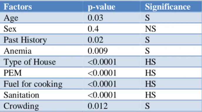
In this study, risk factors for pneumonia were studied. It was found that younger age group, malnutrition, anemia, kutcha house, crowding, poor sanitary facilities, cooking with fuel other than LPG (indoor pollution) and low socio economic status were significant risk factors for pneumonia in children (Table 4).
Table 4:Association of Risk factors with pneumonia.
Factors p-value Significance
Age 0.03 S
Sex 0.4 NS
Past History 0.02 S
Anemia 0.009 S
Type of House <0.0001 HS
PEM <0.0001 HS
Fuel for cooking <0.0001 HS Sanitation <0.0001 HS
Crowding 0.012 S
Mortality
seen in this case and this case belonged to very severe pneumonia.
DISCUSSION
Age distribution
Age is an important predictor of morbidity and mortality in pediatric pneumonias. In the present study, conducted between the age group of one year to five years, majority (64.9%) were less than three years of age. This was in comparison with studies done by Drummond P et al, (63.2%) and Sehgal V et al, (52.2%).3,4
Sex Distribution
In this study it was observed that females (61.7%) outweighed males (38.3%). Female: Male ratio was 1.61. This was in comparison with studies done by Hamid M et al, (68.25) and Rahman et al, (59.2%).5,6
Symptomatology
The WHO protocol puts forward two signs as the “entry criteria” or basis for examining a child below five years of age for possible pneumonia: cough or difficult breathing.
The incidences of presenting symptoms in thisstudy are comparable with studies conducted by Kabra SK et al, and Kumar N et al.7,8
Signs
Tachypnoea has been improved to be a sensitive and specific indicator of the presence of pneumonia. Also, the traditional, method of making a clinical diagnosis of pneumonia has been done by the recognition of auscultatory signs, in particular crepitations, in a child with cough.
In this study, tachypneoa (100%) and chest retractions (73%) were the important signs for making a clinical diagnosis of pneumonia. Crepitations and ronchi (78%) and abnormal breath sounds (29%) were the other associated signs.
Gupta D et al, Margolis P et al, Palafox M et al, and Gadomski AM et al, have observed that tachypnoea and chest retractions were highly specific signs in detecting pneumonia. Reddaiah VP et al, have reported that crepitations and ronchi were found in 76% of patients with pneumonia.9-13
Associated illness
In this study, pneumonia was associated with diarrhea in 1.1% (1 case) and septicemia with shock in 1.1%(1 case). This was in comparison with studies done by Deivanayagam N et al, and Sehgal V et al.4,14
Clinical Diagnosis
In this study Bronchopneumonia was the most common diagnosis made at admission (86.2%), Lobar pneumonia (consolidation) in 12.7%, pneumonia with complications in 2.1% (empyema). In a study conducted by Reddaiah V. P et al, Bronchopneumonia was diagnosed in 64%, Lobar pneumonia in 6.4% and post measles bronchopneumonia in 4.0% of cases.13
Risk factor among children pneumonia
In the present study risk factors among children with pneumonia were studied. It was found that, presence of anemia, malnutrition, poor housing conditions, low socio economic status, poor sanitary facilities and indoor environmental pollution (use of cooking fuel other than LPG) were significant risk factors for pneumonia.
Broor S et al,have reported that lack of breast feeding, severe malnutrition, cooking fuel other than LPG and history of ALRTI in family were significant contributors of severe ALRTI in children under 5 years.15
In a case control study conducted by Hassan MK et al, it was found that age less than 6 months, smoking at home, anemia, lack breast feeding and malnutrition were significant risk factors for severe pneumonia.16
Shah N et al, also have found that young age, immunizations, malnutrition, previous history of severe ARI emerged as significant risk factors for severe pneumonia.17
Mortality
In this study, case fatality rate was 1.06% (1 case). Death occurred within 24 hrs. of presentation to hospital. This is in comparison with studies conducted by Sehgal V et al, Suwanjutha S et al, Reddaiah VP et al, and Mishra S et al, who have reported a case fatality rate of 10.45%, 3.4%, 12.8% and 7.7% respectively.4,13,18,19,
Underlying congenital heart disease (CHD) is a significant risk factor for pneumonia mortality. As we had excluded pneumonia associated with CHD, this may be the probable reason for low case fatality rate seen in this study.
Risk factors for mortality
In the present study it was found that severity of pneumonia (very severe) malnutrition and associated illnesses (septicemia, meningitis) were significantly associated with mortality. Sehgal V et a in their study have reported that younger age group, associated congenital heart disease; very severe pneumonia and malnutrition were significant predictors of mortality4.
and associated illness were significant risk factors for pneumonia.14
CONCLUSION
ARI, especially pneumonia is one of the major causes of morbidity and mortality in children. Bronchopneumonia is the predominant form of presentation in infants and preschool children. Among the risk factors studied, younger age group, malnutrition, poor housing conditions and indoor air pollution (use of cooking fuel other than LPG), low socio economic status and poor sanitary facilities were found significant for pneumonia.
Funding: No funding sources Conflict of interest: None declared
Ethical approval: The study was approved by the Institutional Ethics Committee
REFERENCES
1. Scott JA, Brooks WA, Peiris JM, Holtzman D, Mulholland EK. Pneumonia research to reduce childhood mortality in the developing world. J Clin Invest. 2008 Apr 1;118(4):1291-300.
2. Madhi SA, Klugman KP. Acute respiratory infections. Disease and Mortality in Sub-Saharan Africa. 2006:149.
3. Drummond P, Clark J, Wheeler J, Galloway A, Freeman R, Cant A. Community acquired pneumonia-a prospective UK study. Arch Dis Child 2000;83:408-12.
4. Sehgal V, Sethi GR, Sachdev HP, Satyanarayana L. Predictors of mortality in subjects hospitalized with acute lower respiratory tract infections. Indian pediatr. 1997 Mar 1;34(3):213-9.
5. Hamid M, Qazi SA, Khan MA. Clinical, nutritional and radiological features of pneumonia. J Pak Med Assoc. 1996 May;46(5):95-9.
6. Rahman MM, Rahman AM. Prevalence of acute respiratory tract infection and its risk factors in under five children. Bangladesh Med Res Council Bulletin. 1997 Aug;23(2):47-50.
7. Kabra SK, Verma IC. Acute lower respiratory tract infection: The forgotten pandemic. Indian J Pediat. 1999 Nov 1;66(6):873-5.
8. Kumar N, Singh N, Locham KK, Garg R, Sarwal D. Clinical evaluation of acute respiratory distress and chest wheezing in infants. Indian Pediatr. 2002 May;39(5):478-83.
9. Gupta D, Mishra S, Chaturvedi P. Fast breathing in the diagnosis of pneumonia-a reassessment. J Trop Pediatr. 1996 Aug 1;42(4):196-9.
10. Margolis P, Gadomski A. Does this infant have pneumonia?. JAMA. 1998 Jan 28;279(4):308-13. 11. Palafox M, Guiscafré H, Reyes H, Muñoz O,
Martínez H. Diagnostic value of tachypnoea in pneumonia defined radiologically. Arch Dis Child. 2000 Jan 1;82(1):41-5.
12. Gadomski AM, Aref GH, Hassanien F, el Ghandour S, el-Mougi M, Harrison LH, et al. Caretaker recognition of respiratory signs in children: Correlation with physical examination findings, x-ray diagnosis and pulse oximetry. Int J Epidemiol. 1993 Dec 1;22(6):1166-73.
13. Reddaiah VP, Kapoor SK. Acute Respiratory Infections In Underfives: Experience At Comprehensive Rural Health Services Project Hospital. Ballabgarh. Indian J Comm Med. 1995 Jan 1;20(2):13.
14. Deivanayagam N, Nedunchelian K, Ramasamy S, Ratnam SR. Risk factors for fatal pneumonia: a case control study. Indian Pediatr. 1992 Dec;29(12):1529-32.
15. Broor S, Pandey RM, Ghosh M, Maitreyi RS, Lodha R, Singhal T, Kabra SK. Risk factors for severe acute lower respiratory tract infection in under-five children. Indian Pediatr. 2001 Dec 1;38(12):1361-9.
16. Hassan MA, Al-Sadoon I. Risk factors for severe pneumonia in children in Basrah. Tropic Doctor. 2001 Jul;31(3):139-41.
17. Shah N, Ramankutty V, Premila PG, Sathy N. Risk factors for severe pneumonia in children in south Kerala: a hospital-based case-control study. J Tropic Pediatr. 1994 Aug 1;40(4):201-6.
18. Suwanjutha S, Ruangkanchanasetr S, Chantarojanasiri T, Hotrakitya S. Risk factors associated with morbidity and mortality of pneumonia in Thai children under 5 years. Southeast Asian J Trop Med Public Health. 1994 Mar;25(1):60-6.
19. Mishra S, Kumar H, Anand VK, Patwari AK, Sharma D. ARI control programme: results in hospitalized children. J Trop Pediatr. 1993 Oct 1;39(5):288-92.