C O M M E N T A R Y
Open Access
Where did the motor function of the
cerebellum come from?
Marinella Coco and Vincenzo Perciavalle
*Abstract
Until the end of 18th century, the role of the cerebellum remained obscure. The turning point occurred when Luigi Galvani showed that muscle contraction is due to electricity and Alessandro Volta produced the battery, an apparatus based on the pairing of silver and zinc plates separated by brine soaked paper disks, capable to generate electricity. Luigi Rolando, at beginning of 19th century, was impressed by these two observations. He thought that, since the brain generates the movement, it must contain a device generating electricity. As a battery, it should be formed by overlapping disks and the cerebellum for Rolando seemed to be the right structure for such a
characteristic laminar organization. He argued that, if the cerebellum is the battery that produces electricity for muscle activity, its removal would produce paralysis. Consequently, Rolando removed the cerebellum in a young goat and observed that the animal, before dying, could no longer stand up. He concluded that the cerebellum is a motor structure as it generates the electricity which produces the movement. The conclusions of Rolando were criticized by Marie-Jean-Pierre Flourens who observed that animals undergoing cerebellectomy were still able to move, even if with problems of balance. Flourens concluded that the role of the cerebellum“is to put in order or to coordinate movements wanted by certain parts of the nervous system, excited by others”. It was necessary to wait up to 1891 when Luigi Luciani, observing a dog survived the cerebellectomy, described a triad of symptoms (asthenia, atony and astasis), unquestionably of cerebellar origin.
Keywords:Cerebellum, Motor control, History
Background
Who was the first to think that the cerebellum could play a motor role? In the Middle Ages, both in Europe and in the Islamic world, scholars believed that outer information from the external senses (touch, taste, smell, hearing and sight) was transferred to the brain to be combined into a unified perception, using a faculty called common sense or inner sense (for review, see Manzoni [1]).
Thisinner sensewere believed to be housed not in the nervous tissue but in the ventricles of the brain (ven-tricular theory). It was believed that cerebral ventricles contained the psychic pneuma or vital spirit or animal spirit, a sort of special and light substance endowed with the power to perform sensory, motor and mental activ-ities. The most widely accepted version of this theory was that the synthesized information from the all five
senses was located in the front ventricle. Between the front ventricle and the middle ventricle was a storage space for representing previously perceived objects; the space was called the faculty of imagination or represen-tation. The middle ventricle was believed to be involved incognitionand cognitions were thought to be transferred to the rear ventricle, under the cerebellum, for storage with the faculty ofmemory[1].
The ventricular theory was challenged from the early 16th century by several European scientists, although some remnants of this theory survived in medicine until the 18th century. In fact, up to the end of that century, the role of the brain structures within the posterior cranial fossa, cerebellum included, remained obscure.
Still in the early 1800s, Franz Joseph Gall (1758–1828), the creator of phrenology, argued that cerebellum is the area of self-preservation of the species [2].
* Correspondence:[email protected]
Department of Biomedical and Biotechnological Sciences, Section of Physiology, University of Catania, Via Santa Sofia 64, 95125 Catania, Italy
New knowledge
The turning point occurred thanks to two Italian scien-tists, Luigi Galvani and Alessandro Volta.
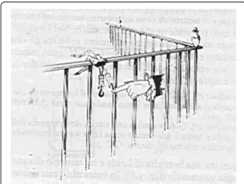
Luigi Galvani (1737–1798), during the 1780’s, per-formed experiments at the University of Bologna involv-ing electricity and frogs. He noticed that frogs’legs hung from brass hooks on his metallic bannister twitched whenever the breeze made them knock against the iron-work (Fig. 1). Moreover, he observed contraction of the frog’s muscles when they were touched with a metallic rod at the moment when an electrostatic machine, in the laboratory, produced a discharge.
Galvani came to the conclusion that some kind of electricity, which he calledanimal electricity, was gener-ated in the tissue of the frog and, flowing through the metal rod, activated the frog’s muscles. He thought of animal electricity as a fluid secreted by the brain, and proposed that flow of this fluid through the nerves acti-vated the muscles. He grew convinced that the vital spirit wasanimal electricity flowing through the nerves and announced this to the Bologna Academy of Science in 1791 [3].
Alessandro Volta (1745–1827) was professor of experi-mental physics at the University of Pavia, from 1779 for al-most 40 years. In 1792, Volta came to know of Galvani’s experiments on animal electricity. He initiated to repeat the experiments and at first his results agreed with those of Galvani. However, analyzing more closely the experi-mental conditions, Volta gradually became convinced that the contractions of the frog’s muscles were not due to the presence of electricity generated in the animal, but to some external electricity caused by the contact of the two metals. He concluded that different kinds of metals had electro-motive power at the point where they are in
contact with the frog. He summarized his ideas with the expression:“It’s the difference in metals that does it”.
In late 1799, Volta produced the apparatus which made him famous: the battery, based on the pairing of silver and zinc plates separated by brine soaked paper disks (Fig. 2). This once more proved that bimetal contact was the real source of electrical power. Volta announced his invention to the scientific community on 20th March 1800 in a letter to Sir Joseph Banks, the President of the Royal Society in London [4].
A new hypothesis
It is at this moment that comes into play another Italian scientist, Luigi Rolando (1773–1831), who in 1804 be-came professor of physiology at the University of Sassari (Fig. 3).
Rolando was impressed by the two main observations of Galvani and Volta: muscle contraction is due to elec-tricity and to generate elecelec-tricity is necessary a battery. His reflection was the following: since the brain gener-ates the movement, it must contain a device generating electricity. In his book published in 1809 [5], he writes “se i fenomeni della locomozione sono l’effetto di un par-ticolare meccanismo, questo non altrove che nell’encefalo andava ricercato”. (if the phenomena of locomotion are the effect of a particular mechanism, this not elsewhere than in the encephalon had to be researched)
For Rolando this part of the brain, as a battery, should be formed by overlapping disks and the cerebellum seemed to be the right structure given its characteristic overlapping laminae, forming the so-called arbor vitae. Probably the term was coined by the Danish anatomist Jacob B. Winsløw (1669 –1760) for the similarity of cerebellar folia with the profile of leaves of the North American treeThuja occidentalisor Eastern Arborvitae, introduced in France in 1534 by French explorers. It
Fig. 1Luigi Galvani observed that the spasms of frog’s muscles occurred when he hooked the frog onto a metal railing (from http://ppp.unipv.it/Volta/Pages/ePage1.html)
seems that the tree was named “l’arbre de vie” by the King Francis I [6] for analogy with the use of this ex-pression in the Book of Proverbs, where the tree of life is associated with wisdom.
Rolando writes “se dunque l’organo elettrico torpedi-nale e quelli del Siluro e del Ginnoto, fatti di sostanza albumino-gelatinoso-cartilaginea e simili attissimi sono a preparare, ed a sviluppare una quantità grandissima di fluido elettrico sufficiente per dare grandissime scosse, perché non potrà separarsi un principio consimile, quale si è il nerveo fluido dalle numerose lamine di sostanza midollare, giallognola, e cinerea del cervelletto? Quale maggiore evidenza potrassi desiderare per stabilire, che il cervelletto è un organo, la cui struttura è affatto consi-mile a quella dell’apparecchio del Volta?” (if the electric organ of torpedo and those of wels catfish and electric eel, made of albuminous-gelatinous-cartilaginous sub-stance, are perfectly suited to prepare and develop a large amount of electric fluid enough to give huge shocks, why a similar principle should not take place in form of nervous fluid from several sheets of yellowish and cinereous substance of the cerebellum? What greater evidence can be desired to establish that the cerebellum is an organ whose structure is absolutely similar to that of the Volta’s device?).
Rolando concluded that, if the cerebellum is the battery that generates electricity for muscle activity, its removal
would produce paralysis. He writes “Qual maggior prova per dimostrare, che dal suddetto viscere si separa un fluido analogo a quello, che dallo strumento citato si sviluppa? Qual più retta conseguenza, se esportato guasto o distrutto il cervelletto cessa ogni influsso del fluido nerveo nei mus-coli destinati alla locomozione?” (What most evidence to prove that the said organ generates a fluid similar to that which develops from the mentioned device? What most direct consequence if removed, destroyed or spoiled the cerebellum ceases any influence of the nervous fluid on the muscles for locomotion?).
Rolando, consequently, removed the cerebellum in a young goat and observed that the animal could no longer stand up “non altrimenti che se fosse paralitico” (not otherwise than if it was paralyzed). The animal survived for 24 h and died probably for postoperative sepsis.
Rolando concluded that the cerebellum is a motor structure as it generates the electricity which produces the movement.
The criticisms
The conclusions of Rolando were criticized by Marie-Jean-Pierre Flourens (1794–1867), professor of physiology at the Collège de France in Paris (Fig. 4). He observed that animals he submitted to cerebellectomy, with the intent to
Fig. 3Luigi Rolando (particular of the paint of Pasquale Baroni in the Museum of Anatomy of the University of Turin)
disprove the hypothesis of Gall, were still able to move, even if mating attempts failed for problems of balance. In his book published in 1824 [7], he concluded that “dans le cervelet réside une propriété dont rien ne don-nait encore l’idée en physiologie, et qui consiste a ordonner ou coordonner le mouvements voulus par cer-taines parties du système nerveux, excités par d’autres” (in the cerebellum lies a property which nothing still gave the idea in physiology, that is to put in order or
to coordinate movements wanted by certain parts of the nervous system, excited by others).
Since also its animals died shortly after the operation, Flourens hoped that the improvement of the surgery would allow to have animals surviving the cerebellect-omy, to clearly distinguish the deficits due to the re-moval of cerebellum from those related to postoperative complications.
Modern knowledge
It was necessary to wait the “germ theory of disease” of Louis Pasteur (1829–1895) and its application in clinical medicine, initially by Joseph Lister (1827–1912), with the use of carbolic acid as an antiseptic, and subsequently by Lawson Tait (1845–1899) and Ernst von Bergmann (1836–1907) which went from antisepsis to asepsis. These medical advances have allowed Luigi Luciani (1840–1919), in that period professor of physiology at the University of Florence, to publish in 1891 [8] his observations on a dog survived the cerebellectomy, with the description of a triad of symptoms (asthenia, atony and astasis), unquestionably of cerebellar origin, that confirmed the intuition of Flou-rens. In the same years (1894), Spanish neuroscientist and future Nobel laureate Santiago Ramón y Cajal (1852–1934) published what is considered the first modern textbook of neuroanatomy [9], with a clear depiction of the cerebellar cortex (Fig. 5).
The first systematic description of the symptoms of cerebellar lesions in man was carried out by the British
neurologist Gordon Morgan Holmes (1876 –1965).
During World War I he was neurologist with the British Expeditionary Forces and working in a field hospital he had the opportunity to investigate the effects of traumatic lesions involving the cerebellum. In 1922 Holmes’ observa-tions on patients with cerebellar wounds as well as tumors
Fig. 5Drawing of Purkinje cells (a) and granule cells (b) from pigeon cerebellum by Santiago Ramón y Cajal. Instituto Santiago Ramón y Cajal, Madrid, Spain
Table 1Major contributions to the current knowledge of the cerebellum
Year Author Contribution
1809 Luigi Rolando The cerebellum is the battery that produces the electricity necessary for generating muscular contraction
1824 Marie-Jean-Pierre Flourens The role of the cerebellum is not that of generating the movement but to regulate it 1891 Luigi Luciani Description, in a dog survived the cerebellectomy, of a triad of symptoms
(asthenia, atony and astasis) unquestionably of cerebellar origin
1894 Santiago Ramón y Cajal Publication of the first modern textbook of neuroanatomy with a clear description of the cerebellar cortex.
1922 Gordon Morgan Holmes Systematic description of the symptoms of cerebellar lesions in man 1967 John C. Eccles, Masao Ito, and János Szentágothai Book: The Cerebellum as a Neuronal Machine
1969 David C. Marr Hypothesis about cerebellum and motor learning: A theory of cerebellar cortex 1971 James S. Albus Hypothesis about cerebellum and motor learning: A theory of cerebellar function 1974 Gary I. Allen and Nakaakira Tsukahara Review: Cerebrocerebellar communication systems
were published in his Croonian Lectures to the Royal College of Physicians [10].
The general conclusion reached before World War II was that the main role of the cerebellum is to detail the different aspects of a movement, not to initiate move-ments or to decide which movemove-ments to execute. After the war, there was a significant increase in knowledge of circuitry and electrophysiology of the cerebellum, summa-rized in 1967 in a book,The Cerebellum as a Neuronal Machine[11], written by the Nobel laureate John C. Eccles (1903–1997), Japanese neuroscientist Masao Ito, and Hungarian anatomist János Szentágothai (1912–1994), followed in 1974 by a review,Cerebrocerebellar communi-cation systems [12], written by two neurophysiologists, the American Gary I. Allen and the Japanese Nakaakira Tsukahara (1933–1985).
In the same years it was suggested that the cerebellum is involved in motor learning. Most theories that attempt to explain the role of cerebellar circuits in motor learning are derived from the ideas of British neuroscientist and psych-ologist David C. Marr (1945–1980) and of American en-gineer James S. Albus (1935–2011). Both attributed an important role to climbing fiber activity capable to cause synchronously activated parallel fiber inputs, to be strengthened for Marr [13] and to be weakened for Albus [14]. In the 1980s, the discovery in the cerebellum of Long Term Depression (LTD) was considered as a form of syn-aptic plasticity involved in motor learning. LTD occurs when impulses of a set of granule cells and one climbing fiber reach the same Purkinje cell synchronously and re-peatedly; synaptic transmission from the granule cells to the Purkinje cell is then persistently depressed [15]. Although LTD is now well characterized, its contribution to motor learning remain controversial [16].
Up to the 1990s the cerebellum was almost universally believed to be primarily involved in movement, but latest results have led to consider that view too restrictive. Im-aging studies have allowed to detect cerebellar activation in relation to cognitive activities and numerous correla-tions between the cerebellum and non-motor regions of the cerebral cortex were highlighted. Moreover, in patients with lesions restricted to the cerebellum, non-motor symptoms have been frequently recognized. In 1998, the American neurologist Jeremy D. Schmahmann [17] de-scribed theCerebellar Cognitive Affective Syndrome, char-acterized by impairment of executive functions, difficulties with spatial cognition, personality change and language deficits. Table 1 summarizes the major contributions to the current knowledge of the cerebellum.
Conclusion
Luigi Rolando devoted his life to the study of the brain. Despite his outlandish theory on the cerebellum, he provided a major contribution to the advancement of
neurosciences and many neural entities are named after him: the substantia gelatinosa of Rolando in the spinal cord, thefissure of Rolandoor central sulcus, theRolandic operculumor post-central operculum, theRolandic artery or central sulcal artery, the Rolandic vein i.e., the vein posterior to Trolard’s vein draining the parietal lobe, thepre-Rolandic artery or precentral sulcal artery, and theRolandic epilepsyor benign childhood epilepsy with centrotemporal spikes (BCECTS), the most common epilepsy syndrome in childhood.
Competing interests
The authors report no conflicts of interest. The authors alone are responsible for the content and writing of the paper.
Authors’contributions
MC and VP wrote the paper and approved the final manuscript.
Received: 22 June 2015 Accepted: 4 August 2015
References
1. Manzoni T. The cerebral ventricles, the animal spirits and the dawn of brain localization of function. Arch Ital Biol. 1998;136:103–52.
2. Gall FJ, Spurzheim JK. Untersuchungen über die Anatomie des Nervensystems überhaupt, und des Gehirns insbesondere: ein dem Französischen Institute überreichtes Memoire; nebst dem Berichte der H.H, Kommissare des Institutes und den Bemerkungen der Verfasser über diesen Bericht. Paris und Strasburg: Treuttel und Würtz; 1809.
3. Galvani L. De viribus electricitatis in motu musculari commentarius, in De Bononiensi Scientiarum et Artium Instituto atque Academia Commentarii, vol VII. Bononiae: Ex Typographia Instituti Scientiarum; 1791.
4. Volta A. On the electricity excited by the mere contact of conducting substances of different kinds. In a letter from Mr. Alexander Volta, F.R.S. Professor of Natural Philosophy in the University of Pavia, to the Rt. Hon. Sir Joseph Banks, Bart. K.B.P.R.S. Phil Trans R Soc Lond. 1800;1:27–9. 5. Rolando L. Saggio sopra la struttura del cervello dell’uomo e degli animali e
sopra le funzioni del sistema nervoso. Sassari. Nella Stamperia da S.S.R.M. Privilegiata. 1809
6. Carpenter AC, Anatomical Etymologies. Web site: http://daphne.palomar.edu/ ccarpenter/anatomywords.htm.
7. Flourens MJP. Recherches expérimentales sur les propriétés et les fonctions du système nerveux dans les animaux vertébrés. Paris: Crevot; 1824. 8. Luciani L. Il cervelletto: nuovi studi di fisiologia normale e patologica.
Firenze: Le Monnier; 1891.
9. Ramón y Cajal S. Les nouvelles idées sur la structure du système nerveux: chez l’homme et chez les vertébrés Paris: Reinwald ;amp Cie. 1894. 234 pp. 10. Holmes GM. Clinical symptoms of cerebellar disease and their
interpretation. Lancet. 1922;202(Vol. 1 for 1922):Lecture I, 1178–1182, and Lecture II, 1232–1237.
11. Eccles JC, Ito MK, Szentágothai J. The Cerebellum as a Neuronal Machine. New York: Springer; 1967. p. 343.
12. Allen GI, Tsukahara N. Cerebrocerebellar communication systems. Physiol Rev. 1974;54(4):957–1006.
13. Marr D. A theory of cerebellar cortex. J Physiol. 1969;202(2):437–70. 14. Albus JS. A theory of cerebellar function. Math. Biogeosciences.
1971;10(1–2):25–61.
15. Ito M, Kano M. Long-lasting depression of parallel fiber-Purkinje cell transmission induced by conjunctive stimulation of parallel fibers and climbing fibers in the cerebellar cortex. Neurosci Lett. 1982;33(3):253–8. 16. Schonewille M, Gao Z, Boele HJ, Veloz MF, Amerika WE, Simek AA, et al.
Reevaluating the role of LTD in cerebellar motor learning. Neuron. 2011;70:43–50.