IDENTIFICATION OF PROTEIN’S COMPLEMENTARY TO THE AUTOANTIGEN PROTEINASE 3 AND THEIR INVOLVEMENT IN THE PATHOGENESIS OF
AUTOIMMUNE DISEASE
David James Bautz
A dissertation submitted to the faculty of the University of North Carolina at Chapel Hill in partial fulfillment of the requirements for the degree of Doctor of Philosophy in the
Department of Biochemistry and Biophysics
Chapel Hill 2008
ABSTRACT
David James Bautz: Identification of Protein’s Complementary to the Autoantigen Proteinase 3 and Their Involvement in the Pathogenesis of Autoimmune Disease
(Under the direction of Ronald Falk, M.D., Gloria Preston, Ph.D. and Alex Tropsha, Ph.D.)
Previous work by our research group showed that PR3-ANCA patients had an antibody response to a recombinant complementary-PR3 protein encoded by the antisense strand of the PR3 mRNA. To follow up on this work, we sought to determine whether the patients also had a T cell response to this recombinant complementary-PR3 protein and whether a protein reactive with those antibodies could be identified in vivo.
Chapter 2 of the dissertation describes the identification of CD4+ TH1 cells that
proliferate in response to a complementary-PR3 peptide. This proliferation was seen by both a CFSE assay as well as by interferon-γ production in an ELISPOT assay. Those patients who had a T cell response to complementary-PR3 peptide also had antibodies to the complementary-PR3 protein.
plasminogen was shown to be a substrate of PR3, indicative of interaction between the two proteins. Lastly, the anti-complementary PR3 antibodies also bound to normal human leukocytes, cells that are known to bind plasminogen on their surface.
Chapter 4 describes the identification of anti-plasminogen autoantibodies in PR3-ANCA positive patients. These antibodies were purified using a complementary-PR3 peptide column, indicating that the anti-cPR3 and anti-plasminogen antibodies are the same. The anti-plasminogen antibodies bound a surface-exposed loop on plasminogen’s catalytic domain. Two in vitro assays confirmed the antibodies affect on plasminogen activity. Serological screening of sera indicated that the anti-plasminogen autoantibodies were more prevalent in those PR3-ANCA patients with a clinical history of venous thrombotic events.
ACKNOWLEDGEMENTS
As the other graduate student in the lab, Dr. Josh Astern could appreciate what life was like as a grad student and it was nice to have someone in the lab going through the same experiences. Josh was also quite adept at providing comic relief to lighten the mood of the lab and there were certainly times when that was needed and appreciated.
Not having to worry about ordering supplies allowed me time to work on my various projects, and for that I am indebted to Nirmal Khandoobai since he took care of all supply ordering and restocking, along with so many other laboratory duties. I also thank Nirmal for making my trip to Cancun for the 2007 ANCA meeting as fun as possible.
I’d like to thank Julie Hamra for identifying patients coming into the clinic, collecting all of our patient samples, and relaying pertinent patient information.
The data generated from this project was made all the more important because of the detailed patient information collated by Dr. Sophia Lionaki. I am very grateful to her for going through stacks and stacks of patient charts, calculating untold numbers of BVAS scores, and preparing tables on patient data
This project involved doing untold number of ELISA’s, and I’d like to thank Dr. JJ Yang for all of her help with those. In addition, JJ was adept at essentially every assay done in this lab and was always willing and eager to train and I thank her for taking time to teach me all those various laboratory techniques.
Having enough recombinant protein available for all the assays performed in these experiments was crucial, and I’d like to thank Frances Belmonte and Therese Tulley for their aid and assistance in making sure there was enough protein available at all times.
I’d like to thank both Dr. Susan Hogan and Hyunsook Chin for their work on the statistical analysis.
Epitope mapping of the anti-plasminogen antibodies was an important piece of data generated during this project, and I’d like to thank Dr. Viorel Mocanu and Dr. Carol Parker for their assistance in epitope mapping of antibody samples using mass spectrometry. I was especially appreciative of their willingness to explain how the epitope mapping worked and to let me sit in on some of those experiments.
Assaying antibodies directed against plasminogen was something that our research group had never done before, thus I am indebted to Dr. Alisa Wolberg for providing ideas for assays to test activity of anti-plasminogen autoantibodies and for training me in those asssays.
TABLE OF CONTENTS
LIST OF TABLES ... xiii
LIST OF FIGURES ... xiv
ABBREVIATIONS ... xv
SYMBOLS ... xix
CHAPTER I ... 1
AUTOIMMUNE DISEASE... 2
SMALL-VESSEL VASCULITIS... 3
WEGENER’S GRANULOMATOSIS... 4
ANCA ... 5
PROTEINASE 3... 7
COMPLEMENTARY PROTEINS... 8
IDIOTYPIC NETWORK THEORY... 12
IDIOTYPES AND AUTOIMMUNITY... 13
COMPLEMENTARY PROTEINS AND THE IDIOTYPIC NETWORK... 15
AUTOANTIGEN COMPLEMENTARITY... 18
ANTIGENIC COMPLEMENTARITY AND THE DEFINITION OF COMPLEMENTARY PROTEINS... 19
CHAPTER II... 25
ABSTRACT... 26
INTRODUCTION... 27
MATERIALS AND METHODS... 30
Patients... 30
Proteins and synthetic peptides... 30
Cell stimulations... 31
CFSE Assay... 31
Enzyme-linked immunospot assay (ELISPOT)... 32
Intracellular cytokine production of CD4+ and CD8+ cells... 33
Detection of anti-cPR3(105-201) antibodies in sera by ELISA... 33
Statistical analysis... 33
RESULTS... 34
Patient study group... 34
T cells produce IFN-γ in response to cPR3(138-169) peptide... 37
Specificity of responses to fragments of cPR3(138-169) peptide... 39
Individual variability among longitudinal samples... 39
Coexistence of cPR3(138-169)-specific T cells with cPR3(105-201)-specific antibodies... 41
DISCUSSION... 44
CHAPTER III ... 48
ABSTRACT... 49
Antigens/Antibodies/Reagents... 52
Recombinant complementary-PR3 protein production... 53
Affinity purification of anti-cPR3(138-169) antibody... 53
Purification of proteins from plasmapheresis fluid... 54
ELISAs showing PLEX proteins reactive to anti-cPR3(138-169) antibody... 55
ELISAs to test patient reactivity to cPR3(105-201)... 56
Western blot analysis of patient plasmapheresis proteins... 56
Protein identification by mass spectrometry... 57
Proteolysis assay... 57
Flow cytometry analysis of normal human leukocytes... 58
RESULTS... 59
A subset of PR3-ANCA patients have antibodies specific for cPR3(105-201)... 59
Plasminogen is a serum protein recognized by anti-cPR3(138-169) antibodies... 59
Protein F from pseudomonas reacts with chicken anti-cPR3(138-169) antibodies... 61
Chicken anti-cPR3(138-169) antibodies purified from yolk react with plasminogen... 66
PR3 and plasminogen are complementary proteins... 66
Anti-cPR3(138-169) antibodies bind to normal human leukocytes... 70
DISCUSSION... 72
CHAPTER IV... 78
ABSTRACT... 79
INTRODUCTION... 80
Antigens/Antibodies/Reagents... 81
Affinity purification of patient anti-cPR3(138-169) antibody... 82
Western blot analysis of patient anti-cPR3(138-169) reactivity... 83
Epitope mapping of anti-plasminogen autoantibodies... 84
In vitro plasminogen assays... 85
ELISAs to test patient reactivity to plasminogen... 86
Statistical Analysis... 86
RESULTS... 87
Anti-cPR3(138-169) antibodies from PR3-ANCA patients react with plasminogen... 87
A target epitope for anti-cPR3(138-169) antibodies on plasminogen... 87
Assessment of anti-plasminogen autoantibodies on plasminogen function... 89
Prevalence of anti-plasminogen autoantibodies... 91
DISCUSSION... 96
CHAPTER V ... 100
LIST OF TABLES
TABLE 1.1. SENSE AND ANTISENSE CODON TABLE... 10
TABLE 2.1. CLINICAL AND DEMOGRAPHIC CHARACTERISTICS OF PATIENTS... 35
TABLE 2.2. RESPONSES TO CPR3(138-169)PEPTIDE FRAGMENTS... 40
TABLE 3.1. IDENTIFICATION OF PROTEIN F FROM PSEUDOMONAS... 65
LIST OF FIGURES
FIGURE 1.1. ANCASTAINING OF HUMAN NEUTROPHILS... 6
FIGURE 1.2. COMPLEMENTARY PROTEINS AND THE IDIOTYPIC NETWORK... 17
FIGURE 1.3. THE THEORY OF AUTOANTIGEN COMPLEMENTARITY... 20
FIGURE 1.4. THE THEORY OF ANTIGENIC COMPLEMENTARITY... 22
FIGURE 2.1.PR3-ANCAPATIENTS T CELLS RESPOND TO CPR3(138-169)... 36
FIGURE 2.2. PR3-ANCAPATIENT T CELLS PRODUCE IFN-Γ IN RESPONSE TO CPR3(138-169)... 38
FIGURE 2.3. PR3-ANCAPATIENT SAMPLE VARIABILITY AND ITS RELATIONSHIP TO BVAS SCORE... 42
FIGURE 3.1. PR3-ANCAPATIENTS REACT WITH CPR3(105-201)... 60
FIGURE 3.2. IDENTIFICATION OF COMPLEMENTARY-PR3PROTEINS USING RABBIT ANTI -CPR3(138-169)ANTIBODIES... 62
FIGURE 3.3. IDENTIFICATION OF COMPLEMENTARY-PR3PROTEINS USING CHICKEN ANTI-... 64
FIGURE 3.4. IDENTIFICATION OF A COMPLEMENTARY-PR3PROTEIN USING CHICKEN ANTI -CPR3(138-169)ANTIBODIES FROM EGG YOLK... 67
FIGURE 3.5. PLASMINOGEN AND PR3 ARE COMPLEMENTARY PROTEINS... 69
FIGURE 3.6. ANTI-CPR3(138-169)ANTIBODIES BIND TO NORMAL HUMAN LEUKOCYTES... 71
FIGURE 4.1. PATIENT IGG,AFFINITY PURIFIED USING A CPR3(138-169)PEPTIDE COLUMN, REACTS WITH PLASMINOGEN... 88
FIGURE 4.2. FUNCTIONAL EFFECTS OF ANTI-PLASMINOGEN AUTOANTIBODIES... 90
ABBREVIATIONS
AChR Acetylcholine receptor
ACTH Adenocorticotropin
AMA Anti-mitochondrial antibody
ANCA Anti-neutrophil cytoplasmic autoantibody
AP Alkaline phosphatase
APGN Anti-plasminogen autoantibodies
AZA Azathioprine
Bis-Q Trans-3,3'-bis[alpha-(trimethylammonia)-methyl]azobenzene
BSA Bovine serum albumin
BVAS Birmingham vasculitis activity score
Ca2+ Calcium ion
c-ANCA ANCA producing a cytoplasmic staining pattern
CDI Cell division index
cDNA complementary DNA
CMV Cytomegalovirus
CNBr Cyanogen bromide
ConA Concavalin A
cPR3 Complementary PR3
CsA Cycolsporine
CSFE Carboxy-fluorescein diacetate succinimidyl ester
CSS Churg-Strauss syndrome
CV Column volumes
CYC Cyclophosphamide
DMSO Dimethyl sulfoxide
DNA Deoxyribonucleic acid
dsDNA Double stranded DNA
DVT Deep vein thrombosis
EDTA Ethylenediamine tetraacetic acid EGFR Epidermal growth factor receptor
ELISA Enzyme-linked immunosorbent assay ELISPOT Enzyme-linked immunospot assay
FITC Fluorescein isothiocyanate
FPLC Fast performance liquid chromatography FSH Human follicle stimulating hormone
g Force of gravity
GAS Group A streptococcus
GC Glucocorticoids
HBS Hepes buffered saline
HCl Hydrochloric acid
HEK 293 Human embryonic kidney cell line
HI Heat inactivated
HRP Horseradish peroxidase
HTCA Complementary ACTH peptide
IFN-γ Interferon gamma
IgG Immunoglobulin G
IgY Immunoglobulin Y
IRB Institutional review board
ITP Idiopathic thrombocytopenia purpura IVIg Intravenous immunoglobulin
kDa Kilodalton
KLH Keyhole limpet hemocyanin
La/SSB Ribonucleoprotein
MALDI-TOF Matrix assisted laser desorpton/ionization-time of flight
MG Myasthenia gravis
MPA Microscopic polyangitis
MMF Mycophenolate mofetil
MPO Myeloperoxidase
MRT Molecular recognition theory
MS Mass spectrometry
NaCl Sodium chloride
NADPH Nicotinamide adenine dinucleotide phosphate
NaN3 Sodium Azide
p-ANCA ANCA producing a perinuclear staining pattern PBS Phosphate buffered saline
PDB Protein data bank
PE Phycoerythrin
pgp 1b Platelet glycoprotein 1b
PLEX Plasmapheresis fluid
PMA Phorbol 12-myristole 13-acetate
PR3 Proteinase 3
PRTN3 Gene encoding PR3
RNA Ribonucleic acid
SDS-PAGE Sodium dodecyl sulfate-polyacrylamide gel electrophoresis SLE Systemic lupus erythematosus
SVV Small vessel vasculitis
tPA Tissue-type plasminogen activator TPCK Tosyl phenylalanyl chloromethyl ketone Tris 2-amino-2-hydroxymethyl-1,3-propanediol TSH Thyroid stimulating hormone
uPA Urokinase-type plasminogen activator
VTE Venous thrombotic event
VWF von Willebrand factor
SYMBOLS
β Beta
© Copyright ° Degrees
γ Gamma
µ Micro
CHAPTER I PROLOGUE
The work described in this dissertation touches upon one of the most complex issues in the biological sciences; how and why does the immune system, which is designed to protect the individual from pathogenic invaders, decide that the host is in fact the enemy? This question is the basis for studying autoimmune disease, and until it can be answered with confidence it will remain incredibly difficult to discover better cures and treatments for patients suffering from these debilitating diseases.
Before describing the rationale for pursuing this project and the data subsequently generated, I will provide a discussion of ideas and theories that were combined in this research. These theories may at first appear to be unrelated; however it will become apparent that as a result of not compartmentalizing theories and ideas a new model for discovering autoantigens and autoantibodies was discovered. The dissertation will begin with an overview of the literature from which the major themes were generated that influenced the design and interpretation of the studies described.
AUTOIMMUNE DISEASE
Discerning the underlying cause of human disease is crucial to develop better and more effective treatments for a leading cause of suffering and mortality, autoimmune disease. While much effort has been put into the study of heart disease, cancer, and stroke, scientists are beginning to focus more on a loose-knit group of poorly understood diseases mediated by the immune system.
Autoimmune diseases are characterized by the apparent unregulated attack by the immune system on self tissue. There are over 80 autoimmune diseases currently identified that affect 14-22 million Americans [1]. They include common conditions such as Grave’s disease (1,150 per 100,000 individuals) as well as less frequent ailments such as Myasthenia gravis (0.4 per 100,000 individuals) [2]. Autoimmune diseases strike people of all ages, ethnicities, and backgrounds, however they typically affect women more frequently than men (>75% of those affected are women) for reasons that remain unclear [3].
Criteria have been introduced to help distinguish autoimmune diseases at three different levels: direct, indirect and circumstantial [4]. Direct evidence for an autoimmune disease requires transmissibility of the characteristic wounds from either human to human or human to animal. Indirect evidence requires an animal model that re-creates the disease. Circumstantial evidence of autoimmune disease relies on “markers”, such as presence of high levels of autoantibodies in serum and/or deposition of antibody/antigen complexes in the affected organ.
Wegener’s granulomatosis (WG), microscopic polyangiitis (MPA), and Churg-Strauss syndrome (CSS), all of which are characterized by vasculitis affecting multiple organ systems, typically the small-sized vessels of the lungs and/or kidneys, as well as the presence of autoantibodies directed against proteins found in neutrophils [6].
SMALL-VESSEL VASCULITIS
WEGENER’S GRANULOMATOSIS
The condition of WG was first reported by Klinger in 1931 [13]. A more detailed description of the disease was later given by Friedrich Wegener [14]. The term “Wegener’s granulomatosis” was not introduced until 1954 by Godman and Churg [11]. Patients suffering from WG typically have inflammation of the small- and medium-sized blood vessels along with a number of different symptoms that affect various organ systems, however over 90% of WG patients have upper or lower respiratory tract disease [15]. Target organs include the upper airway (typically crusting and bleeding in the nasal passages), the lungs (pulmonary nodules and hemoptysis), and the kidneys (rapidly progressing segmental necrotizing glomerulonephritis) [16]. The American College of Rheumatology published acceptance criteria for WG [17], which was followed up by the Chapel Hill Consensus Conference on nomenclature of systemic vasculitis, and concluded that “the term ‘Wegener’s Granulomatosis’ is restricted to patients with granulomatous inflammation” [18].
ANCA
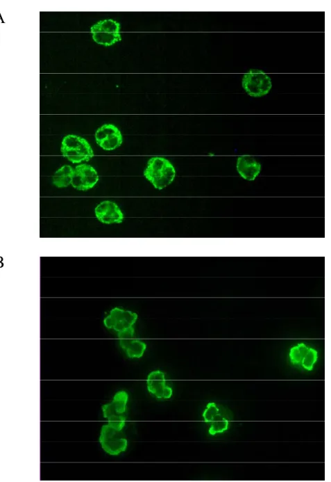
A feature that distinguishes WG, MPA and CSS from other small-vessel vasculitides is the presence of circulating ANCA. ANCA, first described by Davies et al. in 1982 [22] and in WG in 1985 [23], are typically present in 85-95% of patients with small-vessel vasculitides [24]. An indirect immunofluorescence test is used to verify the presence of ANCA. This test involves staining ethanol-fixed neutrophils with patient sera. The antibodies are classified on the basis of their neutrophil staining pattern (Figure 1.1); either c-ANCA (cytoplasmic c-ANCA) or p-c-ANCA (perinuclear c-ANCA). The most common c-c-ANCA antigen is proteinase 3 (PR3), primarily associated with WG [25, 26], while the most common p-ANCA antigen is myeloperoxidase (MPO), which is largely associated with MPA [27]. An atypical staining pattern can also be seen with the target antigens bacterial/permeability increasing protein, elastase and catalase [28], however they are not associated with vasculitic autoimmune diseases. ANCA are present to a lesser degree in other autoimmune diseases such as Sjogren’s syndrome (target antigen is lactoferrin) [29] and ulcerative colitis (target antigen is cathepsin G) [30]. When immunofluorescence staining is combined with ELISA testing the specificity for ANCA small-vessel vasculitis (SVV) is 99% and sensitivity for WG is 73% [31].
A
B
Figure 1.1. ANCA Staining of Human Neutrophils.
to bind PR3-ANCA from Wegener’s patients in a dose-dependent fashion [34]. There is now evidence from experimental animal models that ANCA are pathogenic and can cause disease [35]. Xiao et al. injected MPO into MPO knockout mice and then transferred splenocytes from these animals into mice deficient in B and T lymphocytes. Mice that received anti-MPO splenocytes developed “severe necrotizing and crescentic glomerulonephritis, granulomatous inflammation, and systemic necrotizing vasculitis” [35]. While MPO-ANCA have been shown to be pathogenic, there is still no animal model to prove the same relationship with PR3-ANCA.
PROTEINASE 3
Proteinase 3 (PR3) was first described in 1975 by Dewald et al. [36] and further characterized by Kao et al. during research into whether granulocyte proteins other than elastase could be involved in emphysema formation in humans [37]. PR3, along with elastase and azurocidin, is part of a family of serine proteases whose genes are located in a cluster on chromosome 19 that are synchronously expressed in the early stages of granulopoiesis in the bone marrow [38]. PR3’s function is associated with a number of different cellular activities, including inhibition of NADPH oxidase [39], apoptosis [40], and myeloid differentiation [41]. PR3 is also expressed on the surface of neutrophils in the enzymatically active form, however, unlike the granule localized form, it is resistant to inhibition [42].
addition, the presence of PR3 on the surface of neutrophils has been correlated with disease activity [47]. Taken together, these data suggest that there is amble PR3 antigen available for interaction with ANCA to cause disease in patients.
There appear to be multiple epitopes recognized by ANCA, and most of these are conformational epitopes [48]. Since there is little consensus on what epitopes are important on PR3, our research group became interested in epitope mapping PR3-ANCA. To do this, a random peptide library in bacteria was produced using blunt-ended fragments of the PR3 cDNA. Since the fragments were blunt-ended, they could insert into the vector either in the correct orientation or in the “flipped” orientation, generating PR3 peptides or peptides off the antisense strand of the gene. The expressed peptide fragments were then tested for reactivity with PR3-ANCA. The results of these experiments showed that a subset of PR3-ANCA patients had antibodies not only to PR3, but also to a complementary protein expressed off the antisense strand of the PR3 gene, which is referred to as complementary PR3, or cPR3 [49]. These unexpected findings led to a series of experiments and ultimately to the development of a new theory of autoimmune disease formation, the theory of autoantigen complementarity.
COMPLEMENTARY PROTEINS
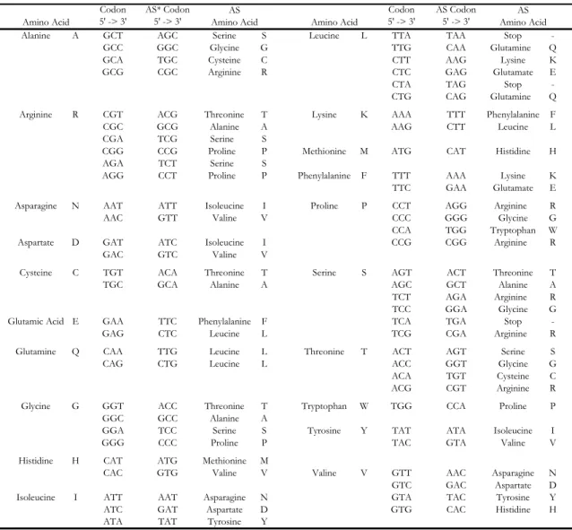
one of the interacting polypeptides in the 5’ -> 3’ direction yielded the amino acid sequence of the other interacting chain. Little work was done with regard to complementary proteins until Mekler’s ideas were reinvented by Biro [51] and Blalock/Smith [52] in the 1980’s. The basic theory put forth by this group of scientists is that peptides and proteins that selectively bind to one another are encoded by in-frame codons on complementary DNA/RNA strands. This idea has also been given the term “sense-antisense” that describes the proteins being encoded by sense and antisense nucleotide strands. Due to the degeneracy of the genetic code, there are multiple antisense partners for most amino acids (Table 1.1).
Based on the Mekler definition of complementary amino acids, a hydrophobic amino acid is always encoded opposite a hydrophilic partner [50, 52]. Thus, theoretically, for complementary proteins to interact it would require hydrophobic and hydrophilic amino acids to interact. While intuitively this seems rather unappealing, there is some evidence to support this arrangement. For example, leucine and lysine have been shown to interact spontaneously in solution [53]. It has also been suggested that binding between complementary proteins could be caused by the interactions between the hydrophobic side chains and the hydrocarbon backbone supporting the polar residues of their complementary partner [54, 55]. A model for the side-chain packing of glutamic acid and leucine has also been reported [56].
Table 1.1. Sense and Antisense Codon Table.
Codon 5' -> 3'
AS* Codon 5' -> 3'
Codon 5' -> 3'
AS Codon 5' -> 3'
Alanine A GCT AGC Serine S Leucine L TTA TAA Stop -GCC GGC Glycine G TTG CAA Glutamine Q
GCA TGC Cysteine C CTT AAG Lysine K
GCG CGC Arginine R CTC GAG Glutamate E
CTA TAG Stop -CTG CAG Glutamine Q Arginine R CGT ACG Threonine T Lysine K AAA TTT Phenylalanine F
CGC GCG Alanine A AAG CTT Leucine L
CGA TCG Serine S
CGG CCG Proline P Methionine M ATG CAT Histidine H AGA TCT Serine S
AGG CCT Proline P Phenylalanine F TTT AAA Lysine K TTC GAA Glutamate E Asparagine N AAT ATT Isoleucine I Proline P CCT AGG Arginine R
AAC GTT Valine V CCC GGG Glycine G
CCA TGG Tryptophan W Aspartate D GAT ATC Isoleucine I CCG CGG Arginine R
GAC GTC Valine V
Cysteine C TGT ACA Threonine T Serine S AGT ACT Threonine T
TGC GCA Alanine A AGC GCT Alanine A
TCT AGA Arginine R TCC GGA Glycine G Glutamic Acid E GAA TTC Phenylalanine F TCA TGA Stop
-GAG CTC Leucine L TCG CGA Arginine R
Glutamine Q CAA TTG Leucine L Threonine T ACT AGT Serine S
CAG CTG Leucine L ACC GGT Glycine G
ACA TGT Cysteine C ACG CGT Arginine R Glycine G GGT ACC Threonine T Tryptophan W TGG CCA Proline P
GGC GCC Alanine A
GGA TCC Serine S Tyrosine Y TAT ATA Isoleucine I
GGG CCC Proline P TAC GTA Valine V
Histidine H CAT ATG Methionine M
CAC GTG Valine V Valine V GTT AAC Asparagine N GTC GAC Aspartate D Isoleucine I ATT AAT Asparagine N GTA TAC Tyrosine Y ATC GAT Aspartate D GTG CAC Histidine H ATA TAT Tyrosine Y
* AS = antisense
Amino Acid Amino Acid
AS Amino Acid
biologically active. Bost et al. produced a complementary peptide to adrenocorticotropin hormone (ACTH) by reading the antisense nucleotide sequence in-frame in the 5’->3’ direction. This peptide, which they termed HTCA, bound to ACTH with high affinity [59]. Ghiso et al. suggested that the interaction between cystatin C, a cysteine proteinase inhibitor, and complement C4 is mediated by segments coded by complementary DNA sequences of the two proteins [60]. Much of the work thus far involving complementary peptides has been with receptor-ligand interactions, for example with angiotensin II [61] and human follicle stimulating hormone (FSH) [62]. However, it should be noted that there is a lot of controversy surrounding the validity of complementary protein interactions and much of the data generated thus far has been debatable and contradictory [63, 64].
IDIOTYPIC NETWORK THEORY
Neils Jerne theorized that the immune system is composed of a large network of interacting antibodies and receptors [67]. He postulated that an antibody molecule contains a paratope, or antibody combining site, that itself is composed of idiotopes, or epitopes displayed by the variable regions of antibodies. Thus, the paratope and idiotopes are expressed together and are dependent upon the primary sequence of the antibody variable region. It is these idiotopes, a group of which is called an idiotype, that are recognized by other antibodies in the immune system.
If an antigen is introduced to the immune system, there will be a response by a group of antibodies. The antigen combining sites on these antibodies will contain unique sequences that are unfamiliar to the immune system, thus an immune response will be generated against these idiotypes. Jerne theorized that these anti-idiotypic antibodies will bear the “internal image” of the original antigen. He also believed that the idiotypic network would play a role in suppressing an antibody response that in turn would lead the entire system back to a state of equilibrium. His idea’s were confirmed in a number of experiments by his research group that showed anti-idiotypic antibodies were produced in rabbits injected with immunoglobulin [68].
of receptor/ligand interactions by anti-idiotypic antibodies has also been extensively reported [70].
IDIOTYPES AND AUTOIMMUNITY
Idiotypic antibody interactions have also been reported in the study of a number of autoimmune diseases. Shechter et al. found that mice injected with insulin developed antibodies not only to insulin, but to insulin receptor [71]. These anti-insulin receptor antibodies bound to insulin receptor, displaced already bound insulin, and mimicked the actions of insulin in stimulating the oxidation of glucose and inhibiting lipolysis. The binding of anti-insulin receptor antibodies to insulin receptor could be blocked by anti-insulin antibodies suggesting the two antibodies formed an idiotypic pair. To characterize these antibodies further, the group produced chemically altered insulin that did not bind to insulin receptor. This altered insulin produced high levels of antibody, however no anti-idiotypic antibody that bound to insulin receptor was produced. The group concluded that the epitope responsible for the specific idiotypic network was most likely the part of insulin recognized by the insulin receptor [72].
anti-mitochondrial antibodies (AMA). When a monoclonal antibody was raised to the E2 complex of pyruvate dehydrogenase complex (the target antigen of AMA), it was able to bind anti-idiotypic antibodies from sera. The specificity of these anti-idiotypic antibodies was shown by their ability to inhibit binding of AMA to mitochondria but not other autoantibodies to their respective autoantigens [75]. The group did not investigate whether these autoantibodies were involved in control of AMA.
Erlanger et al. utilized the same strategy for the identification of anti-idiotypic antibodies in a patient with Grave’s disease, an ailment characterized by autoantibodies directed against thyroid-stimulating hormone (TSH) receptor. Immunization of mice with both bovine and human TSH resulted in the production of hybridomas that produced anti-TSH antibodies as well as anti-idiotypic antibodies. The researchers then used one of the monoclonal anti-TSH antibodies (LE-4) to make an affinity column and passed human plasmapheresis fluid from a Grave’s disease patient over the column. The eluted antibody inhibited the binding of LE-4 to TSH and also bound to the TSH receptor, thus showing the presence of anti-idiotypic antibodies in a human patient [79].
Mice with experimental systemic lupus erythamatosus (SLE) have anti-double stranded (ds) DNA antibodies. Since the idiotypic network has been hypothesized to control autoantibody reactivity, it was hypothesized that anti-anti-dsDNA antibodies would alleviate symptoms in SLE mice. To test this, Shoenfeld et al. affinity purified anti-anti-dsDNA antibodies from IVIg (IVIg-ID) using an affinity column composed of anti-dsDNA antibodies isolated from 55 patients with active SLE. They then treated mice with normal IVIg and IVIg-ID before and after the mice developed anti-dsDNA antibodies. The mice treated with IVIg-ID had significantly lower proteinurea, a longer survival time and a decrease in anti-dsDNA antibodies [80].
COMPLEMENTARY PROTEINS AND THE IDIOTYPIC NETWORK
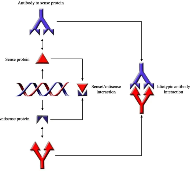
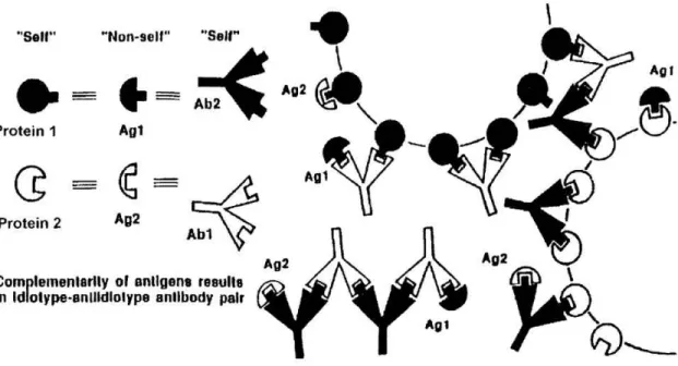
al. tested whether antibodies to complementary peptides would interact in an idiotypic fashion [66]. To test this, they injected a rabbit with ACTH and a second rabbit with HTCA. They then isolated total IgG from both rabbits and showed that antibodies specific to ACTH from one rabbit bound to antibodies to HTCA from the other rabbit through their combining sites; they were an idiotypic pair. This work was followed up by the same group using a pair of arbitrary complementary peptides [81]. Figure 1.2 gives a graphical representation of the relationship between complementary proteins and the idiotypic network.
If anti-idiotypic antibodies can be derived from complementary peptides, one potential application for complementary peptides could be their use in treating autoimmune disease. It follows that if anti-idiotypic antibodies could be induced, above the level already seen naturally in the disease, they could potentially block the action of harmful autoantibodies. This has been shown to be the case in a rat model of MG, where both a monoclonal antibody to an antisense peptide (corresponding to the autoantibody binding site on acetylcholine receptor) and an antisense peptide itself have alleviated symptoms [82, 83].
Antisense protein Sense protein
Antibody to antisense protein Antibody to sense protein
Sense/Antisense interaction
Idiotypic antibody interaction
Antisense protein Sense protein
Antibody to antisense protein Antibody to sense protein
Sense/Antisense interaction
Idiotypic antibody interaction
Figure 1.2. Complementary Proteins and the Idiotypic Network.
(both sense and complementary) and showed that antibodies to La/SSB were produced in both sense and antisense-immunized animals [85]. They also devised a novel ELISA to unmask idiotypic antibody relationships that were interfering with their test. In mice immunized with the complementary peptide they were initially unable to show a response to La/SSB protein. They hypothesized that anti-complementary peptide antibodies were binding to anti-La/SSB antibodies. To circumvent this problem, they heated sera samples at 55°C to disrupt antibody/antibody interactions and then added complementary peptide to block binding between the two antibodies. The samples were re-tested and they showed that the mice immunized with complementary antibody did in fact have antibodies to La/SSB. In addition, they were able to show that T-cell help was required for anti-idiotypic antibody development [86].
AUTOANTIGEN COMPLEMENTARITY
The theory of autoantigen complementarity was conceived based partly on the fact that a subset of PR3-ANCA patients were shown to have antibodies that reacted with a recombinant, complementary-PR3 protein [cPR3(105-201)] [49]. In agreement with the results
seen by Smith et al. [66], antibodies to PR3 were separate and distinct from antibodies to cPR3(105-201) and the two sets of antibodies bound in an idiotypic fashion. In addition, mice injected with cPR3(105-201) developed not only antibodies to cPR3 but also antibodies to human PR3.
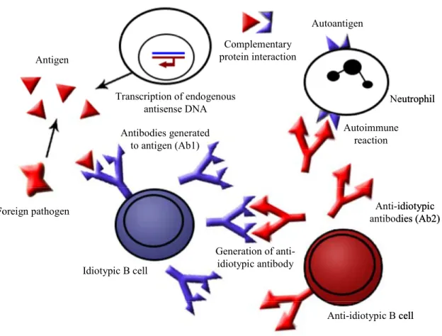
an idiotypic antibody response (in agreement with Jerne’s network theory). This anti-idiotypic antibody then reacts with the autoantigen resulting in autoimmune disease (Figure 1.3).
Where do complementary proteins come from? The theory proposes that the complementary proteins could either be produced endogenously (by aberrant transcription and translation of antisense message) or introduced exogenously by a microbial pathogen. In fact, there is evidence to support both of these routes. There are a large number of antisense transcripts identified in the human genome [87], and in fact a protein encoded by the antisense strand of a ubiquitously expressed gene has been identified in kidney cancer [88]. Antisense transcripts from the PR3 gene were found in 10 of 22 PR3-ANCA patients tested, while no antisense PR3 transcripts were found in a group of SLE patients or healthy controls [49]. In addition to being produced by the host, complementary proteins (or their mimics) could be introduced by microbes and/or viruses. There are a number of bacterial and viral proteins that share sequence homology with proteins that are antisense to known autoantigens [89], including proteins from two microbes, Staphylococcus aureus and Entamoeba histolytica, which have homologies to cPR3(105-201) and have been most closely linked to the
development of PR3-ANCA [90, 91].
ANTIGENIC COMPLEMENTARITY AND THE DEFINITION OF COMPLEMENTARY PROTEINS
Antigen
Transcription of endogenous antisense DNA Foreign pathogen Complementary protein interaction Autoantigen Neutrophil Autoimmune reaction
Anti-idiotypic B cell Idiotypic B cell
Generation of anti-idiotypic antibody Antibodies generated
to antigen (Ab1)
Anti-idiotypic antibodies (Ab2) Antigen
Transcription of endogenous antisense DNA Foreign pathogen Complementary protein interaction Autoantigen Neutrophil Autoimmune reaction
Anti-idiotypic B cell Idiotypic B cell
Generation of anti-idiotypic antibody Antibodies generated
to antigen (Ab1)
Anti-idiotypic antibodies (Ab2)
Figure 1.3. The Theory of Autoantigen Complementarity.
molecularly complementary immune responses that attack each other along with a tissue or organ in the body (Figure 1.4). This theory is similar to the theory of autoantigen complementarity in that it proposes complementary proteins are involved in induction of autoimmune disease; however there is an important difference in how a complementary protein pair is defined. The theory of autoantigen complementarity uses the Mekler definition of complementarity, that is, a peptide or protein produced, in-frame, from the antisense strand of the coding gene. Root-Bernstein et al. define complementary proteins as those able to bind to each other (molecularly complementary surfaces) and capable of inducing the production of complementary antibodies (or T cells) that act like idiotype-anti-idiotype pairs without the necessity of being encoded by sense-antisense codons [92]. Both definitions of complementarity are valid, with the Mekler definition being more stringent while the Root-Bernstein definition allows for a broader group of protein-protein pairs to be considered as complementary. The theory of antigenic complementarity also proposes that the sets of complementary proteins are likely to be microbial and/or viral in nature, and requires that only if one of these proteins is sufficient enough to “self” does an autoimmune reaction occur.
Figure 1.4. The Theory of Antigenic Complementarity.
proteins is reflected in the antibodies) [92]. They also showed that antibodies to cytomegalovirus (CMV) bound to antibodies to group A streptococcus (GAS), two infectious agents that have been associated with the onset of ITP [95, 96]. Proteins from CMV and GAS show significant homologies to both regions of VWF and pgp 1b that are involved in the binding of VWF and pgp 1b. Lastly, antibodies to GAS bind antibodies to VWF, and antibodies to CMV bind antibodies to pgp 1b. Taken together, these data fit their model; however showing this system functions in an animal model of disease is lacking.
PROJECT GOALS AND OUTCOMES
Knowing that patients have circulating antibodies to a protein complementary to PR3, we hypothesized that proteins complementary to PR3 could be purified and identified in vivo. Specifically, the objectives of the research presented here were to identify proteins that are complementary to PR3, to determine if these protein(s) had any functional or clinical significance, and whether these proteins could be implicated in the etiology of PR3-ANCA vasculitis. The hypothesis was that proteins complementary to PR3 could be identified in either patient’s sera or tissue samples, these proteins would interact with PR3, and multiple patients would have antibodies specific to these complementary proteins.
Chapter III of the dissertation will discuss the identification of a novel autoantibody to plasminogen in a subset of PR3-ANCA patients. These autoantibodies were not seen in a group of healthy control subjects, MPO-ANCA patients, or patients who had idiopathic thrombotic events. The antibodies altered normal plasminogen activity in two separate in vitro assays. In addition, these antibodies were found most often in patients who had suffered a thrombotic event.
CHAPTER II
T cell Responsiveness to Complementary-PR3 Protein: A Pathogenic Role for Autoantigen Complementarity in ANCA Disease
Jiajin Yang1, David J Bautz1, Sofia Lionaki1,4, Susan L Hogan1, Hyunsook Chin1, Roland Tisch3, John L. Schmitz2, Barrak M Pressler5, J Charles Jennette1,2 Ronald J
Falk1,2 and Gloria A Preston1,2
1Department of Medicine, Division of Nephrology and Hypertension, UNC Kidney
Center
2Department of Pathology and Laboratory Medicine 3Department of Microbiology and Immunology
University of North Carolina at Chapel Hill, Chapel Hill, NC
4Nephrology and Transplantation Department
Laikon Hospital, Athens, Greece
5Veterinary Clinical Sciences
ABSTRACT
We discovered that patients with PR3-ANCA vasculitis have antibodies reactive against a protein complementary to the autoantigen PR3 [cPR3(105-201)]. Investigations into the etiology and consequences of these anti-cPR3(105-201) antibodies led to the proposal that complementary proteins are involved in inciting autoantibody production, as described in the theory of autoantigen complementarity. The present studies indicate that CD4+ TH1 cells
INTRODUCTION
The concept of complementary protein pairs was first proposed by L.B. Mekler in the late 1960s [97, 98]. He proposed that information embedded within the genetic code could identify proteins that would pair in nature: proteins from sense codons bind proteins from their antisense codons. Skeptics of the validity of this idea are gradually realizing that experimentally this works and many researchers have discovered protein partners by utilizing complementary sequences coded by antisense codons (reviewed in [57, 99]). Of course, not all proteins that form complexes meet Mekler’s definition of a complementary pair. Researchers debate what characteristics truly constitute a complementary pair. A recent and broader definition of a complementary pair states that two proteins are complementary if they are capable of stereospecific binding and inducing an idiotype-antiidiotype antibody response, thus eliminating the restriction of sense and antisense codons [92]. The mechanistic basis for the natural affinity for complementary protein pairs remains largely speculative [64, 92], however, it is thought that inverted hydropathy may be a driving force [52].
reagent, we proved that anti-cPR3(105-201) antibodies were unique and that they bound PR3-ANCA to form an idiotypic pair.
We put forward the theory of autoantigen complementarity, which proposes that inciting antigens for autoimmune diseases are not the autoantigens, but instead proteins complementary to the relevant autoantigens [49]. The first event is an antibody response that targets a complementary protein; subsequently this antibody triggers an anti-idiotypic response. It is the anti-idiotypic antibody that targets the autoantigen.
some against the antigenic complex and some biased towards the individual components. Albeit, immunological responses incited by a complementary protein not complexed to its sense counterpart must be equally considered at this point.
MATERIALS AND METHODS
Patients
Allsubjects gave written informed consent and participated in thestudy according to the guidelines of the UNC Institutional Review Board (IRB # 97-MED-44). The study included 9 females/17 males; mean age 49.8 yrs (26-79 yrs); 3 blacks, 22 Caucasians, 1 Asian (Table 1). Mean of PR3-ANCA titers across samples was 51.7 (range: 3.2-170.0). Healthy controls were recruited on site for blood donations (n = 34). The disease control group of seven MPO-ANCA patients included 5 females /2 males, mean age 45 yrs (21-65 yrs), 1 black, 5 Caucasians and 1 Asian. Limits on the amount of blood obtainable per donation required that the different methodologies in this study be done in tandem. Twelve of 26 patients donated blood more than once during the study’s two-year period. Studies for anti-cPR3(105-201) antibody reactivity required the use of banked sera samples. Healthy control sera were from approved kidney transplant donors (n = 12).
Proteins and synthetic peptides
Recombinant cPR3(105-201) protein was produced as previously described [49]. The sequence of the protein is:
DAGLAARDESANVMWPAEEGDHGDIELLQDLGWGVVGTHAAPAHGQALGAVGH WLVLLWQLDCGDGGTEVGWAAQLDEENVVQFVLRVVVVQKHLSHREVLLGGLLR PHVVGSEHHVHQALGYVPQAVRGRQHEAG (cPR3(138-169) peptide underlined).
Synthetic peptides from Alpha Diagnostic (San Antonio, TX) included:
Fragment 1 - cPR3(138-153): N-DLGWGVVGTHAAPAHG-C (16aa) Fragment 2 - cPR3(146-161): N-THAAPAHGQALGAVGH-C (16aa) Fragment 3 - cPR3(154-169): N-QALGAVGHWLVLLWQL-C (16aa)
Sense-PR3(138-169): N-QLPQQDQPVPHGTQCLAMGWGRVGAHDPPAQV-C (32aa) Scrambled peptide: N-LWAGDWVALGLGAWLAGLHVHAQTPHVQVGGL-C (32aa) Purchased PR3 (Wieslab AB, Lund, Sweden) was passed over an Extracti-Gel AffinityPak detergent-removing column (Pierce, Rockford, IL) and heat inactivated (HI) (100oC/10 min) [106] to linearize the protein [103]. Recall antigen mixture contained tetanus toxoid (2 µg/ml) and diphtheria toxin (2 µg/ml) (LIST LABS, Campbell, CA), plus 15 µg/ml of candida (Allermed Lab, San Diego, CA). Other agents: concavalin A (Con A) (1 µg/ml), phorbol 12-myristate 13-acetate (PMA) (25 ng/ml), and ionomycin (1 µg/ml) (Sigma, St. Louis, MO).
Cell stimulations
Blood was collected into sodium heparin CPTTM Cell Preparation tubes (BD Vacutainer®, Franklin Lakes, NJ) and peripheral blood mononuclear cells isolated per instructions. Stimulants included peptides (2-25 µg/ml), HI-PR3 (2-10 µg/ml), recall antigen mixture and either ConA or PMA plus ionomycin. Peptide solvent dimethyl sulfoxide (DMSO) was added to controls.
CFSE Assay
Subsequent proliferation in the absence of CFSE results in decreased fluorescence intensity by ½ with each cell division. Cells were cultured at 1x10⁶ cell/ml 6 days with proteins (10 µg/ml) or peptides (25 µg/ml). CD3+ cells were labeled with phycoerythrin (PE)-mouse anti-human CD3 monoclonal antibody (BD PharMingen, San Diego, CA) and analyzed by FACScan linked to a CELLQuest software system (Becton Dickinson Immunocytometry Systems, San Jose, CA). The cell division index (CDI) is based on 5000 CFSEbright CD3+ cells as previously described [110].
CFSEbright/5000
CFSEdim treated
CFSEdim
CFSEbright/5000
untreated CDI= CFSEbright/5000
CFSEdim
CFSEbright/5000
CFSEdim
CFSEdim treated
CFSEdim
CFSEbright/5000
CFSEdim
CFSEbright/5000
untreated CDI=
Enzyme-linked immunospot assay (ELISPOT)
Cells were plated at 2 x 10⁶ cells/ml in 100µl in triplicate on MultiScreen 96-Well Filtration Plates (Millipore, Bedford, MA) and coated with anti-human IFN-γ monoclonal antibody (Pierce). Treatments included peptides (5 µg/ml), HI-PR3 (2 µg/ml), recall antigens and Con A. IFN-γ releasing cells were detected with biotinylated-mouse anti-human IFN-γ antibody (Pierce) (2µg/ml), streptavidin (SouthernBiotech, Birmingham, AL) and AEC solution, containing a 3-amino-9-ethylcarbazole tablet, N,N-Dimethyformamide and hydrogen peroxide (Sigma). Data were analyzed using ImmunoSpot reader, ImmunoSpot 3 software, version 3.2 (Cellular Technology Ltd., Cleveland, OH).
Intracellular cytokine production of CD4+ and CD8+ cells
Cells (0.8x10⁶/ml/well) were cultured with HI-PR3 (10 µg/ml) or peptides (5 µg/ml) for four days. PMA and ionomycin (6 hrs) served as a positive control. Brefeldin A (Sigma) was added (10 µg/ml) for 4hrs. Cells were fixed using FACS Lysing Solution and FACS Permeabilizing Solution 2 (Becton Dickinson) and were labeled with FastImmune anti-human IFN-γ FITC antibody (BD PharMingen) and anti-human CD4- or CD8-PerCP labeled antibodies (BD Immunocytometry Systems) and analyzed by FACScan.
Detection of anti-cPR3(105-201) antibodies in sera by ELISA
High-binding plates (Coster, Cambridge, MA) were coated overnight at 4oC with recombinant cPR3(105-201) protein (5 µg/ml). Sera was added (1:100) and reactive antibodies were detected with alkaline phosphatase goat anti-human antibody (Chemicon, Temecula, CA) plus alkaline phosphatase Substrate (Bio-Rad, Hercules, CA, USA) and read on a VERSAmax microplate reader (Molecular Devices, UK). Values are percent of positive control (rabbit anti-his-tag antibody) (Santa Cruz Biotech, Santa Cruz, CA, USA) compared to the mean plus two standard deviations of healthy controls.
Statistical analysis
RESULTS
Patient study group
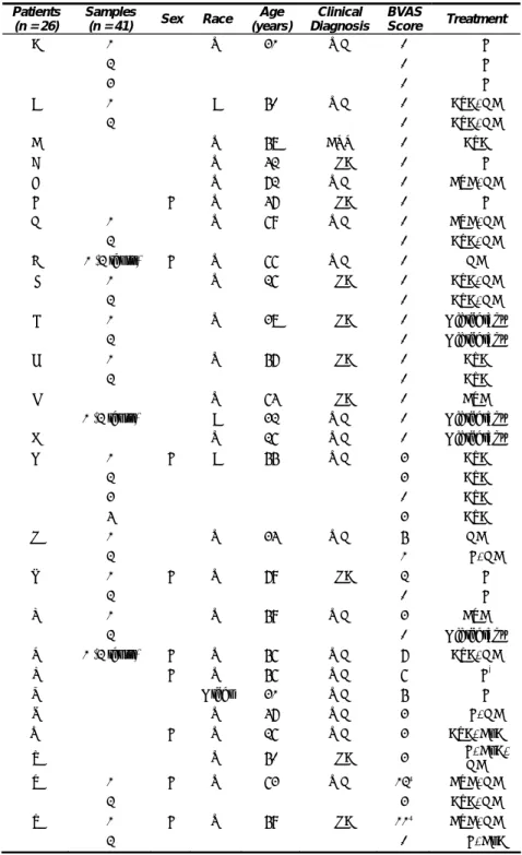
Patients with biopsy-proven PR3-ANCA vasculitis (n = 26) were identified through the Glomerular Disease Collaborative Network between January 2004 and June 2006 (Table 2.1). To avoid the potential of T cell anergy due to immunosuppressive therapies, the patient enrollment was limited to those in remission, slightly active disease on maintenance drugs, and newly diagnosed patients before aggressive treatment. Based on the Birmingham Vasculitis Activity Score (BVAS) 2003, 25 samples were from patients in remission (BVAS = 0) and 17 samples were from patients with active disease (BVAS > 0) (Table 2.1). Because of limitations on the amount of blood obtainable per patient-donation, inclusion in the different analyses required that patients donate more than once during the study’s two-year period.
Proliferative response of cPR3(138-169) peptide reactive T cells
If patients with PR3-ANCA vasculitis have experienced an immunological encounter with a complementary-PR3-like protein, they should possess T cell pools of previously activated, differentiated cells that now exist as long-lived memory cells. The particular amino acid sequence of cPR3(138-169) peptide used in this study was first identified as an epitope of patients’ antibodies during a screen of a bacterial expression library [49]. This specificity was confirmed by mass spectrometry (data not shown). CD3 T cell subsets from patients exhibited increased proliferation upon encounter with cPR3 peptide compared
+
Table 2.1. Clinical and Demographic Characteristics of Patients
Patients (n = 26)
Samples
(n = 41) Sex Race Age (years)
Clinical Diagnosis
BVAS
Score Treatment
A 1 M W 31 WG 0 MMF
2 0 MMF
3 0 MMF
B 1 M B 50 WG 0 AZA, GC
2 0 AZA, GC
C M W 58 CSS 0 AZA
D M W 42 MPA 0 MMF
E M W 72 WG 0 CYC, GC
F F W 47 MPA 0 MMF
G 1 M W 69 WG 0 CYC, GC
2 0 AZA, GC
H 1 (2 tests) F W 66 WG 0 GC
I 1 M W 26 MPA 0 AZA, GC
2 0 AZA, GC
J 1 M W 38 MPA 0 Off therapy
2 0 Off therapy
K 1 M W 57 MPA 0 AZA
2 0 AZA
L M W 64 MPA 0 CYC
M 1 (2 tests) M B 32 WG 0 Off therapy
N M W 26 WG 0 Off therapy
O 1 F B 55 WG 3 AZA
2 3 AZA
3 0 AZA
4 3 AZA
P 1 M W 34 WG 5 GC
2 1 MMF, GC
Q 1 F W 79 MPA 2 MMF
2 0 MMF
R 1 M W 59 WG 3 CYC
2 0 Off therapy
S 1 (2 tests) F W 56 WG 7 AZA, GC
T F W 56 WG 6 MMF*
U M Other 31 WG 5 MMF
V M W 47 WG 3 MMF, GC
W F W 26 WG 3 AZA, CsA
X M W 50 MPA 3 MMF, CsA, GC
Y 1 F W 63 WG 12¶ CYC, GC
2 3 AZA, GC
Z 1 F W 59 MPA 11¶ CYC, GC
2 0 MMF, CsA
MMF: Mycophenolate Mofetil; CYC: Cyclophosphamide; AZA: Azathioprine GC: Glucocorticoids; CsA: Cyclosporine; WG: Wegener’s Granulomatosis MPA: Microscopic Polyangiitis; CSS: Churg Strauss Syndrome;
¶Onset of disease
0 2 4 6 8 0 2 4 6 8
CFSEdim CFSEbright
CD3+
FL1 FL1 FL1 FL1
FL3
Untreated Recall Ags
Complementary (c)
PR3138-169 peptide PR3138-169 Sense peptide
PR3 ANCA PR3 ANCA MPO ANCA HC HC
p= 0.0014*
Proli fera tio n (Cell D iv is ion Ind ex )
p= 0.09
S-PR3138-169
cPR3138-169
MPO ANCA
(25.8)
p= 0.01*
Native PR3 (heat-inactivated) PR3 ANCA HC (13.0) CDI=29.5 CDI=1.08 Recall Ags KLH
B
C
D
A
CD3+
CFSEdim CFSEbright
MPO-ANCA
CDI=6.1 CDI=1.7 PR3-ANCA Recall Ags KLH
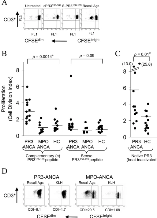
Figure 2.1. PR3-ANCA Patients T cells Respond to cPR3(138-169)
to healthy controls (n = 13) (Wilcoxon rank sum test, P = 0.0014) (Figure 2.1B). A proliferative response to heat-inactivated-PR3 was detected in patients (P = 0.01) (Figure 2.1C), although no differences were found upon encounter with sense-PR3(138-169) peptide (P = 0.09). The CDI of responses to recall antigens was also similar comparing 9.4 ± 5.3 versus controls 11.0 ± 11.5 (P = 0.79). The percent of background proliferation was similar comparing patients (3.0 ± 4.2) versus controls (2.0 ± 2.0).
The proliferative response to cPR3(138-169) peptide occurred without additional costimulation with cytokines, suggesting that these T cells had a memory cell phenotype [111]. Indeed, no significant proliferation in response to KLH was detected in two PR3-ANCA patients and seven MPO-PR3-ANCA patients (mean CDI: recall antigens = 6.55 ± 8.9; KLH = 1.10 ± 0.49) (Figure 2.1D), indicating that under the culture conditions used only previously primed but not naïve T cells are detected.
T cells produce IFN-γ in response to cPR3(138-169) peptide
20 40 60 -10 -5 0 5 10 15 HC
Pts Pts HC Pts HC Pts HC
Complementary (c)PR3138-169 peptide Sense PR3138-169 peptide PR3
p= 0.0002* p= 0.12 p= 0.19
(68.5) Scrambled peptide IFN -γ ELIS PO T (S p ot nu m ber m inus u ntr ea ted)
p= 0.35
IFN -γ ELI SPO T (S pot nu m ber m inus u ntr ea ted) 0 100 200 300 400 500 Pts HC
Recall antigen mixture
0 1 2 3 4 IF N-γ res p ons
e to c
P R 3 138-169 (F o ld-cha nge o f untr eated)
cPR3138-169 (μg/ml)
0 2 5 10
0 1 2 3 4 IF N-γ res p ons
e to c
P R 3 138-169 (F o ld-cha nge o f untr eated)
cPR3138-169 (μg/ml)
0 2 5 10
A
(97.0) (-57.0)B
C
IFN -γ+T ce
lls (m ea n fold-inc reas e )
p= 0.005*
0 2 4 6 8
CD4+ 5.3 1.9
CD8+ 1.3 1.5
Patients HC
IFN
-γ
+T ce
lls (m ea n fold-inc reas e )
p= 0.005*
0 2 4 6 8
CD4+ 5.3 1.9
CD8+ 1.3 1.5
Patients HC
D
p= 0.14
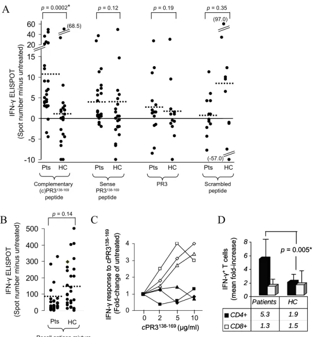
Figure 2.2. PR3-ANCA Patient T cells Produce IFN-γ in Response to cPR3(138-169)
specificity of an IFN-γ-response to this peptide (Figure 2.2C). The cPR3(138-169) peptide
IFN-γ-responders were of the CD4+ subset with a mean increase in patients of 5.3 ± 3.5-fold compared to healthy controls at 1.9 ± 0.75 (Wilcoxon ranked sum test, P = 0.005) (Figure 2.2D). IFN-γ-positive CD8+ cells were not increased (mean of 1.3 ± 1.02) compared to healthy controls (mean of 1.5 ± 0.67) (P = 1.0) (Figure 2.2D).
Specificity of responses to fragments of cPR3(138-169) peptide
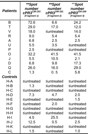
cPR3(138-169) peptide bears some homologies to a number of bacterial proteins [49], and it was questioned whether this 32aa peptide had characteristics similar to a pathogen-derived superagonist. Three 16aa overlapping peptide-fragments were tested for stimulatory characteristics with the supposition that superantigen-like sequences would bias reactivity toward one fragment. IFN-γ responses were random i.e., no cPR3(138-169) specific sequences common to all of patients (Table 2.2). Of healthy controls (n = 12), nine individuals’ T cells were non-reactive, while two had greater than five spots on the assay against fragment 1 and one reacted with fragment 2. The data indicate that cPR3(138-169) is not a superantigen.
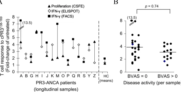
Individual variability among longitudinal samples
Table 2.2. Responses to cPR3(138-169)Peptide Fragments Patients **Spot number cPR3138-153 (Fragment 1) **Spot number cPR3146-161 (Fragment 2) **Spot number cPR3154-169 (Fragment 3)
B 72.6 6.6 24.2 T 29.0 17.0 12.0
V 18.0 ≤untreated 14.0
R 9.0 5.4 5.4 A 6.8 2.5 2.5
U 5.5 3.5 ≤untreated
P 2.5 ≤untreated ≤untreated O 32.2 41.5 41.5
J 5.5 10.5 2.1 D 6.8 9.8 17.3 Q 26.0 16.0 29.0
C 1.3 0. 5 5.8
Controls
H-A ≤untreated ≤untreated ≤untreated H-B 1.3 ≤untreated ≤untreated H-C ≤untreated ≤untreated ≤untreated H-D 7.3 4.7 2.0 H-E ≤untreated ≤untreated 1.5
H-F ≤untreated 2.0 ≤untreated H-G ≤untreated ≤untreated ≤untreated H-H ≤untreated ≤untreated ≤untreated
H-I 4.5 25.5 ≤untreated
H-J 12.5 0.5 2.5 H-K ≤untreated ≤untreated ≤untreated
H-L 1.5 ≤untreated 1.0
**The ELISPOT data are expressed as positive spots of treated wells minus spots on untreated wells.
cPR3(138-169) DLGWGVVGTHAAPAHGQALGAVGHWLVLLWQL
Fragment 1 DLGWGVVGTHAAPAHG
Fragment 2 THAAPAHGQALGAVGH
remission who showed a positive T cell response (BVAS = 0; 11 of 13) (patients A through N - Table 2.1) versus those who were not in remission (with BVAS score > 0; 10 of 13) (patients O through Z - Table 2.1) indicated an equal distribution within the two groups (Fisher’s exact test P = 0.99). Moreover, statistical comparisons of samples taken at times of remission (n = 25), compared to active disease (n = 16) (Table 2.1; Figure 2.3B) gave similar results showing an equal distribution of responders and non-responders (Fisher’s exact test, P = 0.74). The patients with high BVAS scores were newly diagnosed on medications for only a few days. These patients responded to recall antigens and to cPR3(138-169) peptide. Those patients on high dose – long duration medication failed to respond to recall antigen controls and thus were non-informative. Other potential explanations such as type of medication (Table 2.1) or environmental related factors were not identifiable. Variability is inevitable and appears to be a common occurrence in studies of human subjects [104]. The validity of our results is substantiated by use of multiple methodologies and by repetitive patient samplings.
Coexistence of cPR3(138-169)-specific T cells with cPR3(105-201)-specific antibodies
A critical question concerning the functional consequences of cPR3(138-169) peptide-reactive CD4+ TH1 cells is whether they were responsible for cPR3(105-201)-specific B cell
0 2 4 6 8 0 1 2 3 4 5 6 7 8 PR3-ANCA patients (longitudinal samples) T cell r es pons e to cP R3 13 8-16 9 (F old-change o f untr eated) Proliferation (CSFE)
IFN-γ(FACS) IFN-γ(ELISPOT) Proliferation (CSFE)
IFN-γ(FACS) IFN-γ(ELISPOT)
HC (means) (13.5)
A B G H I J K M O P Q R S Y Z BVAS = 0 BVAS > 0
(13.5)
Disease activity (per sample)
A
B
p= 0.74
DISCUSSION
The approach of exploring complementary protein pairs in autoimmune diseases offers a “breakthrough” in understanding mechanisms of pathogenesis. This is the first report of disease-related T cell responsiveness to a protein complementary to a known autoantigen. These cPR3(138-169)-responsive cells were classified as CD4+-TH1 cells, which
are capable of delivering signals for B cell maturation [112]. There was a significant correlation between the presence of anti-cPR3(105-201) antibodies and responsive T cells on an individual basis. These data are consistent with a complementary-protein-specific component in immunological events of PR3-ANCA vasculitis autoimmune disease.
A limitation when studying human T cells is that the only available sample is peripheral blood cells, unlike animial studies where spleens and lymph nodes are available. Using circulating cells, we found that the number of spots in the ELISPOT assay were less than published animal studies using spleens. Others report similarly low numbers of spots from human peripheral cells and propose this is expected for low-frequency reactive cells [113]. These are not unexpected as memory cells are thought to primarily reside in the spleen and peripheral lymphoid tissue, with low numbers of cells found in the circulation. Our efforts to expand the T cells in culture in order to increase the number of spots were unsuccessful. What is comforting is the degree of concordance between the proliferation studies and the ELISPOT assay with repetitive patient samplings. Even with these limitations, we successfully demonstrated a strong statistically significant response in patients compared to healthy controls.
whether PR3-specific B cells require T cell help to produce antibodies using peripheral blood lymphocytes culture system. Their conclusions were that B cells from patients produce PR3-antibodies through a T cell independent pathway or through some non-specific B cell stimulation [114]. Nonetheless, PR3-specific T cells are identifiable. Consider for the moment that anti-idiotypic antibody processes are involved in the generation of PR3-specific antibodies. This possibility was supported when mice immunized with cPR3(105-201) peptide developed not only anti-cPR3(105-201) antibodies, but also antibodies that reacted with human-native PR3. These mouse anti-human-PR3 antibodies produced a cytoplasmic-staining pattern on human neutrophils identical to that produced by patients’ PR3-ANCA. Thus, in these mice the derivation of the anti-human-PR3 reactive antibody must have occurred through an anti-idiotypic response incited by human-specific complementary protein [49]. The anti-idiotypic process is initiated with a T cell and B cell response against a PR3-complementary-protein. Antibodies can regulate each other by suppressing or augmenting the immune reaction in a manner that would perpetuate autoimmune disease [79, 115, 116]. An antibody is immunogenic by virtue of its non-germiline-encoded antigen-binding site. B cells are known to spontaneously display endogenous V region peptides on their HLA class II molecules and acivate CD4+ T cells [117, 118]. Display of immunoglobulin-derived peptides (idiotopes) on APC HLA-II molecules can occur by several routes. Monocytes and dendritic cells phagocytize antigen-antibody complexes bound to surface Fc-receptors, and
proteins for display on HLA-II molecules [120-122]. How can this information be incorporated into understanding PR3-ANCA generation? Experimental evidence indicates that animals immunized with human complementary-PR3 protein not only develop antibodies reactive with the immunogen, but also development of human-specific PR3 antibodies. This has been observed in mice [49], rabbits, and chickens (unpublished data). Likewise, a research group who studies La/SSB-specific autoantibodies associated with Sjogren’s syndrome and systemic lupus erythematosus found that mice immunized with the autoantigen’s complementary-peptide-counterpart elicited antibodies against the immunogen and anti-idiotypic antibodies that reacted with the sense autoantigen [86]. It has been demonstrated in multiple autoimmunity animal models that anti-idiotypes, raised against autoantibodies, induced anti-anti-idiotypes that possessed characteristics of the initial autoantibodies and caused disease after immunization [123, 124].
A crucial question is the source of the actual complementary-PR3 proteins that triggered the immunological responses described here. Ongoing studies are addressing this by probing for proteins from patient material that react with our antibodies from rabbits immunized with complementary peptides. The possibilities remain that it could be carried in by a microbe with proteins homologous to the complementary protein [89, 125] or that patients aberrantly transcribe and translate it [49]. Somewhat encouraging, we have detected antisense transcripts in patients using an antisense specific primer for the reverse transcriptase reaction and PCR [49]. Whether these transcripts are, or even can be, translated is unclear, although there are reports of translated antisense transcripts [126].
CHAPTER III
Isolation and Identification of Complementary-PR3 Proteins in PR3-ANCA Patient Plasmapheresis Fluid
David J. Bautz 1,2, Gloria A. Preston 1,4, Sofia Lionaki 1,4, Peter Hewins 1,4, Jia Jin Yang 1,4, J. Charles Jennette 1,3, and Ronald J. Falk 1,4
1UNC Kidney Center
2Department of Biochemistry and Biophysics
3Department of Pathology and Laboratory Medicine 4Department of Medicine
ABSTRACT
Biophysical interactions of proteins complementary to one another can provide a practical approach for discovery of novel autoimmune responses. Prior studies demonstrated that patients with anti-neutrophil cytoplasmic autoantibody small vessel vasculitis (ANCA SVV) mounted an immune response to proteins complementary to proteinase 3 (PR3). The current study demonstrates that a strategy capitalizing on principles of protein complementarity lead to the discovery of novel complementary-PR3 proteins. Plasma proteins from PR3-ANCA patients were analyzed for proteins complementary to PR3 by chromatography, SDS-PAGE, western blot analysis and mass spectrometry. Plasminogen and Protein F from pseudomonas were identified as putative complementary-PR3 proteins. Plasminogen is a substrate of PR3, indicative of interaction between these two proteins. In prior studies, immunization of mice with complementary-PR3 protein resulted in antibodies produced not only to complementary-PR3 but to human PR3 as well. These antibodies were shown to bind through their variable region; they were an idiotypic pair. A rabbit immunized with PR3 developed antibodies not only to PR3 but to plasminogen as well through the idiotypic network. Antibodies to PR3 were purified from chicken immunized with cPR3 (138-169) peptide, demonstrating an intact idiotypic network that can function in either direction.
INTRODUCTION
Biochemical properties of complementary protein pairs and their respective antibodies are thought to contribute to autoimmunity. A serendipitous discovery by our research group, that patients with proteinase 3 (PR3)-specific anti-neutrophil cytoplasmic autoantibodies (PR3-ANCA) also had antibodies against a protein coded by the antisense strand of the PR3 cDNA [cPR3(105-201)], led to the proposal that autoantigen complementarity is an underlying mechanism of this autoimmune disease [49]. The implications are that molecular complementarity approaches will lead to identification of other, and perhaps proximal, antigens in autoimmune disease. The goal of the present study was to isolate and identify a protein/s from patients’ plasma that might have given rise to these anti-cPR3105-201 antibodies.
conjunction, for over two decades, the idiotypic network has been implicated to be a component of autoimmunity by multiple researchers [79, 80, 116, 123, 124].
MATERIALS AND METHODS
Antigens/Antibodies/Reagents
Complementary-PR3 peptide corresponding to PR3 residues 138-169 [cPR3(138-169)]
(NH2-DLGWGVVGTHAAPAHGQALGAVGHWLVLLWQL-COOH) and the
corresponding sense PR3 peptide [PR3(138-169)] (NH2-QLPQQDQPVPHGTQCLAM
Recombinant complementary-PR3 protein production
We produced a recombinant, complementary-PR3 protein corresponding to PR3 residues 105-201 [cPR3(105-201)] as previously described. Briefly, antisense PRTN3 DNA (nucleotides 166-456; GenBank accession no. X55668) was ligated to a BM40 secretion signal peptide and a 6x histidine tag and inserted into pcDNA3. Protein was expressed and secreted from HEK293 cells. Protein purification was performed using a HisTrap HP column (GE Healthcare, Piscataway, NJ). Cell supernate was applied to the HisTrap column, washed with 5 column volume’s (CV) of binding buffer (PBS with 20 mM histidine, pH 7.6) and protein was removed from the column with 5 CV of elution buffer (PBS with 0.5 M histidine, pH 7.6). Protein elution was monitored by absorbance at 280 nm and verified by both ELISA and western blot using a rabbit anti-histidine antibody. The recombinant protein was dialyzed into phosphate-buffered saline (PBS) and the concentration was obtained with a protein assay using the Bio-Rad protein assay dye reagent and pre-aliquoted BSA standards.
Affinity purification of anti-cPR3(138-169) antibody