Research Article
Socio-demographic profile of prostatic diseases in symptomatic
patients: a prospective cross-sectional study
Praveenkumar P
1*, Chanabasappa V. Chavadi
2INTRODUCTION
Prostatic diseases can be expected to affect most men at some time during their life. They are associated with increased morbidity and mortality in older men. Benign prostatic hyperplasia occurs in most men as an ageing process, so that more than 80% of men will have benign hypertrophy by the age of 80 years. This can lead to various symptoms of urinary tract obstruction and prostatism leading to severe disability in them. Prostatic
carcinoma is the second most common malignancy in male adults next only to lung cancer. It has great impact on health care system, because of its associated morbidity and mortality. Most of them are slow growing, remain sub clinical and undetected. As a result its detection at a later stage will have higher mortality rate. Prostatitis is an inflammatory condition of prostate common in young adults; occur frequently in association with genitourinary infections. Some of the other prostatic conditions such as prostatic cysts which may be congenital or acquired can
ABSTRACT
Background: Prostatic diseases can be expected to affect most men at some time during their life. They are associated with increased morbidity and mortality in older men. Digital rectal examination & serum PSA levels are used for screening of prostatic cancer, while Transrectal ultrasound and MRI are used for diagnosis of different prostatic conditions, to know the extent of carcinoma and to guide prostatic biopsy.
Methods: This prospective study was carried out during the period from September 2011 to September 2014. Data for this study was collected from the patients attending Department of Radio-diagnosis, Hospital attached to the Navodaya Medical College, Raichur.
Results: A total of 50 male patients in the age group of 31-90 years with symptoms related to prostatic diseases like prostatism and infertility due to obstruction were included in our study. Highest incidence of prostatic diseases was found in seventh decade (61-70years) followed by eighth decade. Most common clinical presentation was difficulty in micturition followed by increased frequency of micturition and infertility was the least common presentation. Clinically suspected cases of Ca prostate with or without retention of urine were referred in majority. In BPH, predominant echopattern seen was hypoechoic lesions followed by hyper echoic lesion. In patients with carcinoma prostate, predominant echopattern seen was also hypoechoic lesion (70.00%) followed by hyperechoic and mixed lesions.
Conclusions: Prostatic diseases can be easily evaluated using per-rectal examination and transrectal ultrasonography in symptomatic patients to know the nature of the disease and its prognosis.
Keywords: Prostatic diseases, Per rectal examination, Ca prostate, Transrectal ultrasonography, Benign prostatic hyperplasia
1Department of Radiology, Navodaya Medical College, Raichur, Karnataka, India
2Department of Radiology, Kasturba Medical College, Manipal University, Mangalore, Karnataka, India
Received: 01 March 2015, Revised: 31 March 2015
Accepted: 03 April 2015
*Correspondence:
Dr. Praveenkumar P,
E-mail: pralingoo@rediffmail.com
Copyright: © the author(s), publisher and licensee Medip Academy. This is an open-access article distributed under the terms of the Creative Commons Attribution Non-Commercial License, which permits unrestricted non-commercial use, distribution, and reproduction in any medium, provided the original work is properly cited.
often lead to subfertility and infertility in men, because of its close relation with genital system structures viz, seminal vesicles, ductus deferens, etc. The prostate is divided into distinct anatomical zones and these can be depicted by transrectal ultrasonography.1,2
So, currently digital rectal examination and serum PSA levels are used for screening of prostatic cancer, while Transrectal ultrasound and MRI are used for diagnosis of different prostatic conditions, to know the extent of carcinoma and to guide prostatic biopsy.
Transrectal ultrasonography: Transrectal ultrasound has received increasing attention recently because of its potential for early detection of prostate cancer. It provides greater detail of zonal anatomy of prostate and echo pattern of the gland and its various lesions. Posterior portion of the gland is better delineated where most of prostate carcinoma arises. Besides this, assessment of adjacent structures like seminal vesicles, rectum and urinary bladder for invasion by prostate cancer could be done precisely by transrectal ultrasonography.1
Aims and Objectives
To find out profile of prostatic diseases using per rectal examination, transrectal ultrasound and Doppler evaluation in symptomatic patients with respect to Socio-demographic factors.
METHODS
Source of data:
This prospective study was carried out during the period from September 2011 to September 2014. Data for this study was collected from the patients attending Department of Radio-diagnosis, Hospital attached to the Navodaya Medical College, Raichur.
A total of 50 male patients in the age group of 31-90 years with symptoms related to prostatic diseases like prostatism and infertility due to obstruction were included in our study.
Method of collection of data:
Method of collection of data was by patient evaluation through detailed history as per standard proforma. Special attention was given towards symptoms of prostatism like frequency of micturition, difficulty in micturition, retention of urine, haematuria and infertility and features of distant metastasis like bony pain, abdominal pain, etc. Then necessary physical examination was performed to rule out other causes of symptoms and to see for any signs of distant metastasis in prostatic carcinoma. Then per rectal examination was performed to see for size, consistency, texture of prostate gland and any focal abnormality.
Inclusion criteria: All male patients with clinical and digital rectal examination suggestive of prostatic diseases.
Exclusion criteria:
- All pelvic lesions other than prostatic lesions. - Patients with prostatic diseases but having
contraindications for transrectal evaluation such as rectal masses, anal fissures, etc.
RESULTS
In our study total 50 cases of clinically suspected and digital rectal examination suggestive of prostatic diseases were scanned by transrectal ultrasound and following observations are made.
Out of 50cases final diagnosis was obtained in all 50 cases. The confirmation of transrectal ultrasound findings were done by histopathological examination of prostatic biopsy or whole mount section of the surgical specimen obtained after radical prostatectomy or transurethral resection of prostate.
The following observations are made in our study.
Table 1: Age incidence and percentage of prostatic lesions.
As shown in Table 1, the age range of our patients was from 31-90 years. The highest incidence (36%) of prostatic diseases was found in seventh decade (61-70 years) followed by 26% of cases in eighth decade. In our study youngest patient was of age 31 years and oldest patient was 90 years old. The average age of the patient in our study was 63.46 years.
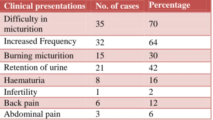
As shown in table 2, the most common clinical presentation was difficulty in micturition (70%) followed by increased frequency of micturition (64%) and infertility was the least common presentation (2%). One case of BPH had abdominal pain due to metastatic lymphadenopathy from seminoma testis and other two cases of abdominal pain were due to metastasis to solid abdominal organs.
Age in years No. of Cases Percentage
31-40 41-50 51-60 61-70 71-80 81-90
2 5 10 18 13 2
Table 2: Clinical presentations.
Table 3: Clinical diagnosis.
As shown in table 3, thus clinically suspected cases of Ca prostate with or without retention of urine were referred in majority (56%) followed by BPH with or without retention (40%) for transrectal ultrasound evaluation.
Table 4: Digital rectal examination in prostatic diseases.
As shown in table 4, firm and smooth prostate (52%) was the most common digital rectal examination finding seen predominantly in BPH. Hard and nodular consistency seen in 48% of patients and predominantly seen in carcinoma prostate.
Among 26 cases of firm and smooth prostate, 18cases (69.2%) were proved to be BPH, 6 cases (23.07%) to be carcinoma prostate and 2 cases (7.7%) to be prostatitis. Among 24 cases of hard nodular prostate, 14 cases (58.3%) were proved to be carcinoma prostate, 8 cases
(33.33%) proved to be BPH, one case each were proved to be prostatitis and prostatic cyst.
Table 5:Prostatic volume/weight.
In our study the prostate gland was assumed to be normal when the prostatic volume / weight is < 20gms. Gland was considered to be enlarged when it measured >20gms, which was further divided in to different grades like mild (20-30gms), moderate (31-70gms) and severe (>70gms) grades.
As shown in table 5, in case of carcinoma prostate, the smallest volume / weight were being 24gms and the largest being 110gms and average volume / weight being 59.4gms. In case of BPH, the smallest volume / weight being 22gms and largest being 140gms and average volume / weight being 47.7gms.
Average prostatic volume / weight being 49.96gms.
Table 6: Zonal incidence of benign and malignant prostatic diseases.
Prostatic
zone BPH
Carcinoma prostate
Prostatic
cyst Prostatitis
Peripheral
(PZ) 5 14 - -
Central
(CZ) 18 2 1 -
PZ + CZ 3 4 - 3
As shown in table 6, the majority of focal lesions in peripheral zone were due to carcinoma prostate comprising of 73.6% of cases and majority of focal lesions in central zone were due to BPH comprising of 85.7% of cases.
Among carcinoma prostate, lesions were most commonly seen in peripheral zone (70%) followed by both zones (20%) and central zone only (10%). Among BPH cases, lesions were commonly seen in central zone only (69.2%) followed by peripheral zone only (19.2%) and both zones (11.5%). So, Carcinoma prostate was seen predominantly in peripheral zone and BPH in central zone.
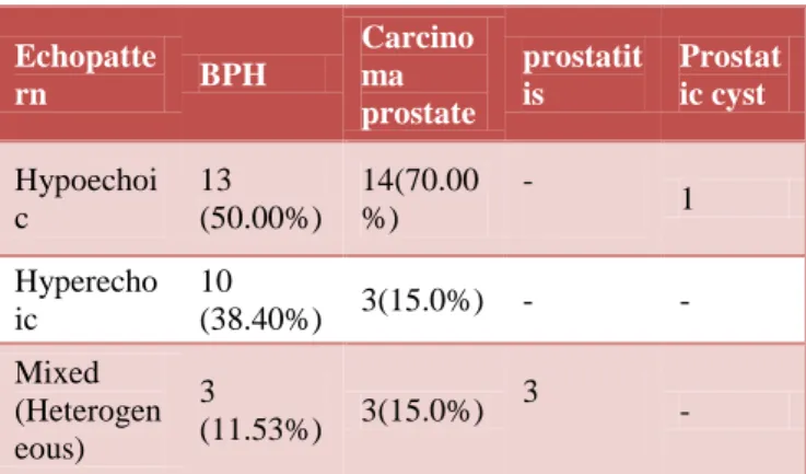
As shown in table 7, in patients with BPH, predominant echopattern seen was hypo echoic lesions (50.00%) followed by hyperechoic lesion (38.40%).
Clinical presentations No. of cases Percentage (%)
Difficulty in
micturition 35 70
Increased Frequency 32 64
Burning micturition 15 30
Retention of urine 21 42
Haematuria 8 16
Infertility 1 2
Back pain 6 12
Abdominal pain 3 6
Clinical diagnosis No. of cases Percentage (%)
Prostatitis 1 2
Prostatic cyst with
infertility 1 2
BPH without
retention 11 22
BPH with retention 9 18
CA prostate
without retention 16 32
CA prostate with
retention 12 24
Digital rectal examinat ion
No. of cases
Percent age (%)
BP H
Ca prosta te
Prostati tis
Prosta tic cyst Firm &
smooth 18 6 2 0 52
Hard &
nodular 8 14 1 1 48
Prostatic volume /
weight (Gms) No. of cases
Percentage (%)
Normal (<20) 4 8
Mild (21-30) 10 20
Moderate (31-70) 26 52
Table 7: Different echo patterns of prostatic diseases.
Echopatte
rn BPH
Carcino ma prostate
prostatit is
Prostat ic cyst
Hypoechoi c
13 (50.00%)
14(70.00 %)
-
1
Hyperecho ic
10
(38.40%) 3(15.0%) - -
Mixed (Heterogen eous)
3
(11.53%) 3(15.0%) 3
-
In patients with carcinoma prostate, predominant echo pattern seen was also hypoechoic lesion (70.00%) followed by hyperechoic and mixed lesions (15.0% each).
Among different echo patterns, hypoechoic lesions were seen commonly in carcinoma prostate (50%) compared to BPH (46.42%). And hyper echoic lesions were seen commonly in BPH (76.9%) compared to carcinoma prostate (23.07%).
Table 8: Capsular status.
Capsular status
No. of cases
Percentag e (%)
BPH Ca prosta te
Prostati tis
Pros tatic cyst
Regular and continuou s
23 8 3 1 70
Irregulara and interrupte d
3 12 - - 30
As shown in table 8, regular and continuous capsule is seen most commonly in 70% of cases, which was seen mainly in BPH (23 patients - 65.70%) and carcinoma prostate (8 patients – 22.85%).
Irregular and interrupted capsule is seen in 30% of patients, which was seen mainly in carcinoma prostate (12 patients – 80%) and BPH (3patients – 20%).
DISCUSSION
In this prospective study, total 50 cases of clinically suspected and digital rectal examination suggestive of prostatic diseases were scanned by transrectal ultrasound using endorectal probe of frequency 6.5 MHZ attached to ultrasound machine present in Department of Radio-diagnosis, Hospital attached to the Navodaya Medical College, Raichur.
Apart from transrectal ultrasound, other radiological investigations like plain radiograms (of pelvis, lumbosacral and thoracic spine), abdominal ultrasound, CT scan of abdomen & pelvis and bone scintigraphy were performed to detect or rule out metastasis from prostatic carcinoma. Serum PSA levels were performed in all necessary cases if patients could afford the cost of test.
Age incidence in prostatic diseases:
The age range of patients included in our present study was 31-90 years with mean age of 63.46 years. The highest incidence of prostatic diseases were present in seventh decade (36%) followed by 28% in eighth decade. In a study carried out by Martin I Resnick et al3 patients ranged from 53 to 78 years old with mean age of 68 years. In a study conducted by Peggy J Frizolhe et al4 prostatic sonogram was done in 228 patients with age range from 43-93 years, with average age of 66 years. In all the studies, including our present study, the patients are belonging to the higher age group of above 50 years with mean age in various studies was in seventh decade. Study of age distribution data suggests that prostatic diseases like BPH and carcinoma are slow growing pathologic entities whose incidences grow slowly but steadily with increasing age.
Clinical presentation of the patient:
Usually patients with prostatic diseases present with prostatism symptoms like difficulty, increased frequency, acute or chronic retention of urine. Some patients may present with haematuria, infertility, abdominal pain and bony pain due to metastasis.
In our present study, difficulty in micturition was the most common presenting symptom (70%) followed by increased frequency of micturition (64%) and infertility was the least common presenting symptom (2%). In the study carried out by Martin Resnick et al3 on 132 apparently normal men, the most common symptom were frequent micturition (27%) followed by sense of residual urine (23.7%). Various disturbances of urination were observed in 25% and nocturia was observed in 15% of cases.
there may be either over diagnosis or under diagnosis of different conditions due to overlapping of clinically symptoms and signs.
Digital Rectal Examination: The digital rectal
examination was carried out in all 50 cases. It was found to be hard and nodular in 24 cases (48%) and out of this 14 cases (58.3%) were carcinoma prostate, 8 cases (33.3%) were BPH and one each case of prostatitis and prostatic cyst. Firm and smooth prostate seen in 26 cases (52%), out of which 18cases (69.2%) were BPH and 6 cases (23.07%) were Carcinoma prostate. M. Resnick6 made the observations that every hard prostate is cancer until proved otherwise. However, they did not provide any data to substantiate the said observation. In a study by Fred Lee et al,7 22 cancers were detected by transrectal USG and ten at digital examination. Overall detection rate for prostate cancer with transrectal USG was two times higher than with DRE (2.6% Vs 1.3%). Kathryn K Hodge et al8 in their study found 10 out of 12 i.e. 83 % prostatic cancer showed abnormal firmness.
Digital rectal examination used commonly for macroscopic examination of prostate provides only indirect and subjective information and is inaccurate.
Volume/weight of the prostate as measured by TRUS:
In the present study we found 4 (8%) cases within the normal range, 10 (20%) cases had mild enlargement, 26 (52%) cases had moderate enlargement and 10 (20%) cases were considerably enlarged with mean volume of 49.96 gm. The smallest prostate gland volume in the cases of carcinoma of prostate was found to be 24 gm and the largest volume was found to be 110 gm. And in BPH smallest was 22gm and largest was 140gms. GJ Griffith et al9 in their 221 cases study found mean prostatic volume of 38 ml (range 6 to 148 ml). Martha K Terris et al10 studied 150 patients with transrectal Sonography and observed that the most accurate method to estimate prostate weight (r=0.94) was a variation of prolate spheroid formula, expressed as ^/6 (transverse dimension) (Anteroposterior dimension) for prostate weighing up to 80 gms and ^/6 (transverse dimension) for prostate weighing more than 80 gms.
In the present study observation suggests that volume should not be the criteria for the diagnosis or biopsy procedure because smallest sized gland could present with the metastases.
Incidence of various benign and malignant lesions in different zones of prostate:
In present study it was observed that the carcinoma of prostate predominantly involved the peripheral zone. In carcinoma prostate 14 out of 20 cases (70.0%) were found in peripheral zone, 4 (20.0%) cases in peripheral + inner glandular zone. In BPH 18 of 26 cases (69.20%) were found in the inner glandular followed by 5 out of 26
cases (19.20%) in peripheral zone and 3 out of 26 (11.50%) in both central and peripheral zone. Thus BPH predominantly involved inner glandular zone. Monzer M Yousef et al11 in their study found 10 (55.5%) cases out of 18 cases in the peripheral zone. Mathew D Rifkin et al12 in their study found that prostate carcinoma originates in the peripheral zone and as it enlarges it envelopes not only the periphery, but it also infiltrates into the central portion of the prostate. A study by Fred Lee et al 13, prostate carcinoma was seen involving the peripheral zone in 68.75% cases.
Carcinoma of prostate occurs in the most active zone of the gland in the old age and peripheral zone in most active at this age; hence disease is predominantly seen in peripheral zone. In Monzer M Abu Yousef et al11, the sample volume of carcinoma cases is very less which decreases specificity in statistical correlation. As shown in the study by Griffith et al9 and Mathew D Rifkin et al12 larger the lesion there is loss in the zonal distribution.
In present study, we found that the lesion of small size had maintained their zonal distribution in the peripheral zone and the larger lesions had infiltrated the periphery as well as central portion of the prostate gland.
Echopattern of various benign and malignant prostatic lesions:
In 50 patients included in the present study, hypoechoic lesions were more common in carcinoma of prostate, of total 20 cases 14 were hypo echoic (70.00%) followed by hyper echoic and mixed echogenic lesions 3 cases each (15.0%).
In BPH, 13 cases (48.1%) were found to be hypoechoic, followed by hyperechoic in 10 cases (37.3%) and mixed echogenic lesions in 4 cases (14.81%).
Findings of the present study nearly match. with the findings of Wolfgang E Dahnert et al14, who in their findings in carcinoma of prostate observed it to be more or less echopenic in 76% (echopenic 54% and hypoechoic 22%,both have decrease echogenicity) and isoechoic in 24% without a single echogenic cancerous lesion.
Mathew D Rifkin et al12 found adenocarcinoma of prostate to be echogenic in 77% and mixed echogenic in 23%. They explicitly state that purely hypoechoic area was never seen in instance of cancer.
lesions located in the peripheral zone were (proved to be carcinoma by histopathological study).
GJ Griffiths et al9 observed ill-defined hypoechoic lesions in 96% cases of carcinoma prostate. Katsuto Shinohara et al16 found hypoechoic area in 42 (60%) cases, isoechoic in 27 (39%) cases and hyperechoic in only one case. Urike M Hamper et al17 studied 151 separate prostatic cancer, of these lesion, 115 lesion (76%) were hypoechoic and 36 lesions (24%) not detected by TRUS because of isoechoic appearance. The study by Mathew D Rifkin et al12 found hyperechoic lesion with larger size. In our study when the size of lesion was smaller it was found to be hypoechoic and confined to one zone. This may be the reason in our finding we found most of the case with hypoechoic areas in the peripheral zone positive for the carcinoma of prostate.
CONCLUSIONS
1. The highest incidence (36%) of prostatic diseases was found in seventh decade (61-70 years) followed by 26% of cases in eighth decade.
2. Most common clinical presentation was difficulty in micturition (70%) followed by increased frequency of micturition (64%) and infertility was the least common presentation (2%).
3. Clinically suspected cases of ca prostate with or without retention of urine were referred in majority (56%) followed by BPH with or without retention (40%) for transrectal ultrasound evaluation.
4. In BPH, predominant echopattern seen was hypoechoic lesions (50.00%) followed by hyperechoic lesion (38.40%).In patients with carcinoma prostate, predominant echopattern seen was also hypoechoic lesion (70.00%) followed by hyperechoic and mixed lesions (15.0% each). 5. Regular and continuous capsule is seen most
commonly in 70% of cases, was seen mainly in BPH (23 patients – 65.70%) and carcinoma prostate (8 patients – 22.85%). Irregular and interrupted capsule is seen in 30% of patients, was seen manly in carcinoma prostate (12 patients – 80%) and BPH (3 patients – 20%).
Funding: No funding sources Conflict of interest: None declared
Ethical approval: The study was approved by the Institutional Ethics Committee
REFERENCES
1. Patel U. The prostate and seminal vesicles. In: Allan P, Baxter G, Weston M, eds. Clinical ultrasound. 3rd edn. Edinburgh, UK: Churchill-Livingstone; 2011:572-92. 2. Patel U, Rickards D. Handbook of Transrectal ultrasound
and biopsy of the prostate. London, UK: Martin Dunitz; 2002.
3. Resnick MI, Willard JW, Boyce WH. Transrectal ultrasonography in the evaluation of patients with prostatic carcinoma. J Urology.1980;124(4):482-4. 4. Fritzsche PJ, Axford PD, Ching VC, Rosenquist RW,
Moore RJ. Correlation of transrectal sonographic findings in patients with suspected and unsuspected prostatic disease. J Urology.1983;130(2):272-4.
5. Watanabe H, Igari D, Tanahasi Y, Harada K, Saito M. Development and application of new equipment for transrectal ultrasonography. J Clin Ultrasound. 1974;2(2):91-8.
6. Resnick MI, Willard JW, Boyce WH. Ultrasonic evaluation of prostatic nodule. J Urology. 1978;12(1):86-9.
7. Lee F, Littrup PJ, McLeary RD, Kumusaka GH, Borlaza GS, McHugh TA. Needle aspiration and core biopsy of prostate cancer: comparative evaluation with biplanar transrectal US guidance. Radiology.1987;163(2):515-20. 8. Hodge KK, McNeal JE, Stamey TA. Ultrasound guided
transrectal core biopsies of the palpably abnormal prostate. J Urol 1989;142(1): 66-70.
9. Griffiths GJ, Clements R, Jones DR, Roberts EE, Peeling WB, Evans KT et al . The ultrasound appearances of prostatic cancer with histological correlation. Clin Radiol. 1987;38(3):219-27.
10. Terris MK, McNeal JE, Stamey TA. Detection of clinically significant prostate cancer by transrectal ultrasound guided systematic biopsies. J Urol. 1992;148(3):829-32.
11. Abu-Yousef MM. Benign prostatic hyperplasia: tissue characterization using suprapubic ultrasound. Radiology.1985;156(1):169-73.
12. Rifkin MD, Friedland GW, Shortliffe L.Prostatic evaluation by transrectal endosonography; detection of carcinoma. Radiology. 1986;158(1):85-90.
13. Lee F, Gray JM, McLeary RD, Lee F Jr, McHugh TA, Solomon MH et al . Prostatic evaluation by transrectal sonography: criteria for diagnosis of early carcinoma. Radiology. 1986;158(1):91-5.
14. Dahnert WF, Hamper UM, Eggleston JC, Walsh PC, Sanders RC. Prostatic evaluation by transrectal sonography with histopathological correlation; the echopenic appearance of early carcinoma. Radiology. 1986;158(1):97-102.
15. Burks DD, Drolshagen LF, Fleischer AC, Liddell HT, McDougal WS, Karl EM et al. Transrectal sonography of benign and malignant prostatic lesions. AJR Am J Roentgenol. 1986;146(6):1187-91.
16. Shinohara K, Wheeler TM, Scardino PT. The appearance of prostate cancer on transrectal ultrasonography: correlation of imaging and pathological examination. J Urol. 1989;142(1):76-82.
17. Hamper UM, Sheth S, Walsh PC, Holtz PM, Epstein JI. Capsular transgression of prostatic carcinoma evaluation with transrectal US with pathologic correlation. Radiology. 1991;178(3):791-5.
DOI: 10.5455/2349-3933.ijam20150512