Structural Similarities and Differences between Two Functionally
Distinct SecA Proteins,
Mycobacterium tuberculosis
SecA1 and SecA2
Stephanie Swanson,aThomas R. Ioerger,bNathan W. Rigel,c*Brittany K. Miller,cMiriam Braunstein,cJames C. Sacchettinia
Department of Biochemistry & Biophysicsaand Department of Computer Science and Engineering,bTexas A&M University, College Station, Texas, USA; Department of
Microbiology and Immunology, University of North Carolina—Chapel Hill, Chapel Hill, North Carolina, USAc
ABSTRACT
While SecA is the ATPase component of the major bacterial secretory (Sec) system, mycobacteria and some Gram-positive pathogens have a second paralog, SecA2. In bacteria with two SecA paralogs, each SecA is functionally distinct, and they cannot compensate for one another. Compared to SecA1, SecA2 exports a distinct and smaller set of substrates, some of which have roles in virulence. In the mycobacterial system, some SecA2-dependent substrates lack a signal peptide, while others contain a signal peptide but possess features in the mature protein that necessitate a role for SecA2 in their export. It is unclear how SecA2 func-tions in protein export, and one open question is whether SecA2 works with the canonical SecYEG channel to export proteins. In this study, we report the structure ofMycobacterium tuberculosisSecA2 (MtbSecA2), which is the first structure of any SecA2 protein. A high level of structural similarity is observed between SecA2 and SecA1. The major structural difference is the absence of the helical wing domain, which is likely to play a role in howMtbSecA2 recognizes its unique substrates. Importantly, struc-tural features critical to the interaction between SecA1 and SecYEG are preserved in SecA2. Furthermore, suppressor mutations of a dominant-negativesecA2mutant map to the surface of SecA2 and help identify functional regions of SecA2 that may pro-mote interactions with SecYEG or the translocating polypeptide substrate. These results support a model in which the mycobac-terial SecA2 works with SecYEG.
IMPORTANCE
SecA2 is a paralog of SecA1, which is the ATPase of the canonical bacterial Sec secretion system. SecA2 has a nonredundant func-tion with SecA1, and SecA2 exports a distinct and smaller set of substrates than SecA1. This work reports the crystal structure of SecA2 ofMycobacterium tuberculosis(the first SecA2 structure reported for any organism). Many of the structural features of SecA1 are conserved in the SecA2 structure, including putative contacts with the SecYEG channel. Several structural differences are also identified that could relate to the unique function and selectivity of SecA2. Suppressor mutations of asecA2mutant map to the surface of SecA2 and help identify functional regions of SecA2 that may promote interactions with SecYEG.
S
ecA is the ATPase component of the bacterial Sec secretion pathway (1). SecA recognizes proteins destined for export from the cytoplasm and provides energy to translocate them across the cytoplasmic membrane by way of the SecYEG translo-case channel. The proteins exported by SecA are synthesized as preproteins with N-terminal signal peptides. Following transloca-tion, the signal peptide is cleaved to release the mature protein species. Both the signal peptide and features of the mature protein are recognized by SecA (2). Some Gram-positive and acid-fast bacteria, including mycobacteria, have a SecA paralog referred to as SecA2. SecA1, the canonical SecA in these organisms, is essen-tial for growth and responsible for the majority of protein export that occurs. In contrast, SecA2 is typically not essential and is required for the export of a more limited subset of proteins (3,4). Studies in mycobacteria show that even when overexpressed, the two SecA proteins are unable to compensate for each other (5). Thus, each SecA protein has distinct functions in protein export. InMycobacterium tuberculosis, SecA2 (MtbSecA2) is not essential for growth in culture, but it is essential for virulencein vivo(6,7). Furthermore, SecA2 is required for intracellular growth ofM. tu-berculosisin macrophages (8). The role of SecA2 in promoting growth in macrophages is attributed to a role in preventing phago-some maturation (9). InMycobacterium marinum, export of pro-tein kinase G (PknG) by the SecA2 pathway is suggested to at least be partially responsible for the SecA2 effect on phagosomematu-ration (10). InM. tuberculosis, the SecA2 pathway is additionally required to restrict apoptosis of infected macrophages. A possible explanation for the latter effect is the SecA2-dependent secretion of superoxide dismutase, which may reduce reactive oxygen spe-cies (ROS)-mediated apoptosis (11,12). An association between SecA2 and the secretion of virulence factors extends to other bac-terial pathogens as well (13–16). There is also an intriguing asso-ciation between the SecA2 pathway and the export of S-layer pro-teins by some Gram-positive bacteria, such asBacillus anthracis (17) andClostridium difficile(18).
Received29 August 2015 Accepted1 December 2015
Accepted manuscript posted online14 December 2015
CitationSwanson S, Ioerger TR, Rigel NW, Miller BK, Braunstein M, Sacchettini JC. 2016. Structural similarities and differences between two functionally distinct SecA proteins,Mycobacterium tuberculosisSecA1 and SecA2. J Bacteriol 198:720 –730.doi:10.1128/JB.00696-15.
Editor:O. Schneewind
Address correspondence to James C. Sacchettini, [email protected].
*Present address: Nathan W. Rigel, Department of Biology, Hofstra University, Hempstead, New York, USA.
Supplemental material for this article may be found athttp://dx.doi.org/10.1128 /JB.00696-15.
It is unclear howMtbSecA2 carries out its unique function in protein export. In some organisms with two SecAs, there is a SecY paralog (SecY2), with which SecA2 likely interacts (19). In SecA2-SecY2 systems, SecA2-SecY2 and several accessory Sec proteins (Asp) are thought to form an accessory protein translocation channel in the cytoplasmic membrane (4). Mycobacteria, however, are in a group of bacteria referred to as “SecA2-only” systems that lack a second SecY ortholog (3). Mycobacteria, as well as several Gram-positive species, includingListeria monocytogenes(13), Corynebac-terium glutamicum(20), andC. difficile(18), are in the “SecA2-only” group. An important but unresolved question is whether SecA2 works with the canonical SecYEG channel to export pro-teins in these systems lacking a second SecY.
The mycobacterial proteins currently known to be exported by SecA2 include examples with typical Sec signal peptides, as well as proteins lacking signal peptides altogether (3). Superoxide dismu-tase (SodA) inM. tuberculosisand PknG inM. tuberculosisandM. marinumare examples of proteins lacking signal peptides that are exported in a SecA2-dependent manner (7,10,21). Of the signal peptide-containing proteins exported by the SecA2 systems of Mycobacterium smegmatis(22),M. marinum(10), andM. tuber-culosis(21), the most thoroughly studied proteins are theM. smeg-matis 1704 (Ms1704) and Ms1712 proteins (22). Studies of Ms1704 andMs1712 demonstrate that they require their signal peptide for export, but it is a feature of the mature portions of these proteins that necessitates export via the SecA2-dependent pathway (23). Interestingly, when fused to a signal peptide for the twin-arginine translocation (Tat) pathway, the mature domain of Ms1704 is exported by the Tat pathway. This result suggests that the defining feature of SecA2 substrates may be a tendency to fold prior to export (23). This is because proteins that get translocated across the membrane by the Tat pathway must be folded in the cytoplasm prior to export (24). In contrast, preproteins exported by the canonical SecA must be unfolded (25), sometimes with the help of export chaperones (26,27), due to the narrow diameter of the SecYEG central channel. Therefore, if SecA2 works with Se-cYEG, the role of SecA2 may be to facilitate the export of proteins that have a tendency to fold prior to export by either helping to maintain such proteins in an unfolded state or assisting in the recognition or export of such problematic substrates.
There is only 38% amino acid sequence identity between the MtbSecA1 andMtbSecA2 proteins. Yet, SecA2, like SecA1, has a DEAD box ATPase domain (28), and ATPase activity is required for SecA2 function (29). Furthermore, SecA2 variants lacking ATPase activity due to an amino acid substitution in the Walker box are dominant negative, and asecA2dominant-negative mu-tant exhibitssecA2mutant phenotypes (a growth defect on rich agar and azide sensitivity) that are more severe than those exhib-ited by a⌬secA2null mutant (29). Extragenic suppressors of this dominant-negativesecA2allele map to thesecY promoter, and increased SecY levels suppress thesecA2dominant-negative phe-notype (30). These findings suggest that the SecA2 dominant-negative protein is locked in a nonproductive interaction with the essential SecYEG channel, which inhibits SecYEG function but can be overcome by increased SecY production. This is consistent with SecA2 working with SecYEG. In a recent study of the SecA2-only system ofL. monocytogenes, suppressors of asecA2mutation also mapped tosecY(74). Furthermore, the behavior of a domi-nant-negative SecA1 mutant in theC. difficilesystem is consistent with the SecYEG translocase used by SecA1 also being used by
SecA2 (18). Thus, it seems likely that in these SecA2-only systems, SecY is involved. However, a direct interaction between SecA2 and SecYEG has not been demonstrated in any system.
Previously, the crystal structure of the canonical SecA1 was solved inM. tuberculosis(31), as well as several other organisms, includingEscherichia coli(32),Bacillus subtilis(33),Thermotoga maritima(34), andThermus thermophilus(35). SecA structures contain five canonical domains, organized roughly in the shape of a barbell: a core helical scaffold domain (HSD), forming the “ax-is”; 2 nucleotide-binding domains (NBD1 and NBD2), which to-gether form a DEAD box, RecA-like, or superfamily II helicase motor domain on one end of the barbell; and a helical-wing do-main (HWD) and preprotein cross-linking dodo-main (PPXD) on the other end of the barbell. In addition, a helix-loop-helix do-main called IRA1 (for “intramolecular regulator of ATPase”) packs against the HSD, with helices aligned in parallel. The loop connecting the helices of IRA1 is known as the two-helix finger (2HF). The 2HF has been shown to insert into the SecYEG pore, and it is proposed to promote forward movement of the prepro-tein through the channel (34,36), although the interaction tween the 2HF and SecYEG could also serve an alternate role be-sides pushing the translocating protein through the channel (37). During preprotein translocation, SecA undergoes significant con-formational changes, one of which involves the orientation of the PPXD domain. According to one model (38), the PPXD likely starts out oriented toward the HWD, forming a hydrophobic “cleft” for binding the signal peptide of the preprotein (39,40), and then rotates toward NBD2 to form a “clamp” around the translocating polypeptide chain, which has been proposed to be initiated by docking with SecYEG (41).
In order to better understand the unique function of SecA2, we solved the crystal structure ofMtbSecA2, which is the first SecA2 structure to be determined in any organism. The structure reveals that the HWD domain is completely absent inMtbSecA2. The HWD could play a role in interacting with protein substrates, as it forms part of a cleft with the PPXD that is implicated in peptide binding (40). Although the residues that directly bind the signal peptide (based on nuclear magnetic resonance [NMR] studies) are contributed by the PPXD and IRA1 domains (40), the HWD would likely be physically proximal to the untranslocated portion of protein substrates. Furthermore, residues in the HWD ofE. coli SecA (along with the PPXD and HSD) have been shown to cross-link with synthetic signal peptides in cysteine substitution exper-iments (42). The lack of an HWD in SecA2 leads to a signal peptide binding cleft that is more highly solvent exposed than in SecA1, which we propose could account for recognition of specific SecA2-dependent substrates and prevent export of the larger number of SecA1-dependent preproteins. The structure also re-veals conservation inMtbSecA2 of features critical to the interac-tion between SecA and SecYEG proteins. Finally, by mapping intragenic suppressor mutations onto the SecA2 structure, we show that the mutated residues appear in surface-exposed re-gions and map to three functional domains that are likely in-volved in mediating interactions with other protein partners, such as SecYEG.
MATERIALS AND METHODS
amino acids (aa) (e.g., NCBI accession no.NP_216337). However, the start site in this annotation is likely to be incorrect, as the first 30 aa are not required for function and represent an N-terminal extension that is not observed in other SecA orthologs (28,43). Therefore, we designate the GTG codon corresponding to residue 31 in the NCBI annotation as the true start codon, yielding a total ORF length of 778 aa. The expression construct pNR14 produces a tag-less form of the protein (28). Selenome-thionyl protein was produced by transforming theE. colimethionine auxotroph B834(DE3) (Novagen) with the pNR14 expression vector. A 6-liter culture was grown under standard conditions to mid-log phase. The cells were pelleted and used to inoculate 12 liters of M9 minimal medium supplemented with 50 mg/liter ofL-selenomethionine (SeMet), 50 mg/liter of standardL-amino acids (excluding methionine), 100 nM vitamin B12, and trace elements (44). Expression was induced with 0.5
mM ITPG (isopropyl--D-thiogalactopyranoside) at 16°C for 12 h. Cells were then harvested and resuspended in lysis buffer containing 50 mM Tris (pH 8.0), 50 mM NaCl, 1 mM dithiothreitol (DTT), 10 mM MgCl2,
20g/ml DNase, and 1⫻protease inhibitor cocktail V (EMD Biosci-ences). The cells were disrupted in a BeadBeater (Biospec) using 0.1-mm-diameter glass beads. Cellular debris was cleared from the lysate by spin-ning at 27,200⫻gfor 2 h. The supernatant was then filtered and loaded onto a Blue Sepharose column (GE Healthcare) that had been equili-brated in a mixture of 50 mM Tris (pH 8.0), 50 mM NaCl, and 1 mM DTT. Protein collected from the flowthrough was further purified by anion-exchange chromatography using a HiTrap Q high-performance (HP) col-umn (GE Healthcare). The purified protein was dialyzed overnight against buffer containing 50 mM Tris (pH 8.0), 50 mM NaCl, and 1 mM DTT and was then concentrated to 10 mg/ml using a Centriprep centrif-ugal concentrator (Milipore) and flash frozen until further use.
Crystallization.Purified protein was crystallized in 20% polyethylene glycol 8000 (PEG 8000), 0.1 M Tris (pH 8.0), 0.2 M NaCl, 3% ethylene glycol, and 8 mM 3-[(3-cholamidopropyl)dimethylammonio]-1-pro-panesulfonate (CHAPS). Wells were set up using sitting-drop vapor dif-fusion at 21°C, with drops consisting of 2 parts buffer and one part pro-tein. Crystals grew to 100m within 3 to 4 days. Perfluoropolyether (Hampton Research) was used as a cryo-protectant. The protein crystal-lized in space group P21with the unit cell parametersa⫽39,b⫽165,c⫽
67 Å, and ⫽97°. The corresponding unit cell volume can accommodate a single molecule in the asymmetric unit.
Crystal dehydration.A crystal dehydration method was developed that significantly improved the mosaic spread and diffraction power of the crystals (45). Both the well and drop solution were replaced with mother liquor that had a 3 to 5% increase in precipitant concentration. Crystals were left to dehydrate for a minimum of 48 h before making another incremental increase in the precipitant. Successfully dehydrated crystals had a reducedbunit cell parameter of up to 15 Å, with the largest differ-ence resulting in a 10.5% decrease in the unit cell volume. The crystal that produced the best diffraction data and led to structure solution had only a 3-Å difference in thebunit cell parameter.
Data collection, structure determination, and refinement. The structure was solved by single-wavelength anomalous dispersion (SAD) using a selenomethionine (SeMet) derivative (46). Anomalous diffraction data were collected at beamline 23-ID of the GM/CA-CAT facilities of the Advanced Photon Source, Argonne National Laboratory. Crystals were partitioned using the 10-m minibeam (47). This prevented global-scale radiation exposure and allowed for more data to be collected from a single crystal. The data were processed and reduced using the HKL2000 soft-ware package (48). The locations of 3 Se sites were found using SHELX C/D and were used as a starting point for locating additional sites in autoSHARP (49,50). The resulting experimental phases extended to a 3.8-Å resolution and produced an electron density map in which approx-imately 60% of the backbone could be placed in NBD1, NBD2, and parts of the HSD. Model building was performed in Coot (51). The phases from the partial model were then combined with the experimental phases using SigmaA and used as a starting point for progressive runs of density
mod-ification in DM (52,53). This facilitated the placement of the backbone in the PPXD as well as in other parts of the model. Initially, sequence was assigned by the positions of the Se atoms and from the density of large side chains. Then a real-space cross-validation procedure called “ping-pong” cross-validation was used to complete the structure (54). Briefly, the model was split into two sets. Side chains that could be identified in the first set of residues were used during phase combination and density mod-ification. The resulting map was used to place side chains for the second set of residues, and the process continued in alternation. Structure refine-ment was carried out in autoBuster (55).
Suppressor screen and reconstruction.Spontaneous suppressors of thesecA2K129Rstrain were isolated by plating onto Mueller-Hinton agar at
37°C, as described previously (30). ThesecA2K129Rstrain has the
chromo-somalsecA2gene deleted and carries a copy of thesecA2gene encoding SecA2K129Rintegrated at the chromosomal L5attsite. ThesecA2K129Rgene
of the suppressors was PCR amplified and sequenced to identify intra-genic suppressor mutations. To confirm that suppressor phenotypes were due to sequenced mutations insecA2K129R, the intragenic suppressors
were recreated in a fresh strain background. PCR-amplifiedsecA2K129R
gene products from the intragenic suppressors were subcloned into pCR2.1 followed by cloning into pMV306. The resulting vectors were electroporated into the⌬secA2mutant ofM. smegmatis, and transfor-mants were tested for sensitivity to sodium azide and SecA2 localization.
Azide sensitivity assay.Cultures were plated for sensitivity to sodium azide as previously described (29). In brief, 200l of saturated (optical density at 600 nm [OD600] of 2.0)M. smegmatisculture was mixed with
3.5 ml of molten 7H9 top agar and then poured onto a 7H10 bottom agar plate lacking Tween. Sterile 6-mm-diameter filter discs were placed onto the surface of the cooled top agar. Ten microliters of 0.15 M sodium azide was then added to the disc. The plates were incubated for 2 days at 37°C, and the resulting zones of growth inhibition were measured. Each strain was tested in triplicate.
Subcellular fractionation and immunoblotting.To determine the subcellular localization of SecA2 inM. smegmatis, we fractionated bacte-rial whole-cell lysates as described previously (22,29). Whole-cell lysates were generated by five passages through a French pressure cell. The lysates were separated into cell envelope (100,000 ⫻ g pellet) and soluble (100,000 ⫻g supernatant) fractions. Protein derived from the same amount of starting cells for each fraction was analyzed by SDS-PAGE and immunoblots using an anti-SecA2 antibody at a 1:20,000 dilution (56). For quantification, secondary antibody conjugated to alkaline phospha-tase was used and detected using the ECF reagent (GE Healthcare). Fluo-rescence was quantified using a phosphorimager and ImageQuant 5.2 (Molecular Dynamics).
Protein structure accession number.The structural coordinates of theMtbSecA2 model used in this study have been deposited in the Protein Data Bank under identification no.4UAQ.
RESULTS
Crystal structure ofMtbSecA2.MtbSecA2 (Rv1821) was crystal-lized in space group P21, and the structure was solved by
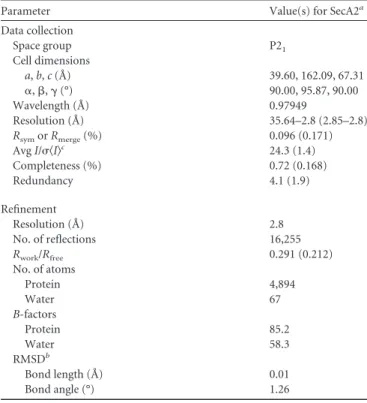
single-wavelength anomalous dispersion (SAD) (57) to a resolution of 2.8 Å. The asymmetric unit of the crystal contains a single mono-mer, and there is no indication of a higher-order oligomer in the crystal lattice. A total of 705 out of 778 residues of the apoprotein were visible in the electron density and could be built. The crys-tallographic statistics are shown inTable 1.
Broad structural similarity betweenMtbSecA1 andMtbSecA2.
DEAD box motor domain with an ATP-binding site between them. Catalytically important residues, such as K115 and R545 are conserved (see Fig. S1 in the supplemental material), consistent with demonstrated ATPase activity of SecA2 (28). As in other SecA
structures, the IRA1 domain consists of a pair of alpha-helices packed in parallel to the HSD (forming a 3-helix bundle) and connected by a 9-aa loop (known as the 2-helix finger [2HF]). SecA2 lacks the⬃70-aa C-terminal domain (CTD) that is present in SecA1 orthologs. However, the short linker to this domain, called the C-terminal linker (CTL [residues 734 to 778]), is re-tained in the SecA2 sequence. The CTL is largely disordered in the crystal structure. However, as observed in previous SecA struc-tures (33), part of the CTL of SecA2 (residues 749 to 759, shown in yellow inFig. 1) forms a third -strand along the outside the preprotein binding site. Note that this region is preceded by a disordered loop (residues 734 to 748), which appears as a discon-tinuity between IRA1 and CTL in the figure, and followed by only 19 residues to the C terminus, which are also disordered. During model building, sequence assignment in this strand was aided by the location of SeMet757 and the density of bulky side chains, which helped to rule out the possibility of a bound preprotein substrate.
Differences between the structures of MtbSecA1 and MtbSecA2.Despite the overall similarity between the structures of SecA1 and SecA2, there are several notable differences. One structural difference between SecA1 and SecA2 is found in the nucleotide-binding region. SecA2 lacks the VAR domain (58), which in other SecA orthologs consists of a pair of helices that reach out from NBD2 and cover over the ATP-binding site (Fig. 2). Consequently, the ATP binding site is more solvent exposed in SecA2. The VAR domain is present in some SecA orthologs, in-cludingMtbSecA1 (31) andE. coliSecA (32), but it is absent in others, such asB. subtilisSecA (33) andT. maritimaSecA ( Tm-SecA) (39). The functional significance of the absence of the VAR domain in SecA2 is unknown.
A second structural difference involves the orientation of the PPXD domain. As in other SecA structures, the PPXD domain consists of an␣⫹-fold that is attached to the NBD1 motor do-main by a pair of anti-parallel-strands that cross over the HSD. The PPXD ofM. tuberculosisSecA2 occupies a distinct orientation TABLE 1Data collection and refinement statistics for SAD (SeMet)
structure of SecA2
Parameter Value(s) for SecA2a
Data collection
Space group P21
Cell dimensions
a,b,c(Å) 39.60, 162.09, 67.31
␣,,␥(°) 90.00, 95.87, 90.00
Wavelength (Å) 0.97949
Resolution (Å) 35.64–2.8 (2.85–2.8)
RsymorRmerge(%) 0.096 (0.171)
AvgI/具I典c 24.3 (1.4)
Completeness (%) 0.72 (0.168)
Redundancy 4.1 (1.9)
Refinement
Resolution (Å) 2.8
No. of reflections 16,255
Rwork/Rfree 0.291 (0.212)
No. of atoms
Protein 4,894
Water 67
B-factors
Protein 85.2
Water 58.3
RMSDb
Bond length (Å) 0.01
Bond angle (°) 1.26
aValues in parentheses are for the highest-resolution shell. b
RMSD, root mean square deviation.
cI, intensity of a reflection.
FIG 1Domain architecture ofMtbSecA2. Orange, NBD1; green, NBD2; cyan, PPXD; magenta, IRA1; black, HSD; yellow, C-terminal linker (CTL).
compared to previous SecA structures, as illustrated inFig. 3. The PPXD in previous SecA structures had been observed in several different orientations, ranging from contact with the HWD (to form a “signal peptide binding-cleft closed” conformation, as ob-served in 1nl3) to contact with NBD2 (to form a “preprotein clamp closed” conformation, as observed in PDB code3DIN) (38, 59) produced by a rigid-body rotation relative to the rest of the protein (38, 39). The PPXD in SecA2 occupies an intermediate position between these two extremes.
The most striking structural difference in SecA2 is that the HWD is missing (Fig. 2) due to deletion of 70 aa that form a helical domain at the end of the HSD, as anticipated from the sequence alignment (see Fig. S2 in the supplemental material). In SecA2, the remaining 23 residues connect the HSD directly to IRA1, bypass-ing the helical wbypass-ing domain. In other SecA structures, includbypass-ing MtbSecA1, the body of the HWD forms a deep hydrophobic cleft with PPXD, which can open or close against it (39), with the signal peptide binding site at the base (formed by residues from PPXD and IRA1) (39,40). The absence of the HWD in SecA2 makes the cleft significantly more open and solvent exposed (illustrated in Fig. S3 in the supplemental material), which could help SecA2 recognize its unique substrates that are distinguished by features of their mature domains—possibly a tendency to fold prior to export (23).
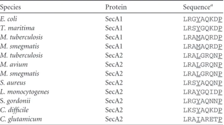
The functionally important two-helix finger (2HF), which is a 9-residue loop connecting two helices in the IRA1 domain that
inserts into the SecYEG pore, is conserved in theMtbSecA2 struc-ture (residues 695 to 703) (Table 2). However, the 2HF loop in MtbSecA2 adopts a different three-dimensional conformation compared to previous structures. In theMtbSecA2 structure, the 2HF is observed to close down approximately 10 Å onto the HSD, like a jaw hinge (Fig. 4), due to differences in how the ends of the helices unwind (even though the 2HF amino acid sequence itself is highly conserved, as shown inTable 2). This orientation contrasts with the conformation observed in most other SecA structures, in which the loop is more flipped out into solvent (Fig. 4); however, the conformations of the 2HF loop are also quite variable among SecA crystal structures (see Fig. S4 in the supplemental material). Fluorescence studies also suggest that the 2HF loop is flexible and can adopt different conformations in solution (37).
Similarities between SecYEG binding regions ofM. tubercu-losisSecA1 and SecA2.The conservation of the overall structure of SecA2 is consistent with a model in which SecA2 works with SecYEG to translocate SecA2-dependent proteins across the membrane. Furthermore, the key regions of SecA2 that would interact with the SecYEG pore are conserved, including the 2HF. The helix-terminating proline in the 2HF is present in SecA2 (Pro703), as it is in all SecA homologs (Table 2). Tyr794 inE. coli FIG 3Comparison of different orientations of the PPXD domain. Shown are
MtbSecA2 (cyan),MtbSecA1 (PDB code1NL3) (dark blue),B. subtilisSecA (PDB code1TF2) (yellow), and theTmSecA-SecYEG complex (PDB code 3DIN) (red). All four PPXD domains are superposed onto the body of MtbSecA2 (orange). At one extreme, in theMtbSecA1 structure (right, dark blue), the PPXD is packed against the HWD (missing in SecA2), representing the signal peptide-recognition site closed conformation. At the other extreme (left, red), the PPXD from theT. maritimacomplex with SecYEG represents the “preprotein clamp closed” configuration, where contact is made with NBD2 (orange, lower left). TheMtbSecA2 PPXD occupies a unique interme-diate position (cyan).
TABLE 2Conservation of the 2HF among SecA homologs
Species Protein Sequencea
E. coli SecA1 LRGYAQKDP
T. maritima SecA1 LRSYGQKDP
M. tuberculosis SecA1 LRAMAQRDP
M. smegmatis SecA1 LRAMAQRDP
M. tuberculosis SecA2 LRALGRQNP
M. avium SecA2 LRALGRQNP
M. smegmatis SecA2 LRALGRQNP
S. aureus SecA2 LRSYAQQNP
L. monocytogenes SecA2 LRAYGQIDP
S.gordonii SecA2 LRGYAQNNP
C. difficile SecA2 LKSYAQKDP
C. glutamicum SecA2 LRAIARETP
aKey residues are underlined.
SecA is another critical residue in the 2HF (36). Although it is substituted for by Leu698 inMtbSecA2, this tyrosine is substituted by large hydrophobic residues in 20% of SecA homologs (methi-onine in MtbSecA1). Furthermore, structural data from the TmSecA-SecYEG complex supports that hydrophobic substitu-tions, such as leucine, can be accommodated at this position, as the side chain sits in a hydrophobic pocket in SecY (34).
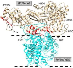
Structural superposition of MtbSecA2 onto TmSecA in the TmSecA-SecYEG complex (PDB code3DIN[34]) (Fig. 5; see Fig. S5 in the supplemental material) further indicates that SecA2 pre-serves many of the structural features of SecA implicated in bind-ing to SecYEG. This includes amino acids in theMtbSecA2 2HF and immediately adjacent regions of IRA1 that contact SecY in the TmSecA-SecYEG complex (aa 687 to 715 in SecA2) (Table 3and Fig. 5). There are also regions of NBD2 and the HSD that are structurally conserved in the SecA2 structure and positioned for contact with SecY (Table 3andFig. 5). These residues in NBD2, IRA1, and the HSD are clustered at the interface with SecY. In addition, although the PPXD of SecA2 is rotated away and does not appear to make direct contact with SecY in the superposition, if it were rotated into an orientation similar to that observed in TmSecA in the complex, it would place additional SecA2 residues (listed inTable 3) in contact with SecY, as shown inFig. 5. It is notable thatMtbSecA2 D607 (in the HSD) corresponds to one of the residues inE. coliSecA (position 640) that can be cross-linked with SecY using photoactivatable unnatural amino acids (60).
Mapping of suppressor mutations on the SecA2 structure.
Prior studies indicate that a SecA2 dominant-negative protein with an amino acid substitution in the ATP binding Walker box, making it unable to bind ATP, is locked in a nonfunctional com-plex, likely with SecYEG, at the membrane (29). In order to iden-tify important residues in SecA2, we identified intragenic suppres-sor mutations that could overcome thesecA2dominant-negative phenotypes (30) with the rationale being that such mutations might map to sites of protein interactions in SecA2 complexes. For convenience, these experiments were performed with the M.
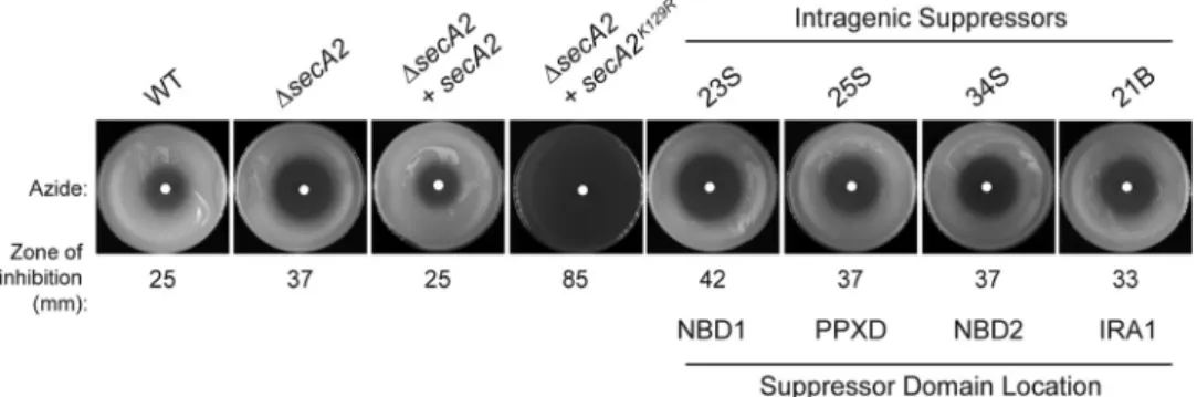
smegmatisortholog of SecA2, which has 83% amino acid identity toMtbSecA2 and is able to substitute for theMtbSecA2 in cross-species complementation experiments (29). An M. smegmatis strain expressing the dominant-negative MsSecA2K129R, which has an amino acid substitution in the Walker box (equivalent to K115 inMtbSecA2) was used. All suppressors identified reversed the severe dominant-negative phenotypes caused by SecA2K129R,
as assessed by azide sensitivity assays (5) and colony size on rich agar (30) (Fig. 6and data not shown), but they still exhibited a phenotype similar to that of a⌬secA2null mutant.
Eight independent suppressors with mutations in the coding sequence ofsecA2K129were identified by sequencing, mapping to four different domains: NBD1, NBD2, PPXD, and IRA1 (Table 4). All eight suppressor mutants produced full-length SecA2 protein at normal levels, as confirmed by Western blot analysis. Each mu-tation was validated to be responsible for the suppression by re-testing the phenotype of individual mutations when introduced into a freshsecA2K129Rmutant background (Fig. 6).
When mapped to the SecA2 structure, all of the suppressor mutations were located on the surface of the protein (Fig. 7). For simplicity, below we will refer to the suppressors using amino acid numbering that corresponds toMtbSecA2 (Table 4). There were three categories of suppressors. The first set of suppressor muta-tions affected the same surface loop of NBD1. There were two suppressors derived from independent cultures with identical mutations in NBD1 and a third suppressor with a different muta-tional alteration that mapped to the same site in NBD1. These NBD1 suppressors involve a 4-residue loop,168STPD172, inM. tuberculosisconnecting a-strand and an␣-helix; this loop was deleted in one mutant and duplicated in another. It is currently unknown what role these residues play, but it is striking that three out of eight suppressor mutations involved this surface-localized loop of the nucleotide binding domain, suggesting it is a function-ally important point of contact for SecA2.
The second group of suppressors (three in total) clustered in the SecA “polypeptide clamp” region made up of PPXD and NBD2 domains. Two suppressor mutations mapped to the SecA2 PPXD domain: a nonsynonymous substitution, D316H, and an insertion of a second glutamate at E354. These amino acids are in separate loops in the PPXD domain, but they are proximal in the three-dimensional structure, approximately 7 Å apart (Fig. 7a). The PPXD is positioned far from the NBD2 domain in the SecA2 structure. (The distance between the closest residues of the two domains is 23 Å, representing a “clamp open” state.) However, in theTmSecA-SecYEG complex, the corresponding PPXD loops to which these suppressor mutations map come in contact with NBD2. Moreover, the TmSecA residue corresponding to the D316H suppressor in theMtbSecA2 PPXD is in direct contact FIG 5Interface residues (red) ofMtbSecA2 (tan) that would contact SecYEG
(cyan), based on superposition withTmSecA (PDB code3DIN). Note that the red residues highlighted in the PPXD correspond to residues ofTmSecA that contact SecYEG as its PPXD is rotated into contact with SecYEG.
TABLE 3Residues ofMtbSecA2 predicted to be in contact with SecY based on structural superposition withTmSecA in complex with SecYEG (PDB code3DIN)
Domain MtbSecA2 residues predicted to contact SecY
NBD1 None
NBD2 E392, R395, Q396
HSD V600, R604, D607, A610, R614
IRA1 Most residues spanning 687–715 (including residues of the 2HF and surrounding IRA1 helices)
with NBD2 in the TmSecA-SecYEG complex (34) (Fig. 7b). It should be noted that thisTmSecA complex with SecYEG repre-sents an extreme conformation (induced by ADP and BeFx in the crystallization buffer) in which the preprotein channel is entirely collapsed (i.e., a loop of the PPXD actually inserts into the tein binding channel). In a structure of SecA bound to a prepro-tein substrate (PDB code3JV2[61]), the PPXD does not rotate quite as far toward NBD2 as in the SecA-SecYEG complex, but the residues corresponding to the suppressor mutations are still on the surface of the PPXD in a region that would be in position to interact with SecYEG or the lipid bilayer (similar to the red resi-dues highlighted in Fig. 5). Thus, these suppressor mutations could disrupt intramolecular interactions when the PPXD rotates to form the “clamp” around the translocating polypeptide (41) or could lock it in the extreme closed state such that the preprotein channel is collapsed altogether. Strikingly, the NBD2 suppressor T449I inM. tuberculosisalso maps to the SecA “preprotein clamp” region and is proximal (within 10 Å) to the two PPXD suppressor mutations when the clamp is closed (based on the analogous res-idues in the TmSecA-SecYEG docked structure [34]) (Fig. 7). Thus, these three suppressors in NBD2 and PPXD could conceiv-ably cause a defect in clamp closure during translocation. In light of past studies suggesting that interactions between SecA2K129R
and SecYEG are responsible for the dominant-negative phenotype (30), these results suggest that a defect in clamp closure may dis-lodge or prevent SecA2 interactions with SecYEG by disrupting interactions with the polypeptide being translocated through the channel.
The final group of intragenic suppressors identified have dele-tions in IRA1. One suppressor has a deletion of residues 714 to 721 inM. tuberculosis, and another suppressor has a very similar, yet distinct, deletion of residues 712 to 719 inM. tuberculosis. These deletions are in the middle of one of the␣-helices, just down-stream from the 2HF that forms part of the interface with SecYEG (Fig. 5and7andTable 3), and similar mutations in IRA1 have previously been shown to disrupt binding to SecYEG (62). Fur-thermore, one of the deleted residues in both of the IRA1 suppres-sors is Phe715, which is a conserved residue predicted to contact SecY (colored red inFig. 5) that is equivalent to the highly con-served Phe798 (inTmSecA). In theTmSecA-SecYEG structure, Phe798 (inTmSecA) forms an aromatic stacking interaction with Tyr418 in the C-terminal tail ofTmSecY (34). This interaction
appears to be crucial to docking as the equivalent tyrosine residue inE. coliSecY (Tyr429) is the location of a cold-sensitive mutation that prevented insertion of SecA into the membrane channel (63). These interacting residues are highly conserved in all Sec systems, includingMtbSecA2 (Phe715) andMtbSecY (Tyr436). The fact that this group of intragenicsecA2suppressor mutants harbors deletions in a structurally conserved and critical SecY-interacting region of IRA1 (Fig. 5andTable 3) is consistent with their mode of suppression being avoidance of complex formation between SecA2K129Rand SecYEG.
Intragenic suppressors alter membrane localization of the dominant-negative SecA2.In the wild type,MsSecA2 is predom-inantly found in the soluble cytoplasm-containing fraction. In contrast, the localization of SecA2K129Ris almost exclusively in the membrane-containing cell envelope pellet (29) (Fig. 8). This is consistent with a model for SecA2K129Rbeing locked in a protein complex with SecYEG at the membrane. Since we predicted that some of the intragenic suppressors alleviate SecYEG interactions, we determined the membrane localization of SecA2K129Rin the
intragenic suppressor mutant background. Strains were lysed and then fractionated into cell envelope (pellet) and soluble (cytoplas-mic) fractions. Western blot analysis with anti-SecA2 antibodies on fractions was then carried out to localize the protein. In each of the representative intragenic suppressors analyzed, the distribu-tion of SecA2K129Rshifted from the envelope, as seen in the
start-ingsecA2K129Rstrain, to the soluble cytoplasmic fraction (Fig. 8). Suppressor mutations in the “clamp” (PPXD and NBD2) and IRA1 domains had the most dramatic effects, restoring partition-FIG 6Intragenic suppressors suppress the azide sensitivity phenotype ofsecA2K129R. Lawns of the indicated strains were plated and tested for sensitivity to 10l
of 0.15 M sodium azide (applied to a paper disk in the center of the plate) for 2 days at 37°C. Average inhibition was calculated by measuring the diameter of the zone of azide inhibition, and values are the means of three biological replicates. The⌬secA2mutantM. smegmatisstrain was transformed with plasmids containing eithersecA2,secA2K129R, or a reconstructed intragenic suppressor with thesecA2K129Rmutation in combination with an intragenic suppressor
mutation located in one of the following domains: NBD1, NBD2, PPXD, or IRA1.
TABLE 4Suppressor mutations observed inM. smegmatisSecA2K129R
Isolate(s)a Effect on residue(s) inMsSecA2 Domain
Corresponding residue(s) in MtbSecA2
6S, 9S Deletion of residues 182–185 NBD1 168–171 (STPD) 23S* Duplication of residues 182–185 NBD1 168–171
2S Asp326¡His PPXD D316
25S* Glu insertion at residue 364 PPXD E354
34S* Thr459¡Ile NBD2 T449
21B* Deletion of residues 734–741 IRA1 714–721
38S Deletion of residues 732–739 IRA1 712–719
ing of SecA2 between the cell envelope and cytoplasm to almost wild-type levels. These data support a model in which the intra-genic suppressor mutations alleviate the dominant-negative phe-notype by disrupting protein-protein interactions involving the SecYEG membrane complex and/or the translocating polypep-tide.
DISCUSSION
Over 30 years ago, SecA was identified as a critical component of the protein export system of bacteria (64). Since that time, there have been extensive genetic, molecular, biochemical, biophysical, and structural studies to understand SecA function. Of the two SecAs inM. tuberculosis, SecA1 is the counterpart of the well-studied canonical SecA, while SecA2 has a distinct function from SecA1 and a nonoverlapping substrate specificity profile. The structure ofMtbSecA2 we report is the first structure of any SecA2 protein. The broad structural similarity observed between the two solvedMtbSecA structures indicates that, even after decades of mechanistic studies, gaps in our understanding of SecA proteins remain.
The smaller size of SecA2 versus SecA1 and canonical SecA proteins appears to come from the absence of the HWD, the VAR domain, and a C-terminal domain (CTD [although it still retains the CTL linker]) reducing the overall size of the protein product from 949 aa to 778 aa (see Fig. S2 in the supplemental material). The lack of an HWD is the most striking structural difference in SecA2. Without the HWD, the signal peptide recognition site of SecA2 is more solvent exposed and thus more accessible to protein substrates. This structural difference may help explain the ability of SecA2 to export substrates with distinctive features of their mature domain, possibly a propensity to fold prior to export (23). The “open” nature of the cleft created by the absence of a HWD could provide a broad surface against which folded proteins could possibly dock and unfold for translocation through the SecYEG transmembrane pore. Several pieces of experimental evidence support the possibility that the HWD could interact with prepro-teins. While a nuclear magnetic resonance (NMR) structure of
SecA bound to a signal peptide did not identify any residues of the HWD that directly interact with the signal peptide (40), several residues of the HWD were found to form cysteine-based cross-links with a synthetic signal peptide (42), which may result from transient states (i.e., alternative conformations of the HWD) sam-pled dynamically in solution. In fact, the HWD is observed to rotate by up to 15° between different crystal structures, depending on the oligomeric state (39). This suggests the HWD itself is mo-bile in solution, which is supported by fluorescence-based (fluo-FIG 7(a) Suppressor mutations fromMsSecA2K129Rmapped ontoMtbSecA2. Mutations are shown as yellow spheres. (b) Suppressor mutations mapped onto theTmSecA complex with SecYEG (PDB code3DIN). In the complex with SecYEG, SecA is in the “preprotein clamp closed” conformation, in which PPXD (cyan) is swung down (arrow) to make contact with NBD2 (green). In this conformation, the residues affected by the suppressor mutations in the PPXD and NBD2 domains can be seen to come into contact (circled).
FIG 8Subcellular localization of SecA2 is altered in intragenic suppressors of secA2K129R. Whole-cell lysates of the indicated strains were fractionated into a
rescence resonance energy transfer [FRET]) studies (65). Further-more, the mobility of the HWD appears to be influenced by the presence of a preprotein (66). Thus, the absence of the HWD in SecA2 could potentially affect substrate recognition. The 70-resi-due deletion of the HWD observed inMtbSecA2 is a general fea-ture among actinomycetes (includingMycobacteriumand Coryne-bacteriumspecies) (see Fig. S2 in the supplemental material). It should be noted that other Gram-positive SecA2 proteins also appear to have a truncated version of this domain (deletions of 13 to 18 residues forStreptococcus gordonii andL. monocytogenes, respectively). Until structures of these other SecA2 orthologs are solved, the potential consequences of these HWD truncations re-main unknown. It is possible that a reduced HWD could open up the signal peptide binding cleft and/or increase the site of interac-tion with preproteins, as we propose forMtbSecA2. To achieve a complete picture of SecA2 function going forward, the conse-quences of a truncated or deleted HWD will need to be explored in both mycobacterial and Gram-positive SecA2 proteins.
The significance of the absence of the VAR domain in the SecA2 NBD region is less clear. The lack of the VAR domain leaves the nucleotide-binding site relatively solvent exposed. While other SecA2 orthologs also lack the VAR domain (see Fig. S2 in the supplemental material), one-third of bacterial SecA (1) proteins lack this domain as well (58). InE. coliSecA, the VAR domain has been shown to regulate ATPase activity and ADP release, assecA ⌬varmutants display higher ATPase activity and faster ADP re-lease rates (58). However,MtbSecA2 (28) was recently reported to release ADP more slowly (not more quickly) than the VAR-con-tainingMtbSecA1 (67).
Mycobacterial SecA2 proteins, as well as SecA2s in many other organisms, lack the C-terminal domain (CTD) (see Fig. S2 in the supplemental material). The CTD in SecA1 proteins consists of a tail of⬃70 aa that is disordered in all previous crystal structures (1). In most bacteria, the CTD of SecA contains a Zn2⫹finger domain that binds to the protein export chaperone SecB (68). Mycobacteria are an exception, in that the CTD of SecA1 does not contain the conserved cysteines of a Zn2⫹finger motif. However, this may not be too surprising because, like Gram-positive bacte-ria (69), no SecB ortholog with a function in protein export has so far been identified in mycobacteria. Thus, because of the lack of Zn2⫹finger motif in the CTD of SecA1 and lack of a SecB or-tholog, the absence of a CTD inMtbSecA2 seems unlikely to be a significant contributing factor to the unique function of SecA2.
In comparison to all prior SecA structures, the SecA2 structure also revealed new orientations of the PPXD and the 2HF loop. However, these differences probably reflect the conformational plasticity of these two structural elements. Given the mobility of the PPXD domain already established for canonical SecA proteins, it seems likely that the PPXD orientation observed in SecA2 rep-resents a previously unobserved structural intermediate in the transition of the preprotein binding clamp from the open to closed position (38). The unique orientation of the 2HF loop ob-served in SecA2, which occurs at a key point of interaction with the translocation channel and varies considerably among SecA struc-tures, is probably a consequence of the flexibility of this loop in solution.
Given that there is no corresponding SecY2 partner in theM. tuberculosisgenome, an important mechanistic question to be an-swered is whether SecA2 works with the canonical SecYEG chan-nel to export proteins. In prior studies, we described a
dominant-negative secA2mutation that exhibits more severe phenotypes than a⌬secA2deletion mutant (29). Such phenotypes often result from a dominant-negative protein being locked in a nonproduc-tive complex with its normal binding partners. Furthermore, we showed extragenic suppressors that overexpress SecY suppress the secA2K129Rdominant-negative phenotype, which argues for an
in-teraction between SecA2K129Rand SecY (30). Here, we identified intragenic suppressors ofsecA2K129R, and all of them mapped to
the surface of the SecA2 structure. One group of suppressors mapped to the IRA1 domain of SecA2 in regions where similar mutations disruptE. coliSecA binding to SecYEG (62). These IRA1 suppressors also restored cytoplasmic localization of SecA2K129R. These results can be explained by the IRA1 suppressor
mutations preventing SecA2K129Rinteractions at the membrane
SecYEG channel, and they support the model for SecA2 working with SecY to promote export of its specific substrates. The sup-pressors that mapped to the “polypeptide clamp” region of SecA could similarly suppress the dominant-negative phenotype. How-ever, in this case, the suppression would result from the inability of SecA2 to trap the translocating polypeptide in the center of the SecYEG channel, causing SecA2 to fail to engage SecYEG (without the substrate) or causing the ternary system (SecYEG-SecA2-pre-protein) to dissociate.
The SecA structure reported here is of a monomer. In other studies, SecA proteins have been crystallized as monomers (54) or dimers (39), and the issue of the oligomeric state of SecA during protein translocation has remained controversial (37,70–72). A recent study demonstrated the ability of recombinantMtbSecA1 andMtbSecA2 to physically interactin vitro(73). If SecA1-SecA2 heterodimers form, it is possible that interactions between SecA1 and SecY avoid the need of SecA2 to directly interact with SecY. However, it is currently unclear if SecA1-SecA2 dimers exist and/or are functional in mycobacteria. Furthermore, the domi-nant-negative SecA2 phenotypes and the intragenic suppressors reported here, combined with structural conservation of SecA-SecY contact sites in SecA2, argue for the ability of SecA2 and SecA-SecY to interact. Ultimately, to clarify the mechanistic details of SecA2-dependent protein export, it will be necessary to study the path-way with anin vitroreconstitution system, as was used to dissect the mechanistic details of theE. coliSec pathway.
Since the SecYEG channel requires that proteins be un-folded for translocation (25), the possibility of SecA2 working with the SecYEG channel is intriguing, in light of experiments suggesting that SecA2 substrates are distinguished by a ten-dency to fold in the cytoplasm (23). The role of SecA2 could be to promote recognition of proteins that would normally be overlooked by the canonical SecA1-SecYEG translocase or to help maintain proteins in an unfolded state prior to or during export. The regions of structural difference and suppressor mutations identified in this study represent exciting new direc-tions for exploring the functional differences between SecA2 and SecA1 proteins.
ACKNOWLEDGMENTS
This work was supported by National Institutes of Health NIAID AI054540 (M.B.), NIH P01 AIO95208 (J.C.S.), and A0015 (J.C.S.) from the Robert A. Welch Foundation.
FUNDING INFORMATION
Robert A. Welch Foundation provided funding to James C. Sacchettini under grant number A0015. HHS | NIH | National Institute of Allergy and Infectious Diseases (NIAID) provided funding to James C. Sacchettini under grant number AIO95208. HHS | NIH | National Institute of Allergy and Infectious Diseases (NIAID) provided funding to Miriam Braunstein under grant number AI054540.
REFERENCES
1. Chatzi KE, Sardis MF, Economou A, Karamanou S. 2014. SecA-mediated targeting and translocation of secretory proteins. Biochim Bio-phys Acta1843:1466 –1474.http://dx.doi.org/10.1016/j.bbamcr.2014.02 .014.
2.Gouridis G, Karamanou S, Gelis I, Kalodimos CG, Economou A.2009. Signal peptides are allosteric activators of the protein translocase. Nature
462:363–367.http://dx.doi.org/10.1038/nature08559.
3.Feltcher ME, Braunstein M.2012. Emerging themes in SecA2-mediated protein export. Nat Rev Microbiol10:779 –789.http://dx.doi.org/10.1038 /nrmicro2874.
4.Bensing BA, Seepersaud R, Yen YT, Sullam PM.2014. Selective transport by SecA2: an expanding family of customized motor proteins. Biochim Biophys Acta1843:1674 –1686.http://dx.doi.org/10.1016/j.bbamcr.2013.10.019. 5.Braunstein M, Brown AM, Kurtz S, Jacobs WR, Jr.2001. Two
nonre-dundant SecA homologues function in mycobacteria. J Bacteriol183:
6979 – 6990.http://dx.doi.org/10.1128/JB.183.24.6979-6990.2001. 6.Sassetti CM, Rubin EJ.2003. Genetic requirements for mycobacterial
survival during infection. Proc Natl Acad Sci U S A100:12989 –12994. http://dx.doi.org/10.1073/pnas.2134250100.
7.Braunstein M, Espinosa BJ, Chan J, Belisle JT, Jacobs WR, Jr.2003. SecA2 functions in the secretion of superoxide dismutase A and in the virulence of Mycobacterium tuberculosis. Mol Microbiol48:453– 464. http://dx.doi.org/10.1046/j.1365-2958.2003.03438.x.
8.Kurtz S, McKinnon KP, Runge MS, Ting JP, Braunstein M.2006. The SecA2 secretion factor of Mycobacterium tuberculosis promotes growth in macrophages and inhibits the host immune response. Infect Immun
74:6855– 6864.http://dx.doi.org/10.1128/IAI.01022-06.
9.Sullivan JT, Young EF, McCann JR, Braunstein M.2012. The Myco-bacterium tuberculosis SecA2 system subverts phagosome maturation to promote growth in macrophages. Infect Immun80:996 –1006.http://dx .doi.org/10.1128/IAI.05987-11.
10. van der Woude AD, Stoop EJ, Stiess M, Wang S, Ummels R, van Stempvoort G, Piersma SR, Cascioferro A, Jimenez CR, Houben EN, Luirink J, Pieters J, van der Sar AM, Bitter W. 2014. Analysis of SecA2-dependent substrates in Mycobacterium marinum identifies pro-tein kinase G (PknG) as a virulence effector. Cell Microbiol16:280 –295. http://dx.doi.org/10.1111/cmi.12221.
11. Break TJ, Jun S, Indramohan M, Carr KD, Sieve AN, Dory L, Berg RE.
2012. Extracellular superoxide dismutase inhibits innate immune re-sponses and clearance of an intracellular bacterial infection. J Immunol
188:3342–3350.http://dx.doi.org/10.4049/jimmunol.1102341.
12. Hinchey J, Lee S, Jeon BY, Basaraba RJ, Venkataswamy MM, Chen B, Chan J, Braunstein M, Orme IM, Derrick SC, Morris SL, Jacobs WR, Jr, Porcelli SA.2007. Enhanced priming of adaptive immunity by a proapo-ptotic mutant of Mycobacterium tuberculosis. J Clin Invest117:2279 – 2288.http://dx.doi.org/10.1172/JCI31947.
13. Lenz LL, Mohammadi S, Geissler A, Portnoy DA. 2003. SecA2-dependent secretion of autolytic enzymes promotes Listeria monocyto-genes pathomonocyto-genesis. Proc Natl Acad Sci U S A100:12432–12437.http://dx .doi.org/10.1073/pnas.2133653100.
14. Siboo IR, Chambers HF, Sullam PM.2005. Role of SraP, a serine-rich surface protein of Staphylococcus aureus, in binding to human platelets. Infect Immun73:2273–2280.http://dx.doi.org/10.1128/IAI.73.4.2273-2280 .2005.
15. Wu H, Mintz KP, Ladha M, Fives-Taylor PM. 1998. Isolation and characterization of Fap1, a fimbriae-associated adhesin of Streptococcus parasanguis FW213. Mol Microbiol 28:487–500. http://dx.doi.org/10 .1046/j.1365-2958.1998.00805.x.
16. Xiong YQ, Bensing BA, Bayer AS, Chambers HF, Sullam PM.2008. Role of the serine-rich surface glycoprotein GspB of Streptococcus gordo-nii in the pathogenesis of infective endocarditis. Microb Pathog45:297– 301.http://dx.doi.org/10.1016/j.micpath.2008.06.004.
17. Nguyen-Mau SM, Oh SY, Kern VJ, Missiakas DM, Schneewind O.2012.
Secretion genes as determinants of Bacillus anthracis chain length. J Bac-teriol194:3841–3850.http://dx.doi.org/10.1128/JB.00384-12.
18. Fagan RP, Fairweather NF.2011. Clostridium difficile has two parallel and essential Sec secretion systems. J Biol Chem286:27483–27493.http: //dx.doi.org/10.1074/jbc.M111.263889.
19. Takamatsu D, Bensing BA, Sullam PM.2004. Four proteins encoded in the gspB-secY2A2 operon of Streptococcus gordonii mediate the intracel-lular glycosylation of the platelet-binding protein GspB. J Bacteriol186:
7100 –7111.http://dx.doi.org/10.1128/JB.186.21.7100-7111.2004. 20. Caspers M, Freudl R.2008. Corynebacterium glutamicum possesses two
secA homologous genes that are essential for viability. Arch Microbiol
189:605– 610.http://dx.doi.org/10.1007/s00203-008-0351-0.
21. Feltcher ME, Gunawardena HP, Zulauf KE, Malik S, Griffin JE, Sassetti CM, Chen X, Braunstein M.2015. Label-free quantitative proteomics reveals a role for the Mycobacterium tuberculosis SecA2 pathway in exporting solute binding proteins and Mce transporters to the cell wall. Mol Cell Proteomics14:1501–1516.http://dx.doi.org/10.1074/mcp.M114 .044685.
22. Gibbons HS, Wolschendorf F, Abshire M, Niederweis M, Braunstein M.
2007. Identification of two Mycobacterium smegmatis lipoproteins ex-ported by a SecA2-dependent pathway. J Bacteriol189:5090 –5100.http: //dx.doi.org/10.1128/JB.00163-07.
23. Feltcher ME, Gibbons HS, Ligon LS, Braunstein M. 2013. Protein export by the mycobacterial SecA2 system is determined by the pre-protein mature domain. J Bacteriol195:672– 681.http://dx.doi.org/10 .1128/JB.02032-12.
24. DeLisa MP, Tullman D, Georgiou G.2003. Folding quality control in the export of proteins by the bacterial twin-arginine translocation pathway. Proc Natl Acad Sci U S A100:6115– 6120.http://dx.doi.org/10.1073/pnas .0937838100.
25. Nouwen N, Berrelkamp G, Driessen AJ.2007. Bacterial Sec-translocase unfolds and translocates a class of folded protein domains. J Mol Biol
372:422– 433.http://dx.doi.org/10.1016/j.jmb.2007.07.003.
26. Weiss JB, Ray PH, Bassford PJ, Jr.1988. Purified SecB protein of Esch-erichia coli retards folding and promotes membrane translocation of the maltose-binding protein in vitro. Proc Natl Acad Sci U S A85:8978 – 8982. http://dx.doi.org/10.1073/pnas.85.23.8978.
27. Kusters I, Driessen AJ.2011. SecA, a remarkable nanomachine. Cell Mol Life Sci68:2053–2066.http://dx.doi.org/10.1007/s00018-011-0681-y. 28. Hou JM, D’Lima NG, Rigel NW, Gibbons HS, McCann JR,
Braun-stein M, Teschke CM.2008. ATPase activity of Mycobacterium tuber-culosis SecA1 and SecA2 proteins and its importance for SecA2 func-tion in macrophages. J Bacteriol190:4880 – 4887.http://dx.doi.org/10 .1128/JB.00412-08.
29. Rigel NW, Gibbons HS, McCann JR, McDonough JA, Kurtz S, Braun-stein M.2009. The accessory SecA2 system of mycobacteria requires ATP binding and the canonical SecA1. J Biol Chem284:9927–9936.http://dx .doi.org/10.1074/jbc.M900325200.
30. Ligon LS, Rigel NW, Romanchuk A, Jones CD, Braunstein M.2013. Suppressor analysis reveals a role for SecY in the SecA2-dependent protein export pathway of mycobacteria. J Bacteriol195:4456 – 4465.http://dx.doi .org/10.1128/JB.00630-13.
31. Sharma V, Arockiasamy A, Ronning DR, Savva CG, Holzenburg A, Braunstein M, Jacobs WR, Jr, Sacchettini JC.2003. Crystal structure of Mycobacterium tuberculosis SecA, a preprotein translocating ATPase. Proc Natl Acad Sci U S A100:2243–2248.http://dx.doi.org/10.1073/pnas .0538077100.
32. Papanikolau Y, Papadovasilaki M, Ravelli RB, McCarthy AA, Cusack S, Economou A, Petratos K.2007. Structure of dimeric SecA, the Esche-richia coli preprotein translocase motor. J Mol Biol366:1545–1557.http: //dx.doi.org/10.1016/j.jmb.2006.12.049.
33. Hunt JF, Weinkauf S, Henry L, Fak JJ, McNicholas P, Oliver DB, Deisenhofer J.2002. Nucleotide control of interdomain interactions in the conformational reaction cycle of SecA. Science297:2018 –2026.http: //dx.doi.org/10.1126/science.1074424.
34. Zimmer J, Nam Y, Rapoport TA.2008. Structure of a complex of the ATPase SecA and the protein-translocation channel. Nature455:936 –943. http://dx.doi.org/10.1038/nature07335.
2008. A role for the two-helix finger of the SecA ATPase in protein trans-location. Nature455:984 –987.http://dx.doi.org/10.1038/nature07439. 37. Whitehouse S, Gold VA, Robson A, Allen WJ, Sessions RB, Collinson
I.2012. Mobility of the SecA 2-helix-finger is not essential for polypeptide translocation via the SecYEG complex. J Cell Biol199:919 –929.http://dx .doi.org/10.1083/jcb.201205191.
38. Chen Y, Bauer BW, Rapoport TA, Gumbart JC.2015. Conformational changes of the clamp of the protein translocation ATPase SecA. J Mol Biol
427:2348 –2359.http://dx.doi.org/10.1016/j.jmb.2015.05.003.
39. Osborne AR, Clemons WM, Jr, Rapoport TA.2004. A large conforma-tional change of the translocation ATPase SecA. Proc Natl Acad Sci U S A
101:10937–10942.http://dx.doi.org/10.1073/pnas.0401742101. 40. Gelis I, Bonvin AM, Keramisanou D, Koukaki M, Gouridis G,
Kara-manou S, Economou A, Kalodimos CG.2007. Structural basis for signal-sequence recognition by the translocase motor SecA as determined by NMR. Cell131:756 –769.http://dx.doi.org/10.1016/j.cell.2007.09.039. 41. Bauer BW, Rapoport TA.2009. Mapping polypeptide interactions of the
SecA ATPase during translocation. Proc Natl Acad Sci U S A106:20800 – 20805.http://dx.doi.org/10.1073/pnas.0910550106.
42. Bhanu MK, Zhao P, Kendall DA.2013. Mapping of the SecA signal peptide binding site and dimeric interface by using the substituted cys-teine accessibility method. J Bacteriol195:4709 – 4715.http://dx.doi.org /10.1128/JB.00661-13.
43. DeJesus MA, Sacchettini JC, Ioerger TR.2013. Reannotation of transla-tional start sites in the genome of Mycobacterium tuberculosis. Tubercu-losis (Edinb)93:18 –25.http://dx.doi.org/10.1016/j.tube.2012.11.012. 44. Studier FW.2005. Protein production by auto-induction in high density
shaking cultures. Protein Expr Purif 41:207–234. http://dx.doi.org/10 .1016/j.pep.2005.01.016.
45. Heras B, Martin JL.2005. Post-crystallization treatments for improving diffraction quality of protein crystals. Acta Crystallogr D Biol Crystallogr
61:1173–1180.http://dx.doi.org/10.1107/S0907444905019451. 46. Leahy DJ, Hendrickson WA, Aukhil I, Erickson HP.1992. Structure of
a fibronectin type III domain from tenascin phased by MAD analysis of the selenomethionyl protein. Science258:987–991.http://dx.doi.org/10.1126 /science.1279805.
47. Fischetti RF, Xu S, Yoder DW, Becker M, Nagarajan V, Sanishvili R, Hilgart MC, Stepanov S, Makarov O, Smith JL. 2009. Mini-beam collimator enables microcrystallography experiments on standard beamlines. J Synchrotron Radiat 16:217–225. http://dx.doi.org/10 .1107/S0909049508040612.
48. Otwinowski Z, Minor W.1997. Processing of X-ray diffraction data collected in oscillation mode. Macromol Crystallogr A276:307–326.http: //dx.doi.org/10.1016/S0076-6879(97)76066-X.
49. Sheldrick GM.2010. Experimental phasing with SHELXC/D/E: combin-ing chain traccombin-ing with density modification. Acta Crystallogr D Biol Crys-tallogr66:479 – 485.http://dx.doi.org/10.1107/S0907444909038360. 50. Vonrhein C, Blanc E, Roversi P, Bricogne G.2007. Automated structure
solution with autoSHARP. Methods Mol Biol364:215–230.
51. Emsley P, Lohkamp B, Scott WG, Cowtan K.2010. Features and devel-opment of Coot. Acta Crystallogr D Biol Crystallogr66:486 –501.http: //dx.doi.org/10.1107/S0907444910007493.
52. Read RJ.1986. Improved Fourier coefficients for maps using phases from partial structures with errors. Acta Crystallogr A42:140 –149.http://dx .doi.org/10.1107/S0108767386099622.
53. Cowtan KD, Main P.1996. Phase combination and cross validation in iterated density-modification calculations. Acta Crystallogr D Biol Crys-tallogr52:43– 48.http://dx.doi.org/10.1107/S090744499500761X. 54. Hunt JF, Deisenhofer J.2003. Ping-pong cross-validation in real space: a
method for increasing the phasing power of a partial model without risk of model bias. Acta Crystallogr D Biol Crystallogr59:214 –224.http://dx.doi .org/10.1107/S0907444902018930.
55. Bricogne GBE, Brandl M, Flensburg C, Keller P, Paciorek W, Roversi PSA, Smart OS, Vonrhein C, Womack TO.2011. BUSTER version 2.8.0. Global Phasing Ltd., Cambridge, United Kingdom.
56. Guo XV, Monteleone M, Klotzsche M, Kamionka A, Hillen W, Braun-stein M, Ehrt S, Schnappinger D.2007. Silencing Mycobacterium smeg-matis by using tetracycline repressors. J Bacteriol189:4614 – 4623.http: //dx.doi.org/10.1128/JB.00216-07.
57. Dauter Z, Dauter M, Dodson E.2002. Jolly SAD. Acta Crystallogr D Biol Crystallogr58:494 –506.http://dx.doi.org/10.1107/S090744490200118X. 58. Das S, Grady LM, Michtavy J, Zhou Y, Cohan FM, Hingorani MM,
Oliver DB.2012. The variable subdomain of Escherichia coli SecA func-tions to regulate SecA ATPase activity and ADP release. J Bacteriol194:
2205–2213.http://dx.doi.org/10.1128/JB.00039-12.
59. Gold VA, Whitehouse S, Robson A, Collinson I.2013. The dynamic action of SecA during the initiation of protein translocation. Biochem J
449:695–705.http://dx.doi.org/10.1042/BJ20121314.
60. Das S, Oliver DB.2011. Mapping of the SecA·SecY and SecA·SecG inter-faces by site-directed in vivo photocross-linking. J Biol Chem286:12371– 12380.http://dx.doi.org/10.1074/jbc.M110.182931.
61. Zimmer J, Rapoport TA.2009. Conformational flexibility and peptide interaction of the translocation ATPase SecA. J Mol Biol394:606 – 612. http://dx.doi.org/10.1016/j.jmb.2009.10.024.
62. Vrontou E, Karamanou S, Baud C, Sianidis G, Economou A.2004. Global co-ordination of protein translocation by the SecA IRA1 switch. J Biol Chem279:22490 –22497.http://dx.doi.org/10.1074/jbc.M40100 8200.
63. Duong F, Wickner W.1999. The PrlA and PrlG phenotypes are caused by a loosened association among the translocase SecYEG subunits. EMBO J
18:3263–3270.http://dx.doi.org/10.1093/emboj/18.12.3263.
64. Oliver DB, Beckwith J.1982. Regulation of a membrane component required for protein secretion in Escherichia coli. Cell30:311–319.http: //dx.doi.org/10.1016/0092-8674(82)90037-X.
65. Ding H, Hunt JF, Mukerji I, Oliver D. 2003. Bacillus subtilis SecA ATPase exists as an antiparallel dimer in solution. Biochemistry42:8729 – 8738.http://dx.doi.org/10.1021/bi0342057.
66. Auclair SM, Oliver DB, Mukerji I.2013. Defining the solution state dimer structure of Escherichia coli SecA using Forster resonance en-ergy transfer. Biochemistry52:2388 –2401.http://dx.doi.org/10.1021 /bi301217t.
67. D’Lima NG, Teschke CM. 2013. ADP-dependent conformational changes distinguish Mycobacterium tuberculosis SecA2 from SecA1. J Biol Chem289:2307–2317.http://dx.doi.org/10.1074/jbc.M113.533323. 68. Zhou J, Xu Z.2003. Structural determinants of SecB recognition by SecA
in bacterial protein translocation. Nat Struct Biol10:942–947.http://dx .doi.org/10.1038/nsb980.
69. Schneewind O, Missiakas DM.2012. Protein secretion and surface dis-play in Gram-positive bacteria. Philos Trans R Soc Lond B Biol Sci367:
1123–1139.http://dx.doi.org/10.1098/rstb.2011.0210.
70. de Keyzer J, van der Sluis EO, Spelbrink RE, Nijstad N, de Kruijff B, Nouwen N, van der Does C, Driessen AJ.2005. Covalently dimerized SecA is functional in protein translocation. J Biol Chem280:35255–35260. http://dx.doi.org/10.1074/jbc.M506157200.
71. Jilaveanu LB, Zito CR, Oliver D.2005. Dimeric SecA is essential for protein translocation. Proc Natl Acad Sci U S A102:7511–7516.http://dx .doi.org/10.1073/pnas.0502774102.
72. Gouridis G, Karamanou S, Sardis MF, Scharer MA, Capitani G, Econo-mou A.2013. Quaternary dynamics of the SecA motor drive translocase catalysis. Mol Cell52:655– 666.http://dx.doi.org/10.1016/j.molcel.2013 .10.036.
73. Prabudiansyah I, Kusters I, Driessen AJ.2015. In vitro interaction of the housekeeping SecA1 with the accessory SecA2 protein of Mycobacterium tuberculosis. PLoS One10:e0128788.http://dx.doi.org/10.1371/journal .pone.0128788.