Sera Antibody Repertoire Analyses Reveal Mechanisms
of Broad and Pandemic Strain Neutralizing
Responses after Human Norovirus Vaccination
Lisa C. Lindesmith,1,7Jonathan R. McDaniel,2,7Anita Changela,3,7Raffaello Verardi,3,7Scott A. Kerr,4,7,9
Veronica Costantini,6Paul D. Brewer-Jensen,1Michael L. Mallory,1William N. Voss,2Daniel R. Boutz,4John J. Blazeck,2 Gregory C. Ippolito,5Jan Vinje,6Peter D. Kwong,3George Georgiou,2,4,5,8,*and Ralph S. Baric1,8,10,*
1Department of Epidemiology, University of North Carolina, Chapel Hill, NC 27599, USA 2Department of Chemical Engineering, University of Texas at Austin, Austin, TX 78712, USA
3Vaccine Research Center, National Institute of Allergy and Infectious Diseases, National Institutes of Health, Bethesda, MD 20892, USA 4Institute for Cellular and Molecular Biology, University of Texas at Austin, Austin, TX 78712, USA
5Department of Molecular Biosciences, University of Texas at Austin, Austin, TX 78712, USA 6Division of Viral Diseases, Centers for Disease Control and Prevention, Atlanta, GA 30333, USA 7These authors contributed equally
8These authors contributed equally
9Present address: BioFire Inc, Salt Lake City, UT 84108, USA 10Lead Contact
*Correspondence:[email protected](G.G.),[email protected](R.S.B.) https://doi.org/10.1016/j.immuni.2019.05.007
SUMMARY
Rapidly evolving RNA viruses, such as the GII.4 strain
of human norovirus (HuNoV), and their vaccines elicit
complex serological responses associated with
pre-vious exposure. Specific correlates of protection,
moreover, remain poorly understood. Here, we
report the GII.4-serological antibody repertoire—
pre- and post-vaccination—and select several
anti-body clonotypes for epitope and structural analysis.
The humoral response was dominated by
GII.4-spe-cific antibodies that blocked ancestral strains or by
antibodies that bound to divergent genotypes and
did not block viral-entry-ligand interactions.
How-ever, one antibody, A1431, showed broad blockade
toward tested GII.4 strains and neutralized the
pandemic GII.P16-GII.4 Sydney strain. Structural
mapping revealed conserved epitopes, which were
occluded on the virion or partially exposed, allowing
for broad blockade with neutralizing activity. Overall,
our results provide high-resolution molecular
infor-mation on humoral immune responses after HuNoV
vaccination and demonstrate that infection-derived
and vaccine-elicited antibodies can exhibit broad
blockade and neutralization against this prevalent
human pathogen.
INTRODUCTION
Human norovirus (HuNoV) is the leading cause of acute gastro-enteritis, a disease responsible for an estimated 200,000 deaths per year, mostly in children less than five years old and the elderly (Collaborators, 2017; Patel et al., 2008). The human
nor-ovirus genome is a single-stranded, plus-sensed RNA with 3 open reading frames (ORFs), including an ORF2 major capsid protein (viral protein 1, VP1) composed of shell and protruding (P) domains. Expression of VP1 in insect and mammalian sys-tems leads to self-assembly of 90 VP1 dimers into icosahedral virus-like particles (VLPs) that are antigenically indistinguishable from virions (Baric et al., 2002; Green et al., 1993; Jiang et al., 1995; Prasad et al., 1999). Despite more than 30 known human norovirus genotypes,60% of norovirus outbreaks are caused by GII.4 genotype strains (Burke et al., 2019; Cannon et al., 2017). Efforts to develop an effective vaccine against human nor-ovirus are complicated by the large number of antigenically distinct genotypes and the rapid evolution of GII.4 viruses (Atmar et al., 2018; Bull et al., 2007; Debbink et al., 2014a; Kocher et al., 2018; Lindesmith et al., 2008). Increased understanding of which capsid epitopes should be targeted to protect against different genotypes and of the impact of pre-exposure history on the elic-itation of protective immunity after vaccination are important considerations in vaccine design and efficacy ( Havenar-Daugh-ton et al., 2018; Lindesmith et al., 2015, 2017; Snijder et al., 2018).
Antigenic drift within GII.4 norovirus strains has prompted serial pandemic outbreaks in 1995, 2002, 2006, 2009, and 2012, each correlating with the emergence of new GII.4 anti-genic variants that appeared to have escaped pre-existing immunity (Debbink et al., 2012b; Lindesmith et al., 2008; Sie-benga et al., 2007). Human norovirus attaches to the cell surface via binding to histoblood group antigens (HBGAs; typically A, B, O, or Lewis antigens) on mucosal surfaces of the gut (Lindesmith et al., 2003; Tan and Jiang, 2011). Exten-sive studies have established that antibodies that block noro-virus VLP binding to HBGAs strongly correlate with protective immunity in chimpanzees and humans (Atmar et al., 2011; Bok et al., 2011; Malm et al., 2014; Reeck et al., 2010). Immune escape frequently relies on mutations at key residues in the immunodominant A, D, and E epitopes targeted by blockade
antibodies, with blockade measured by a surrogate neutrali-zation assay based on the antibody-dependent inhibition of VLP binding to HBGAs (Debbink et al., 2012a; Harrington et al., 2002; Lindesmith et al., 2012a, 2012b). Carbohydrate ligand blockade assays are the primary functional assay used to predict serological immunity and represent the clinical endpoint for evaluating norovirus infection and vaccination outcomes (Atmar et al., 2011; Lindesmith et al., 2015, 2017; Malm et al., 2014; Reeck et al., 2010). Recent development of human intestinal enteroids (HIEs) as an in vitro system that successfully supports the replication of HuNoV also al-lows for direct evaluation of neutralization (Costantini et al., 2018; Ettayebi et al., 2016).
Multivalent VLP immunization broadens the breadth of blockade antibodies produced after immunization of mice ( Deb-bink et al., 2014b; LoBue et al., 2006) and humans (Lindesmith et al., 2015). Currently, the leading human norovirus vaccine candidate is in phase IIb clinical trials (Bernstein et al., 2015; Ler-oux-Roels et al., 2018). This vaccine comprises a mixture of two VLPs: GI.1, the prototypical genogroup 1 strain, and GII.4c (GII.4 consensus), a VLP based on the consensus sequence of the GII.4 strains Houston (2002), Yerseke (2006a), and Den Haag (2006b) (Bernstein et al., 2015; Parra et al., 2012; Treanor et al., 2014). GII.4.2006a was used as the default sequence where all three of the strains diverged, most notably within the evolving immunodominant epitopes A, D, and E. The vaccine induces rapid (evident at day 7 post vaccination) antibody responses against both genogroup I and genogroup II VLPs ( Lin-desmith et al., 2015). The rapid blockade antibody response af-ter vaccination suggests that the vaccine may activate a memory B cell or recall response in adults (Lindesmith et al., 2015), though the mechanisms governing this immune outcome have remained unknown.
Here, we report on the serological repertoire to the GII.4c VLP component of the bivalent human norovirus vaccine pre- and post-immunization. We determined the serological repertoire using immunoglobulin sequencing (Ig-Seq), a prote-omics-based serum antibody repertoire analysis methodology (Georgiou et al., 2014; Lee et al., 2016; Williams et al., 2017). We focused on the GII.4c component of the vaccine because GII.4 strains have proven more clinically significant and display a higher rate of evolutionary drift, which potentially complicates the elicitation of protective immunity. Our results (1) provide clear evidence of the dominant effect of pre-existing immunity arising from earlier exposure on the humoral response to the vaccine; (2) define three classes of HuNoV circulating anti-bodies: one class comprising antibodies with very extensive binding breadth recognizing GI strains and GII strains but hav-ing no blockade activity, a second class that is specific to GII.4 and has blockade activity towards historical pandemic strains, and finally, a third class represented by one virus-neutralizing antibody with potent blockade activity towards historical GII.4 strains and contemporary strains that emerged well after the strains which were used to design the vaccine GII.4c VLP and human trials; (3) characterize in molecular detail VP1 epi-topes that are targeted to enable broad binding or broad blockade with neutralizing activity, and (4) highlight the impact of pre-existing serological repertoire breadth and titers on the response to the vaccine.
RESULTS
The GII.4 HuNoV Serological Repertoire after HuNoV Bivalent Vaccination Is Highly Polarized and Shaped by Previous GII.4 Infection
We analyzed three donors that experienced a significant increase in GII.4 titer after immunization with the bivalent GII.4c + GI.1 VLP vaccine (Figure 1A;Table S1). The serological repertoire was delin-eated using Ig-Seq, a proteomic methodology in which serum antibodies are purified by affinity chromatography against an im-mobilized antigen—in this case, imim-mobilized GII.4c VLP—then proteolytically digested into peptides and analyzed by liquid chro-matography-tandem mass spectrometry (LC-MS/MS). Peptide spectral matches were obtained using a custom database of heavy-chain-variable (VH) genes encoded by peripheral B cells from the respective donor. To construct the VH database, periph-eral blood mononuclear cells (PBMCs) collected at each time point were split into two aliquots: one aliquot was sequenced using high-throughput single-cell flow-focusing technology (DeKosky et al., 2015; McDaniel et al., 2016) to determine the native VH/VL (heavy-chain-variable/light-chain-variable) paired repertoire, and the second aliquot was processed to determine the VH repertoire. Relative quantitation of serum anti-GII.4c antibodies was deter-mined from the LC peak intensity of high-confidence peptide-spectrum matches (PSMs) derived from peptides of the third complementarity determining region of the heavy chain (CDR-H3). Antibodies belonging to a CDR-H3 clonotype likely recognize the same epitope, given the dominant role of CDR-H3 in antigen recognition. As we established earlier, the Ig-Seq work-flow identifies the majority (>70%) of the most abundant antigen-specific antibodies in the serum, and quantitation calibrations using isobaric peptide spike-ins show that peak intensities corre-late well with absolute peptide concentrations (Lavinder et al., 2014). The antibody repertoire composition and relative quantities at the clonotypic level are plotted as a histogram with each bar rep-resenting an individual clonotype and the horizontal axis showing its relative abundance (Figure 1B). All three donors had a titer to GII.4c at day 0, presumably a consequence of prior exposure to GII.4 infection and consistent with earlier data indicating that the vast majority of asymptomatic adults have antibodies to HuNoV (Carmona-Vicente et al., 2015; Nurminen et al., 2011). As has been the case with the serological repertoire to other antigens re-ported so far (e.g., influenza hemagglutinin [Lee et al., 2016], tetanus toxoid [Lavinder et al., 2014], or desmoglin in pemphigus patients [Chen et al., 2017]), the repertoire to GII.4c was found to be oligoclonal and highly polarized, in that <3 antibodies domi-nated the response, contributing more than 50% of the antigen-specific titer (Figure 1B). In particular, at day 0, the dominant CDR-H3 clonotype in each repertoire accounted for between 58% and 86% of the total CDR-H3 peptide intensity, suggesting that the long-term serological memory to GII.4 is dominated by a single antibody. Other examples of highly skewed serological repertoire polarization with one clonotype accounting for >60% of the response have been reported previously (Lee et al., 2016).
Figure 1. The Anti-NoV GII.4c IgG Serum Antibody Repertoire following Vaccination Is Polarized and Shaped by Previous HuNoV Infection
(A) Donors (n = 3) were immunized with experi-mental HuNoV vaccine and serum and PBMCs were collected at the times indicated.
(B) Relative abundance of GII.4c-specific IgG clo-notypes pre- and post-vaccination. Each bar rep-resents a CDR-H3 antibody clonotype observed at a0.5% of the GII.4c antigen-specific antibody repertoire.
(C) Amount of anti-GII.4c serum antibodies (i.e.,
the product of anti-GII.4c IgG ELISA titer3relative antibody abundance as determined by LS-MS/ MS; seeSTAR Methods) pre- and post-vaccina-tion; ELISA titer shown over each bar.
(D) Number of detected antibody clonotypes before and after vaccination.
(E) Anti-GII.4c antibody V-gene somatic hyper-mutation rates for pre-existing and emergent an-tibodies; n.s. represents not statistically significant (two-tailed t test; p > 0.3).
(F) V-gene family usage in the serological repertoire. Related toTable S1.
antibodies (7.1 ± 0.5% mean rate of somatic hypermutation) were slightly less mutated than the pre-existing anti-bodies (8.2 ± 1.1%), this difference did not reach statistical significance. The anti-GII.4c serological repertoire was predominantly comprised of antibodies using the immunoglobulin heavy vari-able 3 (IGHV3) family (80%;Figure 1F) compared to30% in naive or mature B cells (DeKosky et al., 2016; Mroczek et al., 2014). These findings suggest that the serological IgG response to human norovirus vaccination is largely dominated by the boosting of a limited number of pre-existing antibody clonotypes.
Recombinant HuNoV Serum Antibodies Demonstrate Neutralization against GII.4 and Breadth of Binding across Strains
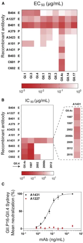
We expressed and characterized a set of 10 representative serum antibodies for which both VH and VL sequences were de-tected by paired VH/VL sequencing. These included the top most abundant clonotypes in donors A and C (A379 and C601), two strongly boosted pre-existing clones (A101 and A1431, boosted by 123 and 1263, respectively, in terms of abundance in the post-vaccination serum relative to day 0), and six emergent antibodies (Figure 1B). Binding and blockade activity were tested using an antigenically diverse panel of noro-virus genogroup I and genogroup II VLPs, including a time-ordered panel of pandemic GII.4 VLPs representing historical variant strains, including US95_96 (GII.4 1997), Farmington Hills (GII.4 2002), and Den Haag (GII.4 2006b), as well as prospective strains that emerged following vaccine design (2006) and the fact that they become detectable in the serum post vaccination
and should not be interpreted in terms of B cell ontogeny (i.e., whether they arise from stimulated naive cells or memory Bcells).Althoughvaccinationincreasedthediversityofthe sero-logical repertoire in all donors, both the number and abundance ofvaccine-elicitedclonotypesweresubstantiallylowerindonor C, who had a 4- to 5-fold higher titer to GII.4c before vaccination (day 0) compared to the other two donors (Figures1B–1D). In this donor,theincreaseintheGII.4ctiter4weekspostvaccination was overwhelmingly due to the boosting of the dominant anti-bodyclonotypeC601,whichaccountedfor>90%oftheserum response and increased in absolute abundance by more than 7-foldrelativetothelevelobservedatday0.Thelowlevelof emergent antibodies in donor C, who had the highest pre-vacci-nationtiter,isconsistentwiththestronginversecorrelation be-tweenday0serumtiterandthenumberofemergentantibody clonotypes in the serological memory after immunization with theseasonalinfluenzavaccine(Leeetal.,2016).
clinical trial (2010), such as GII.Pe-GII.4 Sydney (GII.4 2012) and GII.P16-GII.4 Sydney (GII.4 2015) (Lindesmith et al., 2018a). As expected, all antibodies bound to the vaccine strain VLP ( Fig-ure 2A), with half-maximal effective concentration (EC50) values between 8 and 717 ng/mL. The potential for neutralization by in-hibition of cellular ligand binding was evaluated by determining blockade of VLP-ligand binding (blockade assay) (Figure 2B). The 10 serum antibodies could be categorized into three classes. The first class consisted of 6 antibodies that displayed broad binding but no blocking activity toward multiple GI and GII strains; three of these, namely A1227, B401, and B404, showed binding to all strains tested, including a variety of GI and GII ge-notypes such as GI.I, GII.2, and GII.17. The VP1 protein among these strains shows only 50% amino acid identity (Zheng et al., 2006). The second class consisted of antibodies B403, C601, and C602, which had strong blockade activity towards the vaccine strain and the earlier pandemic GII.4 1997 strain but not to GII.4 strains that emerged post vaccination. Finally, the third class of serum antibodies had a single member, A1431, which had strong blockade activity to all known historical GII.4 strains from 1987 onwards. A1431 had very extensive breadth and potency not only towards all historical strains avail-able but also towards prospective GII.4 strains that appeared after the vaccine trial, including GII.4 2012 and 2015, neither of which had circulated in the general population at the time of sam-ple collection (Figure 2B). Furthermore, A1431 potently neutral-ized GII.P16-GII.4 Sydney virus, a GII.4 strain that emerged 5 years post sample collection, in a human intestinal enteroid cul-ture system (Costantini et al., 2018; Ettayebi et al., 2016) (IC50 5.5 ng/mL), supporting the correlation between blockade of ligand-binding potency and antibody-mediated neutralization (Figures 2C andS1) (Alvarado et al., 2018; Ettayebi et al., 2016). We sought to define biochemically the epitopes recognized by the antibodies with blockade activity to historical strains, namely B403, C601, and C602; as discussed below, the epitope of A1431 that has broad blockade breadth was determined by crystallography. We first evaluated antibody mediated blockade against a panel of VLPs with epitopes A, D, and E exchanged from GII.4 2012 (non-binding VLP) into GII.4 1987 (binding VLP). Exchange of epitope A residues ablated blockade potency of B403 and diminished potency of C601 by 13.5-fold but had lit-tle effect on C602 blockade (1.8-fold decrease). Exchange of epitope D or E residues had little impact on blockade of any of the three antibodies, suggesting that the major binding determi-nants for B403 and C601 likely reside within epitope A (Figures 3A–3C). VLPs displaying an acidic residue at 298, Val at position 356, and His at 357 had lower EC50titers than VLPs without these residues for all three antibodies (Figures 3B and 3C). Exchange of V298E/A356V/E357H into the null backbone VLP 581 ( Linde-smith et al., 2018b) (VLP 581.A4A) gained binding of B403 and C601 and improved binding of C602 by 11.6-fold compared to
Figure 2. HuNoV Recombinant Serum Antibodies Recognize
Conserved Binding, Ligand-Blocking, and GII.4 Neutralizing Epitopes Sigmoidal dose response curves were fit to the mean percent control binding for determination of (A) half-maximum binding (EC50) or (B) half-maximum
blocking of HBGA binding (half-maximum inhibitory concentration, IC50) of
pre-existing (P) and emergent (E) antibodies. Titers >2mg/mL were scored as negative (white box). The HBGA blocking assay was used to characterize A1431 against a panel of pandemic norovirus GII.4 strains. Antibodies were
tested in duplicate in a minimum of two independent experiments. (C) Sigmoidal dose response curves were fit to the mean percent viral genomic copies control for determination of IC50 in a neutralization assay. The
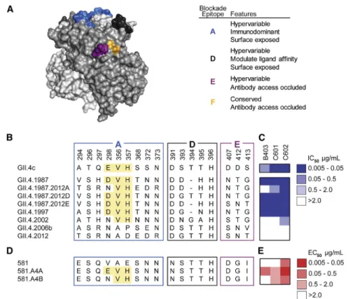
Figure 3. Serum Antibodies that Block His-torical GII.4 Strains Recognize Overlapping Epitopes within Epitope A
(A) Homology model based on PDB: 3SLD (Shanker et al., 2011) of the GII.4 2006a P domain dimer of the HuNoV major capsid protein, VP1 with known P domain blockade antibody epitopes A (blue), D (dark gray), E (purple), and F (orange) highlighted. (B) Amino acid sequences of pandemic strains and select mutants of GII.4 1987.
(C) Blockade potency binned by IC50.
(D) Select mutants of GII.4 2006a.581 VP1. High-lighted areas indicate sequences important for antibody binding.
(E) Antibody binding binned by EC50values.
An-tibodies were tested in duplicate in a minimum of two independent experiments.
GII.4.2002 P domain (P2002) yielded dif-fracting crystals suitable for structural analysis.
Single crystals of the complex between A1227 and P2002 diffracted to 2.6 A˚, and the structure was solved by molecular replacement using the apo P2002 struc-ture (PDB: 4OOV) (Singh et al., 2015) ( Ta-ble S2). A1227 Fab bound to each of the conserved P1 subdomains of the P2002 dimer (Figure 4A). Each Fab bound at the interface between monomers in the P2002 dimer, with the heavy chain recognizing both protomers and the light chain recognizing only a single pro-tomer (Figure 4B). A1227 recognized a region of P2002 which was mostly conserved across genogroups GI and GII (Figures 4C andS2), thus explaining the broad cross-reactivity of anti-body A1227. Overall, each Fab buried950 A˚2by establishing polar contacts and favorable van der Waals interactions with P2002 (Figures 4D andS2). There were no substantial conforma-tional changes upon A1227 binding to P2002, with a root-mean-square deviation (RMSD) of 0.6 A˚ between apo-P2002 and A1227-P2002 complex structures.
To map the A1227 epitope within the context of the VLP, the crystal structure of the A1227-P2002 complex was superim-posed onto the GI.1 capsid structure (PDB: 1IHM) (Prasad et al., 1999). The GI.1 capsid structure was chosen as a template for modeling due to similarities between GI.1 and GII.4 capsid structures indicated by prior electron microscopy (EM) studies (Chen et al., 2004). Modeling revealed that the conserved region recognized by A1227 was located at an occluded site on the norovirus capsid, at the interface between the P domain and shell domain. Overlay of Fab A1227 onto the GI.1 capsid structure (Figures 4E and 4F) indicated that other P domains on the assembled norovirus capsid would clash with both heavy and light chains of the Fab, with only minor clashes with the shell domains (Figure 4G), suggesting that structural rear-rangements of the P domain could either expose the A1227 bind-ing site or allow bindbind-ing at a neighborbind-ing P domain. Its lack of blockade or neutralizing activity likely results from antibody A1227 only recognizing defects or maturation intermediates ( Po-gan et al., 2018).
the581VLP(Figures3Dand3E).SubstitutionofV298N/A356V/ E357H (VLP 581.A4B) did not gain binding for B403, yielded similar binding to that to 581.A4A for C601, and decreased C602 binding by 7.6-fold compared to 591.A4A. These data confirm E or D at position 298 as an anchor residue for B403. Forantibody C601,binding was heavily influencedby V356/ H357, residues that are predicted to contribute to epitope A buthavenotbeenpreviouslyverified.Further,ouranalysisalso revealed that C602 recognizes a previously undefined blockade epitope.Together,thesedatacategorizeantibodiesboostedby HuNoVVLPvaccinationintothreeclasses:(1)genogroupIand genogroup II broadly binding, non-neutralizing antibodies; (2) ancestralGII.4bindingandligand-blockingantibodies;and(3) GII.4 broadly binding and neutralizing antibodies. Genetic map-pingindicated thatantibodiesdisplaying a limitedbreadthof GII.4 ligand-blockade potency (class 2 above) recognized the immunodominantepitopeAantigenicsite(Malloryetal.,2019) and accounted for previously observed back-boost antibody re-sponses in HuNoV vaccinated donors (Lindesmithetal.,2015). These data demonstrate that HuNoV vaccination can elicit serum antibodies capable of neutralizing GII.4 pandemic strains.
StructuralStudiesDefineaHighlyConserved Non-BlockingEpitopeandaConservedGII.4 NeutralizingEpitopewithintheCapsidPDomain
To provide a structural basis for recognition by the GII.4 broad blockade antibody A1431, we solved the crystal struc-ture of A1431 Fab in complex with the GII.4.2002 P domain at 3.1 A˚ resolution (Table S2). The asymmetric unit contained two P-domain-A1431 Fab complexes, with each dimer gener-ated by crystallographic 2-fold symmetry. Both complexes were nearly identical, with an RMSD of 0.3 A˚. Electron density for both P2002 and A1431 variable domains was well defined, although regions of A1431 constant domains were not well ordered.
Antibody A1431 bound each P domain monomer at a cleft be-tween the P1 and P2 subdomains (Figure 5A). Similar to A1227, A1431 binding did not induce substantial conformational changes in the P domain, with an overall RMSD of 0.4 A˚ between the complex and apo (PDB: 4OOV) structures. Antibody A1431 recognized an epitope located near previously mapped blockade epitopes D and F and distinct from the site of HBGA binding (Figure 5B). Both A1431 heavy and light chains contrib-uted to the binding interface with a total buried surface area of 985 A˚2 on the P domain. The CDR-H3 loop of A1431 was Figure 4. The Broadly Binding Non-Blockade Antibody A1227 Binds to a Highly Conserved Epitope within the P Domain
(A) Crystal structure of A1227 Fab bound to GII.4.2002 P domain dimer shown in cartoon representation, with P domain monomers colored pink and orange and A1227 heavy and light chains in gray and blue, respectively.
(B) The A1227 antibody binds near the P1 dimeric interface (P domain dimer in surface representation and A1227 Fab in cartoon).
(C) A1227 Fab footprint is outlined in yellow on the P domain surface, mapped by GI-GII sequence conservation, with degree of conservation colored from variable (white) to conserved (purple).
(D) Detailed view of A1227 Fab-P domain binding interface, with interacting residues depicted in stick and hydrogen bonds shown in yellow dotted lines. (E) Modelling of the GII.4.2002 P domain-A1227 Fab crystal structure onto the GI.1 capsid structure (PDB: 1IHM) reveals an antibody binding site that is occluded on the assembled VLP.
(F) Close-up view from (E) showing the occlusion of A1227 Fab within the GI.1 capsid.
(G) Quantification of volume overlap between A1227 Fab (or VH/VL) and GI.1 capsid proteins. Over 60% of the volume of A1227 Fab overlaps with neighboring P domains when modelled onto either A/B or C/C dimers. Only small overlaps are due to clashing with shell domains. Overlaps from P domain and shell domain are colored in gray and yellow, respectively. Volumes were calculated using Voss-Void-Voxelator program (Voss and Gerstein, 2010).
P1 subdomain. The recognized surface, including P domain residues Gln402, Trp403, Gln504, and Asp506, was nearly completely conserved in GII.4 (Figure 5D) but only semi-conserved between GI and GII genogroups, with Asp506 changed to Pro506 in most other GI and GII strains (Figure S3), Figure 5. Broad GII.4-Blockade Antibody A1431 Recognizes an Accessible Semi-Conserved Site of Vulnerability
(A) Overall structure of A1431 Fab bound to GII.4.2002 P domain dimer is shown in ribbon representation, with P domain monomers in pink and orange and A1431 heavy and light chains in magenta and cyan, respectively.
(B) Mapped blockade epitopes are highlighted on a surface representation of GII.4.2002 P domain dimer (colored as inFigure 3A) with the A1431 Fab shown in cartoon. The crystal structure of GII.4 (Farmington Hills, 2004) P domain complexed to HBGA type B ligand (PDB: 4X05) was superimposed onto the GII.4.2002 P domain + A1431 complex to show location of HBGA binding site.
(C) Details of A1431 Fab interactions with P domain residues are highlighted with interacting residues depicted in stick and hydrogen bonds shown in yellow dotted lines.
(D) A1431 Fab footprint is outlined in yellow on the P domain dimer surface, mapped by GII.4 sequence conservation, with degree of conservation colored from variable (white) to conserved (purple).
(E) The GII.4.2002 P domain-A1431 Fab crystal structure is modeled onto the GI.1 capsid structure (1IHM;Prasad et al., 1999), with a cross-section showing the P domain fit onto the A/B dimer (top) or the C/C dimer (bottom). Overlap between A1431 Fab and P domain, as modeled onto either GI.1 capsid A/B or C/C dimers, is minimal and shown on right, as inFigure 4G.
(F) Modeling of A1431 Fab to the A/B dimer (top) or C/C dimer (bottom) on the GI.1 capsid structure, with A/B dimers colored pink and tan, C/C dimers colored in gray and shell domain shown in yellow.
(G) GI.1 capsid models, colored as in (F), showing occupancy of A1431 binding sites on all A/B dimers (top) or all C/C dimers (bottom). Related toTable S2andFigure S3.
providing the structural basis for the broad recognition of A1431 of GII.4 viruses but not of other norovirus genotypes.
To gain insight into the molecular mechanism of blockade ac-tivity by antibody A1431, we superimposed the crystal structure of the P2002-A1431 Fab complex onto the GI.1 capsid structure (Figure 5E). On the assembled VLP, the P domain dimer exists in two distinct structural environments, defined by A/B and C/C di-mers (Prasad et al., 1999). A1431 Fab binding to the A/B dimer showed no overlap between Fab and either P or S domains, whereas binding to the C/C dimer showed minor overlap (Figures 5E and 5F). Binding of the A1431 Fab to all equivalent sites on the VLP showed recognition at the A/B dimer to cover most of the VLP surface, whereas recognition of the C/C dimer left the 5-fold vertices potentially capable of attaching to HBGAs at the cell surface (Figure 5G). In both cases, binding at all sites would lead to substantial antibody-antibody overlap (Figures 5F and 5G), suggesting that A1431 exerts its blockade and neutralizing activity by binding to only a fraction of potential bind-ing sites on the VLP. Overall, in contrast to the A1227 bindbind-ing site, modeling suggested the A1431 blockade epitope to be mostly accessible on the capsid surface. Notably, the A1431 epitope did not overlap with identified sites of HBGA recognition, and therefore, the observed blockade functionality likely stems either from antibody A1431 sterically inhibiting the approach of the VLP to the >500 kDa multivalent HBGAs found on MUC2 in PGM and in human gastric and duodenal tissues (Park et al., 2015) or from subtle allosteric effects, as reported for another antibody that docks near to the 1431 binding site (Lindesmith et al., 2014, 2018b), as defined by epitope mapping. High-reso-lution structures of broadly reactive and/or neutralizing epitopes expand our understanding of the HuNoV antigenic landscape and narrow the search for optimal vaccine candidates. For instance, whereas antibodies targeting hypervariable P2 epi-topes (i.e., epitope A) have limited cross-strain ligand-blocking potency and antibodies recognizing highly conserved epitopes near the P1-shell-domain interface are highly cross-reactive but have no blocking potency, the antibodies targeting geno-type-conserved epitopes at the P1-P2 interface display broad ligand-binding blocking potency. It should be noted that each of these antibody classes may contribute to vaccine-induced protection, and further studies andin vivomodels are required to fully understand the interplay between the antibody targets and the physiological response.
DISCUSSION
Vaccine-induced immunity relies on the development and main-tenance of long-term protective antibody titers or serological memory. The best studied correlate for protective immunity after HuNoV infection is the development of robust blockade antibody responses (Lindesmith et al., 2015; Malm et al., 2014; Reeck et al., 2010). Immune responses to rapidly evolving viral infec-tions are complex and heavily impacted by pre-exposure his-tories that shape the antibody response to newly evolved strains (Belongia et al., 2017; Gostic et al., 2016; Lessler et al., 2012; Lin-desmith et al., 2017). These studies demonstrate that bivalent HuNoV vaccines can elicit GII.4 neutralizing antibody responses in humans, while also structurally defining the highly conserved GII.4 (1987–2015) epitope recognized by a neutralizing antibody.
Here, we demonstrate that the serological repertoire to HuNoV is oligoclonal and dominated by IGHV3 family antibodies in three pre-immune individuals who received the vaccine. A large fraction (6/10) of the serum antibodies that we biochemically characterized in detail displayed significant binding breadth, with 4/6 recognizing both GI and GII genotype VLPs. Although these high-binding-breadth antibodies did not have blockade activity, they may play a role in protection against HuNoV infec-tion via Fc-mediated mechanisms. Non-neutralizing antibodies that protect against infection in animal models are observed at significant levels in circulation after influenza vaccination (Lee et al., 2016), and recent studies report the isolation of such anti-bodies for other viruses as well (Bootz et al., 2017; Henry Dunand et al., 2016). Current technologies do not allow direct evaluation of HuNoV Fc-mediated protection.
In donor A, we showed that the most dramatically back-boosted blockade antibody following vaccination is capable of blocking VLP-HBGA binding of all examined GII.4 strains circu-lating between 1987 and 2015 and is potently neutralizing in the human intestinal enteroid culture system. This antibody was present in appreciable amounts in the serum prior to vacci-nation, suggesting that it must have been elicited by an earlier HuNoV infection. Although lack of successful propagation of his-toric GII.4 strains limits our ability to test the breadth of GII.4 neutralization, all tested blockade monoclonal antibodies have also been neutralizing antibodies (Alvarado et al., 2018), indi-cating that A1431 is also likely cross-GII.4 neutralizing. These findings reveal that HuNoV infection can induce broad blockade and neutralizing antibodies and that these antibodies can be boosted by vaccination (Lindesmith et al., 2012a, 2015).
HBGAs. These antibodies likely bind to occluded epitopes available for antibody access during phases of particle assembly and/or disassembly, intermediates, or rearrangements. Al-though mechanisms of protection driven by this class of anti-bodies are undefined, short-term boosting of cross-blockade antibodies after vaccination (Lindesmith et al., 2015) and mouse norovirus (MNV) monoclonal antibodies with cross-serotype neutralization (Kolawole et al., 2017) support the potential for HuNoV cross-genotype neutralization.
These data provide structural and mechanistic evidence supporting that the GII.4 component of the Takeda multivalent norovirus VLP vaccine recalls a memory antibody response that persists for at least six months, indicating that, similar to other rapidly evolving RNA viruses, early-life norovirus strain exposure shapes antibody responses to subsequent norovirus strains (Lindesmith et al., 2015, 2017). As with influenza, expo-sure history and genetic background are expected to impact outcomes differently by age groups, potentially impacting vac-cine efficacy (Gostic et al., 2016; Lessler et al., 2012). Future studies are needed to determine additional mechanisms of the antibody boost to prospective GII.4 strains, as well as the anti-body response in key vaccine target populations, including the aged, with a life-time of norovirus exposure but waning immune responsiveness, and the very young, with no pre-exposure history and possibly with maternal antibodies. These data are critical for understanding vaccine performance and population-based immunity against future pandemic isolates. Cross-block-ing antibodies like A1431 provide one mechanism of breadth, and serological repertoire analyses offer robust strategies to dissect the molecular basis for this and other responses in hu-man populations. Coupled with blockade, neutralization potency in the enteroid culture system (Costantini et al., 2018; Ettayebi et al., 2016), and structural determination of antibody binding sites, recombinant serum antibodies can provide invaluable in-formation for immunogen design and vaccination strategies that phenocopy infection and provide broad, long-term protec-tion to HuNoV pandemics.
STAR+METHODS
Detailed methods are provided in the online version of this paper and include the following:
d KEY RESOURCES TABLE
d CONTACT FOR REAGENT AND RESOURCE SHARING
d EXPERIMENTAL MODEL AND SUBJECT DETAILS
B Humans Subjects B Cell lines B Microbes
d METHOD DETAILS
B Ten percent stool filtrate
B Human intestinal enteroid culture (HIE)
B High-throughput sequencing of BCR transcripts B VH Sequencing
B VH/VL Sequencing
B Isolation of HuNoV specific antibodies B Sample preparation and LC-MS/MS analysis B Antibody expression and purification B Production of Virus Like Particles
B Antibody functional assays
B P domain expression and purification B Crystallization of Fab complex with P2002 B Structure determination and analysis B Neutralization Assay
d QUANTIFICATION AND STATISTICAL ANALYSIS
d DATA AND SOFTWARE AVAILABILITY
SUPPLEMENTAL INFORMATION
Supplemental Information can be found online athttps://doi.org/10.1016/j. immuni.2019.05.007.
ACKNOWLEDGMENTS
The authors would like to thank Takeda Vaccines for provision of human sam-ples; the Genome Sequencing and Analysis Facility at the University of Texas at Austin for performing Illumina next-generation sequencing; the Center for Biomedical Research Support Proteomics Facility at the University of Texas at Austin for providing mass spectrometry analysis; and Victoria Madden of Microscopy Services Laboratory, Department of Pathology and Laboratory Medicine, University of North Carolina–Chapel Hill for expert technical sup-port. Support for this work was provided by the Intramural Research Program of’’ the Vaccine Research Center, NIAID, NIH, and the intramural food safety program at CDC and by grants from the NIH (R56AI106006, G.G. and R.S.B. and U19 AI109761 CETR, R.S.B.), Defense Threat Reduction Agency (DTRA) (contract HDTRA1-12-C-0105, G.G.), and the Wellcome Trust [203268/Z/16/ Z] (R.S.B.). Use of sector 22 (Southeast Region Collaborative Access team) at the Advanced Photon Source was supported by the U.S. Department of Energy, Basic Energy Sciences, Office of Science, under contract number W-31-109-Eng-38. The findings and conclusions in this article are those of the authors and do not necessarily represent the official position of the Centers for Disease Control and Prevention. The funders had no role in study design, data collection and interpretation, or the decision to submit the work for publication.
AUTHOR CONTRIBUTIONS
Conceptualization: R.S.B., G.G., L.C.L., J.R.M., A.C., R.V., S.A.K., and P.D.K. Investigation and Formal Analysis: L.C.L., J.R.M., A.C., R.V., S.A.K., V.C., P.D.J.B., M.L.M., W.N.V., D.R.B., J.J.B., G.C.I., and J.V. Writing, All Drafts: L.C.L., J.R.M., A.C., R.V., P.D.K., G.G., and R.S.B. with input from all authors. Funding Acquisition and Supervision: R.S.B., G.G., and P.D.K.
DECLARATION OF INTERESTS
J.R.M., S.A.K., A.C., R.V., V.C., P.D.B.J., M.L.M., W.N.V., D.R.B., J.J.B., G.C.I., J.V., P.D.K., and G.G. declare no competing interests. L.C.L. and R.S.B. have consulted and collaborated with Takeda on norovirus vaccines. They are also inventors on patents filed on issues relevant to noroviruses. G.G. is an inventor on patents related to the analysis of the serological antibody repertoire.
Received: January 3, 2019 Revised: March 15, 2019 Accepted: May 15, 2019 Published: June 13, 2019
SUPPORTING CITATIONS
The following references appear in the Supplemental Information: Word et al. (1999).
REFERENCES
Recent developments in the PHENIX software for automated crystallographic structure determination. J. Synchrotron Radiat.11, 53–55.
Agnihothram, S., Menachery, V.D., Yount, B.L., Jr., Lindesmith, L.C., Scobey, T., Whitmore, A., Sch€afer, A., Heise, M.T., and Baric, R.S. (2018). Development of a Broadly Accessible Venezuelan Equine Encephalitis Virus Replicon Particle Vaccine Platform. J. Virol.92, e00027–e00018.
Ahmed, S.F., Klena, J.D., Mostafa, M., Dogantemur, J., Middleton, T., Hanson, J., and Sebeny, P.J. (2012). Viral gastroenteritis associated with genogroup II norovirus among U.S. military personnel in Turkey, 2009. PLoS ONE7, e35791.
Alvarado, G., Ettayebi, K., Atmar, R.L., Bombardi, R.G., Kose, N., Estes, M.K., and Crowe, J.E., Jr. (2018). Human Monoclonal Antibodies That Neutralize Pandemic GII.4 Noroviruses. Gastroenterology155, 1898–1907.
Atmar, R.L., Bernstein, D.I., Harro, C.D., Al-Ibrahim, M.S., Chen, W.H., Ferreira, J., Estes, M.K., Graham, D.Y., Opekun, A.R., Richardson, C., and Mendelman, P.M. (2011). Norovirus vaccine against experimental human Norwalk Virus illness. N. Engl. J. Med.365, 2178–2187.
Atmar, R.L., Ramani, S., and Estes, M.K. (2018). Human noroviruses: recent advances in a 50-year history. Curr. Opin. Infect. Dis.31, 422–432.
Baric, R.S., Yount, B., Lindesmith, L., Harrington, P.R., Greene, S.R., Tseng, F.C., Davis, N., Johnston, R.E., Klapper, D.G., and Moe, C.L. (2002). Expression and self-assembly of norwalk virus capsid protein from venezuelan equine encephalitis virus replicons. J. Virol.76, 3023–3030.
Belongia, E.A., Skowronski, D.M., McLean, H.Q., Chambers, C., Sundaram, M.E., and De Serres, G. (2017). Repeated annual influenza vaccination and vaccine effectiveness: review of evidence. Expert Rev. Vaccines16, 1–14.
Bernstein, D.I., Atmar, R.L., Lyon, G.M., Treanor, J.J., Chen, W.H., Jiang, X., Vinje´, J., Gregoricus, N., Frenck, R.W., Jr., Moe, C.L., et al. (2015). Norovirus vaccine against experimental human GII.4 virus illness: a challenge study in healthy adults. J. Infect. Dis.211, 870–878.
Bok, K., Parra, G.I., Mitra, T., Abente, E., Shaver, C.K., Boon, D., Engle, R., Yu, C., Kapikian, A.Z., Sosnovtsev, S.V., et al. (2011). Chimpanzees as an animal model for human norovirus infection and vaccine development. Proc. Natl. Acad. Sci. USA108, 325–330.
Bootz, A., Karbach, A., Spindler, J., Kropff, B., Reuter, N., Sticht, H., Winkler, T.H., Britt, W.J., and Mach, M. (2017). Protective capacity of neutralizing and non-neutralizing antibodies against glycoprotein B of cytomegalovirus. PLoS Pathog.13, e1006601.
Bull, R.A., Tanaka, M.M., and White, P.A. (2007). Norovirus recombination. J. Gen. Virol.88, 3347–3359.
Burke, R.M., Shah, M.P., Wikswo, M.E., Barclay, L., Kambhampati, A., Marsh, Z., Cannon, J.L., Parashar, U.D., Vinje´, J., and Hall, A.J. (2019). The Norovirus Epidemiologic Triad: Predictors of Severe Outcomes in US Norovirus Outbreaks, 2009-2016. J. Infect. Dis.219, 1364–1372.
Cannon, J.L., Barclay, L., Collins, N.R., Wikswo, M.E., Castro, C.J., Magan˜a, L.C., Gregoricus, N., Marine, R.L., Chhabra, P., and Vinje´, J. (2017). Genetic and Epidemiologic Trends of Norovirus Outbreaks in the United States from 2013 to 2016 Demonstrated Emergence of Novel GII.4 Recombinant Viruses. J. Clin. Microbiol.55, 2208–2221.
Cardemil, C.V., Parashar, U.D., and Hall, A.J. (2017). Norovirus Infection in Older Adults: Epidemiology, Risk Factors, and Opportunities for Prevention and Control. Infect. Dis. Clin. North Am.31, 839–870.
Carmona-Vicente, N., Ferna´ndez-Jime´nez, M., Ribes, J.M., Te´llez-Castillo, C.J., Khodayar-Pardo, P., Rodrı´guez-Diaz, J., and Buesa, J. (2015). Norovirus infections and seroprevalence of genotype GII.4-specific antibodies in a Spanish population. J. Med. Virol.87, 675–682.
Chen, R., Neill, J.D., Noel, J.S., Hutson, A.M., Glass, R.I., Estes, M.K., and Prasad, B.V. (2004). Inter- and intragenus structural variations in caliciviruses and their functional implications. J. Virol.78, 6469–6479.
Chen, J., Zheng, Q., Hammers, C.M., Ellebrecht, C.T., Mukherjee, E.M., Tang, H.Y., Lin, C., Yuan, H., Pan, M., Langenhan, J., et al. (2017). Proteomic Analysis of Pemphigus Autoantibodies Indicates a Larger, More Diverse, and More Dynamic Repertoire than Determined by B Cell Genetics. Cell Rep.18, 237–247.
Collaborators, G.B.D.D.D.; GBD Diarrhoeal Diseases Collaborators (2017). Estimates of global, regional, and national morbidity, mortality, and aetiologies of diarrhoeal diseases: a systematic analysis for the Global Burden of Disease Study 2015. Lancet Infect. Dis.17, 909–948.
Costantini, V., Morantz, E.K., Browne, H., Ettayebi, K., Zeng, X.L., Atmar, R.L., Estes, M.K., and Vinje´, J. (2018). Human Norovirus Replication in Human Intestinal Enteroids as Model to Evaluate Virus Inactivation. Emerg. Infect. Dis.24, 1453–1464.
Debbink, K., Donaldson, E.F., Lindesmith, L.C., and Baric, R.S. (2012a). Genetic mapping of a highly variable norovirus GII.4 blockade epitope: poten-tial role in escape from human herd immunity. J. Virol.86, 1214–1226.
Debbink, K., Lindesmith, L.C., Donaldson, E.F., and Baric, R.S. (2012b). Norovirus immunity and the great escape. PLoS Pathog.8, e1002921.
Debbink, K., Costantini, V., Swanstrom, J., Agnihothram, S., Vinje, J., Baric, R., and Lindesmith, L. (2013). Human norovirus detection and production, quan-tification, and storage of virus-like particles. Curr Protoc Microbiol 31, 15K.1.1–15K.1.45.
Debbink, K., Lindesmith, L.C., and Baric, R.S. (2014a). The state of norovirus vaccines. Clin. Infect. Dis.58, 1746–1752.
Debbink, K., Lindesmith, L.C., Donaldson, E.F., Swanstrom, J., and Baric, R.S. (2014b). Chimeric GII.4 norovirus virus-like-particle-based vaccines induce broadly blocking immune responses. J. Virol.88, 7256–7266.
DeKosky, B.J., Kojima, T., Rodin, A., Charab, W., Ippolito, G.C., Ellington, A.D., and Georgiou, G. (2015). In-depth determination and analysis of the hu-man paired heavy- and light-chain antibody repertoire. Nat. Med.21, 86–91.
DeKosky, B.J., Lungu, O.I., Park, D., Johnson, E.L., Charab, W., Chrysostomou, C., Kuroda, D., Ellington, A.D., Ippolito, G.C., Gray, J.J., and Georgiou, G. (2016). Large-scale sequence and structural comparisons of hu-man naive and antigen-experienced antibody repertoires. Proc. Natl. Acad. Sci. USA113, E2636–E2645.
Diskin, R., Scheid, J.F., Marcovecchio, P.M., West, A.P., Jr., Klein, F., Gao, H., Gnanapragasam, P.N., Abadir, A., Seaman, M.S., Nussenzweig, M.C., and Bjorkman, P.J. (2011). Increasing the potency and breadth of an HIV antibody by using structure-based rational design. Science334, 1289–1293.
Emsley, P., and Cowtan, K. (2004). Coot: model-building tools for molecular graphics. Acta Crystallogr. D Biol. Crystallogr.60, 2126–2132.
Ettayebi, K., Crawford, S.E., Murakami, K., Broughman, J.R., Karandikar, U., Tenge, V.R., Neill, F.H., Blutt, S.E., Zeng, X.L., Qu, L., et al. (2016). Replication of human noroviruses in stem cell-derived human enteroids. Science353, 1387–1393.
Georgiou, G., Ippolito, G.C., Beausang, J., Busse, C.E., Wardemann, H., and Quake, S.R. (2014). The promise and challenge of high-throughput sequencing of the antibody repertoire. Nat. Biotechnol.32, 158–168.
Gostic, K.M., Ambrose, M., Worobey, M., and Lloyd-Smith, J.O. (2016). Potent protection against H5N1 and H7N9 influenza via childhood hemagglutinin imprinting. Science354, 722–726.
Green, K.Y., Lew, J.F., Jiang, X., Kapikian, A.Z., and Estes, M.K. (1993). Comparison of the reactivities of baculovirus-expressed recombinant Norwalk virus capsid antigen with those of the native Norwalk virus antigen in serologic assays and some epidemiologic observations. J. Clin. Microbiol. 31, 2185–2191.
Hansman, G.S., Biert€umpfel, C., Georgiev, I., McLellan, J.S., Chen, L., Zhou, T., Katayama, K., and Kwong, P.D. (2011). Crystal structures of GII.10 and GII.12 norovirus protruding domains in complex with histo-blood group anti-gens reveal details for a potential site of vulnerability. J. Virol.85, 6687–6701.
Hansman, G.S., Taylor, D.W., McLellan, J.S., Smith, T.J., Georgiev, I., Tame, J.R., Park, S.Y., Yamazaki, M., Gondaira, F., Miki, M., et al. (2012). Structural basis for broad detection of genogroup II noroviruses by a mono-clonal antibody that binds to a site occluded in the viral particle. J. Virol.86, 3635–3646.
Lindesmith, L.C., Costantini, V., Swanstrom, J., Debbink, K., Donaldson, E.F., Vinje´, J., and Baric, R.S. (2013). Emergence of a norovirus GII.4 strain corre-lates with changes in evolving blockade epitopes. J. Virol.87, 2803–2813.
Lindesmith, L.C., Donaldson, E.F., Beltramello, M., Pintus, S., Corti, D., Swanstrom, J., Debbink, K., Jones, T.A., Lanzavecchia, A., and Baric, R.S. (2014). Particle conformation regulates antibody access to a conserved GII.4 norovirus blockade epitope. J. Virol.88, 8826–8842.
Lindesmith, L.C., Ferris, M.T., Mullan, C.W., Ferreira, J., Debbink, K., Swanstrom, J., Richardson, C., Goodwin, R.R., Baehner, F., Mendelman, P.M., et al. (2015). Broad blockade antibody responses in human volunteers after immunization with a multivalent norovirus VLP candidate vaccine: immu-nological analyses from a phase I clinical trial. PLoS Med.12, e1001807.
Lindesmith, L.C., Mallory, M.L., Jones, T.A., Richardson, C., Goodwin, R.R., Baehner, F., Mendelman, P.M., Bargatze, R.F., and Baric, R.S. (2017). Impact of Pre-exposure History and Host Genetics on Antibody Avidity Following Norovirus Vaccination. J. Infect. Dis.215, 984–991.
Lindesmith, L.C., Brewer-Jensen, P.D., Mallory, M.L., Debbink, K., Swann, E.W., Vinje´, J., and Baric, R.S. (2018a). Antigenic Characterization of a Novel Recombinant GII.P16-GII.4 Sydney Norovirus Strain With Minor Sequence Variation Leading to Antibody Escape. J. Infect. Dis.217, 1145–1152.
Lindesmith, L.C., Mallory, M.L., Debbink, K., Donaldson, E.F., Brewer-Jensen, P.D., Swann, E.W., Sheahan, T.P., Graham, R.L., Beltramello, M., Corti, D., et al. (2018b). Conformational Occlusion of Blockade Antibody Epitopes, a Novel Mechanism of GII.4 Human Norovirus Immune Evasion. MSphere3, e00518–e17.
LoBue, A.D., Lindesmith, L., Yount, B., Harrington, P.R., Thompson, J.M., Johnston, R.E., Moe, C.L., and Baric, R.S. (2006). Multivalent norovirus vac-cines induce strong mucosal and systemic blocking antibodies against multi-ple strains. Vaccine24, 5220–5234.
Mallory, M.L., Lindesmith, L.C., Graham, R.L., and Baric, R.S. (2019). GII.4 Human Norovirus: Surveying the Antigenic Landscape. Viruses11, 177.
Malm, M., Uusi-Kerttula, H., Vesikari, T., and Blazevic, V. (2014). High serum levels of norovirus genotype-specific blocking antibodies correlate with pro-tection from infection in children. J. Infect. Dis.210, 1755–1762.
McCoy, A.J., Grosse-Kunstleve, R.W., Adams, P.D., Winn, M.D., Storoni, L.C., and Read, R.J. (2007). Phaser crystallographic software. J. Appl. Cryst.40, 658–674.
McDaniel, J.R., DeKosky, B.J., Tanno, H., Ellington, A.D., and Georgiou, G. (2016). Ultra-high-throughput sequencing of the immune receptor repertoire from millions of lymphocytes. Nat. Protoc.11, 429–442.
McLellan, J.S., Pancera, M., Carrico, C., Gorman, J., Julien, J.P., Khayat, R., Louder, R., Pejchal, R., Sastry, M., Dai, K., et al. (2011). Structure of HIV-1 gp120 V1/V2 domain with broadly neutralizing antibody PG9. Nature480, 336–343.
Mroczek, E.S., Ippolito, G.C., Rogosch, T., Hoi, K.H., Hwangpo, T.A., Brand, M.G., Zhuang, Y., Liu, C.R., Schneider, D.A., Zemlin, M., et al. (2014). Differences in the composition of the human antibody repertoire by B cell sub-sets in the blood. Front. Immunol.5, 96.
Nurminen, K., Blazevic, V., Huhti, L., R€as€anen, S., Koho, T., Hyto¨nen, V.P., and Vesikari, T. (2011). Prevalence of norovirus GII-4 antibodies in Finnish children. J. Med. Virol.83, 525–531.
Otwinowski, Z., and Minor, W. (1997). Processing of X-ray diffraction data collected in oscillation mode. Methods Enzymol.276, 307–326.
Park, J.S., Yeom, J.S., Seo, J.H., Lim, J.Y., Park, C.H., Woo, H.O., Youn, H.S., Jun, J.S., Park, J.H., Ko, G.H., et al. (2015). Immunohistochemical Expressions of MUC2, MUC5AC, and MUC6 in Normal, Helicobacter pylori Infected and Metaplastic Gastric Mucosa of Children and Adolescents. Helicobacter20, 260–268.
Parra, G.I., Bok, K., Taylor, R., Haynes, J.R., Sosnovtsev, S.V., Richardson, C., and Green, K.Y. (2012). Immunogenicity and specificity of norovirus Consensus GII.4 virus-like particles in monovalent and bivalent vaccine formu-lations. Vaccine30, 3580–3586.
Havenar-Daughton, C., Sarkar, A., Kulp, D.W., Toy, L., Hu, X., Deresa, I., Kalyuzhniy, O., Kaushik, K., Upadhyay, A.A., Menis, S., et al. (2018). The hu-man naive B cell repertoire contains distinct subclasses for a germline-target-ingHIV-1vaccineimmunogen.SciTranslMed10,eaat0381.
Henry Dunand, C.J., Leon, P.E., Huang, M., Choi, A., Chromikova, V., Ho, I.Y., Tan, G.S., Cruz, J., Hirsh, A., Zheng, N.Y., et al. (2016). Both Neutralizing and Non-Neutralizing Human H7N9 Influenza Vaccine-Induced Monoclonal Antibodies Confer Protection. Cell Host Microbe 19, 800–813.
Ippolito, G.C., Hoi, K.H., Reddy, S.T., Carroll, S.M., Ge, X., Rogosch, T., Zemlin, M., Shultz, L.D., Ellington, A.D., Vandenberg, C.L., and Georgiou, G. (2012). Antibody Repertoires in Humanized NOD-scid-IL2Rgamma(null) Mice and Human B Cells Reveals Human-Like Diversification and Tolerance Checkpoints in the Mouse. PLoS One 7, e35497.
Jiang, X., Matson, D.O., Ruiz-Palacios, G.M., Hu, J., Treanor, J., and Pickering, L.K. (1995). Expression, self-assembly, and antigenicity of a snow mountain agent-like calicivirus capsid protein. J. Clin. Microbiol. 33, 1452–1455. Kocher, J.F., Debbink, K., Lindesmith, L.C., Graham, R.L., Hugues, B., Goodwin, R.R., and Baric, R.S. (2018). Noroviurs Vaccines. In Plotkin’s Vaccines, P.A.O, S.A. Plotkin, W.A. Orenstein, and K.M. Edwards, eds. (Philadelphia, PA: Elsevier), pp. 698–703.
Kolawole, A.O., Smith, H.Q., Svoboda, S.A., Lewis, M.S., Sherman, M.B., Lynch, G.C., Pettitt, B.M., Smith, T.J., and Wobus, C.E. (2017). Norovirus Escape from Broadly Neutralizing Antibodies Is Limited to Allostery-Like Mechanisms. MSphere 2, e00334–e17.
Koromyslova, A.D., Morozov, V.A., Hefele, L., and Hansman, G.S. (2018). Human Norovirus Neutralized by a Monoclonal Antibody Targeting the Histo-BloodGroupAntigenPocket.J.Virol.93,e02174–e18.
Krissinel, E., and Henrick, K. (2007). Inference of macromolecular assemblies fromcrystallinestate.J.Mol.Biol.372,774–797.
Kwon, Y.D., Chuang, G.Y., Zhang, B., Bailer, R.T., Doria-Rose, N.A., Gindin, T.S., Lin, B., Louder, M.K., McKee, K., O’Dell, S., et al. (2018). Surface-Matrix Screening Identifies Semi-specific Interactions that Improve Potency of a Near Pan-reactive HIV-1-Neutralizing Antibody. Cell Rep. 22, 1798–1809. Lavinder, J.J., Wine, Y., Giesecke, C., Ippolito, G.C., Horton, A.P., Lungu, O.I., Hoi, K.H., DeKosky, B.J., Murrin, E.M., Wirth, M.M., et al. (2014). Identification and characterization of the constituent human serum antibodies elicited by vaccination. Proc. Natl. Acad. Sci. USA 111, 2259–2264.
Lee, J., Boutz, D.R., Chromikova, V., Joyce, M.G., Vollmers, C., Leung, K., Horton, A.P., DeKosky, B.J., Lee, C.H., Lavinder, J.J., et al. (2016). Molecular-level analysis of the serum antibody repertoire in young adults beforeandafterseasonalinfluenzavaccination.Nat.Med.22,1456–1464.
Leroux-Roels, G., Cramer, J.P., Mendelman, P.M., Sherwood, J., Clemens, R., Aerssens,A.,DeCoster,I.,Borkowski,A.,Baehner,F.,andVanDamme,P. (2018). Safety and Immunogenicity of Different Formulations of Norovirus Vaccine Candidate in Healthy Adults: A Randomized, Controlled, Double-Blind Clinical Trial. J. Infect. Dis. 217, 597–607.
Lessler, J., Riley, S., Read, J.M., Wang, S., Zhu, H., Smith, G.J., Guan, Y., Jiang, C.Q., and Cummings, D.A. (2012). Evidence for antigenic seniority in influenza A (H3N2) antibody responses in southern China. PLoS Pathog. 8, e1002802.
Lindesmith, L., Moe, C., Marionneau, S., Ruvoen, N., Jiang, X., Lindblad, L., Stewart, P., LePendu, J., and Baric, R. (2003). Human susceptibility and resis-tance to Norwalk virus infection. Nat. Med. 9, 548–553.
Lindesmith, L.C., Donaldson, E.F., Lobue, A.D., Cannon, J.L., Zheng, D.P., Vinje, J., and Baric, R.S. (2008). Mechanisms of GII.4 norovirus persistence in human populations. PLoS Med. 5, e31.
Patel, M.M., Widdowson, M.A., Glass, R.I., Akazawa, K., Vinje´, J., and Parashar, U.D. (2008). Systematic literature review of role of noroviruses in sporadic gastroenteritis. Emerg. Infect. Dis.14, 1224–1231.
Pei, J., and Grishin, N.V. (2001). AL2CO: calculation of positional conservation in a protein sequence alignment. Bioinformatics17, 700–712.
Petrignani, M., Verhoef, L., de Graaf, M., Richardus, J.H., and Koopmans, M. (2018). Chronic sequelae and severe complications of norovirus infection: A systematic review of literature. J. Clin. Virol.105, 1–10.
Pettersen, E.F., Goddard, T.D., Huang, C.C., Couch, G.S., Greenblatt, D.M., Meng, E.C., and Ferrin, T.E. (2004). UCSF Chimera–a visualization system for exploratory research and analysis. J. Comput. Chem. 25, 1605–1612.
Pogan, R., D€ulfer, J., and Uetrecht, C. (2018). Norovirus assembly and stability. Curr. Opin. Virol.31, 59–65.
Prasad, B.V., Hardy, M.E., Dokland, T., Bella, J., Rossmann, M.G., and Estes, M.K. (1999). X-ray crystallographic structure of the Norwalk virus capsid. Science286, 287–290.
Reeck, A., Kavanagh, O., Estes, M.K., Opekun, A.R., Gilger, M.A., Graham, D.Y., and Atmar, R.L. (2010). Serological correlate of protection against noro-virus-induced gastroenteritis. J. Infect. Dis.202, 1212–1218.
Shanker, S., Choi, J.M., Sankaran, B., Atmar, R.L., Estes, M.K., and Prasad, B.V. (2011). Structural analysis of histo-blood group antigen binding specificity in a norovirus GII.4 epidemic variant: implications for epochal evolution. J. Virol.85, 8635–8645.
Shanker, S., Czako´, R., Sapparapu, G., Alvarado, G., Viskovska, M., Sankaran, B., Atmar, R.L., Crowe, J.E., Jr., Estes, M.K., and Prasad, B.V. (2016). Structural basis for norovirus neutralization by an HBGA blocking human IgA antibody. Proc. Natl. Acad. Sci. USA113, E5830–E5837.
Siebenga, J.J., Vennema, H., Renckens, B., de Bruin, E., van der Veer, B., Siezen, R.J., and Koopmans, M. (2007). Epochal evolution of GGII.4 norovirus capsid proteins from 1995 to 2006. J. Virol.81, 9932–9941.
Singh, B.K., Leuthold, M.M., and Hansman, G.S. (2015). Human noroviruses’ fondness for histo-blood group antigens. J. Virol.89, 2024–2040.
Snijder, J., Ortego, M.S., Weidle, C., Stuart, A.B., Gray, M.D., McElrath, M.J., Pancera, M., Veesler, D., and McGuire, A.T. (2018). An Antibody Targeting the Fusion Machinery Neutralizes Dual-Tropic Infection and Defines a Site of Vulnerability on Epstein-Barr Virus. Immunity48, 799–811.e9.
Tan, M., and Jiang, X. (2011). Norovirus-host interaction: multi-selections by human histo-blood group antigens. Trends Microbiol.19, 382–388.
Treanor, J.J., Atmar, R.L., Frey, S.E., Gormley, R., Chen, W.H., Ferreira, J., Goodwin, R., Borkowski, A., Clemens, R., and Mendelman, P.M. (2014). A novel intramuscular bivalent norovirus virus-like particle vaccine candidate– reactogenicity, safety, and immunogenicity in a phase 1 trial in healthy adults. J. Infect. Dis.210, 1763–1771.
Voss, N.R., and Gerstein, M. (2010). 3V: cavity, channel and cleft volume calcu-lator and extractor. Nucleic Acids Res.38, W555–W562.
Williams, L.D., Ofek, G., Schatzle, S., McDaniel, J.R., Lu, X., Nicely, N.I., Wu, L., Lougheed, C.S., Bradley, T., Louder, M.K., et al. (2017). Potent and broad HIV-neutralizing antibodies in memory B cells and plasma. Sci Immunol2, eaal2200.
Word, J.M., Lovell, S.C., LaBean, T.H., Taylor, H.C., Zalis, M.E., Presley, B.K., Richardson, J.S., and Richardson, D.C. (1999). Visualizing and quantifying mo-lecular goodness-of-fit: small-probe contact dots with explicit hydrogen atoms. J. Mol. Biol.285, 1711–1733.
STAR
+
METHODS
KEY RESOURCES TABLE
REAGENT or RESOURCE SOURCE IDENTIFIER
Antibodies
Goat anti-human IgG Fcperoxidase antibody Invitrogen Cat# A18817
Donkey anti-rabbit IgG-HRP GE Healthcare Cat# NA934; RRID: AB_772206
Bacterial and Virus Strains
E.coli One Shot TOP10 Invitrogen Cat# C404010
E.coli BL21 (DE3) Agilent Technologies Cat# 200131
Biological Samples
Rabbit anti-norovirus capsid protein polyclonal serum CoCalico Biologicals Custom Synthesis
N/A
GII.P16-GII.4 Sydney Positive stool sample N/A
Chemicals, Peptides, and Recombinant Proteins
GI.1 Virus like particles R. Baric, UNC-CH GenBank: AFJ38516.1
GI.3 Virus like particles R. Baric, UNC-CH GenBank: AFK75851.1 GI.4 Virus like particles R. Baric, UNC-CH GenBank: AFK75852.1
GII.4 1987 Virus like particles R. Baric, UNC-CH GenBank: AAK50355.1 GII.4 1997 Virus like particles R. Baric, UNC-CH GenBank: AFJ04707.1
GII.4 2002 Virus like particles R. Baric, UNC-CH GenBank: AFJ04708.1 GII.4 2006b Virus like particles R. Baric, UNC-CH GenBank: AFJ04709.1
GII.4 2009 Virus like particles R. Baric, UNC-CH GenBank: ADD10375.1 GII.4 2012 Virus like particles R. Baric, UNC-CH GenBank: AFV08795.1
GII.4 2015 Virus like particles R. Baric, UNC-CH GenBank: APA31970.1 GII.1 Virus like particles R. Baric, UNC-CH GenBank: AFS33555.1
GII.2 Virus like particles R. Baric, UNC-CH GenBank: AAN08112.1 GII.3 Virus like particles R. Baric, UNC-CH GenBank: AFK75854.1
GII.14 Virus like particles R. Baric, UNC-CH GenBank: AAN05735.1 GII.17 Virus like particles R. Baric, UNC-CH GenBank: AKB94547.1
GII.4c Virus like particles This study GenBank: MK614455 GII.4 2006a.581 Virus like particles R. Baric, UNC-CH GenBank: MK614084
GII.4 2006a.581.A4A Virus like particles This study GenBank: MK614085 GII.4 2006a.581.A4B Virus like particles This study GenBank: MK614086
GII.4 1987.2012A Virus like particles This study GenBank: MK614081
GII.4 1987.2012D Virus like particles This study GenBank: MK614082 GII.4 1987.2012E Virus like particles This study GenBank: MK614083
Pig gastric mucin Sigma-Aldrich Cat# M1778
Matrigel Corning Cat# 356231
IntestiCult Organoid Growth Medium (Human) STEMCELL Technologies Cat# 06010
Y-27632 Sigma-Aldrich Cat# Y0503-1MG
Advanced Dulbecco Modified Eagle Medium (DMEM)/F12
Invitrogen Cat# 12634-028
GlutaMAX Invitrogen Cat# 35050-061
Penicillin/Streptomycin Invitrogen Cat# 15140-122
1M HEPES Invitrogen Cat# 15630-080
B27 supplement Invitrogen Cat# 17504-044
N2 supplement Invitrogen Cat# 17502-048
mouse epidermal growth factor Invitrogen Cat# PMG8043
Continued
REAGENT or RESOURCE SOURCE IDENTIFIER
n-Acetyl-L-cysteine Sigma-Aldrich Cat# A9165-5G
[Leu15]-Gastrin Sigma-Aldrich Cat# G9145
Noggin media Ettayebi et al 2016 N/A
A83-01 Tocris Cat# 2939
DPBS, no calcium, no magnesium Invitrogen Cat# 14190-250 Sodium glycochenodeoxycholate Sigma-Aldrich Cat# G0759-100MG
Collagen from human placenta (collagen type IV) Sigma-Aldrich Cat# C5533-5MG
C2 Ceramide Santa Cruz Biotechnology Cat# sc-201375A
5 M Ethylenediaminetetraacetic (EDTA) Invitrogen Cat# 15575-020
0.05% Trypsin/0.5mM EDTA Gibco Cat# 25300054
Fetal bovine serum certified Life Technologies Cat# 16000-044 cOmplete, EDTA-free Protease Inhibitor Cocktail Sigma-Aldrich Cat# 5056489001
Lysozyme from chicken egg white Sigma-Aldrich Cat# L6876 cOmplete His-Tag Purification Resin Sigma-Aldrich Cat# 5893801001
HRV-3C Protease Sigma-Aldrich Cat# SAE0045
Turbo293 Transfection Reagent Speed Biosystems Cat# PXX1002
AbBooster, Antibody Expression Enhancer Abi Scientific Cat# PB2668 Expi293 Expression Medium Thermo Fisher Scientific Cat# A1435101
GE MabSelect Protein A Resin Sigma-Aldrich Cat# GE17-5199-01 Pierce IgG Elution Buffer Thermo Fisher Scientific Cat# 21004
Polyethylene Glycol 4000 Rigaku Cat# 1008059
Tris-HCl buffer Rigaku Cat# HR2-237
0.1 M L-Proline Hampton Research Cat# HR2-428-26
Polyethylene Glycol 8000 Rigaku Cat# 1008062
CHES/Sodium Hydroxide Rigaku Cat#1008158
Critical Commercial Assays
mMESSAGE MACHINE T7 Transcription kit Invitrogen Cat# AM1344
Deoxyribonuclease I Sigma Aldrich Cat# D4513-1VL
TRIzol Thermo Fisher Scientific Cat# 15596-026
Rneasy Kit Qiagen Cat# 74004
SuperScript II Invitrogen Cat# 11904-018
Human Memory B Cell Isolation Kit Miltenyi Cat# 130-094-350
Oligo d(T)25 magnetic beads NEB Cat# S1419S
One step fast qRT-PCR solution VWR Cat# 95039-940
N-hydroxysuccinimide (NHS)-agarose beads Thermo Fisher Scientific Cat# 26197
Melon gel purification Thermo Fisher Scientific Cat# 45212 Tris(2-carboxyethyl)phosphine Thermo Fisher Scientific Cat# 77720
Iodoacetamide Sigma Aldrich Cat# I1149-5G
Trifluoroethanol Thermo Fisher Scientific Cat# AC13975-1000
Trypsin Gold Thermo Fisher Scientific Cat# PR-V5280
5x MagMAX - 96 Viral RNA Isolation Kit Applied Biosystems Cat# AMB1836-5
AgPath-ID One-Step RT-PCR Applied Biosystems Cat# 4387391
Deposited Data
Crystal structure of GII.4.2002 P domain in complex with A1227 Fab
This paper PDB: 6N81
Crystal structure of GII.4.2002 P domain in complex with A1431 Fab
This paper PDB: 6N8D
GII.4c Virus like particles This study GenBank: MK614455
Continued
REAGENT or RESOURCE SOURCE IDENTIFIER
GII.4 2006a.581 Virus like particles R. Baric, UNC-CH GenBank: MK614084 GII.4 2006a.581.A4A Virus like particles This study GenBank: MK614085
GII.4 2006a.581.A4B Virus like particles This study GenBank: MK614086 GII.4 1987. 2012A Virus like particles This study GenBank: MK614081
GII.4 1987. 2012D Virus like particles This study GenBank: MK614082 GII.4 1987. 2012E Virus like particles This study GenBank: MK614083
Experimental Models: Cell Lines
BHK cell line ATCC Cat# CCL-10
Human intestinal enteroids (J3 cell line) Ettayebi et al., 2016 N/A Human: Expi293F cells Thermo Fisher Scientific Cat# A14527
Oligonucleotides
Cog 1F (50-CGYTGGATGCGITTYCATGA-30) Cannon et al., 2017 N/A Cog 1R (50-CTTAGACGCCATCATCATTYAC-30) Cannon et al., 2017 N/A Probe Ring 1E (50
-FAM-TGGACAGGRGAYCGC-MGBNFQ-30)
Cannon et al., 2017 N/A
MS2.F (50-TGGCACTACCCCTCTCCGTATTCACG-30) Cannon et al., 2017 N/A MS2.R (50-GTACGGGCGACCCCACGATGAC-30) Cannon et al., 2017 N/A Probe MS2.P (50-HEX-CACATCGATAGATCAAGG
TGCCTACAAGC-BHQ1-30)
Cannon et al., 2017 N/A
Cog 2F (50-CARGARBCNATGTTYAGRTGGATGAG-30) Cannon et al., 2017 N/A Cog 2R (50-TCGACGCCATCTTCATTCACA-30) Cannon et al., 2017 N/A Probe Ring 2 (50
-Cy5-TGGGAGGGCGATCGCAATCT-BHQ2-30)
Cannon et al., 2017 N/A
Recombinant DNA
pVR21 R. Baric, UNC N/A
pUC57 with norovirus capsid inserted BioBasic Custom Synthesis N/A
pVRC8400 Vector NIH/VRC N/A
pMal-C5X plasmid New England Biolabs Cat# N8108S
Software and Algorithms
Prism https://www.graphpad.com/
scientific-software/prism/
RRID: SCR_002798
Sequest, Percolator on Proteome Discoverer 1.4 Thermo Fisher Scientific https://www.thermofisher.com/order/ catalog/product/OPTON-30795
HKL2000 HKL Research http://www.hkl-xray.com/
Phenix Adams et al., 2004 https://sbgrid.org/software/
Coot Emsley and Cowtan, 2004 https://sbgrid.org/software/
Pymol Schro¨dinger https://pymol.org/2
Chimera Pettersen et al., 2004 https://www.cgl.ucsf.edu/chimera/ AL2CO Pei and Grishin, 2001 http://prodata.swmed.edu/al2co/al2co.php PDBePISA Krissinel and Henrick, 2007 http://www.ebi.ac.uk/pdbe/pisa/
Other
KingFisher Flex Purification System ThermoFisher Scientific Cat# 5400610
GII.4 Sydney RNA transcripts CDC N/A
7500 Real-Time PCR System Applied Biosystems Cat# 4351104
CONTACTFORREAGENTANDRESOURCESHARING
EXPERIMENTAL MODEL AND SUBJECT DETAILS
Humans Subjects
Serum and PBMC samples
This study used de-identified human samples under University of North Carolina institutional review board exemption approval #11-0883. Serum and PBMC samples were collected from two female ages 25 and 46 and one male age 30 participants in a study conducted by Takeda Vaccines Inc. (Deerfield, IL) (Treanor et al., 2014). Participants in Group A1 received an intramuscular injection of 5mg of both GI.1 and GII.4c VLP adjuvanted with 3-O-deacylated monophosphoryl lipid A (MPL, GlaxoSmithKline) and alhydrogel on day 0 (dose 1) followed by an identical booster vaccination on day 28 (dose 2). This study was done under IRB approval and sub-jects provided informed consent. The trial was registered atClinicalTrials.gov(NCT01168401).
Cell lines
BHK -21 cells (ATCC CCL-10, derived from unsexed hamsters) were grown at 37C in 5% CO2in Minimum Essential Medium with Earles salts and L-glutamine (Gibco) supplemented with 7% fetal Clone II (Hyclone), non-essential amino acids (Gibco), sodium pyruvate (Gibco) and Antibiotic-Antimycotic (Gibco).
Microbes
E coliTop 10 with pVR21 plasmid containing norovirus capsid genes were propagated at 30C in the presence of 0.1mg/ml
carbe-nicillin in Luria-Bertani broth. For full IgG expression and characterization, VH and VL plasmids were transfected into 30 mL cultures of Expi293F cells (Invitrogen) at a 1:2 ratio and incubated at 37C and 8% CO2for 7 days.
For protein crystallization, P-domains were transfected intoE.coliBL21 (DE3) (Agilent Technologies) and plated onto Luria-Bertani plates with 50mg/mL Ampicillin (LB-Amp). VH and VL chains were cotransfected into Expi293F cells (ThermoFisher) using a 3:1 ratio of Turbo293 transfection reagent (Speed Biosystem) to plasmid. Cells were incubated at 37C, 5% CO2, 130 rpm shaking, 70% hu-midity for 18 h and subsequently boosted with 80mL of AbBooster (Abi Scientific).
METHOD DETAILS
Ten percent stool filtrate
This investigation was determined by CDC to be public health non-research, and therefore was not subject to institutional review board review. Ten percent stool suspension was prepared by adding 0.5 g of GII.P16-GII.4 Sydney positive whole stool to 4.5 mL of PBS. The stool suspension was vortexed for 30 seconds, kept at room temperature for 5 min and vortexed again. Sample was sonicated and solids were removed by centrifugation for 10 min at 10,000 x g. The supernatant was serially filtered through 5mm, 1mm, 0.45mm, and 0.22mm filters. The resulting 10% stool filtrate was aliquoted and stored at70C.
Human intestinal enteroid culture (HIE)
HIE cells were kindly provided by Dr. Mary K. Estes, Baylor College of Medicine, Houston, Texas. Adult (female) secretor positive jejunal HIE cultures were grown as undifferentiated 3D cultures as described previously with minor modifications (Costantini et al., 2018; Ettayebi et al., 2016). Briefly, HIEs were recovered from liquid nitrogen (LN2), suspended in 20ml of Matrigel (Corning), plated in a single well of a 24 well plate, and grown as 3D cultures in 500mL IntestiCult Organoid Growth Medium (Human) (STEMCELL Technologies) supplemented with 10mM Y-27632 (Sigma-Aldrich). Media was refreshed every other day. Highly dense 3D cultures were passaged once per week. To passage, HIE were taken up in ice-cold complete medium without growth factors (CMGF-), broken up by repeating pipetting cycles, suspended in Matrigel (Corning), and plated in 24 well plates. CMGF- compro-mises Advanced Dulbecco Modified Eagle Medium (DMEM)/F12 supplemented with 1% GlutaMAX, 1% Penicillin/Streptomycin, 1% 1M HEPES. If single cell suspension was required, HIE were taken up in ice-cold 0.5mM EDTA and broken up by repeating pipet-ting cycles. After centrifugation (200 x g for 5 min at 4C), single cell suspension was obtained upon treatment of the HIE with 0.05% Trypsin/0.5mM EDTA (Invitrogen) for 3–4 min at 37C. Trypsin was then inactivated by adding CMGF- /10% FBS to the cell suspen-sion. Cell suspensions were prepared by pipetting with a P1000 pipet and passing the cells through a 40mm cell strainer. The cells were pelleted for 5 min at 400 xg, suspended in 100ml of in IntestiCult Organoid Growth Medium (Human) (STEMCELL Technologies) supplemented with 10 mM Y-27632 (Sigma-Aldrich) and plated as undifferentiated monolayers in collagen IV (Sigma-Aldrich) pre-coated 96 well plates. After 24 h, culture medium was replaced with differentiation medium to induce cell differentiation. Differentiation media compromises CMGF- ,1:100 B27 supplement, 1:50 N2 supplement, 1:1000 mouse epidermal growth factor (all Invitrogen), 1 mM n-Acetyl-L-cysteine, 10 nM [Leu15]-Gastrin, 10mM Y-27632 (all Sigma-Aldrich), Noggin 20%, (conditioned home-made medium), 500 nM A83-01 (Tocris). Cells were differentiated for 4 days. Media was refreshed every other day.
High-throughput sequencing of BCR transcripts