COMPUTATIONAL DESIGN OF ZINC BINDING SITES AT PROTEIN INTERFACES AND ENZYME ACTIVE SITES
Bryan Shroyer Der
A dissertation submitted to the faculty of the University of North Carolina at Chapel Hill in partial fulfillment of the requirements for the degree of Doctor of Philosophy in the
Department of Biochemistry and Biophysics.
Chapel Hill 2013
Approved by:
Brian Kuhlman
Nikolay Dokholyan
Richard Wolfenden
Edward Collins
ABSTRACT
Bryan Shroyer Der: Computational design of zinc binding sites at protein interfaces and enzyme active sites
(Under the direction of Dr. Brian Kuhlman)
Engineered proteins will continue to expand the molecular toolkit for applications
in medicine, biotechnology, and basic research. While protein engineering efforts often
use a “parts list” limited to the twenty amino acids, metal ions expand the parts list and
are critical for the folding and function of 30-40% of known proteins. In particular, zinc
ions are common as structural metal sites and catalytic metal sites. Thus, the work
described here uses and develops computational methods to engineer structural zinc sites
at protein interfaces and catalytic zinc sites at potential active sites. The first chapter
discusses the design of a de novo zinc-mediated heterodimeric interaction that targets
wild-type ubiquitin. Although zinc binding was successful, a lack of cooperativity
resulted in a modest effect of zinc on ubiquitin binding affinity. The second chapter
presents a de novo zinc-mediated homodimer as an alternative protein interface design
strategy with more cooperative metal binding. Zinc binding improved the homodimer
binding affinity by >100-fold, and crystal structures demonstrate moderate accuracy in
the design of the zinc sites and the protein-protein interaction. The third chapter reveals
homodimer. This discovery emphasizes the usefulness of protein interfaces for active
site formation, the power of zinc for catalysis, and the modest rates achieved thus far in
the field of de novo enzyme design. The fourth chapter introduces our efforts to
purposefully design a new catalytic motif in a deeper protein cleft. Our approach differs
from most enzyme design studies that instead rely on existing catalytic motifs and modify
substrate-binding residues. A conformational change shown in the crystal structure of a
designed zinc site in a TIM-barrel scaffold emphasizes the importance of second-shell
hydrogen bonds to support the primary coordination shell for robust metal binding in
deeper protein clefts. In summary, we have endeavored to better understand and more
reliably engineer protein structure and function using a predictive computational
approach, and as we improve our ability to design zinc sites in proteins, more
ACKNOWLEDGEMENTS
Chapter 1:
Bryan Der wrote the manuscript and generated the figures. Brian Kuhlman edited
the manuscript.
Chapter 2:
Bryan Der and Ramesh Jha performed the computational design and initial
experimental testing. Bryan Der performed additional mutagenesis and NMR analysis,
and wrote the manuscript. Peter Thompson assisted with NMR analysis. Gurkan Guntas
worked on directed evolution, which is not discussed here. Brian Kuhlman oversaw the
study and edited the manuscript.
This work was supported by the National Institutes of Health [GM073960 and
T32GM008570]; the National Science Foundation graduate research fellowship
[2009070950 to B.D. and 2008072760 to P.T.]; and the University of North Carolina
Royster Society Pogue fellowship to S.L. and B.D. We also thank Dr. Ashutosh Tripathy
at the UNC Macromolecular Interactions Facility, Dr. Greg Young at the UNC
Biomolecular NMR Laboratory Core Facility, and Dr. Mike Miley and Dr. Mischa
Machius at the UNC Molecular X-ray Crystallography Core Facility for their valuable
Chapter 3:
Bryan Der performed the computational design, experimental testing,
crystallization, crystallographic refinement, and wrote the manuscript. Mischa Machius
collected diffraction data and assisted with crystallographic refinement. Mike Miley
assisted with crystallization. Jeffrey Mills and Thomas Szyperski performed NMR
analysis. Brian Kuhlman oversaw the study and edited the manuscript.
This work was funded by the NIH grant GM073960 and by the National Science
Foundation graduate research fellowship (2009070950 to B.D.). We thank Jenny
Williams and ITS Research Computing at UNC for their important assistance in running
extensive design simulations on the Topsail supercomputing cluster. We thank Dr.
Ashutosh Tripathy of the UNC Macromolecular Interaction Facility for MALS data
collection and analysis, and Dr. Greg Young of the UNC Biomolecular NMR Lab. We
thank Andrew Leaver-Fay for help with RosettaMatch and Steven Lewis for helping to
write the Rosetta applications SurfaceGroups and ZincMatchFilter. We thank SER-CAT
for resources provided for the crystallography studies, and we thank NESG for resources
provided for the NMR studies in this work.
Chapter 4:
Bryan Der performed the kinetic analysis and wrote the manuscript. David
Edwards of the Wolfenden lab helped plan experiments, advised the kinetic analysis, and
edited the manuscript. Brian Kuhlman oversaw the study and edited the manuscript.
We thank Dr. Richard Wolfenden for his expertise in determining rate
Young of the UNC Biomolecular NMR Lab for his contributions. This work was funded
by the NIH grant GM073960 and by the National Science Foundation graduate research
fellowship (2009070950 to B.D.).
Chapter 5:
Bryan Der performed the computational design, experimental testing, and
crystallography. Mischa Machius collected diffraction data. Bobby Bayne assisted with
experimental testing. Andrew Leaver-Fay assisted with RosettaMatch input file
generation. Brian Kuhlman oversaw the study.
We thank Andrew Leaver-Fay for his help with secondary matching in
RosettaMatch for design of second-shell hydrogen bonds. We also thank Dr. Ashutosh
Tripathy at the UNC Macromolecular Interactions Facility for assisting with isothermal
titration calorimetry. We thank SER-CAT for resources provided for the crystallography
studies. We thank UNC ITS for computing support on the Killdevil cluster. We also
thank Sharon Guffy for her work to continue the ongoing effort to successfully design
TABLE OF CONTENTS
ACKNOWLEDGEMENTS...v
TABLE OF CONTENTS... viii
LIST OF TABLES...x
LIST OF MAIN FIGURES... xi
LIST OF SUPPORTING FIGURES... xiii
CHAPTER 1 INTRODUCTION TO PROTEIN INTERFACE DESIGN AND ENZYME DESIGN...1
1.1 Protein interface design...1
1.2 Enzyme design ...8
1.3 References ...14
CHAPTER 2 COMBINED COMPUTATIONAL DESIGN OF A ZINC BINDING SITE AND A PROTEIN-PROTEIN INTERACTIONS: ONE OPEN ZINC COORDINATION SITE WAS NOT A ROBUST HOTSPOT FOR DE NOVO UBIQUITIN BINDING ...21
2.1 Overview ...21
2.2 Introduction ...22
2.3 Methods...24
2.4 Results...31
2.5 Discussion ...44
2.6 Supporting Information...47
2.7 References ...68
3.1 Overview ...73
3.2 Introduction ...74
3.3 Methods...77
3.4 Results...88
3.5 Discussion ...104
3.6 Supporting Information...108
3.7 References ...124
CHAPTER 4 CATALYSIS BY A DE NOVO ZINC-MEDIATED PROTEIN INTERFACE: IMPLICATIONS FOR NATURAL ENZYME EVOLUTION AND RATIONAL ENZYME DESIGN ...128
4.1 Overview ...128
4.2 Introduction ...129
4.3 Methods...133
4.4 Results...136
4.5 Discussion ...145
4.6 Supporting Information...150
4.7 References ...159
CHAPTER 5 SECOND SHELL HYDROGEN BONDS REARRANGE THE INTENDED PRIMARY COORDINATION SPHERE IN A DESIGNED ZINC SITE...163
5.1 Overview ...163
5.2 Introduction ...164
5.3 Methods...168
5.4 Results...174
5.5 Discussion ...186
5.6 Supporting Information...188
LIST OF TABLES
Table 2.1. The designed 3-residue zinc binding site ...36
Table 2.2. The designed zinc binding site and other designed residues...43
Table 3.1. Summary of crystal formation of MID1 variants...87
Table 3.2. Parameters describing homodimer binding orientation ...98
Table 4.1. Parameters of MID1-zinc hydrolysis of 4NPA...139
Table 4.2. Comparison of rates of 4NPA hydrolysis by artificial esterases...148
Table 5.1. Novel metal binding sites by computational design. * ...167
Table S2.1. Experimentally tested zinc-mediated ubiquitin binders...59
Table S2.2. HNCACB chemical shift assignments of ubiquitin. ...60
Table S3.1. Computed parameters for eight designs selected for experimental testing ...108
Table S3.2. Experimental end-results for the eight tested designs...108
Table S3.3. Crystallographic data collection and refinement statistics...109
Table S3.4. Zinc-coordination geometry as observed in the MID1-zinc crystal structure compared to the designed MID1-zinc model ...110
LIST OF MAIN FIGURES
Figure 1.1. A computationally engineered protein binds the conserved
stem region of influenza hemagglutinin as a potential flu treatment...3
Figure 1.2. Examples of homomultimer formation and function in nature. ...4
Figure 1.3. Validated strategies for computational design of new protein-protein interactions...6
Figure 1.4. Illustrative summary of the dissertation chapters. ...13
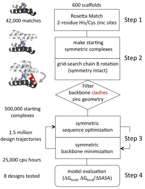
Figure 2.1. Zinc heterodimer computational design protocol...33
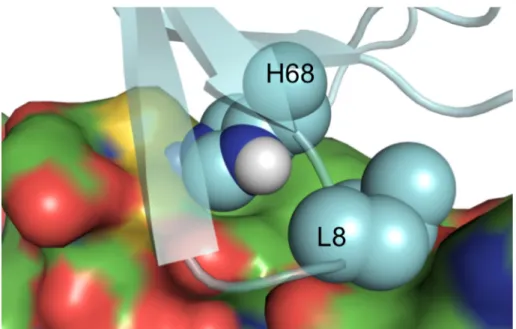
Figure 2.2. Zinc binding by a 2-Cys/1-His mutant, 2D4X-CCH...37
Figure 2.3. Binding affinities of Spelter and Spelter mutants for Bodipy-labeled ubiquitin measured by fluorescence polarization...39
Figure 2.4. NMR peak intensity and chemical shift changes of 15N- and 13C-labeled ubiquitin upon titration of zinc-bound Spelter...42
Figure 3.1. Computational model of MID1 (metal interface design 1). ...89
Figure 3.2. Flow chart of the protocol for the design of the symmetric metal-mediated interface...90
Figure 3.3. Biophysical characterization of MID1 metal binding. ...94
Figure 3.4. Binding orientation of apo1/2, zinc, MID1-cobalt...98
Figure 3.5. Comparison of the MID1-zinc model to crystal structures. ...101
Figure 3.6. Circular dichroism thermal denaturation of MID1, MID1-H12E, and MID1-H35E with and without zinc. ...102
Figure 3.7. Superimposition of four metal-bound MID1 crystal structures...104
Figure 4.1. A metal-mediated protein interface as a minimalist route to a new active site...132
Figure 4.2. Michaelis-Menten kinetics of MID1-zinc hydrolysis of 4NPA. ...137
Figure 4.4. Enzymatic hydrolysis of 4NPA by MID1-zinc requires
three-histidine-coordinated zinc with an open zinc coordination site...141
Figure 4.5. Crystallographic evidence for the catalytic mechanism. ...142
Figure 4.6. MID1-zinc catalyses 4NPP hydrolysis. ...143
Figure 5.1. Design concept and motivation for designing a zinc site in a
deeper native cleft. ...165
Figure 5.2. Zinc binding by ZE2...176
Figure 5.3. Hydrolysis of 4NPA by ZE2. ...178
Figure 5.4. Inaccuracy of the ZE2 computational model compared to the
crystal structure...179
Figure 5.5. Second shell hydrogen bonds to zinc-coordinating histidines
at native zinc sites. ...181
Figure 5.6. The second shell of cysteine residues...182
Figure 5.7. Computational design models featuring three-histidine zinc
LIST OF SUPPORTING FIGURES
Figure S2.1. Illustration of the geometric hashing algorithm in
RosettaMatch used to design a three-residue zinc binding sites on existing
protein scaffolds...47
Figure S2.2. Ideal geometry for zinc coordination by histidine and cysteine. ...48
Figure S2.4. Depictions of four experimentally tested designs of zinc-mediated ubiquitin binding proteins. ...49
Figure S2.5. Comparison of interaction motifs featuring a hydrophobic helix targeting the hydrophobic surface patch of ubiquitin. ...50
Figure S2.6. Zinc binding data for the Spelter design. ...51
Figure S2.7. The designed metal site is specific for zinc...52
Figure S2.8. Fluorescence polarization binding curves of point mutants...53
Figure S2.9. The 3-D NMR experiment HNCACB used to assign our construct of ubiquitin. ...54
Figure S2.10. Possible mechanism by which zinc improved Spelter binding to ubiquitin-H68A...55
Figure S2.11. The backside H68 delta nitrogen is partially buried without a hydrogen bond...56
Figure S2.12. Spelter binds ubiquitin with higher affinity than many naturally occurring ubiquitin-binding interactions. ...57
Figure S3.1. Ideal geometry for zinc coordination by histidine and cysteine. ...111
Figure S3.2. Ribbon diagrams of the eight experimentally tested designs. ...112
Figure S3.3. Size exclusion chromatography of 1YZM-WT, MID1-apo, and MID1-zinc provides an initial indication of dimer formation...113
Figure S3.4. NMR 1H15N HSQC of MID1-zinc. ...114
Figure S3.5. High-resolution crystal structure of MID1-apo1...115
Figure S3.7. High-resolution electron density reveals carboxylate-metal
interactions in the zinc- and cobalt-coordination spheres...116
Figure S3.8. Comparison of the MID1-zinc model to the MID1-cobalt
crystal structure...117
Figure S3.9. The position of phenylalanine at residue 42 deviates from
the computational model due to helix unwinding...117
Figure S3.10. Glutamate point mutations result in four-coordination of
zinc...118
Figure S3.11. QUILT analysis of hydrophobic patch size of the 1YZM
wild-type scaffold compared to the MID1 design. ...118
Figure S4.1. Crystal structure of MID1-zinc (PDB code 3V1C), a
computationally designed de novo zinc-mediated interface [22]. ...150
Figure S4.2. UV-Vis spectra monitor the MID1-zinc catalyzed hydrolysis
of 4NPA. ...151
Figure S4.3. MID1 is fully bound to zinc to form MID1-zinc at 5 µM
concentration...152
Figure S4.4. MID1-zinc catalysis of 4NPA is first-order in enzyme
concentration...153
Figure S4.5. Absorbance and catalytic activity correlate closely in
fractions collected by gel filtration. ...154
Figure S4.6. Indication that the MID1-zinc enzyme has one active site
instead of two...155
Figure S4.7. Brønsted analysis of MID1-zinc hydrolysis of aryl acetate
substrates...156
Figure S4.8. Proposed reaction coordinate for the mechanism of MID1-zinc catalysis of aryloxy acetate hydrolysis based on limited Brønsted
analysis data. ...157
Figure S5.1. Ideal geometries for zinc coordination determined by
statistical measurements of native zinc sites...188
Figure S5.2. Computational models of the three zinc site designs that
were experimentally tested. ...189
Figure S5.4. Electron density of the three histidines and succinate
coordinating zinc in the ZE2 crystal structure. ...191
Figure S5.5. Illustration of the primary shell, second shell, and third shell
from the ZE2 crystal structure. ...192
Figure S5.6. Residue preferences for second shell hydrogen bonding
interactions...193
LIST OF ABBREVIATIONS
MID1 metal interface design 1
ZE2 zinc esterase design 2
HHH three-histidine zinc coordination
CCH two-cysteine one-histidine zinc coordination
TS transition state
HisT target histidine
4NPA 4-nitrophenyl acetate
4NPP 4-nitrophenyl phosphate
CD circular dichroism
HSQC heteronuclear single quantum coherence
ITC isothermal titration calorimetry
CHAPTER 11
INTRODUCTION TO PROTEIN INTERFACE DESIGN AND ENZYME DESIGN
1.1 Protein interface design
Nature has provided a wealth of protein interfaces and enzyme active sites that
support life, and Nature’s binders and catalysts can be used for applications in medicine
and biotechnology. However, most protein drugs and enzymes, even if they are derived
from natural proteins, require some level of engineering to optimize activity,
pharmacokinetics, solubility, thermostability, specificity, and other properties. For
example, subunit interaction affinities were altered to generate fast-acting and
slow-acting variants of insulin for improved treatment of diabetic patients [1, 2]. More
ambitiously, new protein drugs and catalysts can be designed from scratch rather than by
incremental modification of native proteins. Working toward this long-term goal, I used
and developed computational methods to design zinc-binding sites as the basis for new
protein interfaces and new enzyme active sites.
Designed protein-protein interactions can have myriad applications, serving as
biosensors, biomaterials, competitive inhibitors, cell signaling molecules, and tools for
co-localization and cell targeting. Novel protein-protein interactions are primarily
1 Some content previously appeared in an article in Current Opinion in Structural Biology. The original
generated by rational design, directed evolution, and computational design. Rational or
intuitive design has a limited level of complexity and reproducibility, though rational
charge swaps can alter specificity and other rational mutations can disrupt undesired
interactions. Directed evolution is the most common route to new binders: widely used
library selection strategies such as phage display are effective for generating new
target-binding interactions using antibody scaffolds [3] and non-antibody scaffolds [4], and
binders identified with these approaches have shown promise in clinical trials [5].
Computational interface design is an alternative approach that is currently not as robust
as experimental selection and screening, but it is advantageous in certain design scenarios
and successes have emerged rapidly in recent years. Considering cases of computational
de novo interface design (excluding classical coiled-coils [6]), only three examples prior
to 2011 were reported [7-9], and these have weak affinity and/or lack crystallographic
validation. However, since 2011, more than ten published results with crystallographic
validation have emerged, some with high affinity [10-19]. The recent momentum in
computational interface design highlights the promise of this method to transition from
basic science to applied science, thus renewing motivation to innovate in this emerging
field.
1.1.1 Motivation for computational protein interface design
Computational design is advantageous over directed evolution for precise control
of binding location and binding mode, and for design goals without selection methods
such as homo-multimeric assemblies [11, 16, 17], arrays [15], fibril caps [20],
[14]. For example, with the goal of developing a protein-based inhibitor of the flu,
protein interactions can be engineered to target the hemagglutinin receptor on the viral
surface, which is required for viral entry. Most directed evolution approaches generate
binders for the large, glycosylated, variable head domain. Viruses can easily change the
composition of this head domain, so targeting the small, conserved stem region would
lead to a more effective and broadly neutralizing protein therapeutic. This goal was
recently accomplished using computational methods, and the protein engineered to bind
the conserved stem region could become a flu treatment [12] (Figure 1.1).
Figure 1.1. A computationally engineered protein binds the conserved stem region of influenza hemagglutinin as a potential flu treatment.
The variable head region (gray, left side) is typically targeted by directed evolution methods, but computational methods were used to generate a binder (red) that targets the conserved stem to more broadly inhibit flu entry.
Secondly, computational design is advantageous when generating
homo-multimeric assemblies. Homodimers and homo-multimers are very common in nature,
representing an estimated one-third of known proteins [22]; functional reasons for
catalysis at the subunit interface, membrane insertion, nucleic acid binding, and steric
protection from proteases (Figure 1.2). Computational methods have successfully generated homomultimers, including a homodimer [11], classical coiled-coils with three
to six subunits [23, 24], and protein cages with 12 or 24 subunits [16]. These design
goals would be challenging for directed evolution methods, which are better suited for
heterodimers.
Figure 1.2. Examples of homomultimer formation and function in nature.
1.1.2 Previous strategies for computational protein interface design
In most computational protein interface design studies, new protein interactions
have been created by mutating the amino acids on the surfaces of naturally occurring
proteins so that favorable interactions occur upon formation of the target complex. The
computational task involves iterating between sequence optimization and energy
evaluation. Despite continued improvements in conformational searching and energy
evaluation [25], limited conformational sampling and inaccurate energy evaluation are
major pitfalls for computational interface design. In particular, the design of interfaces
that make use of cooperative hydrogen bonding networks has been especially challenging
[26]. As a result, successful designs to date have relied on carefully chosen strategies in
which a desired high-probability binding mode limits the conformational search and is
somewhat robust to inaccuracies in energy calculations.
Classical coiled-coils are amenable to computational design due to the relative
simplicity of the scaffold and empirical sequence-structure relationships. The history of
coiled-coil design using computational methods dates back to 1993 [27], though the first
known classical coiled-coil hexamer was engineered recently [24]. Another early
strategy for protein interface design is ‘grafting’, reusing a known hotspot to generate a
new interacting partner [7, 18, 28]. Grafting has since matured into hotspot-based design,
in which novel hotspots anchor the new interacting pair [29]. For example, a tyrosine
residue can simultaneously form a hydrogen bond and hydrophobic interactions and was
successfully used as an engineered hotspot [10]. Beyond hotspot-based design, targeting
a hydrophobic groove with a hydrophobic helix is also a friendly design goal [9, 12, 13].
and knobs-into-holes side-chain interactions can confer some interaction specificity.
Design of hydrogen bonds will often be required for interaction specificity, but design of
new hydrogen bonds remains one of the major challenges in protein design. One proven
strategy uses beta-strand pairing at the interface for cooperative hydrogen bond formation
without entropic penalties associated with side-chain hydrogen bonds [11]. Coiled-coils,
grafting, hydrophobic grooves, and beta-strand pairs offer a limited set of strategies for
reliable protein interface design, so additional strategies must be developed (Figure 1.3).
Figure 1.3. Validated strategies for computational design of new protein-protein interactions.
1.1.3 My contributions to computational protein interface design
My work aims to design metal binding sites instead of hydrogen bonds at protein
interfaces to enhance orientation specificity and binding affinity. Metal coordination
geometry is well-defined [30, 31], coordination bonds are stronger than hydrogen bonds,
and metal binding sites were an early success in computational protein design [32, 33].
My first design goal was to generate a new binder for wild-type ubiquitin using an
interfacial zinc-binding site. Structural zinc sites feature four coordination bonds, so the
designed binder had three coordinating residues, and the surface histidine on ubiquitin
was intended as the fourth coordinating residue. Overall this design was successful
because the three designed residues bound zinc with high-affinity and the designed binder
targeted the intended surface of ubiquitin [34]. However, the zinc site did not contribute
much to ubiquitin binding affinity, and we concluded that a single zinc coordination bond
is not a robust hotspot due to a lack of cooperativity, or ‘chelate effect’.
To better capture the cooperative chelate effect in zinc binding, my next design
goal was a homodimer with two interface zinc sites [17]. Rather than one coordination
bond bridging the interface, this design model featured four coordination bonds bridging
the interface (two at each zinc site). The binding affinity for homodimerization was <30
nM, representing the highest reported affinity for a de novo interface designed only with
computation and without directed evolution. Hydrophobic interactions led to weak
affinity without zinc (4 µM), but zinc improved the binding affinity by >100-fold.
Furthermore, crystal structures validated the overall binding orientation and designed
structure at the atomic level [17]. Thus, computationally designed interface zinc sites
offer a new strategy for computational protein interface design.
1.2 Enzyme design
Novel enzyme active sites also have broad applications in medicine and
biotechnology. Due to their impressive rate accelerations and specificity under mild,
aqueous conditions, enzymes are used for ‘green’ chemistry synthesis of high-value
products, natural products, and small molecule drugs, as well as biofuel production and
bioremediation of polluted environmental sites.
1.2.1 Rational design and directed evolution of enzymes
As with protein interfaces, the available strategies for design of novel enzymes
include rational design, directed evolution, and computational design. Rational methods
have been used to synthesize small macrocycles that mimic metal coordination sites in
proteins [35-38], though these cannot desolvate the substrate and have limited potential
as enzyme-like catalysts. Rational design of catalytic peptides also received early
attention, but again these catalysts lack capacity for substrate binding and desolvation
[39-44]. A unique rational design study used a single aspartate mutation in a
hydrophobic pocket of calmodulin, which demonstrated Kemp elimination activity [45].
Directed evolution [46-49] has been the most common and most successful
method for enzyme design [50]; mainstream directed evolution methods combine library
generation methods (often error-prone PCR and site-saturation mutagenesis) with
including non-natural reactions [51]. Directed evolution is suited for optimization of
existing enzymes, rather than generating new enzymes de novo. The major exception is
directed evolution of catalytic antibodies, in which the non-catalytic antibody scaffold is
imparted with catalytic function by varying a small number of residues. More
specifically, the variable CDR loops of an antibody scaffold are varied in large libraries
and screened for binding of transition state analogues. Many successful catalytic
antibodies have been engineered in this way [52, 53], though product inhibition is a
common pitfall because high affinity often requires recognition of substrate components
far from the site of reaction [53].
1.2.2 Computational enzyme design
Computational enzyme design provides an alternative to directed evolution, but
given the proven effectiveness of directed evolution for enzyme design, why are
computational methods for enzyme design important to pursue? First, directed evolution
proceeds through incremental changes to substrate specificity and/or rate enhancement,
and additive accumulation of point mutations leads to large net changes in specificity or
rate [54]. However, for designing drastically different enzymes or de novo enzymes,
large changes in conformation or sequence are more accessible with computational
modeling. Secondly, effective selection schemes and screens are not always available
[55], so testing a smaller number of discrete designs is sometimes necessary instead of
screening large libraries. Thirdly, mutations generated by directed evolution can occur
predictive design offers a better route to rigorously test and improve our understanding of
enzymatic catalysis.
The Baker lab has developed an enzyme engineering workflow featuring
computational design followed by directed evolution. The computational step
reengineers natural enzyme active sites to perform a different type of reaction,
incorporating many simultaneous mutations that would be challenging to evolve.
Typically, 50 to 100 variants are individually tested to find one or two variants with
detectable activity, which is required for subsequent optimization by directed evolution.
This workflow has successfully generated retroaldolases [56, 57], a Kemp eliminase [58,
59], Diels Alderase [60], and organophosphatase [61], and a catalyst for carbon addition
by the Morita-Baylis-Hillman mechanism [62, 63].
1.2.3 My contributions to enzyme design
My work in computational enzyme design attempts to harness the catalytic power
of metal ions, specifically mononuclear zinc ions. To briefly summarize my
contributions, the previously described de novo zinc-mediated homodimer [17]
serendipitously functioned as a zinc hydrolase with 104 - 105 rate enhancements of model
carboxyester and phosphoester substrates [64]. Secondly, I designed a catalytic zinc
motif in the deep cleft of a TIM-barrel scaffold, though this design demonstrated an
unexpected conformational change upon zinc binding and had low activity. In ongoing
work, I am designing zinc sites containing two second-shell hydrogen bonds to support
a new metal center and new substrate binding residues, my efforts focus on designing
new three-histidine zinc sites and ignore target substrates.
My work in enzyme design, albeit in early stages of development, is motivated by
current limitations in the field of enzyme design. Regarding the serendipitous
zinc-mediated hydrolysis in a designed protein [64], this discovery puts into perspective the
moderate rates that have been achieved thus far in de novo enzyme design, and the
impressive rates achieved by some of Nature’s best enzymes [65, 66]. This discovery
also emphasizes the catalytic power of metal ions, the usefulness of protein interfaces for
active site formation, and suggests a plausible route for the natural evolution of
primordial enzymes. Secondly, computational design of new metal sites alone (structural
or catalytic) remains a difficult challenge, despite intermittent successes over the course
of ~25 years. Most reported successes over the years have lacked crystallographic
validation [32-34, 67-76], often because detection of activity was emphasized over
structural accuracy. While co-crystallization with a transition state analogue is very
challenging, crystallization of the ground state enzyme has also been rare in
metalloenzyme design. Thirdly, most computationally designed metalloenzymes are
oxidoreductases [39, 40, 77-81] containing iron, diiron, iron, copper, or
heme-copper metal sites, rather than mononuclear zinc sites that most commonly confer
hydrolytic activity. One exception is the computational conversion of a native deaminase
into an organophosphatase [61], but this work repurposed a native mononuclear zinc site
and only altered substrate-binding residues, whereas my work seeks to design new metal
centers. Likewise, directed evolution of metalloenzymes such as cytochrome P450
typically leaves the native metal center intact and alters substrate-binding residues [82,
83]. Fourthly, my work seeks to design second-shell hydrogen bonds that support the
metal coordinating residues. This phenomenon is well-known in natural proteins [84-90],
but these are often anecdotal accounts of individual metal sites. My work adds a more
comprehensive analysis of second-shell interactions among zinc-containing proteins in
the Protein Data Bank. Furthermore, despite awareness of second-shell effects,
computational design and crystallographic validation of metal sites supported by
second-shell hydrogen bonds remains an outstanding challenge.
In summary, my work in enzyme design started with a serendipitous discovery
but developed into a purposeful pursuit to design new catalytic zinc sites. My work aims
to add to the field of computational enzyme design by focusing on catalytic residues and
hydrolysis reactions, emphasizing second-shell hydrogen bond support of the metal
coordination site, and including crystallographic validation.
The following chapters present a designed heterodimer and designed homodimer
that establish zinc binding as a useful route to protein interface design, and they present
two de novo catalytic zinc sites with crystal structures that provide important lessons and
Figure 1.4. Illustrative summary of the dissertation chapters.
1.3 References
1. Phillips, N.B., et al., Supramolecular Protein Engineering: Design of zinc-stapled insulin hexamers as a long acting depot. Journal of Biological Chemistry, 2010. 285(16): p. 11755-11759.
2. Noble, S.L., E. Johnston, and B. Walton, Insulin lispro: A fast-acting insulin analog. American Family Physician, 1998. 57(2): p. 279-286.
3. Sidhu, S.S., Antibodies for all: The case for genome-wide affinity reagents. Febs Letters, 2012. 586(17): p. 2778-2779.
4. Gilbreth, R.N. and S. Koide, Structural insights for engineering binding proteins based on non-antibody scaffolds. Current Opinion in Structural Biology, 2012. 22(4): p. 413-420.
5. Wurch, T., A. Pierre, and S. Depil, Novel protein scaffolds as emerging therapeutic proteins: from discovery to clinical proof-of-concept. Trends in Biotechnology, 2012. 30(11): p. 575-582.
6. Woolfson, D.N., The design of coiled-coil structures and assemblies. Fibrous Proteins: Coiled-Coils, Collagen and Elastomers, 2005. 70: p. 79-112.
7. Liu, S., et al., Nonnatural protein-protein interaction-pair design by key residues grafting. Proceedings of the National Academy of Sciences of the United States of America, 2007. 104(13): p. 5330-5335.
8. Huang, P.S., J.J. Love, and S.L. Mayo, A de novo designed protein protein interface. Protein Sci, 2007. 16(12): p. 2770-4.
9. Jha, R.K., et al., Computational Design of a PAK1 Binding Protein. Journal of Molecular Biology, 2010. 400(2): p. 257-270.
10. Karanicolas, J., et al., A De Novo Protein Binding Pair By Computational Design and Directed Evolution. Molecular Cell, 2011. 42(2): p. 250-260.
11. Stranges, P.B., et al., Computational design of a symmetric homodimer using beta-strand assembly. Proceedings of the National Academy of Sciences of the United States of America, 2011. 108(51): p. 20562-20567.
12. Fleishman, S.J., et al., Computational Design of Proteins Targeting the Conserved Stem Region of Influenza Hemagglutinin. Science, 2011. 332(6031): p. 816-821.
13. Sammond, D.W., et al., Computational Design of the Sequence and Structure of a Protein-Binding Peptide. Journal of the American Chemical Society, 2011. 133(12): p. 4190-4192.
15. Lanci, C.J., et al., Computational design of a protein crystal. Proceedings of the National Academy of Sciences of the United States of America, 2012. 109(19): p. 7304-7309.
16. King, N.P., et al., Computational Design of Self-Assembling Protein Nanomaterials with Atomic Level Accuracy. Science, 2012. 336(6085): p. 1171-1174.
17. Der, B.S., et al., Metal-Mediated Affinity and Orientation Specificity in a Computationally Designed Protein Homodimer. Journal of the American Chemical Society, 2012. 134(1): p. 375-385.
18. Azoitei, M.L., et al., Computational Design of High-Affinity Epitope Scaffolds by Backbone Grafting of a Linear Epitope. Journal of Molecular Biology, 2012. 415(1): p. 175-192.
19. Procko, E., et al., Computational design of a protein-based enzyme inhibitor. J Mol Biol, 2013. 425(18): p. 3563-75.
20. Sievers, S.A., et al., Structure-based design of non-natural amino-acid inhibitors of amyloid fibril formation. Nature, 2011. 475(7354): p. 96-U117.
21. Humphris, E.L. and T. Kortemme, Design of multi-specificity in protein interfaces. Plos Computational Biology, 2007. 3(8): p. 1591-1604.
22. Ali, M.H. and B. Imperiali, Protein oligomerization: How and why. Bioorganic & Medicinal Chemistry, 2005. 13(17): p. 5013-5020.
23. Ogihara, N.L., et al., The crystal structure of the designed trimeric coiled coil coil-V(a)L(d): Implications for engineering crystals and supramolecular assemblies. Protein Science, 1997. 6(1): p. 80-88.
24. Zaccai, N.R., et al., A de novo peptide hexamer with a mutable channel. Nature Chemical Biology, 2011. 7(12): p. 935-941.
25. Leaver-Fay, A., et al., Scientific Benchmarks for Guiding Macromolecular Energy Function Improvement. Methods in Protein Design, 2013. 523: p. 109-143.
26. Stranges, P.B. and B. Kuhlman, A comparison of successful and failed protein interface designs highlights the challenges of designing buried hydrogen bonds. Protein Science, 2013. 22(1): p. 74-82.
27. Oshea, E.K., K.J. Lumb, and P.S. Kim, Peptide Velcro - Design of a Heterodimeric Coiled-Coil. Current Biology, 1993. 3(10): p. 658-667.
29. Fleishman, S.J., et al., Hotspot-centric de novo design of protein binders. J Mol Biol, 2011. 413(5): p. 1047-62.
30. Auld, D.S., Zinc coordination sphere in biochemical zinc sites. Biometals, 2001. 14(3-4): p. 271-313.
31. Lee, Y.M. and C. Lim, Physical basis of structural and catalytic Zn-binding sites in proteins. J Mol Biol, 2008. 379(3): p. 545-53.
32. Regan, L. and N.D. Clarke, A Tetrahedral Zinc(II)-Binding Site Introduced into a Designed Protein. Biochemistry, 1990. 29(49): p. 10878-10883.
33. Klemba, M., et al., Novel Metal-Binding Proteins by Design. Nature Structural Biology, 1995. 2(5): p. 368-373.
34. Der, B.S., et al., Combined computational design of a zinc-binding site and a protein-protein interaction: One open zinc coordination site was not a robust hotspot for de novo ubiquitin binding. Proteins-Structure Function and Bioinformatics, 2013. 81(7): p. 1245-1255.
35. Kimura, E., et al., A Zinc(Ii) Complex of 1,5,9-Triazacyclododecane ([12]Anen3) as a Model for Carbonic-Anhydrase. Journal of the American Chemical Society, 1990. 112(15): p. 5805-5811.
36. Kimura, E., et al., Carboxyester Hydrolysis Promoted by a New Zinc(Ii) Macrocyclic Triamine Complex with an Alkoxide Pendant - a Model Study for the Serine Alkoxide Nucleophile in Zinc Enzymes. Journal of the American Chemical Society, 1994. 116(11): p. 4764-4771.
37. Kou, F.P., et al., A Zinc(II) Complex of Tripyridylamine(TPA) as a Model for Carbonic Anhydrase. Acta Physico-Chimica Sinica, 1996. 12(9): p. 804-808.
38. Konig, B., et al., 1,4,7,10-Tetraazacyclododecane metal complexes as potent promoters of carboxyester hydrolysis under physiological conditions. Inorganic Chemistry, 2007. 46(10): p. 4336-4356.
39. Benson, D.E., M.S. Wisz, and H.W. Hellinga, Rational design of nascent metalloenzymes. Proceedings of the National Academy of Sciences of the United States of America, 2000. 97(12): p. 6292-6297.
40. Nanda, V. and R.L. Koder, Designing artificial enzymes by intuition and computation. Nature Chemistry, 2010. 2(1): p. 15-24.
41. Broo, K.S., et al., Catalysis of hydrolysis and transesterification reactions of p-nitrophenyl esters by a designed helix-loop-helix dimer. Journal of the American Chemical Society, 1997. 119(47): p. 11362-11372.
nucleophilic and general-acid catalysis. Chemistry-a European Journal, 2000. 6(12): p. 2214-2220.
43. Rossi, P., et al., De novo metallonucleases based on helix-loop-helix motifs. Chemistry-a European Journal, 2004. 10(17): p. 4163-4170.
44. Davie, E.A.C., et al., Asymmetric catalysis mediated by synthetic peptides. Chemical Reviews, 2007. 107(12): p. 5759-5812.
45. Korendovych, I.V., et al., Design of a switchable eliminase. Proceedings of the National Academy of Sciences of the United States of America, 2011. 108(17): p. 6823-6827.
46. Stemmer, W.P.C., Rapid Evolution of a Protein in-Vitro by DNA Shuffling. Nature, 1994. 370(6488): p. 389-391.
47. Arnold, F.H., Directed evolution: Creating biocatalysts for the future. Chemical Engineering Science, 1996. 51(23): p. 5091-5102.
48. Campbell, J.H., J.A. Lengyel, and J. Langridge, Evolution of a second gene for beta-galactosidase in Escherichia coli. Proc Natl Acad Sci U S A, 1973. 70(6): p. 1841-5.
49. Arnold, F.H., Design by directed evolution. Accounts of Chemical Research, 1998. 31(3): p. 125-131.
50. Turner, N.J., Directed evolution of enzymes for applied biocatalysis. Trends in Biotechnology, 2003. 21(11): p. 474-478.
51. Brustad, E.M. and F.H. Arnold, Optimizing non-natural protein function with directed evolution. Current Opinion in Chemical Biology, 2011. 15(2): p. 201-210.
52. Wentworth, P. and K.D. Janda, Catalytic antibodies - Structure and function. Cell Biochemistry and Biophysics, 2001. 35(1): p. 63-87.
53. Hilvert, D., Critical analysis of antibody catalysis. Annual Review of Biochemistry, 2000. 69: p. 751-793.
54. Tracewell, C.A. and F.H. Arnold, Directed enzyme evolution: climbing fitness peaks one amino acid at a time. Current Opinion in Chemical Biology, 2009. 13(1): p. 3-9.
55. Leemhuis, H., R.M. Kelly, and L. Dijkhuizen, Directed Evolution of Enzymes: Library Screening Strategies. IUBMB Life, 2009. 61(3): p. 222-228.
56. Jiang, L., et al., De novo computational design of retro-aldol enzymes. Science, 2008. 319(5868): p. 1387-1391.
58. Rothlisberger, D., et al., Kemp elimination catalysts by computational enzyme design. Nature, 2008. 453(7192): p. 190-U4.
59. Khersonsky, O., et al., Bridging the gaps in design methodologies by evolutionary optimization of the stability and proficiency of designed Kemp eliminase KE59. Proceedings of the National Academy of Sciences of the United States of America, 2012. 109(26): p. 10358-10363.
60. Siegel, J.B., et al., Computational Design of an Enzyme Catalyst for a Stereoselective Bimolecular Diels-Alder Reaction. Science, 2010. 329(5989): p. 309-313.
61. Khare, S.D., et al., Computational redesign of a mononuclear zinc metalloenzyme for organophosphate hydrolysis. Nature Chemical Biology, 2012. 8(3): p. 294-300.
62. Bjelic, S., et al., Computational Design of Enone-Binding Proteins with Catalytic Activity for the Morita-Baylis-Hillman Reaction. Acs Chemical Biology, 2013. 8(4): p. 749-757.
63. Nivon, L., et al., Computational Design of enone-binding proteins with catalytic activity for the Morita-Baylis-Hillman reaction. Protein Science, 2012. 21: p. 178-178.
64. Der, B.S., D.R. Edwards, and B. Kuhlman, Catalysis by a De Novo Zinc-Mediated Protein Interface: Implications for Natural Enzyme Evolution and Rational Enzyme Engineering. Biochemistry, 2012. 51(18): p. 3933-3940.
65. Miller, B.G., T.W. Traut, and R. Wolfenden, A role for zinc in OMP decarboxylase, an unusually proficient enzyme. Journal of the American Chemical Society, 1998. 120(11): p. 2666-2667.
66. Radzicka, A. and R. Wolfenden, A Proficient Enzyme. Science, 1995. 267(5194): p. 90-93.
67. Handel, T. and W.F. Degrado, Denovo Design of a Zn-2+-Binding Protein. Journal of the American Chemical Society, 1990. 112(18): p. 6710-6711.
68. Hellinga, H.W. and F.M. Richards, Construction of New Ligand-Binding Sites in Proteins of Known Structure .1. Computer-Aided Modeling of Sites with Predefined Geometry. Journal of Molecular Biology, 1991. 222(3): p. 763-785.
69. Pessi, A., et al., A Designed Metal-Binding Protein with a Novel Fold. Nature, 1993. 362(6418): p. 367-369.
71. Benson, D.E., A.E. Haddy, and H.W. Hellinga, Converting a maltose receptor into a nascent binuclear copper oxygenase by computational design. Biochemistry, 2002. 41(9): p. 3262-3269.
72. Dwyer, M.A., L.L. Looger, and H.W. Hellinga, Computational design of a Zn2+ receptor that controls bacterial gene expression. Proceedings of the National Academy of Sciences of the United States of America, 2003. 100(20): p. 11255-11260.
73. Yang, W., et al., Design of a calcium-binding protein with desired structure in a cell adhesion molecule. Journal of the American Chemical Society, 2005. 127(7): p. 2085-2093.
74. Ambroggio, X.I. and B. Kuhlman, Computational design of a single amino acid sequence that can switch between two distinct protein folds. Journal of the American Chemical Society, 2006. 128(4): p. 1154-1161.
75. Grzyb, J., et al., De novo design of a non-natural fold for an iron-sulfur protein: Alpha-helical coiled-coil with a four-iron four-sulfur cluster binding site in its central core. Biochimica Et Biophysica Acta-Bioenergetics, 2010. 1797(3): p. 406-413.
76. Zhu, C., et al., Engineering a zinc binding site into the de novo designed protein DS119 with a beta alpha beta structure. Protein & Cell, 2011. 2(12): p. 1006-1013.
77. Sigman, J.A., B.C. Kwok, and Y. Lu, From myoglobin to heme-copper oxidase: Design and engineering of a Cu-B center into sperm whale myoglobin. Journal of the American Chemical Society, 2000. 122(34): p. 8192-8196.
78. Kaplan, J. and W.F. DeGrado, De novo design of catalytic proteins. Proceedings of the National Academy of Sciences of the United States of America, 2004. 101(32): p. 11566-11570.
79. Nanda, V., et al., De novo design of a redox-active minimal rubredoxin mimic. Journal of the American Chemical Society, 2005. 127(16): p. 5804-5805.
80. Faiella, M., et al., An artificial di-iron oxo-protein with phenol oxidase activity. Nature Chemical Biology, 2009. 5(12): p. 882-884.
81. Yeung, N., et al., Rational design of a structural and functional nitric oxide reductase. Nature, 2009. 462(7276): p. 1079-U144.
82. Romero, P.A. and F.H. Arnold, Exploring protein fitness landscapes by directed evolution. Nature Reviews Molecular Cell Biology, 2009. 10(12): p. 866-876.
84. Kiefer, L.L., S.A. Paterno, and C.A. Fierke, Hydrogen-Bond Network in the Metal-Binding Site of Carbonic-Anhydrase Enhances Zinc Affinity and Catalytic Efficiency. Journal of the American Chemical Society, 1995. 117(26): p. 6831-6837.
85. Christianson, D.W. and R.S. Alexander, Carboxylate Histidine Zinc Interactions in Protein-Structure and Function. Journal of the American Chemical Society, 1989. 111(16): p. 6412-6419.
86. Lesburg, C.A. and D.W. Christianson, X-Ray Crystallographic Studies of Engineered Hydrogen-Bond Networks in a Protein-Zinc Binding-Site. Journal of the American Chemical Society, 1995. 117(26): p. 6838-6844.
87. Lin, I.J., et al., Changes in hydrogen-bond strengths explain reduction potentials in 10 rubredoxin variants. Proceedings of the National Academy of Sciences of the United States of America, 2005. 102(41): p. 14581-14586.
88. Stone, E.M., L. Chantranupong, and G. Georgiou, The Second-Shell Metal Ligands of Human Arginase Affect Coordination of the Nucleophile and Substrate. Biochemistry, 2010. 49(49): p. 10582-10588.
89. Karlin, S., Z.Y. Zhu, and K.D. Karlin, The extended environment of mononuclear metal centers in protein structures. Proceedings of the National Academy of Sciences of the United States of America, 1997. 94(26): p. 14225-14230.
CHAPTER 22
COMBINED COMPUTATIONAL DESIGN OF A ZINC BINDING SITE AND A PROTEIN-PROTEIN INTERACTIONS: ONE OPEN ZINC COORDINATION SITE WAS NOT A ROBUST HOTSPOT FOR DE NOVO UBIQUITIN BINDING
2.1 Overview
We computationally designed a de novo protein-protein interaction between wild-type ubiquitin and a redesigned scaffold. Our strategy was to incorporate zinc at the
designed interface to promote affinity and orientation specificity. A large set of
monomeric scaffold surfaces were computationally engineered with three-residue zinc
coordination sites, and the ubiquitin residue H68 was docked to the open coordination
site to complete a tetrahedral zinc site. This single coordination bond was intended as a
hotspot and polar interaction for ubiquitin binding, and surrounding residues on the
scaffold were optimized primarily as hydrophobic residues using a rotamer-based
sequence design protocol in Rosetta. From thousands of independent design simulations,
four sequences were selected for experimental characterization. The best performing
design, called Spelter, binds tightly to zinc (Kd < 10 nM) and binds ubiquitin with a Kd of
2 This chapter previously appeared as an article in the journal of PROTEINS: Structure Function and
20 µM in the presence of zinc and 68 µM in the absence of zinc. Mutagenesis studies
and NMR chemical shift perturbation experiments indicate that Spelter interacts with
H68 and the target surface on ubiquitin, however, H68 does not form a hotspot as
intended. Instead, mutation of H68 to alanine results in tighter binding. While a 3/1 zinc
coordination arrangement at an interface cannot be ruled out as a means to improve
affinity, our study led us to conclude that 2/2 coordination arrangements or multiple-zinc
designs are more likely to promote high-affinity protein interactions.
2.2 Introduction
Understanding the physical basis of protein-protein interaction is a continued
pursuit in molecular biology. A ground-up approach for understanding protein binding
will help clarify mechanisms of cellular functions and lead to new therapeutic and
diagnostic uses of proteins in medicine. Studies of natural interactions have provided
valuable insights into how proteins interact, from detailed dissection of individual
binding partners such as barnase and barstar [1-3], to broad studies of hundreds of
complexes [4-9]. Although much research has been aimed at studying protein
interactions observed in nature, a complementary approach is to rationally design and
build new interactions [10].
Redesigning existing interactions for improved affinity or altered specificity is a
test of our understanding is to design new protein-protein interactions from scratch. De novo computational interface design is still a young endeavor but has already seen a number of successes. Many of these studies strategically use pre-existing knowledge of
patterns of recognition by using sequence profiles [14], augmenting a native complex [15,
16], using known binding grooves [17-19], side-chain interaction motifs [19-21], or
backbone interaction motifs (strand-strand pairing, linear epitopes, glycine helix-helix
contact, helix stacking) [21-24]. Karanicolas et al. used ankyrin repeat protein as a known versatile binding protein for design, but they ambitiously avoided using
pre-existing interaction motifs already observed in natural protein-protein interactions [25].
Although efforts in computational interface design have been encouraging, there
is a significant need for improvement for reliable computational engineering of new
interactions. Broad conclusions cannot be reliably drawn from a small number of
attempts in de novo interface design, so continued efforts that explore different approaches and different modes of interaction will be critical in accumulating deeper
knowledge about the physical basis of protein-protein interactions [26]. One repeated
lesson from protein-protein interaction studies is that a few hotspot residues dominate the
binding event [27, 28] – hotspot-based approaches have been used to design new
interfaces, and these hotspots can be grafted from natural interfaces [19, 20, 29] and
developed from scratch [19, 30]. Here we designed a three-residue zinc site from scratch
where the open coordination site was the intended hotspot for target protein binding.
Computational methods have been used to design new tetrahedral zinc binding
sites [31, 32], and zinc sites have previously been shown to promote affinity and
self-assembly, metal-mediated binding modes have been determined empirically but not
predicted rationally [33-35]. In our previous homodimer design, two metal sites were
used to promote binding in the desired orientation [36]. In this work, our one-zinc
approach is for a heterodimeric interaction with a wild-type target.
We chose ubiquitin as a target because it has one surface histidine that may
participate in zinc binding and because it is a small, stable protein that has been
structurally characterized by crystallography and NMR. We observed a moderate
binding affinity between wild-type ubiquitin and our redesigned scaffold named Spelter,
where the presence of zinc resulted in 3-fold increase in affinity (Kd = 20 µM in the
presence of zinc, Kd = 68 µM in the absence of zinc). Despite successful zinc binding in
the redesigned scaffold (Kd < 10 nM), we conclude that this engineered zinc site did not
provide a robust hotspot for target binding. Moderate affinity in a one-sided de novo
interface design is a significant achievement for computation-only protein interface
design. However, micromolar affinity has thus far been an affinity barrier for successful
small hydrophobic designs, and designing polar interface contacts from scratch remains a
significant challenge in protein interface design.
2.3 Methods
2.3.1 Scaffolds and target for protein interface design
A set of 635 scaffold proteins from the Protein Data Bank [37] were used in the
these scaffolds to be listed as monomeric, expressible in E. coli, <2.5 Å resolution, without disulfides, without ligands, and between 80-250 residues. The list of scaffolds
can be obtained from the Supporting Material of our previous work [36].
2.3.2 RosettaMatch
These 635 scaffolds were used in RosettaMatch to search for possible
three-residue histidine/cysteine zinc binding sites on the scaffold surfaces. RosettaMatch is a
protocol typically used in enzyme design [38, 39] within the Rosetta modeling suite [40].
It uses a transition state model (TS) to search for designable residue sets on a scaffold
protein that might stabilize the TS and hence catalyze the reaction. In our case, the
pseudo-TS consisted of a HisT (histidine from ‘target’) positioned at an optimal
coordination distance and orientation with a zinc atom consistent with the geometry of
zinc coordination.
Distance, angle, and torsion constraints were used to correctly build the HisT
downstream of all possible histidine or cysteine rotamers at each allowed residue
position. Each HisT placement was followed by clash detection between the HisT and the
scaffold backbone, and the clash-free HisT locations were recorded in a 6-dimensional bin
(3 dimensions for Cartesian position, 3 dimensions for rotational orientation) (Figure S2.1). If HisT locations from three different residues were hashed into the same 6-D bin,
this was a match.
After matching, a geometry-based evaluation of all output matches was performed
to select the matches with coordination bond lengths, angles, and dihedrals that were
Rosetta. Among 635 scaffolds, ~2,000 high-quality tetrahedral three-residue zinc
matches were identified. Half of these matches, however, featured cysteine residues at i,
i+1 positions. We excluded these matches from the final list of 1,015 because we did not identify the Cys/Cys at i, i+1 as a naturally occurring zinc coordination motif. The Rosetta 2.3 version of RosettaMatch was used for this study, however, the current version
of Rosetta (3.4) contains an updated implementation of the RosettaMatch algorithm.
Input files and command lines for the equivalent Rosetta3 implementation of
RosettaMatch are also given in the Supporting Material.
2.3.3 ZincHeterodimerDesign
ZincHeterodimerDesign is the name of the Rosetta protocol that was written for
the protein interface design stage. Reorganization of the Rosetta code into discrete
protocols simplified the addition of new design protocols [40]. The required inputs for
ZincHeterodimerDesign are the atomic coordinates of a scaffold protein, a three-residue
zinc match from the scaffold, and ubiquitin. Other command-line options are given in the
Supporting Information. In Step 1 of the protocol, the zinc match atomic coordinates
were grafted onto the scaffold. In Step 2, the HisT transition state residue was replaced
by the H68 residue of ubiquitin, which docked the ubiquitin to the scaffold with a zinc
coordination site bridging the interface. This pseudo-docking step ignored protein
complementarity, so in Step 3, a Monte Carlo rigid-body search was performed to relieve
steric clashes between the scaffold and ubiquitin while preserving the zinc binding site.
The zinc binding site was conserved by limiting the degrees of freedom to rotation about
angles were limited to rotamers from the 2002 Dunbrack library used in Rosetta [41, 42],
and “inverse rotamer” sampling moved the ubiquitin and kept the imidazole ring fixed in
space. In this rigid-body search, both scaffold and ubiquitin side chains were scored as
centroids to evaluate overall shape complementarity and ignore clashes between side
chains that could be redesigned. These centroid protein representations were
kinematically coupled to the full-atom representations required to explore the torsional
rotations of the H68 side chain. The lowest-energy centroid docking arrangement was
chosen from the Monte Carlo search, and Step 4 of the protocol was full-atom design of
the scaffold interface residues and repacking of the ubiquitin interface residues. Interface
residues were identified as those within 10 Å of the other partner based on Cβ-Cβ
distances, and this design step used the standard fixed-backbone rotamer packing
functionality in Rosetta. In summary, this protocol used zinc binding and emphasized
docking and side-chain degrees of freedom but did not include backbone sampling. The
best models were chosen based on computed binding energy per Å2 of interface surface
area, followed by visual inspection.
2.3.4 Cloning, Expression, Purification, Mutagenesis
We synthesized genes of the four designs, the wild-type scaffold (PDB code
2D4X, the bacterial flageller hook-filament junction protein), and wild-type ubiquitin by
oligo-assembly [43]. The genes were cloned into the pQE-80L vector (Qiagen)
supplemented with an N-terminal MBP (maltose binding protein) fusion to aid expression
and solubility. Ubiquitin with a G76C mutation for fluorescence polarization
were performed by overlapping PCR using internal primers encoding the desired
mutations. These proteins were expressed in BL21(DE3)pLysS (Invitrogen) cells with
induction using 333 µM IPTG for six hours at 25 oC. The 6xHis-MBP-design fusions
were purified by Ni2+-NTA high-affinity chromatography (HisTrap columns, GE
Healthcare Biosciences). The eluent was supplemented with 1 mM DTT to prevent
disulfide formation and 0.5 mM EDTA to scavenge metal ions. The 6xHis-MBP tag was
cleaved by overnight incubation by TEV protease. For ubiquitin G76C, the His-tag was
cleaved by overnight thrombin proteolysis. The proteolyzed samples were dialyzed back
into HisTrap column buffer, and a second HisTrap purification removed the 6xHis-MBP
tag or uncleaved protein. The flow-through containing the desired protein was again
supplemented with 1 mM DTT and 0.5 mM EDTA before concentration to <4 ml for gel
filtration using column Superdex 75 Hiload 16/60 (Amersham Biosciences). The final
protein buffer was 20 mM MOPS, pH 6.9, 50 mM NaCl, 0.5 mM TCEP (thiol-free
reducing agent).
2.3.5 Isothermal Titration Calorimetry
Isothermal titration calorimetry experiments to measure zinc binding were
performed using a MicroCal VP-ITC (GE Healthcare). 2.3 ml of 20 µM protein was
loaded into the sample chamber, and 250 µM ZnSO4 injectant was diluted from a high
concentration stock using the protein dialysis buffer. 29 titrations of 10 µl volume were
performed with 150 seconds equilibration, and the resulting titration curves were fit using
2.3.6 Circular Dichroism
Thermal denaturation experiments were performed using circular dichroism with
a JASCO J-815 CD spectrometer. Temperature was ramped at 1 oC/min under the
control of a JASCO Peltier device and water bath. Protein concentration for the
2D4X-variants was 15 µM in a 1-mm quartz cuvette, and for experiments containing zinc,
ZnSO4 was added to 16.5 µM (protein to metal ratio of 1 to 1.1). Protein unfolding was
monitored at wavelength 222 nm with units converted to mean molar ellipticity to
provide apparent melting temperatures as a measure of thermostability.
2.3.7 Fluorescence Polarization
Wild-type ubiquitin was altered with a G76C mutation for covalent attachment of
thiol-reactive Bodipy (507/545, Molecular Probes). Bodipy conjugation was performed
as previously described [17], and labeling efficiency of 20% was observed. Fluorescence
polarization binding assays were performed using a SPEX FluoroLog-3 instrument. The
180 µl starting sample in a 3x3-mm quartz cuvette contained 5 µM ubiquitin (1 µM
fluorescently labeled ubiquitin). Slits of 2.5 nm were used with excitation/emission
wavelengths of 508/545 nm. Each polarization reading had 0.1 s integration time, and
readings were taken in triplicate. Plots of polarization versus concentration of titrant
were modeled with a single-site binding equation with nonlinear fitting using SigmaPlot
2.3.8 NMR: HNCACB backbone assignment
Uniformly labeled 15N- and 13C- ubiquitin was expressed using minimal media as
described in our previous work [36] and was purified as described above. Ubiquitin
concentration was 1.1 mM, and the sample buffer contained 20 mM MOPS, pH 6.9, 50
mM NaCl, 0.5 mM TCEP, and 10% D2O. To assign sequence-specific resonances of
1H
N, 15N, 13Cα, and 13Cβ, we performed the three-dimensional HNCACB experiment
[44]. Data collection proceeded for 70 hours on a Varian INOVA 700 MHz spectrometer
with a cold probe. Data were processed using the NMRPipe/NMRDraw software [45],
and assignments were made using the Sparky software [46]. A subset of strips showing
peak quartets spanning residues 63-73 and a list of chemical shift values are given in the
Supporting Material.
2.3.9 NMR: 15N- and 13C-HSQC
2D [15N, 1H]- and [13C, 1H]-HSQC spectra were recorded at 25 oC on a Varian
INOVA 700 MHz spectrometer with a cold probe. Titration of Spelter with equimolar
zinc was performed to monitor chemical shift perturbations upon binding. Data were
processed using the NMRPipe/NMRDraw software [45], and peak-picking was
performed using the NMRViewJ software [47]. Peaks in the 15N-HSQC spectra were
assigned based on chemical shift values from our HNCACB 3-D backbone assignment.
Peaks in the 13C-HSQC were assigned using CA and CB chemical shift values from our
HNCACB experiment combined with published values for proton, CG, CD, and CE
values [48]. Compound changes in chemical shift (Δδcomp) were measured as distances
atom – for example, two peaks for a CB side-chain atom in histidine – these measured
distances were averaged. Due to peak broadening upon formation of a complex larger
than 30 kDa, we compared chemical shifts from the ubiquitin-only spectrum with a
limited titration in which Spelter was present at a 30% molar ratio with ubiquitin. Our
limited titration along with intermediate exchange resulted in small changes in chemical
shift (Eq. S1), so we also analyzed changes in peak intensities as a second method to determine the ubiquitin residues at the Spelter binding interface. Peak intensities were
calculated using the NMRViewJ software [47].
€
Δδcomp=
(
Δδproton)
2
+
(
Δδheavy/Rscale)
2
15N:R
scale=6.5
13C:R
scale=5.4
Eq. 1
2.4 Results
2.4.1 Choosing a target and scaffold for interface design
We aimed to bind a wild-type target protein using a computationally redesigned
scaffold protein where one metal coordination group is supplied by the target protein and
the other three coordinating groups are supplied by the redesigned scaffold (Figure 2.1A). This 3/1 metal coordination arrangement seemed attractive for designing a heterodimeric interaction with a wild-type target because many target surfaces will have
one or more metal-coordinating side chains (histidine, aspartate, glutamate, cysteine).
The criterion for a good target protein was a surface histidine for metal binding near