i
VIRAL CHARACTERISTICS ASSOCIATED WITH HUMAN
IMMUNODEFICIENCY VIRUS TYPE 1 INFECTION OF THE CENTRAL
NERVOUS SYSTEM EARLY FOLLOWING TRANSMISSION
Christa Buckheit Sturdevant
A dissertation submitted to the faculty of the University of North Carolina at Chapel Hill in partial fulfillment of the requirements for the degree of Doctor of Philosophy in the
Department of Microbiology and Immunology (Virology).
Chapel Hill 2013
Approved by: Ronald Swanstrom Blossom Damania Kristina Abel
Mark Heise
Annelies Van Rie
© 2013
Christa Buckheit Sturdevant ALL RIGHTS RESERVED
ABSTRACT
Christa Buckheit Sturdevant: Viral Characteristics Associated with Human
Immunodeficiency Virus Type 1 Infection of the Central Nervous System Early Following Transmission
(Under the direction of Dr. Ronald Swanstrom)
Human immunodeficiency virus type 1 (HIV-‐‑1) infection of the central nervous system (CNS) can occur shortly after transmission and compartmentalized HIV-‐‑1 variants, genetically distinct from virus in the blood, can be detected in the cerebrospinal fluid (CSF) in some individuals throughout the course of infection. HIV-‐‑1 replication can induce neuropathogenesis and may also represent a distinct reservoir from that found in CD4+ T cells in the blood and lymphoid tissue. We do not know the time course of the virologic events that lead to neurological dysfunction and the potential establishment of a CNS reservoir, or the extent to which these long-‐‑term outcomes are predicted by the initial aspects of virus-‐‑host interaction. In this dissertation, I examined the viral genetic and phenotypic determinants associated with CNS compartmentalization early following both vertical and horizontal transmission. Additionally, I examined CNS compartmentalization over the full course of disease in simian immunodeficiency virus (SIV)-‐‑infected rhesus macaques.
predominantly associated with a longer time since HIV-‐‑1 infection. Compartmentalization was also shown to persist and evolve over a period of up to two years in the adult cohort. Through the use of Bayesian Evolutionary Analysis by Sampling Trees (BEAST), I identified and examined multiple variant transmission events and defined two distinct pathways to compartmentalization: the early sequestration of a transmitted virus in the CNS; and the later establishment of an independently replicating population within the CNS that originated from the periphery. I also concluded via entry tropism analyses that in most children and adults, replication in the CNS is sustained by growth in CD4+ T cells, while in some young children the virus in the CNS evolves to replicate in cells with low CD4 surface expression, potentially macrophages and/or microglia. The observation of significant
To my husband Dan, thank you for always being there for me and for your unwavering love, encouragement, and support. I could not have done this without you.
ACKNOWLEDGEMENTS
The studies presented in this dissertation were carried out under the supervision of Dr. Ronald Swanstrom. I am so thankful to have had the opportunity to work under such a wonderful mentor and am grateful for his guidance, encouragement, and patience during my graduate career. I also want to thank all of my thesis committee members, Blossom Damania, Kristina Abel, Mark Heise, and Annelies Van Rie, for providing invaluable scientific guidance and discussions for the work presented in this dissertation.
To my collaborators, Dr. Annelies Van Rie, Dr. Richard W. Price, and Dr. Serena Spudich, I am extremely thankful for the opportunity to work on such exciting and
impactful projects. These studies would have not been possible without your expertise and the clinical samples that you provided. Thank you for your scientific guidance, support and critical input on manuscripts. I also want to thank Dr. Swanstrom, Dr. Van Rie, Dr.
Spudich, and the UNC Virology Training Grant for providing funding for these studies. In addition, for the Malawi pediatric study (Chapter II), we thank the parents and infants for their participation. We also thank the Malawi study coordinator, Chawanangwa Mahebere Chirambo, and study nurse, Edith Kafoteka. We thank Richard Price for helpful discussion. For the adult primary infection study (Chapter III), we thank the staff at the UCSF Options study and Magnet for referral of participants, Julia Peterson and Evelyn Lee for
participation. We also thank Sarah B. Joseph for helpful discussion and assistance with the Affinofile assay.
Finally, to the Swanstrom Lab, thank you for making these years at UNC so
memorable. I especially want to thank Sarah Joseph for all of her help with experiments and the numerous fruitful discussions, and Maria, Marc, and Lauren for all of the pranks and laughs – you made it fun to come into work every day.
TABLE OF CONTENTS
ABSTRACT ... iii
LIST OF TABLES ... xiv
LIST OF FIGURES ... xv
CHAPTER I. INTRODUCTION ... 17
HIV-‐‑1 BACKGROUND ... 17
Genome organization and virion structure ... 17
HIV-‐‑1 envelope binding and fusion ... 18
HIV-‐‑1 replication strategy ... 19
HIV-‐‑1 DISEASE PROGRESSION: ACUTE INFECTION THROUGH END-‐‑STAGE AIDS ... 21
HIV-‐‑1 PATHOGENESIS: OTHER AGENTS AND OPPORTUNISTIC INFECTIONS ... 23
The earliest signs of immunodeficiency ... 23
HIV-‐‑1 and cancer ... 25
Opportunistic infections of the CNS ... 26
HIV/HCV co-‐‑infection ... 28
HIV-‐‑1 EFFECTS ON VARIOUS ORGAN SYSTEMS ... 29
HIV-‐‑1 and the immune system ... 29
HIV-‐‑1 and the gut-‐‑associated lymphoid tissue ... 31
HIV-‐‑1-‐‑associated nephropathy ... 32
The blood-‐‑brain barrier ... 33
HIV-‐‑1 CNS infection: timing and mechanisms of entry ... 34
CNS infection of other neurovirulent viruses ... 35
CNS COMPLICATIONS ASSOCIATED WITH HIV-‐‑1 INFECTION ... 37
HIV-‐‑1-‐‑associated dementia ... 37
HIV-‐‑1 encephalitis ... 38
Mild-‐‑to-‐‑moderate HIV-‐‑1-‐‑associated neurocognitive disorders ... 39
HIV-‐‑1 neurological complications in children ... 40
CNS inflammatory response and CNS injury ... 41
HIV-‐‑1 COMPARTMENTALIZATION ... 43
HIV-‐‑1 CNS compartmentalization is observed throughout the course of infection ... 43
Compartmentalization versus clonal amplification ... 44
HIV-‐‑1 compartmentalization in other biological compartments ... 45
Compartmentalization and SIV ... 46
CNS compartmentalization: T cell versus macrophage tropism ... 47
SIGNIFICANCE AND OBJECTIVES ... 48
CHAPTER II. CENTRAL NERVOUS SYSTEM COMPARTMENTALIZATION OF HIV-‐‑1 SUBTYPE C VARIANTS EARLY AND LATE IN INFECTION IN YOUNG CHILDREN ... 50
OBJECTIVE ... 50
AUTHOR SUMMARY ... 52
RESULTS ... 56
Study design ... 56
Relationship of viral populations in blood and CSF ... 57
Compartmentalization is significantly related to older age and a higher CSF/blood viral load ratio ... 58
Evolutionary history of viral populations within the first three years of life ... 59
Evidence of multiple transmitted variants ... 60
Evolution of CSF viruses to infect cells with low levels of CD4 surface expression ... 61
DISCUSSION ... 63
MATERIALS AND METHODS ... 68
Study subject population ... 68
Single genome amplification ... 68
Phylogenetic analysis of env viral sequences ... 69
Bayesian analysis ... 70
Construction of HIV-‐‑1 env clones ... 70
Cells ... 71
Env-‐‑pseudotyped viruses ... 71
293-‐‑Affinofile cellular surface xxpression of CD4 and CCR5 ... 72
Single-‐‑cycle infection of 293-‐‑Affinofile cells ... 72
CHAPTER III. COMPARTMENTALIZED REPLICATION OF R5 T CELL-‐‑TROPIC HIV-‐‑1 IN THE CENTRAL NERVOUS
SYSTEM EARLY IN THE COURSE OF INFECTION ... 92
OBJECTIVE ... 92
AUTHOR SUMMARY ... 94
INTRODUCTION ... 95
RESULTS ... 98
Study population and analysis parameters ... 98
Approximately two-‐‑thirds of subjects have low CSF viral RNA concentrations during the first two years of infection ... 100
Pleocytosis can be associated with elevated viral load in the CSF and an influx of virus from the blood ... 101
Compartmentalized viral populations early are associated with higher CSF viral loads, and local replication and/or inflammation can persist or reoccur over time ... 103
Genetic evidence for the persistence of HIV-‐‑1 CNS replication ... 104
Timing of introduction of HIV-‐‑1 into the CNS ... 106
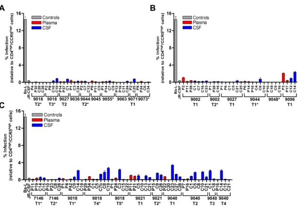
All transmitted variants are R5 T cell-‐‑tropic and are predominantly selected to use high levels of CD4 for entry ... 107
DISCUSSION ... 109
MATERIALS AND METHODS ... 115
Ethics statement ... 115
Study design ... 115
Single genome amplification ... 116
Bayesian analysis ... 117
Construction of HIV-‐‑1 env clones ... 118
Cells ... 118
Env-‐‑pseudotyped viruses ... 118
293-‐‑Affinofile cellular surface expression of CD4 and CCR5 ... 119
Single-‐‑cycle infection of 293-‐‑Affinofile cells ... 119
Statistical analysis ... 120
CHAPTER IV. COMPARTMENTALIZED SIMIAN IMMUNODEFICIENCY VIRUS POPULATIONS OBSERVED IN THE CEREBROSPINAL FLUID OF RHESUS MACAQUES EXAMINED OVER THE FULL COURSE OF INFECTION ... 135
OBJECTIVE ... 135
INTRODUCTION ... 137
RESULTS ... 140
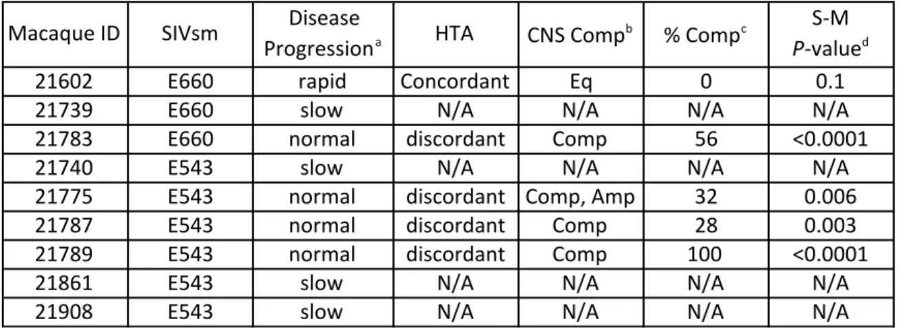
Study design ... 140
Detection of discordant blood and CSF viral populations within macaques using HTA ... 141
Compartmentalized CSF populations observed in SIVsm E660 and E543 infected macaques ... 143
DISCUSSION ... 145
MATERIALS AND METHODS ... 150
Viruses, animals, and specimen collection ... 150
Viral RNA isolation ... 150
Single genome amplification ... 151
Phylogenetic analysis of env viral sequences ... 152
CHAPTER V. DISCUSSION AND FUTURE DIRECTIONS ... 160
REFERENCES ... 177
LIST OF TABLES
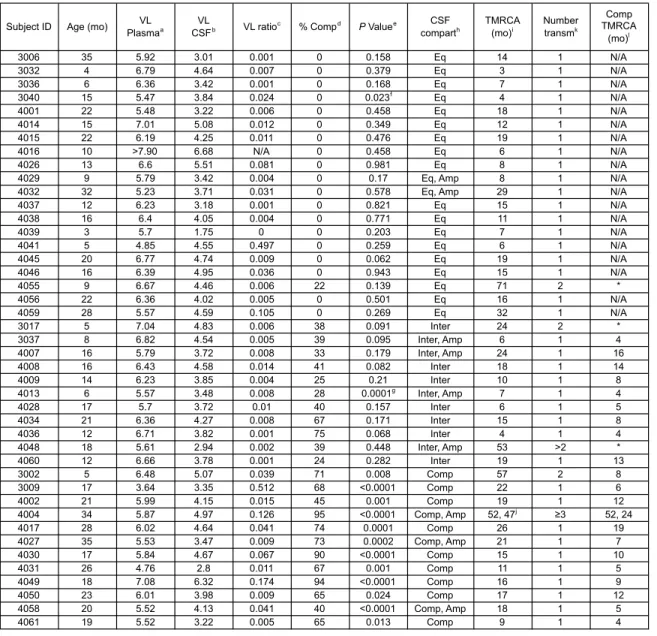
Table 1: Subject population virologic and phylogenetic characteristics ... 74
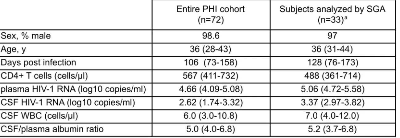
Table 2: Background demographic and clinical characteristics of study participants at baseline ... 121
Table 3: Subject population virologic, clinical and phylogenetic characteristics ... 122
Table 4: Virologic and clinical characteristics for subjects not analyzed by SGA ... 124
Table 5: Summary of HTA and SGA analysis ... 153
LIST OF FIGURES
Figure 1: HIV-‐‑1 blood and CSF populations in infected children can be well equilibrated, intermediate or compartmentalized ... 76
Figure 2: Older age and a higher CSF/blood viral load ratio are strong
determining factors for compartmentalization ... 77
Figure 3: No relationship between viral load and subject classification ... 78
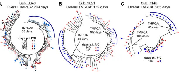
Figure 4: Evolutionary history of HIV-‐‑1 populations within the first three years of pediatric CNS infection ... 79
Figure 5: Evidence of multiple transmission events during pediatric HIV-‐‑1 subtype C infection ... 80
Figure 6: Compartmentalized subject 4004 exhibiting three or more
transmitted viruses ... 82
Figure 7: Intermediate subject 3017 exhibiting two transmitted viruses ... 84
Figure 8: Intermediate subject 4048 exhibiting greater than two
transmitted viruses ... 86
Figure 9: Equilibrated subject 4055 exhibiting two transmitted viruses ... 88
Figure 10: Compartmentalized HIV-‐‑1 CSF populations in children can have a low CD4 entry phenotype ... 90
Figure 11: No contamination was observed between subjects ... 91
Figure 12: HIV-‐‑1 blood and CSF populations early during infection can be equilibrated, intermediate or compartmentalized ... 126
Figure 13: Correlates of CSF HIV-‐‑1 RNA levels, CNS inflammation, and CNS phylogenetic state ... 127
Figure 14: An assessment of the viral populations in the two compartments
Figure 15: Compartmentalization can persist and evolve independently within the CSF over time ... 131
Figure 16: All viruses required high levels of CD4 for entry, indicative of
selection for replication in CD4+ T cells ... 132
Figure 17: Four states can define the relationship between virus in the
CSF/CNS and blood early during infection ... 133
Figure 18: Plasma viral RNA copies/mL indicate various types of disease
progression ... 154
Figure 19: HTA analysis over the full course of infection shows discordant viral populations ... 155
Figure 20: Equilibrated and compartmentalized populations observed
SIVsmE660 infection ... 157
Figure 21: Significant compartmentalization observed in SIVsm E543 infections ... 158
CHAPTER I. INTRODUCTION
HIV-1 BACKGROUND
Genome organization and virion structure
Human immunodeficiency virus type 1 (HIV-‐‑1) is a member of the Lentivirus genus within the Retroviridae family. HIV-‐‑1 is classified into four groups (M, N, O and P), each representing independent cross-‐‑species transmission events. The M group, the first to be discovered and encompassing 95% of the circulation viral strains worldwide, is further divided into nine subtypes (A, D, C, D, F, G, H, J, and K). Subtype C, the most dominant subtype worldwide, in predominant in Africa, while subtype B isolates are the most prevalent in Europe and North America (101).
The full-‐‑length dimeric RNA genome associates with the NC protein through a short packaging signal located within the 5’ end of the unspliced RNA. The NC-‐‑RNA complex is surrounded by the CA protein, which is arranged into a conical core that also contains several copies of viral protease (PR), IN, and RT enzymes required for productive infection. The conical core of CA is surrounded by a spherical shell composed of the matrix (MA) protein, which is embedded in the viral envelope, a lipid bilayer gained during budding from the infected cell, that contains the Env protein associated in trimers, as well as various cellular proteins (101, 115).
HIV-1 envelope binding and fusion
variable domains evolve rapidly in response to antibody selection and shield the conserved domains (321).
Env first binds the cellular surface receptor CD4 (67), a member of the
immunoglobulin family that is highly expressed on CD4+ T cells and expressed at low levels on macrophages (64, 169), followed by binding with the chemokine coreceptors CCR5 (56, 75) or CXCR4 (93). The binding of HIV-‐‑1 Env protein determines the cellular tropism of HIV-‐‑1 variants. Attachment factors such as heparin sulfate proteoglycans, α4β7 integrin, or dendritic cell-‐‑specific intercellular adhesion molecule 3-‐‑grabbing non-‐‑integrin (DC-‐‑SIGN) help bring Env into close proximity to CD4, although these factors are not essential for entry (336). Within the gp120 monomer, the CD4 binding site is located within a deep binding pocket that is likely protected from antibodies by glycosylated variable loops (143, 165). The sequential receptor/coreceptor binding induces a conformation change that exposes the hydrophobic gp41 N-‐‑terminal fusion peptide, promoting its insertion into the target membrane and forming the stable gp41 six-‐‑helix bundle that mediates viral-‐‑cell membrane fusion (52, 103, 203, 333).
HIV-1 replication strategy
reaction preformed by the viral RT. HIV-‐‑1 RT contains two crucial domains; a DNA polymerase responsible for incorporation of deoxynucleotides onto a nucleotide template, and an RNase H activity responsible for degradation of the RNA strand of the RNA:DNA replication intermediate. HIV-‐‑1 is genetically very heterogeneous due to the error prone viral DNA polymerase and the high recombination frequency observed during reverse transcription when a single cell is infected by multiple virions. The linear DNA is then integrated into the cellular DNA via the actions of the viral IN, most often into areas of the cellular genome experiencing active gene expression (288). Integration into host cells enables the virus to persist in the infected cell and form latent reservoirs when infected cells become dormant.
Integration of the provirus signals the transition from the early to the late stages of HIV-‐‑1 replication. The synthesis of viral RNA from the integrated viral DNA, performed by the cellular RNA polymerase II, generates RNA transcripts. The viral Tat protein binds the transactivation response region (TAR), a bulged hairpin structure on the 5’ end of the nascent viral RNA, and recruits host proteins that enhance the ability of RNA polymerase II to elongate down the genome with high processivity (115, 173, 186). RNA transcripts are exported from the nucleus into the cytoplasm for translation or packaging into the virion particle. RNA export is mediated by HIV-‐‑1 Rev, which binds the Rev-‐‑responsive elements (RRE) and recruits Crm1, a cellular nuclear export factor, to the viral RNA (65, 115). The
further processed/cleaved during and after viral assembly and budding by the HIV-‐‑1 PR (156).
The HIV-‐‑1 accessory proteins serve various functions in HIV-‐‑1 replication. Nef downregulates the surface expression of CD4 receptor (105) and the major
histocompatibility complex (MHC) class I (62) , facilitating virus release and inhibiting the cytotoxic T lymphocyte (CTL)-‐‑mediated lysis of HIV-‐‑1 infected cell, respectively. The Vpr protein, packaged within virion particles, facilitates the import of the preintegration complex into the nucleus in nondividing cells (245) and can induce cell cycle arrest (155). HIV-‐‑1 Vpu enhances virus production, mediates the degradation of CD4 by the ubiquitin-‐‑ conjugating pathway, and antagonizes tetherin (215, 289, 314, 338). Lastly, Vif neutralizes the potent antiviral activity of APOBEC3G, found within nonpermissive cells including human T lymphocytes, enhancing the infectivity of HIV-‐‑1 (187, 295, 344).
HIV-1 DISEASE PROGRESSION: ACUTE INFECTION THROUGH END-STAGE
AIDS
Viral sequences during the earliest stages of horizontal infection are typically highly homogenous, representing productive clinical infection by one variant (3, 158, 273).
Following transmission, there is an initial eclipse phase, up to ten days long, during which time infection is localized to the exposure site (172) and systemic circulation has not yet occurred.
Acute HIV-‐‑1 infection (AHI) is associated with an exponential increase and then subsequent decline in HIV-‐‑1 RNA in the plasma (61, 201). Peak plasma HIV-‐‑1 viremia is reached between fourteen to twenty-‐‑one days post infection (175) and levels above one million RNA copies/mL can be measured. During this time, individuals have a robust inflammatory response, characterized by a “cytokine storm” (310), and can develop
symptoms of HIV-‐‑1 acute retroviral syndrome, such as fever, rash, severe fatigue, headache, and diarrhea. HIV-‐‑1 replicates rapidly and spreads throughout the body, resulting in a drastic decline in CD4+ T cells, particularly in the gut-‐‑associated lymphoid tissue (GALT) (34, 194).
Several weeks post infection the HIV-‐‑1 viral load begins to decrease in response to virus-‐‑specific CD8+ CTLs (32, 229) and a partial rebound of CD4+ T cell counts occurs. Patients progress from AHI to early chronic infection as the plasma viral load begins to plateau and a viral set point is obtained (142, 281). The acute and early phases of infection are referred to as primary HIV-‐‑1 infection (PHI) (66, 174, 343).
Control and Prevention (CDC), HIV-‐‑1 infected individuals are clinically diagnosed with AIDS when CD4+ T cells counts drop below 200 cells/µμl, compared to approximately 1,000 cells/µμl in healthy individuals, or when one or more AIDS-‐‑defining illness (opportunistic infection) is recorded. Early symptomatic conditions of immunodeficiency include candidiasis and shingles, while more severe AIDS-‐‑defining conditions include HIV-‐‑1-‐‑ associated dementia (HAD), cryptococcosis, pneumonia caused by Pneumocystis jiroveci
(PCP), tuberculosis, Toxoplasma gondii infection of the brain, Kaposi’s sarcoma, and wasting syndrome. Eventually, HIV-‐‑1 infected individuals succumb to the opportunistic infections caused by severe immunodeficiency.
HIV-1 PATHOGENESIS: OTHER AGENTS AND OPPORTUNISTIC INFECTIONS
The earliest signs of immunodeficiency
Mucosal infections including Candida albicans and Pneumocystis jiroveci pneumonia (PCP) are often early signs of immunodeficiency. Candida species are normal inhabitants of the human gastrointestinal tract and can be recovered from the mouth in up to one-‐‑third of normal individuals and two-‐‑third of HIV-‐‑1 infected individuals. Mucocutaneous
bacterial flora also play a role (196, 197). Candida albicans may also disseminate to other organs, including the CNS where it can result in other complications such as meningitis.
Pneumocystis jiroveci pneumonia (PCP) of the lungs is a presenting feature of
previously undiagnosed HIV-‐‑1 infection in up to 85% of infected individuals. A ubiquitous fungus, PCP rarely causes disease in the absence of underlying conditions, such as
immunosuppression. While the incidence of PCP has decreased in the HAART era (227), PCP remains a major concern for HIV-‐‑1 infected individuals who do not know their infection status or are not on/cannot tolerate therapy (157). PCP infection has been
hypothesized to result from reactivation of latent infection or via direct exposure from the environment or an infected individual (206). While not widely studied, polymorphisms in the gene encoding the Fc segment of IgG have been predicted to influence the risk of PCP (98). Previous PCP, oral candidiasis, and persistent fevers are also risk factors for PCP.
Herpes simplex virus 2 (HSV-‐‑2) infection is also a significant opportunistic infection in individuals with HIV-‐‑1, resulting in HIV-‐‑1/HSV-‐‑2 coinfections in up to 90% of HIV-‐‑1 infected individuals (246). Primarily transmitted via sexual contact and frequently
HIV-‐‑1 often occurs at mucosal surfaces, prior or co-‐‑infection with HSV-‐‑2 is a major health concern for HIV-‐‑1 infected individuals.
HIV-1 and cancer
Cancers will occur in approximately 40% of individuals with HIV-‐‑1 infection, often associated with infection with other pathogenic viruses. The connection between HIV-‐‑1 and malignancies is likely the result of immune suppression, which leads to reduced immune surveillance and an inability to control viral replication or the growth of transformed calls, as well as due to a disrupted balance of cytokine levels (170). Additionally, immune suppression seems to be a driving factor in HIV-‐‑1 associated malignancies, as cancers developing in the presence of HIV-‐‑1 are much more aggressive than those found in uninfected patients (181).
Kaposi’s Sarcoma (KS) is a well-‐‑demarcated angioproliferative multifocal
with the rare occurrence of body cavity lymphomas in AIDS patients (50). Studies have suggested that various cytokines and growth factors, including IL-‐‑1β, IL-‐‑6, TNF-‐‑α, and GM-‐‑ CSF, as well as the HIV-‐‑1 Tat gene, are involved in the development of KS (92).
HIV-‐‑1 infection is associated with an increased susceptibility to B-‐‑cell lymphomas and anogenital carcinomas. HIV-‐‑1 infection increases the risk of developing B-‐‑cell
lymphomas over 60-‐‑fold compared to the general population (24). Infected individuals heterozygous for the CCR5 Δ32 deletion have been shown to have a reduced risk of
developing B-‐‑cell lymphomas (71, 249) while subjects with SDF-‐‑1 polymorphisms may have increased susceptibility (170). EBV is the causative agent in approximately 40-‐‑60% of these lymphomas outside of the brain and in virtually all CNS lymphomas (182, 296, 301). EBV can induce B-‐‑cell proliferation and enhance production of B-‐‑cell cytokines (i.e. IL-‐‑6 and IL-‐‑ 10), starting the process of cellular transformation to lymphomas. Anal and cervical
carcinomas are also AIDS-‐‑defining conditions. In approximately 90% of such cancers, HPV promotes carcinoma development (347) through suppression of cellular proteins that control cell division (p53 tumor suppression protein and retinoblastoma gene product) by viral proteins E6 and E7 (170).
Opportunistic infections of the CNS
ART, are vulnerable to neurological opportunistic infections as a result of persistent immune suppression and maintained HIV-‐‑1 CNS replication. Viruses, bacteria, fungi, and parasites can cause CNS opportunistic infections.
CMV, a DNA virus of the herpesvirus family, can cause AIDS-‐‑associated CMV encephalitis associated with confusion, disorientation, apathy, and withdrawal, and is difficult to distinguish from HAD. Between 40-‐‑100% of the general population is infected with CMV and reactivation of endogenous CMV accounts for much of the CMV-‐‑associated diseases in immunocompromised individuals (124, 200). Acquired by close contact at mucosal surfaces, CMV is spread via infected circulating leukocytes and has a wide tropism for neural cells (200). While less common, HSV infection can also cause encephalitis, often concurrent with a CMV infection (25). PML is a demyelinating disease that results from lytic infection of oligodendrocytes with the John Cunningham (JC) virus, a DNA
polyomavirus. The incidence of the normally rare PML increased 50-‐‑fold with the HIV-‐‑1 epidemic and lesions can be found throughout the brain (21, 255). Patients with PML experience weakness, gait abnormalities, speech disorders, visual effects, and cognitive abnormalities (36). The natural disease course is progressive and leads to death within months if a patient remains immunocompromised (21).
functions such as resist phagocytosis, inhibit neutrophil migration, deplete complement, dysregulate cytokine production, and interfere with antigen presentation (20). Patients with cryptococcal meningitis typically experience headache, fever, nausea, occasionally vision and behavior changes as well as an elevation of intercranial pressure (ICP). The parasite
Toxoplasma gondii, acquired through ingestion of undercooked meats or items contaminated by cat excrement (253), is also a common cause of various neurological diseases, including encephalitis, in AIDS patients (37). Gamma interferon (IFN-‐‑γ)-‐‑dependent cell-‐‑mediated immunity is important in combating T. gondii infection as it activates CNS cells that suppress the parasite through production of oxygen free radicals and reactive nitrogen intermediates (145).
HIV/HCV co-infection
hepatocellular carcinoma. Similar to HIV-‐‑1, HCV is characterized by high genetic
heterogeneity, contributing to the development of variants capable of evading immune and therapeutic defenses.
HCV and HIV-‐‑1 appear to do more than coexist within dually infected individuals. For example, mother-‐‑to child-‐‑transmission (MTCT) of HCV is more common in mothers infected with both HIV-‐‑1 and HCV, as opposed to only HCV (120, 188) and HIV-‐‑1 infection accelerates the development of severe liver disease (106, 256). As a result of the additive effects of HIV/HCV dual infection, end-‐‑stage liver disease now accounts for nearly half of the deaths among HIV-‐‑positive patients (307). HCV infection has also been shown to affect the CNS, with HCV RNA detected in the cerebrospinal fluid (CSF) of both HIV-‐‑positive and HIV-‐‑negative patients (183, 207). Additionally, HCV infection has been shown to contribute to HIV-‐‑1-‐‑associated minor cognitive/motor disorders and HIV-‐‑1-‐‑associated dementia
complex (265).
HIV-1 EFFECTS ON VARIOUS ORGAN SYSTEMS
HIV-1 and the immune system
Macrophage abnormalities primarily include reduced chemotaxis and antigen-‐‑presenting function, which results in reduced lymphocyte proliferation (170). Some studies suggest that HIV-‐‑1 infection reduces phagocytosis in macrophages, but the small number of these cells infected prevents detailed analysis. HIV-‐‑1 can also induce damage to precursor cells or stromal cells in the thymus, reducing the production of new lymphocytes (108). There is also a reduction in number and cytotoxic activity of NK cells and decreased chemokine response and phagocytic activity of neutrophils (170).
HIV-‐‑1 viral proteins have been shown to have detrimental effects on the immune response. The cytotoxic Env protein gp120 can increase apoptosis of T cells and alter cytokine levels, including inhibiting IL-‐‑2 and thereby T cell proliferation and regulation (216, 226). HIV-‐‑1 gp120 also induces IL-‐‑10 production by monocytes, resulting in anergy in human peripheral blood lymphocytes (286). The viral protein Gag also inhibits T cell function through binding cyclophilins (180). Nef downregulates the major
histocompatibility complex (MHC) class I levels from the surface of T cells and APCs, thereby inhibiting the CTL-‐‑mediated lysis of HIV-‐‑1 infected cell (62, 290). Tat induces apoptosis of T cells, B cell depletion, as well as reduces mannose receptor expression on APCs crucial for phagocytosis and antigens delivery to the MHC class II pathway (47).
HIV-1 and the gut-associated lymphoid tissue
The GALT is the largest lymphoid tissue in the human body, containing over 50% of all lymphoid cells (34). Approximately 20% of CD4+ T cells in the GALT are infected during acute HIV-‐‑1 and SIV infection (194, 328), attributed to their high coreceptor expression levels and predominantly active state (12). Additionally, up to 60% of uninfected CD4+ T cells die by apoptosis (107), resulting in an overall depletion of approximately 80% of CD4+ T cells in the GALT. This decrease coincides with an increase in CD8+ T cells, a
dysregulation in T cell homeostasis that results in increased inflammatory cytokine levels, inflammation and chronic immune activation (198, 276). Additional CD4+ T cells are then recruited from the periphery into the GALT, providing new targets for HIV-‐‑1 replication, and contributing to the maintenance of a viral reservoir. While CD4+ T cell counts in the blood rebound following peak viremia (201), there is a significant delay in the restoration of CD4+ T cell levels in the GALT (34, 125).
despite therapy, the sustained local inflammation and damage, and the extensive CD4+ T cell killing pose major challenges for complete mucosal immune restoration and eradication of viral reservoirs.
HIV-1-associated nephropathy
HIV-‐‑1 associated nephropathy (HIVAN) is one of the major causes of end-‐‑stage renal disease (ESRD) in HIV-‐‑1 infected individuals (202). HIV-‐‑1 infects resident renal epithelial cells (REC), which may constitute a distinct viral compartment, as viral RNA can be detected in REC despite suppression in the plasma (39), and phylogenetic analysis
comparing HIV-‐‑1 sequences from REC and peripheral blood mononuclear cells (PBMC) in patients with HIVAN found evidence for independent viral evolution within the kidney (191). However, RECs do not express the HIV-‐‑1 surface receptor CD4 or co-‐‑receptors CCR5 or CXCR4; therefore, the mechanism of entry of HIV-‐‑1 into REC remains perplexing. One study proposed that HIV-‐‑1 RNA is transferred to RECs in a CD4 and Env independent manor via virological synapses, requiring the attachment receptor heparin sulfate proteoglycans (55).
proliferation and differentiation of podocytes within the kidney (146, 147) through activation of the Stat3 and Ras-‐‑c-‐‑Raf-‐‑MAPK 1, 2 pathways (133) and a disruption of the RhoA/Rac1 balance (179). Several studies have also suggested roles of NFκB and oxidant stress in HIV-‐‑1-‐‑induced podocyte injury (202). Vpr has been shown to induce dysregulation of cytokinesis and apoptosis (261), a characteristic of HIVAN, via increased expression of FAT10 (303) and facilitation of Bax-‐‑mediated mitochondrial injury and apoptosis (304).
HIV-1 NEUROINVASION
The blood-brain barrier
Brain microvascular endothelial cells (BMVEC) within the CNS form the blood-‐‑brain barrier (BBB), a selectively permeable barrier that regulates brain homeostasis and restricts the movement of molecules, ions, and pathogens between the blood and the brain. The BBB, formed during embryogenesis, is composed of and regulated by interactions between
BMVEC, pericytes, and astrocytes along the capillaries in the brain (204). Pericytes, vascular support cells associated with BMVEC as nascent vessels are generated, are critical during BBB development (13, 70). Astrocyte foot processes, which surround BMVEC, have been shown to regulate BBB permeability through the secretion of compounds that alter tight and adherin junction proteins (1, 91). Molecules such as MCP-‐‑1 (311), TNF-‐‑α (100),
Substances cross the BBB through several mechanisms, many of which can be exploited by HIV-‐‑1 for entry into the CNS. Serum proteins, such as albumin, can enter the brain at a low rate through specific points within the BBB termed extracellular pathways (16, 35). Substances can also enter the brain when the BBB is disrupted, for example by trauma, hypertension, infection, and cytokine damage (178, 195). Transportation via endocytosis through clathrin-‐‑coated vesicles (absorptive endocytosis) as well as simple membrane diffusion are also possible mechanisms for crossing the BBB (17). Additionally, immune cells cross the BBB through diapedesis. (315).
HIV-1 CNS infection: timing and mechanisms of entry
HIV-‐‑1 appears in the CNS shortly after transmission, with the detection of HIV-‐‑1 in both the CSF and brain tissue during the weeks after initial exposure (70, 89, 238, 284, 308, 318), although it is not clear if the virus detected is actually replicating in the CNS. Several different mechanisms have been proposed for HIV-‐‑1 CNS entry. Two theories hypothesize infection of BMVEC (90), the first by direct infection and the second via the trancytosis of HIV-‐‑1 by BMVEC, with the subsequent infection of additional parenchyma cells or release of free virus into the CNS (18, 231, 292). However, these two theories are controversial as HIV-‐‑1 infection of BMVEC in vitro is highly restrictive and non-‐‑productive (208, 242, 291).
adhesion molecules, such as ICAM-‐‑1 and VCAM-‐‑1, is upregulated on the surface of BMVEC (87, 123, 277). As leukocytes roll along the endothelial surface, they are tethered via the interactions between BMVEC adhesion molecules and corresponding ligands on their surface, such as LFA-‐‑1 (219). This tethering is then followed by diapediesis (90). Leukocyte recruitment and transmigration into the CNS and BBB permeability is enhanced via the expression of chemoattractant cytokines and chemokines, including MCP-‐‑1 (102, 332), MIP-‐‑ 1α, MIP-‐‑1β (33, 282), TNF-‐‑α and IL-‐‑1β (220). It has also been suggested that HIV-‐‑1 infected cells can enter the CNS via migration across the choroid plexus, and on rare occasions, via migration from the peripheral blood to the subarachnoid space (45).
CNS infection of other neurovirulent viruses
The CNS is particularly vulnerable to viral infection due to the longevity of CNS cells and the immune-‐‑privileged nature of the CNS. There are five important cell types critical for proper CNS function, all of which can be infected by neurotropic viruses. Astrocytes, the most abundant cell in the CNS, execute numerous essential functions including managing healthy synaptic transmission, maintaining BBB function, protecting neurons from injury and removing toxic materials (305). Microglia, the brain-‐‑resident macrophages, play an important role in local immune response, while perivascular
motor functions. Oligodendrocytes generate and maintain the myelin sheath surrounding the axons of neurons, helping to support efficient transmission of electrical signals between neurons (123).
Neurotropic viruses can differ in their tropism, route of entry, and mechanism of spread. Human herpesvirus 6 and JC virus primarily target oligodenrocytes, while retroviruses such as HIV-‐‑1 and Human T lymphotropic virus 1 primarily infect
microglia/macrophages and astrocytes, respectively. Viruses such as rabies virus (RV), West Nile virus (WNV), poliovirus, adenovirus and cytomegalovirus (CMV), which infect
neurons, are more likely to cause fatal diseases as these cells are long-‐‑lived and terminally differentiated. Some neurotropic viruses, such as RV spread from neuron to neuron via axonal transport (95). Other neurotropic viruses, such as poliovirus, invade the CNS by directly infecting BMVECs within the BBB (63), while measles virus and lentiviruses enter the CNS via diapedesis of infected leukocytes (99, 128).
The clinical manifestations and disease severity resulting from CNS infection can also vary between neurotropic viruses. For example, RV infection of neurons and the
resulting neuronal impairment is 100% lethal in the absence of early treatment (95) and PML caused by JC virus is progressive and can lead to death within months if a patient remains immunocompromised (21). Alternatively, mumps has a high neuroinvasiveness but neurological disease is mild.
CNS COMPLICATIONS ASSOCIATED WITH HIV-1 INFECTION
HIV-1-associated dementia
The consequences of HIV-‐‑1 infection of the CNS have been well documented since the beginning of the epidemic (213, 214, 302). The spectrum of HIV-‐‑1-‐‑associated
neurocognitive disorders (HAND) spans from minor HIV-‐‑1-‐‑associated minor cognitive-‐‑ motor disorders (MCMD) to the more severe HAD (31, 116, 117). Clinically, HAD is
characterized by motor deficits (leg weakness, unsteady gait, tremor, and loss of fine-‐‑motor coordination), cognitive impairment (mental slowness, forgetfulness and poor
concentration), and behavior abnormalities (apathy, lethargy, withdrawal, diminished emotional responses, and sometime psychosis (214)), significantly impairing an individual’s ability to execute daily functions. Histological observation of brain tissue from patients with HAD revealed that less than 10% of the brains were normal, most revealing abnormalities including perivascular infiltrates of lymphocytes and macrophages as well as
multinucleated giant cells (40, 117).
Prior to HAART, 20-‐‑30% of HIV-‐‑1 infected individuals developed dementia: with the introduction of HAART, this number has been reduced to approximately 10% of infected adults (116, 268). The insufficient CNS penetration of some antiretroviral drugs, potentially enabling HIV-‐‑1 to persist within the CNS throughout infection despite therapy, may
susceptibility to HAD (26, 149). While HAD is most common in severely immune-‐‑
compromised individuals, individuals can be diagnosed with dementia prior to the onset of AIDS: in approximately 3% of infected individuals, dementia is an AIDS-‐‑defining illness (121).
HIV-1 encephalitis
In some HIV-‐‑1 infected individuals suffering from more sever neurological complications, such as HAD, significant tissue damage can occur, leading to HIV-‐‑1 encephalitis (HIVE). This neurodegenerative condition is associated with productive infection of macrophages and microglia, reactive astrocytes, as well as damage to gray and white matter. HIVE brain lesions can be widespread but are primarily found within the corpus callosum, anterior commissure, basal ganglia, and brainstem within the white matter (41). While HIVE is frequently associated with HAD, HIVE refers to the pathological
features identified in the brain and not the clinical syndrome; therefore, HIVE can occur in the absence of HAD.
A hallmark of HIVE is the formation of microglial nodules containing
patients with HIVE (68). Microgliosis and astrogliosis can also occur in HIVE and neuron damage has been observed, although neurons themselves are rarely infected (42, 43). The neurodegeneration associated with HIVE has also been linked to synaptic loss and
dysfunction that culminates in neuronal loss, and there is also growing interest in the role of cell-‐‑cycle proteins such as p53, pRb and E2F1 in HIVE (153).
Mild-to-moderate HIV-1-associated neurocognitive disorders
While HAART has markedly reduced the incidence of HAD, the prevalence of milder HAND, including asymptomatic HIV-‐‑1-‐‑associated neuropsychological (NP) impairment (ANI) and MCMD, appears to have increased despite treatment (10, 96) and affect the quality of life for infected individuals (269). Several factors have been suggested to account for the consistently high rates of HAND despite HAART including irreversible brain injury prior to initiation of HAART, neuronal injury or dysfunction due to a
prolonged CNS inflammatory response to persistent CNS viral replication, and long-‐‑term exposure to neurotoxic viral proteins (134)
Standardized NP testing is used to support clinical diagnosis of MCMD, normalized for age, education and other demographic variables (10). Impairments in daily activities such as employment, medication management, and driving are also documented (135). The contributions of preexisting comorbidities including drug use, depression, learning
diagnosis of MCMD. Neuroimaging is a valuable tool for characterizing MCMD. Proton-‐‑ magnetic resonance spectroscopy (MRS) is often utilized, a noninvasive imaging method that monitors neuronal injury via analysis of metabolite abnormalities. Additionally, as MRS has generally been limited to clinical studies, there has been an increase in the use of blood oxygen level-‐‑dependent (BOLD) functional magnetic resonance imaging to evaluate HIV-‐‑1 related brain dysfunction (10).
HIV-1 neurological complications in children
Since the introduction of HAART, the incidence of encephalopathy has been significantly reduced, but is still present in a subset of children, especially during the first two to three years of life (327) during which time young children are more susceptible to CNS disease compared to older children and adolescents (28, 88, 177). HIV-‐‑1 infection in utero increases a child’s risk for neurodevelopmental delay and encephalopathy within the first 30 months of life (300). After the first two years of infection, the incidence of
encephalopathy in children compared to adults is similar, indicating that in contrast to young children and infants, the pathophysiology of CNS disease in older children and adolescence is more similar to what has been observed in adults (320). HIV-‐‑1 infection in young children affects an immature brain and as a result, HIV-‐‑1 CNS disease is often an AIDS-‐‑defining illness (104, 329).
CNS inflammatory response and CNS injury
can be elevated during primary HIV-‐‑1 infection, corresponding with established markers of neuronal injury, suggesting that in some individuals, neurodegeneraton can start early (232).
Pro-‐‑inflammatory cytokines such as TNF-‐‑α, IFN-‐‑γ, IL-‐‑1, and IL-‐‑6 have all been shown to cause neurotoxicity (221), and TGF-‐‑β has been shown to cause astrocytosis (236) and alter BMVEC permeability (189, 204). Additionally, multiple pro-‐‑inflammatory cytokines and chemokines such as MCP-‐‑1, IP-‐‑10 and M-‐‑CSF are amplified in the CSF of HAD patients compared to neurologically asymptomatic patients (235). Additionally, the increased production of protanoids by infiltrating immune cells, and nitric oxide (NO), a nitrogen free radical produced by nitrogen oxide synthase that is upregulated on activated microglia and astrocytes, have also been shown to contribute to neurodegeneration in HAD subjects (176, 221).
CNS, causing cellular dysfunction and toxicity, and if released in the periphery, can often cross the BBB, causing neurotoxicity. Both Tat (144) and gp120 (152) can be found by immunostaining in the CNS perivascular regions.
HIV-1 COMPARTMENTALIZATION
HIV-1 CNS compartmentalization is observed throughout the course of
infection
A limitation to studying viral evolution in the CNS is that direct sampling of HIV-‐‑1 in brain tissue is usually possible only once, at biopsy or autopsy. Therefore, to examine HIV-‐‑1 viral populations in the CNS, studies often rely upon repeated sampling of virus in the CSF. In the CSF of HIV-‐‑1 infected subjects, it is possible to detect CSF virus populations that are undergoing independent, or compartmentalized replication (82, 132, 225, 238, 285). These CSF viral populations are genetically distinct from virus in the periphery.
Compartmentalized CNS populations have also been detected using autopsy tissues from HIV-‐‑1 infected patients (164, 341).
Intermediate levels of compartmentalization have been associated with either an asymptomatic state or less severe forms of HAND (132, 284), while extensive CSF
compartmentalization has been shown to be a strong indicator of pathogenesis contributing to HAD (82, 132, 225, 239, 258, 313). Compartmentalized variants have also been detected in the brains of HAD subjects at autopsy (82, 83, 119, 225, 247, 262, 322).
Compartmentalization versus clonal amplification
Two types of CNS compartmentalization have been observed. The first involves the independent replication of a viral population within the CNS, or significant
compartmentalization, characterized by high diversity and indicative of a prolonged period of isolated replication. These more complex populations can enter cells using low levels of CD4 and are presumably replicating in macrophages and/or microglia (283). The second form of CNS compartmentalization is characterized by the rapid expansion of identical or nearly identical variants, generating a population of low complexity and indicating recent expansion within the CNS. This less complex method of compartmentalization, or clonal amplification, is comprised of virus that requires high levels of CD4 for entry, presumably replicating in CD4+ T cells within the CNS.
is uncertain. Clonal amplification is often associated with CSF pleocytosis indicative of an inflammatory response, potentially providing a cellular target that can support rapid
expansion of an HIV-‐‑1 variant. This clonally amplified variant may have only recently been transported into and expanded by CD4+ T cells within the CNS, or the influx of CD4+ T cells may support the rapid expansion of a compartmentalized CNS variant already replicating independently within the CNS.
HIV-1 compartmentalization in other biological compartments
There is growing interest in compartmentalization in other biological compartments besides the CNS. Sexual transmission is the most common mode of HIV-‐‑1 transmission and HIV-‐‑1 is detected in the male genital tract (MGT) seminal plasma as well as in the female genital tract (FGT) mucus early during infection, indicating that there is a mechanism for introducing virus into genital tract secretions (141, 241, 324). One study analyzing the viral dynamics between the peripheral blood and semen found that in addition to the direct import of virus from the periphery, resulting in genetically similar populations between the two compartments, clonal amplification as well as significant compartmentalization could be observed within the seminal tract (11). Separate studies analyzing the genetic
While still controversial due to the lack of HIV-‐‑1 receptor and coreceptor expression on REC, studies have suggested compartmentalization can occur within the kidney in HIV-‐‑1 infected patients with HIVAN (39, 191). HIV-‐‑1 RNA and proviral DNA have also been detected in liver biopsies from persons with HIV-‐‑1 infection (48, 77). There has recently been some preliminary evidence to suggest that compartmentalized HIV-‐‑1 populations can be detected in the liver of infected individuals, suggesting adaptation to the liver as an isolated compartment for HIV-‐‑1 replication (27).
Compartmentalization and SIV
Nonhuman primate/Simian immunodeficiency virus (SIV) models are powerful tools to study the pathogenesis of HIV-‐‑1, including compartmentalization. Infections using certain strains of SIV can result in CNS disorders, such as viral meningitis and encephalitis, and the Rhesus macaque/SIV model of CNS infection enables sampling of CSF throughout the course of infection and allows rapid sampling and tissue preservation at autopsy, thereby removing the limitations of human/HIV-‐‑1 infections (46, 316). Studies have reported SIV infection of macrophage/microglia in the CNS (161, 339) and SIV RNA/DNA has been isolated from both CSF and brain tissue of infected macaques (60, 267), indicating that SIV infection of local cells in the CNS occurs during the course of infection.
infections (130). Additionally, this discordance in population dynamics was associated with elevated MCP-‐‑1 levels and increased numbers of infiltrating perivascular macrophages, a common feature of SIV-‐‑ and HIV-‐‑1-‐‑associated neurological disease (116, 166, 339, 346). In contrast, uninfected macaques or macaques with concordant viral populations between the two compartments had little to no increase in MCP-‐‑1 levels or infiltration of inflammatory cells compared to baseline levels. Overall, with similar population dynamics observed in SIV infection, the SIV model could potentially be used to better understand CNS disease over the full course of infection.
CNS compartmentalization: T cell versus macrophage tropism
Within the periphery, HIV-‐‑1 decays rapidly following initiation of HAART (86, 142, 234, 285, 331), indicating replication in a short-‐‑lived cell type, such as CD4+ T cells. Within the CNS, some virus also decays rapidly following the initiation of therapy, while others exhibit a slow decay, indicating replication in a long-‐‑lived cell type such as macrophages (86, 127, 131, 285). This rapid decay of virus (i.e. T cell tropism) has been observed in the CSF of asymptomatic subjects, while genetically compartmentalized T cell-‐‑tropic and macrophage-‐‑tropic HIV-‐‑1 populations have been shown to be associated with neurological complications, such as HAD, in adults (283, 285).
tropic HIV-‐‑1 variants are characterized by the ability to infect cells with low CD4 surface expression (119, 262, 322) and are poorly represented in the blood (241, 283). Additionally, while the transmitted variant is predominantly R5-‐‑tropic, macrophage-‐‑tropic variants are not transmitted (7, 148, 171, 223, 273).
SIGNIFICANCE AND OBJECTIVES
HIV-‐‑1 infection of the CNS can occur shortly after transmission (70, 238, 308), and genetically distinct, compartmentalized populations have been detected in the CSF of infected individuals throughout the course of infection, most strongly associated with HAD (132, 258). Without therapy approximately one-‐‑third of those infected will eventually progress to HAD (31, 116). While cART has markedly reduced the incidence of HAD, the prevalence of milder HAND has increased despite treatment (10, 96). Independent
Several areas required further examination to expand our understanding of early CNS pathogenesis. First, information on the genetic and phenotypic characteristics of HIV-‐‑1 within the CNS of infants and young children is scarce. Second, while extensive research has been conducted on HIV-‐‑1 subtype B CNS infections, little is known about CNS compartmentalization of HIV-‐‑1 subtype C (the most common subtype worldwide) or the ability of HIV-‐‑1 subtype C populations to evolve to use low levels of CD4 for entry. Third, while studies have shown that HIV-‐‑1 compartmentalization can occur during primary infection, these studies were limited by sample size and/or by the use of less sensitive approaches to study compartmentalization: thus, a more comprehensive analysis is needed. Finally, studies have begun to examine compartmentalization in an SIV model, but a larger sample size and more sensitive approaches to analyze compartmentalization are needed to make more generalizable conclusions regarding the viral population dynamics in the CSF and blood during SIV infection.
Overall, using tools such as compartmentalization and entry tropism that have helped define the more severe stages of HIV-‐‑1 neurological involvement, we have expanded our understanding of the subtype B and C virus/host interactions early in disease.