rsbl.royalsocietypublishing.org
Research
Cite this article:
Fitak RR, Wheeler BR, Ernst
DA, Lohmann KJ, Johnsen S. 2017 Candidate
genes mediating magnetoreception in rainbow
trout (
Oncorhynchus mykiss
).
Biol. Lett.
13
:
20170142.
http://dx.doi.org/10.1098/rsbl.2017.0142
Received: 6 March 2017
Accepted: 30 March 2017
Subject Areas:
biophysics, ecology, evolution
Keywords:
magnetic pulse, magnetite, ferritin,
transcriptomics
Author for correspondence:
Robert R. Fitak
e-mail: [email protected]
Electronic supplementary material is available
online at https://dx.doi.org/10.6084/m9.
figshare.c.3738152.
Physiology
Candidate genes mediating
magnetoreception in rainbow trout
(
Oncorhynchus mykiss
)
Robert R. Fitak
1, Benjamin R. Wheeler
1, David A. Ernst
2, Kenneth J. Lohmann
2and So¨nke Johnsen
11Department of Biology, Duke University, Durham, NC 27708, USA
2Department of Biology, University of North Carolina, Chapel Hill, NC 27599, USA
RRF, 0000-0002-7398-6259; DAE, 0000-0001-5134-1401; SJ, 0000-0002-3943-8320
Diverse animals use Earth’s magnetic field in orientation and navigation, but little is known about the molecular mechanisms that underlie magnetorecep-tion. Recent studies have focused on two possibilities: (i) magnetite-based receptors; and (ii) biochemical reactions involving radical pairs. We used RNA sequencing to examine gene expression in the brain of rainbow trout (Oncorhynchus mykiss) after exposure to a magnetic pulse known to disrupt magnetic orientation behaviour. We identified 181 differentially expressed genes, including increased expression of six copies of thefrimgene, which encodes a subunit of the universal iron-binding and trafficking protein ferri-tin. Functions linked to the oxidative effects of free iron (e.g. oxidoreductase activity, transition metal ion binding, mitochondrial oxidative phosphoryl-ation) were also affected. These results are consistent with the hypothesis that a magnetic pulse alters or damages magnetite-based receptors and/or other iron-containing structures, which are subsequently repaired or replaced through processes involving ferritin. Additionally, some genes that function in the development and repair of photoreceptive structures (e.g. crggm3, purp, prl, gcip, crabp1 and pax6) were also differentially expressed, raising the possibility that a magnetic pulse might affect structures and processes unrelated to magnetite-based magnetoreceptors.
1. Introduction
Numerous animals detect Earth’s magnetic field and use it to guide movements over a wide range of spatial scales [1]. Despite this, the molecular mechanisms underlying magnetoreception have remained enigmatic. Most recent studies have focused on two proposed mechanisms. The first is chemical magneto-reception, in which complex biochemical reactions are influenced by Earth’s magnetic field [1,2]; the second involves crystals of the mineral magnetite, which interact with the ambient magnetic field and might transduce magnetic stimuli to the nervous system in several different ways (e.g. via hair cells or stretch receptors) [1,3,4].
One technique that has been used to distinguish between these mechanisms involves subjecting animals to a strong, brief magnetic pulse. This treatment is expected to have no lasting effect on chemical magnetoreception but might dis-rupt magnetite-mediated magnetoreception by altering the structure or pattern of magnetization in magnetite crystals [1,3,4]. Behavioural experiments have demonstrated that magnetic pulses do indeed alter or disrupt the magnetic orientation behaviour of several animals [1,3,5]. These findings are consistent with an effect on magnetite-based magnetoreception, although the possibility of a more general effect on the health or physiology of animals cannot be excluded with certainty [1].
The rainbow trout Oncorhynchus mykiss is a promising species for studies of magnetoreception because trout respond to magnetic stimuli, candidate magnetoreceptor cells have been proposed and a draft genome sequence is available [6,7]. These factors make possible new approaches for investigating magnetoreception. Here, we report the first use of transcriptome sequencing to examine the effects of a pulsed magnetic field on gene expression, with a view towards identifying candidate genes that might function in the production or repair of magnetoreceptors.
2. Material and methods
Methods are summarized below; see the electronic supplemen-tary material for a complete description of all procedures.
We randomly selected and exposed individual, captive-reared rainbow trout to either a 0.085 T magnetic field for 5 ms ( pulsed
group) or a sham exposure (control group) that included identical handling but lacked any pulsed magnetic field. Afterwards, fish were transferred to a new container, allowed to rest for 5 min,
then euthanized. The brain was removed and stored at2808C.
Total RNA was extracted from the brains of 20 individuals
(10 controlþ10 pulsed) and pooled in pairs for library preparation.
RNA libraries (5 controlþ5 pulsed) were barcoded, pooled and
sequenced using two lanes of an Illumina HiSeq 2500 (125 bp, single-end reads). Raw sequence data were deposited into GenBank under accession number PRJNA324102 (table 1). All sequences were quality trimmed [8] and mapped [9] to the reference genome (GenBank accession CCAF010000000) [7]). Transcript abundance was quantified as the expected fragments per kilobase of exon per million mapped fragments (FPKM)
and differences between groups were calculated as log2of the
expression ratio of pulsed relative to control groups (log2FC) [10].
We performed a de novo functional annotation of all proteins in the reference genome using [11]. Results were stored as gene ontology (GO) terms and functional enrichment of differentially
Table 1.
Summary of the RNA libraries generated for this study. Mapped reads passed quality criteria, aligned to the genome, and overlapped a protein-coding
region. SRA, sequence read archive.
library
SRA accession
group
test date
raw reads (310
6)
mapped reads (310
6)
C9
SRR3623970
control
19 Aug 2015
27.0
10.0
C4
SRR3623965
control
26 Aug 2015
31.0
11.6
C10
SRR3623971
control
27 Aug 2015
28.0
10.2
C11
SRR3623958
control
28 Aug 2015
29.0
11.0
C12
SRR3623959
control
2 Sep 2015
27.9
10.3
P13
SRR3623960
pulsed
19 Aug 2015
26.4
9.6
P7
SRR3623968
pulsed
26 Aug 2015
30.3
11.1
P14
SRR3623961
pulsed
27 Aug 2015
30.4
11.1
P15
SRR3623962
pulsed
28 Aug 2015
28.9
10.8
P16
SRR3623963
pulsed
2 Sep 2015
28.5
9.7
4
0
–4
0 1 2
mean log10FPKM + 1
3 4
log
2
FC
purp pax6
frim
prl
gcip
crggm3
Figure 1.
MA plot of the expression level (log
10FPKM
þ
1) and ratio
(log
2FC) for each gene in pulsed relative to control trout. Genes with
signifi-cantly different expression are shown in red. A smoothed function generated
using a generalized additive model is provided (blue line). Genes mentioned
in the text are labelled.
1 1
10 100 1000
pulsed FPKM
10
control FPKM
100 1000
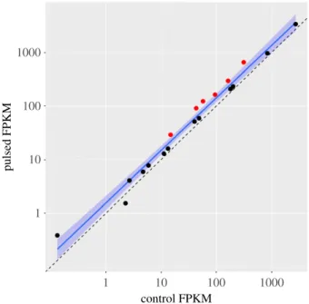
Figure 2.
Expression of 19 annotated ferritin genes in the trout genome,
including six copies of the gene
frim
, whose expression increased significantly
(red points). The dashed line indicates equal expression between groups. The
locally weighted scatterplot smoother (LOWESS, solid blue line) and its 95%
CI (blue shaded region) are shown.
rsbl.r
oy
alsocietypublishing.org
Biol.
Lett.
13
:
20170142
expressed genes was assessed using a Fisher’s exact test. Only functions with a false discovery rate (FDR) less than 0.05 were retained [11]. We used the non-parametric approach in [12] to identify GO features containing genes whose expression profiles can best cluster the groups of samples.
3. Results
We generated more than 285 million sequencing reads from 10 RNA libraries (electronic supplementary material, figure S1). On average, 10.5 (s.d. 0.67) million reads per library passed quality control and mapped to annotated genes in the reference genome (table 1). Of the 46 585 annotated genes, 38 550 (83%) contained at least 10 overlapping reads across all libraries and were assessed for expression level (electronic supplementary material, figure S2). We identified 181 genes differentially expressed in response to the pulsed magnetic field (figure 1; electronic supplementary material, figure S3). These genes were enriched for 15 GO annotations (electronic supplementary material, table S1), most speci-fically ferric iron binding (FDR¼21026
), iron ion transport (FDR¼31025
) and cellular iron ion homeostasis (FDR¼31025
). Eighteen (95%) of 19 ferritin-coding genes in the trout genome increased expression, including signifi-cant increases in six copies of the genefrim,which encodes the middle subunit of the ferritin protein (figure 2). Of the 6899 GO annotations assigned to the genome, 69 (1%) were significantly linked to differences between control and pulsed groups (table 2; electronic supplementary material, table S2). The highest-ranking terms were the functions of oxidoreductase activity (p¼0.0005) and fibroblast growth
factor-activated receptor activity (p¼0.0009).
4. Discussion
(a) Iron regulation and magnetoreception
If the receptors for the magnetic sense are based on magne-tite, then they may be disrupted by a magnetic pulse
[1,3– 5]. Results revealed increased expression of ferritin in fish exposed to a magnetic pulse. Ferritin is a polymeric protein that acts as a repository to store excess iron within cells [13]. Inside ferritin, up to 4500 iron atoms are oxidized and stored as hydrated iron oxides, including superparamag-netic, ferromagnetic and ferrimagnetic crystals [13,14]. It is possible that the magnetic pulse resulted in the disruption or liberation of iron oxide crystals from ferritin [4] or other iron-containing structures (e.g. MagR, MagR/Cry) [15]. If so, then excess free iron might account not only for the increase in ferritin expression (for the purpose of sequestra-tion), but also for the differences in activities associated with the oxidative consequences of free iron. The latter include oxidoreductase activity, transition metal ion binding, electron transport chain complexes and DNA damage repair. Previous studies have implicated ferritin in the biomineraliza-tion of magnetite [16,17]. Thus, ferritin might be involved in producing or repairing magnetite-based magnetoreceptors after a magnetic pulse.
(b) Genes associated with photosensitive structures
Several genes that function in the development, maintenance or repair of photosensitive structures and pathways (crggm3,
prl,purp, crabp1 pax6andgcip) were differentially expressed. Meanwhile, no significant differences in the expression of cryptochromes, photosensitive proteins hypothesized to function in chemical magnetoreception [2], were observed. Among the genes affected were some implicated in develop-ment of the optic nerve and habenula, two neural structures that link photoreceptive organs (retinae and pineal gland, respectively) to the brain. Both the retina and pineal gland have been considered possible locations of magnetoreception [1– 3,18,19], although only the pineal gland, including non-visual encephalic photoreceptors, was included in the brain tissue sampled in this study.
Both purp and crabp1 bind and transport vitamin A derivatives between and within, respectively, cells of the reti-nal pigment epithelium and interphotoreceptor matrix [20].
Table 2.
Top 10 ranked GO terms from [11]. Each gene was scored and ranked by its ability to differentiate control and pulse-magnetized groups, and each
term is then ordered by the average ranking (rank) of its associated genes. The number of genes assigned to each term (count) and their average score (score)
are given. All GO terms are of three domains: biological process (BP), molecular function (MF) and cellular component (CC). The complete list of significant
terms can be found in the electronic supplementary material.
GO term
average
rank
average score
(310
3)
count
p
-value
description
type
GO: 0016614
724.6
2.1
7
0.0001
oxidoreductase activity, acting on CH-OH group of donors
MF
GO: 0005007
724.6
2.1
7
0.0009
fibroblast growth factor-activated receptor activity
MF
GO: 0072669
748.6
1.5
6
0.0047
tRNA-splicing ligase complex
CC
GO: 0034464
757.3
1.2
8
0.0030
BBSome
CC
GO: 0003094
775.6
1.0
5
0.0291
glomerular filtration
BP
GO: 0021554
775.6
1.0
5
0.0295
optic nerve development
BP
GO: 0030517
775.6
1.0
5
0.0296
negative regulation of axon extension
BP
GO: 0005847
775.6
0.10
5
0.0304
mRNA cleavage and polyadenylation specificity factor
complex
CC
GO: 0046914
779.8
1.1
9
0.0041
transition metal ion binding
MF
GO: 0038062
785
1.2
8
0.0043
protein tyrosine kinase collagen receptor activity
MF
rsbl.r
oy
alsocietypublishing.org
Biol.
Lett.
13
:
20170142
These vitamin A derivatives are eventually metabolized into 11-cis-retinal, the chromophore required for vision. Also known as retinol binding protein 4, purpacts as a neurite-sprouting factor and is important for optic nerve regeneration [21]. One of the largest reductions in expression was in the hormone prolactin (prl). Prolactin has been shown to stimu-late the synthesis of visual pigments in the retinae of rainbow trout [22]. However, this hormone has a broad range of functions, thus making its role in responding to the magnetic pulse unclear.
Why a magnetic pulse altered expression of genes related to photosensitive structures is not known. One possibility is that magnetic particles are closely associated with these struc-tures so that pulse-induced movement of the particles damaged surrounding tissue and elevated gene expression needed to repair this ‘collateral damage’. Alternatively or additionally, the pulse might have generated magneto-phosphenes by activating photosensitive structures, or otherwise exerted an effect through unknown mechanisms.
5. Conclusion
Our study is the first to use a transcriptomic approach to investigate the effect of magnetic pulses on gene expression, with a view towards identifying candidate genes involved in magnetoreception. Results demonstrate that expression of ferritin genes is elevated after a pulse, a finding consistent with the hypothesis that ferritin is involved in generating or
repairing magnetite-based magnetoreceptors. In addition, the magnetic pulse altered expression of genes implicated in the development, function and repair of visual elements. Additional studies examining the response of candidate genes (e.g. using quantitative polymerase chain reaction, qPCR) [23] are needed to extend the work to other relevant tis-sues (e.g. retina, pineal gland and habenula) and to other magnetically sensitive species, as well as to elucidate the func-tional significance of the patterns of gene expression observed.
Ethics. Experimental methods were approved by Duke University’s IACUC ( protocol A175-15-06).
Data accessibility. Raw sequence data were deposited into GenBank under accession PRJNA324102 (also see table 1). All mapping, expression and annotation data are available in Dryad (http://dx. doi.org/10.5061/dryad.73b86) [24].
Authors’ contributions.R.R.F. designed the study, collected and analysed data, and drafted the manuscript. B.R.W. collected data and helped draft the manuscript. D.A.E. and K.J.L. helped design the study, collect data and draft the manuscript. S.J. conceived, designed and coordinated the study and helped draft the manuscript. All authors gave final approval for publication and agree to be held accountable for the content herein.
Competing interests.We declare we have no competing interests.
Funding.Funding was provided by the Air Force Office of Scientific Research (grant no. FA9550-14-1-0208) to S.J. and K.J.L.
Acknowledgements.We thank Cantrell Creek Trout Farm for providing trout, the Duke Shared Cluster Resource for computational resources, and Alex Ochoa, Eleanor Caves, Laura Bagge and Katie Thomas for comments on earlier drafts.
References
1. Johnsen S, Lohmann KJ. 2005 The physics and neurobiology of magnetoreception.Nat. Rev. Neurosci.6, 703 – 712. (doi:10.1038/nrn1745) 2. Ritz T, Adem S, Schulten K. 2000 A model for
photoreceptor-based magnetoreception in birds.
Biophys. J.78, 707 – 718. (doi:10.1016/S0006-3495(00)76629-X)
3. Kirschvink JL, Walker MM, Diebel CE. 2001 Magnetite-based magnetoreception.Curr. Opin. Neurobiol.11, 462 – 467. (doi:10.1016/S0959-4388(00)00235-X)
4. Davila AF, Winklhofer M, Shcherbakov VP, Petersen N. 2005 Magnetic pulse affects a putative magnetoreceptor mechanism.
Biophys. J.89, 56 – 63. (doi:10.1529/biophysj.104. 049346)
5. Ernst DA, Lohmann KJ. 2016 Effect of magnetic pulses on Caribbean spiny lobsters: implications for magnetoreception.J. Exp. Biol.219, 1827 – 1832. (doi:10.1242/jeb.136036)
6. Walker MM, Diebel CE, Haugh CV, Pankhurst PM, Montgomery JC, Green CR. 1997 Structure and function of the vertebrate magnetic sense. Nature390, 371 – 376. (doi:10.1038/ 37057)
7. Berthelot Cet al.2014 The rainbow trout genome provides novel insights into evolution after whole-genome duplication in vertebrates.Nat. Commun.5, 3657. (doi:10.1038/ncomms4657)
8. Bolger AM, Lohse M, Usadel B. 2014 Trimmomatic: a flexible trimmer for Illumina sequence data.
Bioinformatics30, 2114 – 2120. (doi:10.1093/ bioinformatics/btu170)
9. Dobin A, Davis CA, Schlesinger F, Drenkow J, Zaleski C, Jha S, Batut P, Chaisson M, Gingeras TR. 2013 STAR: ultrafast universal RNA-seq aligner.
Bioinformatics29, 15 – 21. (doi:10.1093/ bioinformatics/bts635)
10. Trapnell C, Williams BA, Pertea G, Mortazavi A, Kwan G, van Baren MJ, Salzberg SL, Wold BJ, Pachter L. 2010 Transcript assembly and quantification by RNA-Seq reveals unannotated transcripts and isoform switching during cell differentiation.Nat. Biotechnol.28, 511 – 515. (doi:10.1038/nbt.1621)
11. Conesa A, Go¨tz S, Garcı´a-Go´mez JM, Terol J, Talo´n M, Robles M. 2005 Blast2GO: a universal tool for annotation, visualization, and analysis in functional genomics research.Bioinformatics21, 3674 – 3676. (doi:10.1093/bioinformatics/bti610)
12. Rue-Albrecht K, McGettigan PA, Herna´ndez B, Nalpas NC, Magee DA, Parnell AC, Gordon SV, MacHugh DE. 2016 GOexpress: an R/Bioconductor package for the identification and visualisation of robust gene ontology signatures through supervised learning of gene expression data.BMC
Bioinformatics17, 126. (doi:10.1186/s12859-016-0971-3)
13. Torti FM, Torti SV. 2002 Regulation of ferritin genes and protein.Blood99, 3505 – 3516. (doi:10.1182/ blood.V99.10.3505)
14. Pannalal SJ, Crowe SA, Cioppa MT, Symons DTA, Sturm A, Fowle DA. 2005 Room-temperature magnetic properties of ferrihydrite: a potential magnetic remanence carrier?Earth Planet. Sci. Lett.236, 856 – 870. (doi:10.1016/j.epsl.2005.05. 019)
15. Qin Set al.2016 A magnetic protein biocompass.Nat. Mater.15, 217 – 226. (doi:10. 1038/nmat4484)
16. Baumgartner J, Morin G, Menguy N, Gonzalez TP, Widdrat M, Cosmidis J, Faivre D. 2013 Magnetotactic bacteria form magnetite from a phosphate-rich ferric hydroxide via nanometric ferric (oxyhydr)oxide intermediates.Proc. Natl Acad. Sci. USA110, 14 883 – 14 888. (doi:10.1073/pnas. 1307119110)
17. Hsu CY, Chan YP. 2011 Identification, and localization of proteins associated with biomineralization in the iron deposition vesicles of honeybees (Apis mellifera).
PLoS ONE6, e19088. (doi:10.1371/journal.pone. 0019088)
19. Deutschlander ME, Borland SC, Phillips JB. 1999 Extraocular magnetic compass in newts.Nature400, 324 – 325. (doi:10.1038/ 22450)
20. Rhinn M, Dolle´ P. 2012 Retinoic acid signalling during development.Development139, 843 – 858. (doi:10.1242/dev.065938)
21. Kato S, Matsukawa T, Koriyama Y, Sugitani K, Ogai K. 2013 A molecular mechanism of optic nerve
regeneration in fish: the retinoid signaling pathway.
Prog. Retin. Eye Res.37, 13 – 30. (doi:10.1016/j. preteyeres.2013.07.004)
22. Allen DM, Cristy M. 1978 Thiourea does not block visual pigment responses to prolactin in trout.
Vision Res.18, 859 – 860. (doi:10.1016/0042-6989(78)90129-3)
23. Rajkumar APet al.2015 Experimental validation of methods for differential gene expression
analysis and sample pooling in RNA-seq.BMC Genomics16, 548. (doi:10.1186/s12864-015-1767-y)
24. Fitak RR, Wheeler BR, Ernst DA, Lohmann KJ, Johnsen S. 2017 Data from: Candidate genes mediating magnetoreception in rainbow trout (Oncorhynchus mykiss). Dryad Digital Repository. (doi:10.5061/dryad. 73b86)
![Table 2. Top 10 ranked GO terms from [11]. Each gene was scored and ranked by its ability to differentiate control and pulse-magnetized groups, and each term is then ordered by the average ranking (rank) of its associated genes](https://thumb-us.123doks.com/thumbv2/123dok_us/8319087.2204681/3.892.59.834.153.454/ability-differentiate-control-magnetized-ordered-average-ranking-associated.webp)
