CHARACTERIZATION OF HEPARAN SULFATE-PROTEIN INTERACTIONS FOR SYNTHETIC HEPARIN DESIGN
Elizabeth Pempe Chappell
A dissertation submitted to the faculty of the University of North Carolina at Chapel Hill in partial fulfillment of the requirements for the degree of Doctor of Philosophy in the
Department of Pharmaceutical Sciences (Chemical Biology and Medicinal Chemistry).
Chapel Hill 2013
Approved by: Jian Liu, Ph.D.
ii ABSTRACT
ELIZABETH PEMPE CHAPPELL: Characterization of heparan sulfate-protein interactions for synthetic heparin design
(Under the direction of Jian Liu, Ph.D.)
Heparin is a widely prescribed anticoagulant that has been in clinical use for over 70 years. It is a natural product and a special form of heparan sulfate, a heterogeneous
polysaccharide that is expressed as a proteoglycan on the surface of all animal tissues. In recent years, the development of a chemoenzymatic method to synthesize specific heparan sulfate polysaccharides and oligosaccharides has enabled studies of the structure-based interactions between negatively charged heparan sulfate and its protein binding partners.
iii
ACKNOWLEDGEMENTS
Several people were involved in the completion of this work, and many others supported me throughout my years of research. I am grateful and fortunate to have these people in my life.
Thanks to Tom, my husband, who convinced me that I was capable of doing a Ph.D., who supported me throughout every step of graduate school and who gave me a full life and so much happiness outside of the university.
Thanks to my family (Treats, Pempes, Chappells, Evanses and Cederholms), who inspire me to be capable, interesting, grounded and loving through their example.
Thank you to my labmates in the Liu laboratory: Sherket, Courtney, Heather, Ryan, Justin, Yongmei, Zhou, Sheng, Kai, Lan, Renpeng, Joyce, Susan, T, Tim and Chunhui. I have learned from each of them and have enjoyed their company every day that I was in the lab. Special thanks to Joyce Chandarajoti and Danielle Cook for their scientific help,
laughter and endless moral support.
iv
TABLE OF CONTENTS
List of Tables……….………vii
List of Figures………..………viii
List of Abbreviations………ix
Chapter I. Introduction……….……….………...…... 1
Heparan sulfate………..…….1
Heparan sulfate structure……….…….……1
HS biosynthetic enzymes……….….………2
Heparin vs. HS………..………..7
Current anticoagulant therapies………...…9
Strategies for improved heparin drugs………..………13
Statement of problem………...…………...16
II. Materials and Methods………...…...……17
Expression of HS biosynthetic enzymes………..…………....17
Preparation of 35S-labeled polysaccharides………..……....…..17
Expression of recombinant Sulf-2 in CHO cells ………17
Western blotting………...18
Sulf-2 enzymatic assay ………..………18
Analysis of synthetic polysaccharides……….…….19
Preparation of 35S-labeled HS from CHO cells……….………….….19
v
Preparation of mPF4……….………..20
Dot blot assay for PF4 binding……….…….21
Affinity co-electrophoresis………..21
AT binding assay……….22
Preparation of enzyme cofactors………..22
Preparation of oligosaccharide backbone……….………..22
N-detrifluoroacetylation………..……23
N-sulfation of oligosaccharides……….………24
Sulfation and epimerization modifications of oligosaccharide backbones…....….…24
Mass spectrometric analysis of oligosaccharides………..24
Inhibition of the activities of factor Xa and IIa……….……25
The neutralization effect of PF4 on anti-Xa activity……….………..26
HPLC analysis of oligosaccharides………..26
Nitrous acid-degraded disaccharide analysis of 35S-labeled oligosaccharides……..26
Materials, solutions and buffers for Flp-In 293-based assays………..…27
Preparation of 35S-labeled HS constructs for Stabilin binding studies……….…27
Stabilin expression plasmids.…...……….…29
Endocytosis assays……….29
Direct ectodomain binding assays………...………….30
Assessment of liver clearance……….………..30
Synthesis of pure pnp-tagged oligosaccharides……….31
Protamine reversibility assays………...……31
Clearance profile of anti-Xa activity in rats………..32
Anti-Xa activity in mice………....32
III. Reduction of platelet factor 4 binding to heparan sulfate……..………33
Substrate specificity of Sulf-2……….……35
vi
PF4 binding of oligosaccharides with anti-IIa activity………44
Conclusions………..…47
IV. Identification of Stabilin-binding structural motifs……….……49
Polysaccharide constructs……….…50
Cell internalization assays……….…51
Direct binding assay………...…………53
Antithrombin competition………...……55
Oligosaccharide constructs………...………57
Conclusions………..…62
V. Design of homogeneous heparins with controlled clearance pathways………...…65
Design and synthesis of 6-, 8-, 10- and 12-mer…………..………...……66
Antithrombin binding affinities………...…67
Protamine reversibility………...……….……68
Pharmacokinectic profile in rats...………....……69
Anti-Xa activity and elimination route in mice………...……...………..…71
Conclusions……….……74
VI. Conclusions………..…76
Appendix I. Curriculum vitae………..…..80
vii
LIST OF TABLES
Table
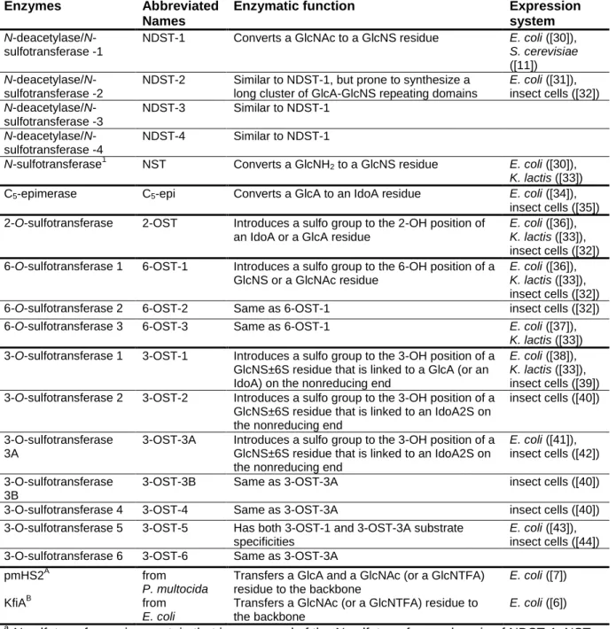
1.1. List of HS biosynthetic enzymes………..……8
3.1. Substrate specificity of Sulf-2……….…40
3.2. Binding affinities of Sulf-2-treated and untreated HS to PF4 and AT………...…43
3.3. Chemical structure and anti-IIa/-Xa ratio of synthetic oligosaccharides………...…45
4.1. Summary of the polysaccharide and oligosaccharide constructs………...58
viii
LIST OF FIGURES
Figure
1.1. Chemical structure of the disaccharides present in HS………...2
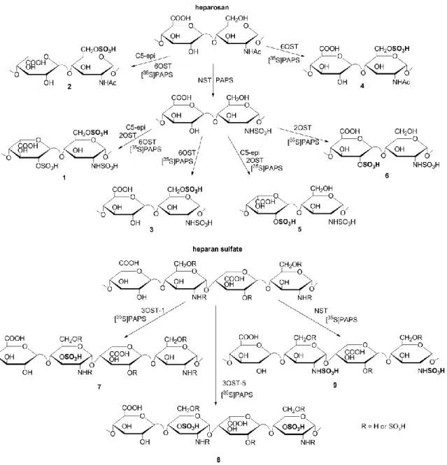
1.2. Enzyme reactions in the synthesis of heparan sulfate……….3
1.3. Substrate recognition of C5-epimerase and 3OST………...………6
1.4. Interaction of anticoagulant drugs with the coagulation cascade………..…...11
3.1. Disaccharide composition of Sulf-2-treated and untreated [35S]HS……….36
3.2. Synthetic scheme for the 35S-labeled HS constructs………..38
3.3. Disaccharide analysis of construct 1 before and after Sulf-2 treatment………..39
3.4.Effect of Sulf-2 treatment on PF4 and AT binding………..42
3.5. Disaccharide analysis of 3-O-sulfated HS with and without Sulf-2 treatment……….…44
3.6. Determination of the binding of PF4 to O-sulfated oligosaccharides………..….46
3.7. Determination of the binding of compounds 10-13 to AT using ACE………..…….47
4.1. Domain structure of Stabilin-2………50
4.2. Chemical structures of the polysaccharide constructs………..….51
4.3. Internalization of 3-O-sulfated heparin by Stab-1 and -2………...52
4.4. Internalization of modified HS polysaccharides……….….53
4.5. Direct binding of HS constructs and Stabilin ectodomains………...54
4.6. Inhibition of Stabilin-mediated endocytosis by antithrombin……….……....56
4.7. Effect of size on endocytosis……….…59
4.8. A 3-O-sulfated decasaccharide is required for binding to Stabilin receptors…….…….60
4.9. 3-O-sulfation leads to efficient liver retention………..…61
4.10. The structure of 28b, the shortest HS construct that displayed robust Stab binding...61
5.1. Susceptibility of HS oligosaccharides to protamine neutralization………...68
ix
x
LIST OF ABBREVIATIONS
ΔUA Δ4,5-unsaturated uronic acid 2-OST 2-O-sulfotransferase
3-OST 3-O-sulfotransferase 6-OST 6-O-sulfotransferase AnMan anhydromannitol
AT antithrombin
ATP adenosine triphosphate BSA bovine serum albumin C5-epi C5-epimerase
CHO Chinese hamster ovary CM conditioned medium ConA concanavalin A CS chondroitin sulfate DEAE diethylaminoethyl
DMEM Dulbecco's Modified Eagle Medium
ESI-MS electrospray ionization mass spectrometry
Ext exostosin
EV empty vector
FBS fetal bovine serum GlcA glucuronic acid GlcN glucosamine
xi
HPLC high-performance liquid chromatography HS heparan sulfate
HUVEC human umbilical vein endothelial cells
IACUC Institutional Animal Care and Use Committee IdoA iduronic acid
IPTG isopropyl β-D-1-thiogalactopyranoside
KfiA N-acetylglucosaminyltransferase from E. coli K5 LMWH low-molecular-weight heparin
MES 2-(N-morpholino)ethanesulfonic acid NDST N-deacteylase/N-sulfotransferase NST N-sulfotransferase
PAMN polyamine
PAPS 3’-phosphoadenosine 5’-phosphosulfate PBS phosphate-buffered saline
PF4 platelet factor 4
pmHS2 heparosan synthase-2 from P. multocida
pnp para-nitrophenol
SDS-PAGE sodium dodecyl sulfate polyacrylamide gel electrophoresis SNAC sodium N-[8-(2-hydroxybenzoyl)amino]caprylate
Stab Stabilin
Sulf 6-O-endosulfatase
TBST Tris-buffered saline with Tween-20 TEV tobacco etch virus
CHAPTER I INTRODUCTION
Heparan sulfate
Heparan sulfate (HS) is a widely expressed carbohydrate. As the body’s most negatively charged molecule, it interacts with numerous proteins to regulate many biological functions relevant to human health and disease. These functions range from embryonic development and coagulation to inflammation and cancer metastasis. Heparan sulfate is part of the glycosaminoglycan family, the members of which contain long unbranched carbohydrates made up of repeating disaccharide units. Other members include keratan sulfate, dermatan sulfate, hyaluronan and chondroitin sulfate [1].
Heparan sulfate structure
The bioactivity of HS is dependent on its structure, namely the location of
electronegative sulfo groups along its backbone and the presence of iduronic acid (IdoA), glucuronic acid (GlcA) and glucosamine (GlcN) residues (Fig. 1.1). HS exists on the surface of animal cells and within the extracellular matrix as a proteoglycan consisting of long carbohydrate chains attached to a core protein. Cell surface HS proteoglycans include syndecans and glypicans; perlecan and agrin are the primary extracellular examples [2-4]. The HS chains are composed of repeating disaccharide units of uronic acid and
-2
sulfated, and its amine group can be acetylated, sulfated or unsubstituted (Fig. 1.1). The biological functions of HS proteoglycans are dominated by the HS side chains.
Figure 1.1. Chemical structure of the disaccharides present in HS. GlcA, glucuronic acid; IdoA, iduronic acid; GlcN, glucosamine.
This broad structural variation in the location of negative groups, in addition to variation in length and glycosidic bond position, allows HS to interact with different binding protein partners to display many biological functions. Although nonspecific ionic interactions between HS and proteins exist, the binding of HS to proteins can be specific. Thus, the preparation of unique HS chains with defined sulfation patterns and length is highly desirable, as they allow researchers to investigate the substrate specificity of HS-protein interactions and provide numerous therapeutic opportunities.
HS biosynthetic enzymes
HS biosynthesis occurs in the Golgi apparatus and is carried out by a series of enzymes (Figure 1.2). The synthesis of HS has two main components: chain elongation and modification of the individual sugars. In vivo, the HS chain is elongated by the Exostosin genes, Ext1 and Ext2 [5].This process can be mimicked in vitro by two bacterial
3
Figure 1.2. Enzyme reactions in the synthesis of heparan sulfate. N-deacetylase/N-sulfotransferase replaces N-acetyl groups with N-sulfo groups. C5-epimerase converts glucuronic acid to iduronic acid.
4
KfiA [6], and heparosan synthase-2 from Pasteurella multocida, or pmHS2 [7].In laboratory syntheses, KfiA and pmHS2 are incubated with uridine 5’-diphospho-N
-trifluoroacetylglucosamine (UDP-NTFA) and UDP-GlcA, respectively; with each cycle of incubation, the oligosaccharide is elongated by one sugar unit [8]. GlcNTFA is used because it is easily chemically deacetylated to GlcNH2 for later N-sulfation.
The N-deacetylase/N-sulfotransferase (NDST) enzyme is the first to modify an intact HS chain during biosynthesis, and its action is believed to direct the location of all
subsequent sulfation reactions [9]. The 325-aa C-terminal region (constituting the N -sulfotransferase domain) of NDST is commonly expressed and used for HS synthesis in vitro following chemical deacetylation [10], although recent studies have focused on
expression of the entire NDST enzyme [11].Sulfation reactions are carried out by incubating HS with a sulfotransferase and 3’-phosphoadenosine 5’-phosphosulfate (PAPS), a natural sulfate donor.
Characterization of NDST-1 by Sheng et al. uncovered a unique substrate specificity for this enzyme [12]. Treatment of a synthetic dodecasaccharide substrate with NDST-1 produced a variety of N-sulfated products containing clusters of GlcNS, suggesting that NDST-1 binds to HS at a random position, converts consecutive GlcNAc to GlcNS from the non-reducing to reducing end, then releases the substrate when it is five sugars away from the reducing end. There are a total of four isoforms of NDST; NDST-2, which is highly expressed in mast cells, is proposed to be involved in the synthesis of highly-sulfated heparin but not HS [13]. This was principally confirmed by the absence of heparin in mast cells from NDST-2 knockout mice [14]. The N-deacetylase and N-sulfotransferase activities of NDST-2 through -4 have been investigated, but their use in HS synthesis specifically has not been fully explored [15].
After N-sulfation, C5-epimerase (C5-epi) converts some D-glucuronic acid residues to
5
shown to exhibit a biphasic catalytic mode: depending on the substitution groups of the surrounding saccharide residues, the epimerization reaction is either reversible or
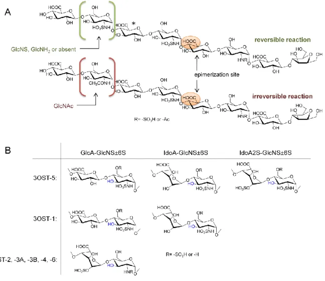
irreversible. Using structurally defined oligosaccharides, Sheng et al. identified that C5-epi will act on a GlcA residue if the residue immediately upstream (towards the non-reducing end) is a GlcNS residue. If the residue three sugars upstream is GlcNS, GlcNH2 or not present, the reaction is reversible; if it is GlcNAc, the reaction is irreversible (Figure 1.3A) [17]. This finding will enable researchers to “lock” IdoA sugars in place and synthesize pure HS oligosaccharides containing IdoA during in vitro synthesis.
A series of O-sulfotransferases then sulfate their respective positions on HS. Heparan sulfate 2-O-sulfotransferase (2-OST) catalyzes the transfer of an -OSO3H group from PAPS to IdoA or GlcA. It is present in one isoform and has approximately five-fold greater affinity for IdoA than for GlcA [18], although mutational analyses have suggested that the preference for IdoA over GlcA can be controlled through site-specific mutations [19].
6-OST sulfates both GlcNAc and GlcNS to form GlcNAc6S and GlcNS6S, respectively. 6-OST isoforms, of which there are three, appear to sulfate the same substrates [20]; however, placement of 6-O-sulfo groups in oligosaccharides can be controlled somewhat by the enzymatic reaction time and by elongating oligosaccharides already containing 6-O-sulfated glucosamine [21]. A combination of 6-OST-1 and -3 was demonstrated to prefer GlcNAc residues close to the reducing end of oligosaccharide substrates, but placement of a single 6-O-sulfo group in an oligosaccharide remains a challenge [21, 22].
6
Figure 1.3. Substrate recognition of C5-epimerase and 3OST. A. The epimerization reaction of C5-epi
is reversible or irreversible depending on the GlcN three sugars towards the nonreducing end of the glucuronic acid to be modified. The asterisk indicates a GlcA that could be epimerized if the sugar to the immediate left is GlcNS. B. Substrates of the seven 3OST isoforms. The position that will carry the 3-O-sulfo group is shown in blue.
7
The remaining 3OSTs (-2, -3A, -3B, -4 and -6) will sulfate a GlcNS or GlcNH2 linked to an IdoA2S at the non-reducing end (Figure 1.3B). These different substrate specificities allow placement of 3-O-sulfo groups in specific locations depending on the identity of the
neighboring residue. Based on their ease of production in E. coli, 3-OST-1, -3 and -5 are most commonly used in chemoenzymatic syntheses; 3-OST-3 and -5 will produce HS that binds to gD [24]. A complete list of the enzymes used in HS synthesis is given in Table 1.1.
Heparin vs. HS
Heparin and HS have very similar structures; however, heparin refers to a special form of HS that has more sulfo groups and a higher level of iduronic acid residues. Heparan sulfate is produced by virtually all cells in species ranging from simple invertebrates to humans [23]. Heparin, on the other hand, is produced by mast cells and is present only in some tissues of select members of the animal kingdom. Interesting examples include the observations that rabbit tissues do not contain heparin [26] and that chicken skin contains relatively high levels of heparin [27].
Heparan sulfate proteoglycans appeared early in metazoan evolution, and their common structural motifs are conserved in modern organisms [23]. Uncharacterized proteoglycans have been identified in ancient multicellular organisms such as Hydra [28], and lower organisms such as Drosophila and C. elegans contain homologs of syndecan, glypican and perlecan [29]. The essential role of HS proteoglycans (and heparins) in the development and physiology of living organisms is supported by their prevalence throughout the animal kingdom.
8
Enzymes Abbreviated
Names
Enzymatic function Expression
system
N-deacetylase/N -sulfotransferase -1
NDST-1 Converts a GlcNAc to a GlcNS residue E. coli ([30]),
S. cerevisiae
([11])
N-deacetylase/N -sulfotransferase -2
NDST-2 Similar to NDST-1, but prone to synthesize a long cluster of GlcA-GlcNS repeating domains
E. coli ([31]), insect cells ([32])
N-deacetylase/N -sulfotransferase -3
NDST-3 Similar to NDST-1
N-deacetylase/N -sulfotransferase -4
NDST-4 Similar to NDST-1
N-sulfotransferase1 NST Converts a GlcNH2 to a GlcNS residue E. coli ([30]), K. lactis ([33]) C5-epimerase C5-epi Converts a GlcA to an IdoA residue E. coli ([34]),
insect cells ([35]) 2-O-sulfotransferase 2-OST Introduces a sulfo group to the 2-OH position of
an IdoA or a GlcA residue
E. coli ([36]),
K. lactis ([33]), insect cells ([32]) 6-O-sulfotransferase 1 6-OST-1 Introduces a sulfo group to the 6-OH position of a
GlcNS or a GlcNAc residue
E. coli ([36]),
K. lactis ([33]), insect cells ([32])
6-O-sulfotransferase 2 6-OST-2 Same as 6-OST-1 insect cells ([32])
6-O-sulfotransferase 3 6-OST-3 Same as 6-OST-1 E. coli ([37]),
K. lactis ([33]) 3-O-sulfotransferase 1 3-OST-1 Introduces a sulfo group to the 3-OH position of a
GlcNS±6S residue that is linked to a GlcA (or an IdoA) on the nonreducing end
E. coli ([38]),
K. lactis ([33]), insect cells ([39]) 3-O-sulfotransferase 2 3-OST-2 Introduces a sulfo group to the 3-OH position of a
GlcNS±6S residue that is linked to an IdoA2S on the nonreducing end
insect cells ([40])
3-O-sulfotransferase 3A
3-OST-3A Introduces a sulfo group to the 3-OH position of a GlcNS±6S residue that is linked to an IdoA2S on the nonreducing end
E. coli ([41]), insect cells ([42])
3-O-sulfotransferase 3B
3-OST-3B Same as 3-OST-3A insect cells ([40])
3-O-sulfotransferase 4 3-OST-4 Same as 3-OST-3A insect cells ([40])
3-O-sulfotransferase 5 3-OST-5 Has both 3-OST-1 and 3-OST-3A substrate specificities
E. coli ([43]), insect cells ([44]) 3-O-sulfotransferase 6 3-OST-6 Same as 3-OST-3A
pmHS2A from
P. multocida
Transfers a GlcA and a GlcNAc (or a GlcNTFA) residue to the backbone
E. coli ([7])
KfiAB from
E. coli
Transfers a GlcNAc (or a GlcNTFA) residue to the backbone
E. coli ([6])
a
N-sulfotransferase is a protein that is composed of the N-sulfotransferase domain of NDST-1. NST is an unnatural protein, and it is used in the chemoenzymatic synthesis to convert a GlcNH2 residue
to a GlcNS residue.
b
Both pmHS2 and KfiA are bacteria enzymes, not HS biosynthetic enzymes. However, they are used for building the backbone structure of HS during chemoenzymatic synthesis.
9
sulfates from many different sources have been found to contain these domains in addition to a short linker that is positioned between the domains [45]. Heparan sulfate is ubiquitous in invertebrates as well; a survey of 23 invertebrates from 13 different phyla identified heparan sulfate in all of the species studied [46]. Studies in crustaceans and mollusks have
characterized heparan sulfate in various tissues and found that the concentration of heparan sulfate is directly proportional to the salinity of the habitat [26].
There is much greater variation in the distribution of heparin, both among different organisms and within the tissues of a given species. In mammals, heparin is generally found in tissues that are in direct contact with the external environment, such as the skin,
intestines and lungs, but it has low prevalence in the brain, muscle and kidneys of most species [26]. Several mollusks have been shown to contain heparin-like polysaccharides that possess anticoagulant activity [47, 48], but heparin is not present in all invertebrates. Like HS, heparin is understood to be comprised primarily of two different regions: a highly sulfated region that is subject to degradation by heparinase and a lowly sulfated region that is susceptible to heparitinase II, and the length and quantities of these regions depend on the species of origin and the specific tissue [26]. For example, bovine lung heparin is rich in the heparinase-cleavable region, while bovine intestinal heparin and mollusk heparins contain more of the heparitinase II-cleavable region [49, 50]. The roles of heparan sulfate and heparin appear to be the same in vertebrates and invertebrates.
Current anticoagulant therapies
Anticoagulants prevent the formation of blood clots by interfering with the blood coagulation cascade. Commonly used anticoagulants include vitamin K antagonists,
10
classified as unfractionated heparin, low-molecular-weight heparin or synthetic pentasaccharides.
Warfarin, the primary vitamin K antagonist, is a synthetic derivative of a natural product of plant and fungal origin, dicumarol [51]. It functions by inhibiting the biosynthesis of vitamin K-dependent procoagulant factors II (also called prothrombin), VII, IX and X [52]. It is given orally, and due to the long half-lives of some of these factors, it can take several days for it to reach its full antithrombotic effect. Warfarin is a mixture of R and S enantiomers (which are both active, but to different degrees) [53], and it is influenced by drug interactions and diet, especially the intake of vitamin K-rich greens. Thus, warfarin drugs require close monitoring and can cause bleeding complications [52]. Available reversal agents include vitamin K, fresh frozen plasma and bypassing agents such as prothrombin complex concentrates or recombinant factor VIIa. Warfarin is most typically administered for thrombosis treatment or prophylaxis.
Full-length unfractionated heparin (UFH) is isolated as a mixture from animal mast cells, primarily porcine intestine. It was discovered in 1916, before the establishment of the Food and Drug Administration, and it is one of the oldest drugs still in clinical use [54]. Heparin is a potent activator of antithrombin, a protein that inhibits factors Xa and IIa to prevent the downstream formation of fibrin clots (Fig. 1.4). Its inexpensive production, short half-life and reversibility with protamine make UFH an advantageous drug for surgery and for handling blood in laboratory settings. It also has certain drawbacks, including an
11
Figure 1.4. Interaction of anticoagulant drugs with the coagulation cascade. Coagulation is initiated by the intrinsic or extrinsic pathway. The two pathways converge on the activation of factor X to Xa and result in the formation of fibrin clots. Target sites for current anticoagulant drugs are shown in red.
Low-molecular-weight heparin (LMWH) drugs, such as enoxaparin (Lovenox), are a mixture of products from depolymerized UFH. The average length is about 15 saccharide units, compared to 40 for UFH. LMWH inhibits IIa to a lesser extent than UFH, as not all components of the mixture are long enough to form an antithrombin:Xa:IIa complex. It tends to be safer than UFH, having a lower incidence of HIT, and its longer half-life and
12
LMWH is cleared by the kidneys, so anti-Xa monitoring is recommended for patients with renal impairment as well as for elderly, pregnant or obese patients.
The shortest and most recently developed heparin drugs, such as fondaparinux (Arixtra), are analogs of the antithrombin-binding heparin pentasaccharide. They inhibit Xa and do not have anti-IIa activity. Arixtra is produced through a lengthy chemical synthesis and, unlike UFH and LWMH, it is a pure compound, not a mixture. Like LMWH, it is dosed subcutaneously. Arixtra has a half-life of approximately 17 hours, allows for unmonitored once-daily dosing and is currently approved for deep vein thrombosis and pulmonary embolism [55]. A hypermethylated version of fondaparinux known as idraparinux was developed in the late 1990s [56]. It has a 30-fold higher affinity for antithrombin than Arixtra, and its 120-hour half-life permits once-weekly administration [55]. Like Arixtra, it does not have an antidote. However, a biotinylated version of idraparinux was developed, and its anti-Xa activity is effectively reversed by avidin [57]. Both fondaparinux and idraparinux do not bind to PF4 and do not show evidence of causing heparin-induced thrombocytopenia. Despite these promising developments, idraparinux was associated with an increased risk of major hemorrhage in clinical trials [58], and it was withdrawn from further development.
Thrombin, or factor IIa, is a serine protease that is central to hemostasis. Direct thrombin inhibitors function independently of antithrombin and do not bind PF4 [52]. The drugs lepirudin, desirudin and bivalirudin are derivatives of hirudin, a naturally occurring peptide found in the salivary glands of medicinal leeches, and they are approved for
13
Direct Xa inhibitors have the advantage of bypassing intermediary molecules like antithrombin, which may result in more consistent anticoagulation. Razaxaban and apixaban are both oral small-molecule direct Xa inhibitors developed by Bristol-Myers Squibb.
Razaxaban was abandoned in favor of apixaban due to its better safety profile, and apixaban (Eliquis) was approved in December 2012. Rivaroxaban is another FDA-approved Xa inhibitor; it is also a small molecule developed by Bayer that can be given in once-daily oral doses. Of all oral anticoagulants, rivaroxaban has been evaluated the most extensively and in largest patient populations [55]. It has a half-life that is considerably shorter than other oral Xa inhibitors (about 5 to 9 hours) [52], and it has a low risk of drug-drug interactions [60]. Although they have many promising characteristics, a disadvantage of the oral Xa inhibitors is their lack of a reversal agent.
Selective direct inhibitors of factors IXa and XIa are also in development, but they have yet to complete clinical trials [55].
Strategies for improved heparin drugs
Although unfractionated, low-molecular-weight and pentasaccharide heparin drugs are presently available, there is a need for more advanced heparins that suit the
requirements of distinct patient populations. As long as binding to antithrombin is maintained, the structure of heparin can be rationally designed to allow for optimal interactions with other molecules and desirable pharmacological properties.
14
gastrointestinal lining, penetration enhancers such as SNAC (sodium N
-[8-(2-hydroxybenzoyl)amino]caprylate) have been administered with heparin, with promising results [63]. Other approaches include novel drug delivery systems, such as nanoparticles, lipid conjugates and enteric coatings [64-66]. Despite experimental successes, no oral heparin formulations have reached the market. A more relevant issue for rational synthesis of the heparin carbohydrate chain would be to focus on structures that were bioavailable via subcutaneous injection (like Arixtra and LMWHs) rather than intravenously. Although
unfractionated heparin can be administered subcutaneously, its bioavailability is reduced to about 30% (compared to nearly 100% for LMWH), so much higher doses are required [67, 68]. Thus, heparins of reduced molecular weight are likely to have high subcutaneous bioavailability.
After administration, side effects of heparin drugs come into consideration. For many drugs, unwanted side effects can be avoided by modifying the structure of the active
15
predictable, and second, because the compounds could be designed to avoid interaction with immunogenic molecules.
For certain heparin applications, particularly surgery, the availability of an antidote is imperative. Additionally, unstopped hemorrhaging as a result of heparin therapy can lead to long-term debilitating diseases and may be life threatening [74]. Protamine sulfate can be used to reverse unfractionated heparin, although heparin rebound is a concern, but it is ineffective with low-molecular-weight heparin and Arixtra [75]. Excessive bleeding due to Arixtra, although rare, can be very difficult to manage because of its long half-life [76]. It has been successfully managed with fresh frozen plasma and prothrombin complex
concentrates [75], but the only systematically evaluated reversal for pentasaccharide heparins is the use of recombinant FVIIa [76]; however, the use of rFVIIa for hemorrhage is still controversial [77]. Novel heparin drugs would ideally be reversible by protamine to evade life-threatening bleeding episodes.
Finally, the metabolic route and rate by which heparin is cleared from the body can make it ideal or dangerous depending on the patient population. Long-acting heparins suit patients that require long-term therapy, and short-acting heparins better treat those
16
Statement of Problem
Heparin has been widely used for decades, but it has several limitations as a drug. Unfractionated heparin has contamination issues due to its sourcing and causes side effects like heparin-induced thrombocytopenia. Low-molecular-weight heparin can cause severe bleeding due to its structural heterogeneity. Pentasaccharides like Arixtra are unsafe for kidney impaired patients due to their renal clearance route [79]. The goals of this
dissertation were to characterize the protein interactions underlying some of these drawbacks and to propose improved heparin structures.
Recent advances in heparan sulfate synthesis using a chemoenzymatic approach have enabled the production of novel molecules with desired sulfation patterns and lengths (for a review, see [80]). First, this synthetic approach was used to investigate methods to reduce binding between heparan sulfate and PF4, an interaction that initiates heparin-induced thrombocytopenia. In addition, we sought to characterize the structural
CHAPTER II
MATERIALS AND METHODS
Expression of HS biosynthetic enzymes
Several enzymes were used for HS synthesis, including NST, C5-epi, 2-OST, OST-1, 6-OST-3, 3-OST-1, 3OST-5, KfiA, and pmHS2. All enzymes were expressed in E. coli and purified by appropriate affinity chromatography as described previously [82].
Preparation of 35S-labeled polysaccharides
Radiolabeled polysaccharide substrates were prepared using approximately 1 µg heparosan, a capsular polysaccharide isolated from the E. coli K5 strain, as a starting material [83]. Other substrates were prepared from bovine kidney heparan sulfate. The starting materials were modified with 5-10 µg of C5-epi, NST, 6OST-1/3, 2OST, 3OST-1 and 3OST-5 enzymes in sequential 200-µL reactions containing approximately 1 x 10⁶ cpm [35S]PAPS and 10 nmol unlabeled PAPS in 50 mM MES (pH 7) and 0.5% triton X-100. The enzymatic reactions were incubated at 37°C for 60 min, heat inactivated and purified using a DEAE column [83].
Expression of recombinant Sulf-2 in CHO cells
18
plasmid designated as pcDNA3.1A-myc/His-HSulf-2. Wild-type CHO cells were transiently transfected with the expression plasmid pcDNA3.1A-myc/His-HSulf-2 or an empty
pcDNA3.1A vector according to a standard protocol. Briefly, CHO cells were seeded in 6-well plates in F-12 media supplemented with 10% fetal bovine serum (FBS) and were maintained in a 5% CO2 humidified incubator at 37°C. When the cells reached 90-95% confluence, they were transfected using Lipofectamine 2000 reagent (Invitrogen) and Opti-MEM Reduced Serum Media (Invitrogen) according to the manufacturer’s protocol. After 4-6 h, the medium was replaced with F-12 media containing 10% FBS. The conditioned medium was collected after 48-72 h of incubation and centrifuged at 4,000 rpm for 15 minutes to remove cellular debris.
Western blotting
Sulf-2 and EV-transfected cells were collected using trypsin and washed with PBS. Cells were lysed with 1.5 M sucrose, 1% triton X-100 and 1 mM PMSF. Sulf-2 and EV CM and lysates were separated by 12% SDS-PAGE and visualized with an anti-myc primary antibody (Cell Signaling Technology) and the SuperSignal detection system (Thermo Scientific).
Sulf-2 enzymatic assay
A 100-µL reaction containing 35S-labeled substrate, 50 mM MES (pH 6.5), 10 mM CaCl2, 0.1% triton X-100 and 50 µL Sulf-2 enzyme was incubated overnight at 37°C.
19
Analysis of synthetic polysaccharides
To determine the structural compositions of radiolabeled substrates, disaccharide analyses were performed using heparin lyase digestion. Polysaccharide substrates were incubated overnight with a mixture of heparin lysase I, II and III (0.1 mg/mL each) in 200 µL of 50 mM sodium phosphate (pH 7) at 37°C. The reaction was terminated by boiling at 100°C for 5 min and was loaded onto a Bio-Gel P-2 column (Bio Rad) to isolate disaccharides. These
disaccharides were analyzed using reverse-phase ion-pairing HPLC [31].
Preparation of 35S-labeled HS from CHO cells
A T-75 flask of wild-type CHO cells was grown to confluence in F-12 media supplemented with 10% FBS. The cells were then incubated with 1 mL of 1.0 mCi/mL sodium [35S]sulfate (Perkin Elmer) for 6 h at 37°C with 5% CO2. Two hundred microliters of a pronase stock solutioncontaining 1 mg/mL Pronase (Sigma), 240 mM NaAcO (pH 6.5) and 1.92 M NaCl was added to the cells, and the flask was incubated overnight at 37°C. The
pronase-digested sample was centrifuged at 10,000 rpm for 15 min and filtered using a 0.45-µm filter, then purified using a DEAE-Sepharcel column (Sigma), which was equilibrated using a buffer containing 20 mM NaAcO (pH 5) and 150 mM NaCl. The [35S]HS was eluted from the column with 1 M NaCl in 20 mM NaAcO and was dialyzed overnight against 50 mM
20
Preparation of 3OST-1-treated [35S]HS
To 3-O-sulfate [35S]HS using unlabeled PAPS for affinity co-electrophoresis, approximately 100,000 cpm of [35S]HS was added to a 50-µL reaction containing 0.05 ng 3OST-1, 10 mM MnCl2, 5 mM MgCl2, 75 ug/mL protamine chloride, 0.4 mg/mL chondroitin sulfate, 0.12 mg/mL BSA, 1% triton X-100 and 0.5 mM PAPS. The reaction was incubated at 37°C for 20 min, inactivated at 80°C for 10 min, then diluted with 60 µL H2O and centrifuged at 2000 x g for 10 min. Substrates used for antithrombin binding studies were then isolated using a concanavalin A-Sepharose column (Sigma).
Preparation of mPF4
The full-length murine PF4 cDNA was obtained from Open Biosystems (Clone ID: 582960). The heparin-binding domain of PF4 (Ala33-Ser105) was cloned into a PET32 vector
(Novagen) using NcoI and HindIII sites to give a plasmid named as PF4-PET32/TEV. In this plasmid, a tobacco etch virus protease (TEV) cleavage hexapeptide sequence, EQLYFQG, was constructed between thioredoxin and PF4. The design permitted cleavage of the thioredoxin-PF4 fusion protein to release PF4. The resultant recombinant PF4 protein has five extra amino acid residues (GSRHG) at the N-terminus. A bacterial strain, BL21(DE3)-RIL/pRK793, expressing TEV protease was a generous gift from Dr. Lars Pedersen (National Institute of Environmental Health Sciences).
21
The purity of PF4 was greater than 90% as determined by NuPAGE 12% Bis-Tris Gel (Invitrogen). PF4 was eluted as a protein with a MW of 30 kDa on a Sephadex G75 column, suggesting that PF4 is present in a tetrameric form that was consistent with a previous report [84]. The concentration of PF4 was determined by amino acid compositional analysis.
Dot blot assay for PF4 binding
A dot blot assay was used to determine the binding affinities of HS to PF4. 35S-labeled HS (approximately 6,000 cpm) was incubated with 0-15 µg/mL PF4 for 30 min at 37°C in 130 mM NaCl, 50 mM Tris (pH 7) buffer (for the [35S]HS polysaccharide studies) or in 250 mM NaCl, 50 mM Tris (pH 7.3) buffer (for the oligosaccharide studies) to allow complex
formation. The mixture was then spotted onto a nitrocellulose membrane (GE Healthcare), which binds to proteins nonspecifically, allowing the capture of PF4-[35S]HS complexes. The membrane wells were washed with buffer, then excised, and the bound radioactivity was quantified using a scintillation counter.
Affinity co-electrophoresis
Affinity co-electrophoresis gels were prepared using a standard protocol [85] with lanes containing 0-1.41 µM PF4 or 0-3.2 µM AT and approximately 50,000 cpm [35S]HS per gel. Gels were run for 2 (AT) or 2.5 (PF4) hours and the bands were imaged using a Storm 860 phosphorimager (Molecular Dynamics) and ImageQuant TL v2005 software (GE Healthcare Life Sciences). Kd values were determined by plotting R/[protein] vs. R, where R = (Mo – M)/Mo; Mo = the mobility of free HS and M = the mobility of HS at each protein
22
AT binding assay
To quantify the binding of Sulf-2-treated and untreated HS to AT, 3OST-1-labeled substrates were incubated with 0.1 mg/mL AT, and the complex of AT and HS was captured using concanavalin A-Sepharose beads (Sigma). For this experiment, the AT-binding [35S]HS was prepared by incubating HS from bovine kidney with the 3OST-1 enzyme and [35S]PAPS.
Preparation of enzyme cofactors
A sulfo donor, 3’-phosphoadenosine 5’-phophosulfate (PAPS), was prepared using
adenosine phosphokinase and ATP-sulfurylase [82]. The preparation of UDP-GlcNTFA was started from glucosamine (Sigma), which was first converted to GlcNTFA by reacting with S -ethyl trifluorothioacetate (Sigma-Aldrich) following the protocol described previously [82]. The resultant GlcNTFA was converted to GlcNTFA-1-phosphate using N-acetylhexosamine 1-kinase [86]. The plasmid expressing N-acetylhexoamine 1-kinase was a generous gift from Prof. Peng Wang (Georgia State University), and the expression of the enzyme was carried out in E. coli as reported [86]. The UDP-GlcNTFA synthesis was completed by transforming GlcNTFA-1-phosphate using glucosamine-1-phosphate acetyltransferase/N -acetylglucosamine-1-phosphate uridyltransferase (GlmU) as described [82]. The resultant UDP-GlcNTFA was then ready for the elongation reaction using KfiA.
Preparation of oligosaccharide backbone
23
was monitored by the disappearance of UDP-GlcNTFA using a silica-based polyamine HPLC column (PAMN-HPLC, Waters). Once the reaction was completed, pmHS2 (30 µg/mL) and UDP-GlcA (750 μM) were added, and the reaction was incubated for another 24 hours at room temperature. The resultant product was a tetrasaccharide, which was purified by a Biogel P-2 column (0.75 × 200 cm) that was equilibrated with 0.1 M ammonium
bicarbonate at a flow rate of 6 mL/h. The fractions were then subjected to ESI-MS analysis. The fractions containing the tetrasaccharide were pooled. The procedures for synthesizing the N-TFA pentadecasaccharide (15-mer), N-TFA heptadecasaccharide (17-mer), N-TFA nonadecasaccharide (19-mer) and N-TFA henicosasaccharide (21-mer) were essentially the same completed by repeating the above cycle with the designated times.
When the backbone was elongated beyond octasaccharide, a special method was employed to deplete the UDP-GlcNTFA and UDP-GlcNAc prior to the further elongation by pmHS2. If unreacted UDP-GlcNTFA of UDP-GlcNAc was present in the GlcA elongation step involving pmHS2, an uncontrolled elongation occurred, resulting in a mixture. It is, therefore, especially important to avoid the formation of mixtures when the synthesis reached octasaccharide and beyond because the P-2 column cannot separate
oligosaccharides larger than octasaccharides. To this end, disaccharide (GlcA-AnMan) was added to the reaction to exhaust the residue UDP-GlcNTFA and GlcNAc at the reaction, and the resultant trisaccharide was removed by the P-2 column.
N-detrifluoroacetylation
24
N-sulfation of oligosaccharides
N-sulfation of oligosaccharide was carried out by incubating the oligosaccharide substrates with NST and PAPS. The reaction mixture typically contained 1-2 mg de-N
-trifluoroacetylatedoligosaccharide, 500 µM PAPS, 50 mM MES, pH 7.0 and 1 mg of NST in a total volume of 15 mL. The reaction mixture was incubated at 37°C overnight.
Sulfation and epimerization modifications of oligosaccharide backbones
The conversion of N-sulfo oligosaccharidesto final products involved three steps, including C5-epimerization/2-O-sulfation, 6-O-sulfation and 3-O-sulfation. N-sulfo oligosaccharides (1-2 mg) were incubated with a reaction mixture containing 50 mM MES, pH 7.0, 0.03 mg/mL C5-epi and 2 mM CaCl2 in a total volume of 40 mL. After incubating 30 min at 37°C, 2-OST (0.03 mg/mL) and 200 µM PAPS were added, and the reaction was incubated overnight at 37°C. The products were purified by a DEAE column described previously (14). For 6-O -sulfation, the substrate was incubated with a reaction mixture containing 50 mM MES, pH 7.0, and 500 µM PAPS overnight at 37°C in the presence of OST-1 (0.03 mg/mL) and 6-OST-3 (0.03 mg/mL) in a total volume of 20 mL overnight at 37°C. The products were
purified by a DEAE column. For 3-O-sulfation, the reaction mixture contained 3-OST-1 (0.03 mg/mL), 10 mM MnCl2, 5 mM MgCl2,and PAPS 500 µM in a total volume of 20 mL overnight at 37°C.
Mass spectrometric analysis of oligosaccharides
25
set to 5 KV and 275°C. Sulfated oligosaccharide (1 µL) was diluted in a different working solution containing 200 µL of 70% acetonitrile and 10 mM imidazole. Experiments for sulfated oligosaccharides were carried out in negative ionization mode with the electrospray source set to 2 KV and 200°C. The automatic gain control was set to 1 X 10⁷ for full scan MS. The MS data were acquired and processed using Xcalibur 1.3.
Inhibition of the activities of factor Xa and IIa
Assays were based on a previously published method [87, 88]. Briefly, factor Xa (Enzyme Research Laboratories, South Bend, IN) and thrombin (Sigma) were diluted at 80 nM and 100 nM, respectively, with PBS containing 1 mg/mL BSA. Human AT (Cutter Biological) was diluted with PBS containing 1 mg/mL BSA to give a stock solution at the concentration of 0.4 µM. The chromogenic substrates, S-2765 (for factor Xa assay) and S-2238 (for factor IIa assay) were prepared at 1.3 mM and 1.5 mM in water. The synthesized oligosaccharides or heparin was dissolved in PBS at various concentrations (1 to 30 nM). The reaction mixture, which consisted of 70 µL of AT stock solution and 15 µL of the sample solution, was
26
The neutralization effect of PF4 on anti-Xa activity
HS oligosaccharides were incubated in a 96-well plate with 70 µL BSA (1 mg/mL in PBS), 10 µL AT (0.2 mg/mL in PBS) and 0-16 µL PF4 (0.45 mg/mL) for two minutes at room temperature. Ten microliters of Xa was then added, and after four minutes, 30 µL of chromogenic substrate S-2765 (1 mg/mL) was added to initiate the color change reaction. Sequential absorbance readings at 405 nm were started immediately using an ELx808 plate reader (BioTek). The rate of increased absorbance relative to the rate of a control sample was used to define Xa activity. Quantities of oligosaccharides showing approximately 8% Xa activity in the absence of PF4 were used.
HPLC analysis of oligosaccharides
Both DEAE-HPLC and polyamine-based anion exchange (PAMN)-HPLC were used to determine the purity of the oligosaccharides. The elution conditions for the HPLC analysis are described in the literature [82].
Nitrous acid-degraded disaccharide analysis of 35S-labeled oligosaccharides
The 35S-labeled compound was deacetylated and degraded with nitrous acid at pH 4.5, then
at pH 1.5, followed by reduction with sodium borohydride as described by Shively and
colleagues[89]. The resultant 35S-labeled disaccharides were resolved by a C18
reverse-phase column (0.46 × 25 cm) (Vydac) under reverse-reverse-phase ion pairing HPLC conditions.
The identities of the disaccharides were determined by coeluting with appropriate 35
27
Materials, solutions and buffers for Flp-In 293-based assays
Heparin (or unfractionated heparin) was from Sigma (St. Louis, MO). Fondaparinux
(Arixtra®) was purchased from a local pharmacy. Flp-In 293 cells, serum, and high glucose Dulbecco’s Modified Eagle Medium (DMEM) were from Gibco, Hygromycin B, Zeocin, and glutamine were from Invitrogen/Gibco (Carlsbad, CA). Western blot analysis was completed by either colorimetric or chemiluminescence detection of blotted protein. Anti-V5 antibodies and resins were from Bethyl Laboratories (Montgomery, TX). Other materials, reagents and kits were obtained as described recently [90]. Tris-buffered saline with Tween-20 (TBST) contains 20 mM Tris-HCl, pH 7.0, 150 mM NaCl, and 0.1% Tween-20. TBST/BSA is TBST with 1.0% (w/v) bovine serum albumin (BSA). Phosphate buffered saline (PBS) contains 137 mM NaCl, 8 mM Na2HPO4, 1.5 mM KH2PO4, 2.7 mM KCl, pH 7.2. Hank’s buffered saline solution (HBSS) contains 5 mM KCl, 0.4 mM KH2PO4, 0.8 mM MgSO4, 137 mM NaCl, 0.3 mM Na2HPO4, 5.5 mM glucose, 1.26 mM CaCl2, 0.5 mM MgCl2, and 28 μM phenol red; at the time of use, 3.5 g/100 mL of NaHCO3 was added and the pH was adjusted to 7.2 with HCl. Endocytosis Medium contains DMEM supplemented with 0.05% BSA.
Preparation of 35S-labeled HS constructs for Stabilin binding studies
A total of 27 HS constructs were prepared using a chemoenzymatic approach published previously [91, 92]. Constructs 14 through 23 are polysaccharide constructs differing in sulfation types and IdoA content, while construct 24 through 36 are oligosaccharide constructs ranging from hepta- to nonadeca-saccharides (Table 4.1). A representative structure of a decasaccharide (28b) is shown in Fig. 4.10. For the synthesis of
28
analysis to confirm the anticipated sulfations [93]. To prepare the oligosaccharide constructs (24 through 36), both elongation and modification steps were involved. During the
elongation step, a disaccharide starting material (GlcA-AnMan) was first elongated to the desirable size with KfiA (N-acetyl glucosaminyl transferase of E. coli K5 strain) and pmHS2 (Pasteurella multocida heparosan synthase 2) in the presence of GlcA and UDP-GlcNAc or UDP-GlcNTFA. The elongated products were confirmed by electrospray ionization mass spectrometry (ESI-MS). The oligosaccharides were then converted to N -sulfo oligosaccharides by treating with triethylamine followed by N-sulfotransferase modification. The products were demonstrated to have the anticipated molecular size and purity by ESI-MS.
The oligosaccharides were then modified by C5-epimerase (C5-epi), 2-O
-sulfotransferase (2-OST), 6-O-sulfotransferase 1 and 3 (6-OST-1 and 6-OST-3) and 3-O -sulfotransferase 1(3-OST-1). After the modifications, a mixture of oligosaccharides with different levels of sulfation was obtained as determined by DEAE (diethylaminoethyl)-HPLC. To introduce a 35S-label to the polysaccharides or oligosaccharides, [35S]PAPS replaced unlabeled PAPS. 3-O-[35S]sulfated heparin was prepared by incubating heparin with 3-OST-5 enzyme and [35S]PAPS, and the product was purified by DEAE chromatography.
29
Stabilin expression plasmids
The cDNA for human Stab1 (a kind gift of J. Kzhyshkowska, University of Heidelberg) and Stab2/315-HARE were ligated into the MCS of the pcDNA5/FRT/V5-6xHIS-TOPO vector. The Stab2/190-HARE cDNA encoding the C-terminal 1416 aa is cloned in
pSecTag/FRT/V5-6xHIS-TOPO, which provides a secretion signal for the 190-HARE protein [95]. Plasmids encoding the secreted ecto-domain were generated by single primer deletion mutagenesis [96] in which the transmembrane and cytoplasmic domain encoding regions were deleted, and the resulting plasmids were then used to create stable cells which secreted properly folded and functional ecto-domains in the medium. The ecto-domains comprised amino acids M1-P2475 for Stab1, M1-T2458 for Stabilin-2/315-HARE, and S1136-V2453 for Stab2/190-HARE.
Endocytosis assays
Stably transfected cells expressing Stab-1 or Stab-2 receptors or only Hygromycin B
resistant (empty vector, EV) were plated in 24-well dishes and grown in DMEM with 8% FBS and 50 μg/mL Hygromycin B for at least 2 days prior to the experiments. The cells were incubated at 37˚C for 3 h with fresh Endocytosis Medium supplemented with labeled 35S-HS constructs (2.0 x 10⁴ cpm/mL). For those experiments utilizing AT, the 35S-HS constructs were pre-incubated with AT (0.2 mg/mL) for 30 min prior to diluting ten-fold in endocytosis medium. Specific binding or endocytosis was assessed in the presence of excess
30
solution, lysed in 0.3 N NaOH, and radioactivity and protein content were determined and expressed as cpm/µg protein ± standard deviation of the mean. 35S radioactivity of all samples was measured by a Beckman-Coulter LS6500 scintillation counter. Non-specific binding/background radiation levels were consistently between 16 and 20 cpm for all experiments.
Direct ectodomain binding assays
To assess direct protein-HS binding, ecto-domains of each receptor were expressed and secreted in stable cell lines. The ecto-domains were immunoprecipitated with a goat anti-V5 resin (Bethyl labs), washed with TBS and then incubated with 4.0 x 10⁵ cpm of each 35S-HS construct for 1.5 h under rotation. The resin was centrifuged, washed 3 times with TBS, then placed in scintillation fluid and quantified by a Beckman-Coulter LS6500 scintillation counter. The amount of protein on the resin was quantified by separation with 5% SDS-PAGE,
blotted, probed with rabbit anti-V5 antibody (Bethyl labs), and images were captured on film.
Assessment of liver clearance
All animal procedures were approved by the IACUC of the University of Nebraska under the Association for Assessment and Accreditation of Laboratory Animal Care (AAALAC)
guidelines. BALB/c mice were placed under general anesthesia (isoflurane) on a warming platform during the entire procedure. Once unconsciousness was confirmed, the mice were injected via the lateral tail vein using a 27G x 1/2 needle with 0.053 µCi 35S-labeled HS construct. The radiolabeled material was allowed to circulate for 10 min, followed by
31
pieces, weighed and then homogenized with a PowerGen 125 (Fisher) tissue homogenizer in 0.75 mL 1% NP-40. Homogenized tissue was then spun at 12,000 x g for 2 min to clear out insoluble material, and supernatants were mixed with 4 mL scintillation fluid and counted.
Synthesis of pure pnp-tagged oligosaccharides
Oligosaccharides were elongated from a 1-O-(para-nitrophenyl) glucuronide (GlcA-pnp) monosaccharide (Sigma) using KfiA and pmHS2. The oligosaccharides were modified using 2OST, C5-epi, 6OSTs and 3OST-1. Controlled additions of GlcNAc and GlcNTFA sugars in the backbone to prepare a GlcNTFA-GlcA-GlcNS-GlcA-GlcNS pentasaccharide region allowed exploitation of the irreversible reaction mode of C5-epi on the underlined GlcA. This IdoA was then 2-O-sulfated. Next, the GlcNTFA was converted to a GlcNS and another GlcNTFA-GlcA was added to the nonreducing end so that the next GlcA could be converted to IdoA2S. Subsequent cycles prepared pure oligosaccharides that contained repeating GlcNS6S-IdoA2s regions. The reactions were purified using a C18 column (0.75 × 20 cm, Biotage), and the product was identified by its absorbance at 310 nm and later by mass spectroscopy.
Protamine reversibility assays
32
Clearance profile of anti-Xa activity in rats
Male Sprague-Dawley rats were dosed with amounts of Lovenox or Arixtra having equivalent anti-Xa activity: 2.27 mg/kg for Lovenox and 0.2 mg/kg for Arixtra. The drugs were diluted in saline and injected subcutaneously at a volume of approximately 100 µL. Blood (approximately 50 µL) was collected from unanesthetized rats at set time intervals by the tail clip method, then spun down to isolate plasma. The plasma was subjected to anti-Xa activity assays. All rat procedures were approved by the UNC IACUC.
Anti-Xa activity in mice
CHAPTER III
REDUCTION OF PLATELET FACTOR 4 BINDING TO HEPARAN SULFATE
One of the most dangerous side effects associated with the use of heparin is heparin-induced thrombocytopenia, or HIT. HIT results in platelet degradation and a
prothrombotic state and occurs in up to 5% of patients receiving unfractionated heparin [97]. HIT is initiated by the formation of HS-platelet factor 4 complexes that are recognized by platelet-activating antibodies to invoke an immune response [98]. Thus, decreased binding between HS and PF4 could prevent the formation of these complexes and the likelihood of HIT.
Platelet factor 4 (PF4) belongs to the CXC family of chemoattractant chemokines. The interaction between HS and PF4 is mediated by negatively charged sulfo groups on the HS backbone interacting with positively charged residues on the PF4 protein, which is released from alpha-granules upon vascular injury [99]. However, little is known about the specific HS sulfation patterns and length required for this interaction. Based on a preliminary examination of HS-PF4 binding in our laboratory, we hypothesized that the removal of 6-O -sulfation should reduce the binding of HS to PF4. Sulf-2, a 6-O-endosulfatase, is known to remove these negatively charged 6-O-sulfo groups from HS, and its role in mediating HS-PF4 binding was investigated. The substrate specificity of Sulf-2 was also explored.
34
promote some signaling pathways, such as Wnts [100, 105, 106], bone morphogenic protein [107] and glial cell-derived neurotrophic factor [108] while inhibiting others, such as
fibroblast growth factor-2 [109, 110] and transforming growth factor-β [111]. Gene
knockdown [100] and knock-out studies [108, 112-114] have shown the importance of Sulfs in development; these mice show aberrant growth, muscle innervation, skeletal tissue, and lung development. In cancer, Sulfs are believed to possess both pro-oncogenic [115-117] and tumor suppressing [110, 118, 119] activities. Overexpression of Sulf-2 in particular was recently found to promote carcinogenesis in non-small-cell lung carcinomas, pancreatic cancer and hepatocellular carcinoma [120, 121]. Sulf-2 also regulates receptor tyrosine kinase pathways and tumor growth in glioblastoma [122], making it an attractive target for cancer therapy.
The Sulfs are understood to cleave 6-O-sulfo groups from trisulfated (N-, 2-O- and
6-O-sulfated) disaccharides [101]. However, the extent of their substrate specificity and their ability to recognize other disaccharides is not well understood, largely due to the fact that polysaccharides with defined sulfation types were unavailable. In previous studies, only substrates containing this trisulfated motif have been used to test Sulf-2 activity [101, 123]. Utilizing our ability to control the sulfation types in HS polysaccharides using biosynthetic enzymes [37], we enzymatically synthesized 35S-labeled polysaccharides that allowed us to test Sulf-2’s ability to recognize other disaccharide motifs. In addition, we investigated the effect of Sulf-2 treatment on the ability of HS to bind to antithrombin (AT) and PF4. Binding to antithrombin is what confers anticoagulant activity to HS: when heparin binds
antithrombin, it induces a conformational change that increases antithrombin’s affinity for proteases in the blood coagulation cascade, including factors Xa and IIa. This inhibits the downstream production of fibrin and fibrous clots.
35
length of HS oligosaccharides and their interaction with PF4 and coagulation factors. An AT-binding pentasaccharide sequence is sufficient for anti-Xa activity, but if the HS chain is long enough, it is capable of forming a ternary complex with antithrombin and factor IIa (also called thrombin). The ratio between factor IIa and Xa activity is an important component of heparin drugs: activity against both coagulation factors is preferred, as it is expected to infer more reliable anticoagulation. Previous studies indicated that to obtain anti-IIa activity, a heparin fragment of approximately 5,000 Da (roughly an 18-mer) was necessary [124]. Our group prepared a set of oligosaccharides ranging from a 15- to 21-mer to investigate the minimum length requirement for anti-IIa activity, and the PF4 binding capabilities of these oligosaccharides were tested. This information could be useful in designing heparins with a high anti-IIa/-Xa ratio and reduced PF4 binding.
Substrate specificity of Sulf-2
The Sulf-2 enzyme was transiently expressed in CHO cells to obtain the Sulf-2 protein for the substrate specificity study. The recombinant Sulf-2 enzyme was detected in both the cell lysate and in the conditioned medium, as measured by western blot analysis (Fig. 3.1D). The recombinant Sulf-2 enzyme was harvested from the conditioned medium of transfected CHO cells.To test the activity of the crude protein, [35S]HS isolated from CHO cells that had been metabolically labeled with sodium [35S]sulfate was subjected to Sulf-2 treatment. The resultant HS was digested with heparin lyases to disaccharides for
36
Figure 3.1. Disaccharide composition of Sulf-2-treated and untreated [35S]HS. [35S]HS from CHO cells was digested with heparin lyases and analyzed by RPIP-HPLC. A. Disaccharide composition of untreated [35S]HS. Peak 2, ΔUA-GlcNS; peak 3, ΔUA-GlcNac6S; peak 4, ΔUA-GlcNS6S; peak 5, ΔUA2S-GlcNS; peak 6, ΔUA2S-GlcNS6S. B. Disaccharide composition of EV-treated [35S]HS. Peak 2, ΔUA-GlcNS; peak 4, ΔUA-GlcNS6S; peak 5, ΔUA2S-GlcNS; peak 6, ΔUA2S-GlcNS6S. C. Disaccharide composition of Sulf-2-treated [35S]HS. Peak 1, free sulfate; peak 2, ΔUA-GlcNS; peak 3, ΔUA-GlcNac6S; peak 4, ΔUA-GlcNS6S; peak 5, ΔUA2S-GlcNS; peak 6, ΔUA2S-GlcNS6S. D. Western blot showing presence of the Sulf-2 protein. Cell lysates (30 µg) and CM were separated on 12% SDS-PAGE, blotted to nitrocellulose and probed with anti-myc antibody. EV, empty vector-tranfected cells. E. Lyase degradation products of [35S]HS. R1=H/SO3H, R2=H/SO3H/Ac. F. Lyase
degradation products of Sulf-2-treated [35S]HS. R1=H/SO3H, R2=H/SO3H/Ac. Cleavable groups are
37
Having determined that Sulf-2 was active, we sought to determine with greater detail which polysaccharide substrates could function as substrates for the enzyme. To this end, a series of polysaccharides carrying different sulfation types with or without IdoA were
prepared using an enzymatic approach, as demonstrated in Fig. 3.2. A 35S-label was
strategically introduced to a specific site by a sulfotransferase, facilitating the identification of which sulfo groups were removed by Sulf-2. For example, construct 1, IdoA2S-[6-O
-35S]GlcNS6S, carried the 6-O-[35S]sulfo group at the N,6-O-sulfo glucosamine (GlcNS6S) unit. Thus, a release of [35S]sulfo groups from construct 1 after Sulf-2 treatment
unambiguously indicated that a 6-O-desulfation reaction occurred. Construct 1 was
prepared by incubating deacetylated heparosan with unlabeled PAPS and NST, 2OST and C5-epi in subsequent steps. This compound was 35S-labeled at the 6-O position using [35S]PAPS and 6OST. The rest of the constructs (2-9) were prepared in a similar fashion using recombinant enzymes, as shown in the synthetic scheme (Fig. 3.2). The structure of each substrate was confirmed by disaccharide analysis as described previously [125].
38
39
Figure 3.3. Disaccharide analysis of construct 1 before and after Sulf-2 treatment. Construct 1 is IdoUA2S-[6-O-35S]GlcNS6S, the best Sulf-2 substrate. A. Disaccharide composition of construct 1. B. Disaccharide composition of Sulf-2-treated construct 1; disaccharides without 35S-labeled groups are not detected. C. Lyase degradation products of construct 1. D. Lyase degradation products of Sulf-2-treated construct 1. Cleavable groups are shown in bold.
40
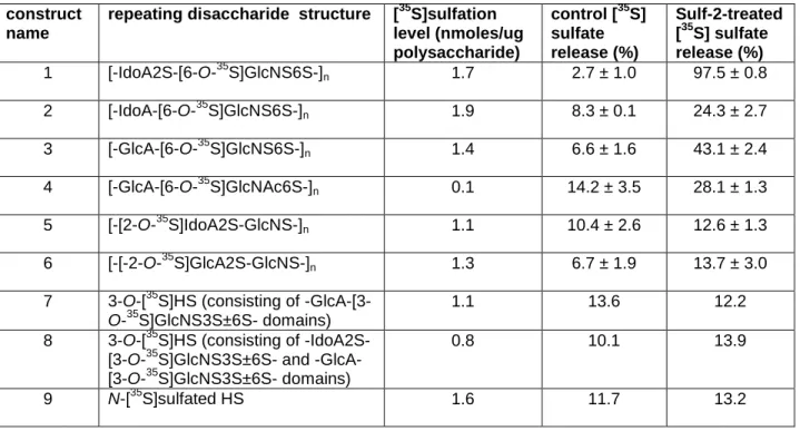
containing N-, 2-O- and 6-O-sulfations. Further analysis of the susceptibility of the different sulfated polysaccharides to [35S]sulfate release permitted us to dissect the substrate specificities of Sulf-2.
construct name
repeating disaccharide structure [35S]sulfation level (nmoles/ug polysaccharide)
control [35S] sulfate release (%)
Sulf-2-treated [35S] sulfate release (%) 1 [-IdoA2S-[6-O-35S]GlcNS6S-]n 1.7 2.7 ± 1.0 97.5 ± 0.8
2 [-IdoA-[6-O-35S]GlcNS6S-]n 1.9 8.3 ± 0.1 24.3 ± 2.7
3 [-GlcA-[6-O-35S]GlcNS6S-]n 1.4 6.6 ± 1.6 43.1 ± 2.4
4 [-GlcA-[6-O-35S]GlcNAc6S-]n 0.1 14.2 ± 3.5 28.1 ± 1.3
5 [-[2-O-35S]IdoA2S-GlcNS-]n 1.1 10.4 ± 2.6 12.6 ± 1.3
6 [-[-2-O-35S]GlcA2S-GlcNS-]n 1.3 6.7 ± 1.9 13.7 ± 3.0
7 3-O-[35S]HS (consisting of -GlcA-[3-O-35S]GlcNS3S±6S- domains)
1.1 13.6 12.2
8 3-O-[35S]HS (consisting of -IdoA2S-[3-O-35S]GlcNS3S±6S- and -GlcA-[3-O-35S]GlcNS3S±6S- domains)
0.8 10.1 13.9
9 N-[35S]sulfated HS 1.6 11.7 13.2
Table 3.1. Substrate specificity of Sulf-2. Synthetic substrates were incubated with or without the Sulf-2 enzyme and isolated from released sulfate groups by QuickSpin column. The remaining [35S]sulfate on the polysaccharide was quantified with a scintillation counter.
The susceptibility of the nine HS constructs to sulfatase activity indicated that in addition to trisulfated disaccharides, Sulf-2 is able to desulfate the 6-O-sulfo group on a GlcNS6S disaccharide that is flanked by a nonreducing-end GlcA or IdoA residue
41
Effect of Sulf-2 treatment on PF4 and antithrombin binding
To assess whether Sulf-2 could decrease the binding of HS to PF4, [35S]HS prepared from CHO cells was used due to its high specific [35S] radioactivity. A dot blot membrane binding assay was used to compare the PF4 binding capabilities of Sulf-2-treated and unSulf-2-treated HS (Fig. 3.4A). The wells contained 6,000 cpm of Sulf-2-Sulf-2-treated or untreated [35S]HS with increasing amounts of PF4. [35S]HS bound to PF4 was captured by the nitrocellulose membrane. The untreated samples reached a maximum binding of 66% with 152 nM PF4, but the Sulf-2-treated samples bound only up to 3.1% with 608 nM PF4. From the two binding curves, it is apparent that Sulf-2 treatment can reduce the binding of HS to PF4 by over ten-fold.
AT-binding HS was prepared by incubating HS from bovine kidney with 3OST-1 and [35S]PAPS. A Sulf-2-treated fraction was prepared by incubating this material with Sulf-2. Concanavalin A (ConA)-Sepharose beads were incubated with AT and HS, and the AT-bound HS fraction was eluted using a 1 M NaCl solution (Fig. 3.4). When the treated and untreated fractions were incubated with ConA-Sepharose and AT, 52% of the untreated fraction and 44% of the Sulf-2-treated fraction were recovered, suggesting that Sulf-2 does not remove critical 6-O-sulfo groups from the AT-binding pentasaccharide within the polysaccharide. [N-35S]HS, which does not considerably bind AT, was used as a negative control.
42
and 50,000 cpm of the AT-binding fraction of 3OST-1-treated [35S]HS from CHO cells was added. The gel was run for 2 h, dried overnight, imaged and analyzed using ImageQuant TL software. For the untreated and treated samples, a plot of R/[AT] vs. R gave linear slopes of y=-0.0949x + 0.048 (R2=0.87) and y=-0.094 + 0.44 (R2=0.95), respectively. These
correspond to Kd values of 10.5 and 10.6 nM, indicating that the binding affinity of HS to AT is unaffected by treatment with Sulf-2.
Figure 3.4. Effect of Sulf-2 treatment on PF4 and AT binding. A. PF4 binding assay comparing Sulf-2-treated and untreated radiolabeled heparan sulfate. [35S]HS obtained from CHO cells was incubated with varying amounts of PF4, and the solutions were directly applied to a nitrocellulose membrane. The membrane was then washed and subjected to radioactivity analysis using a scintillation counter. Filled circles: untreated HS; open circles: Sulf-2-treated HS. B. [35S]HS was incubated with 0.1 mg/mL AT, and the complex of AT and HS was captured using ConA-Sepharose beads. The AT-binding [35S]HS was prepared by incubating HS from bovine kidney with the 3OST-1 enzyme and [35S]PAPS.
43
respectively. These correspond to Kd values of 5.47 and 117.6 nM, showing that the binding affinity of HS for PF4 is decreased approximately 20-fold by treatment with Sulf-2. Taken together, our data suggest that Sulf-2 treatment decreases the binding affinity of HS to PF4 while the affinity to AT remains intact.
untreated [35S]HS Sulf-2-treated [35S]HS
AT 10.5 nM 10.6 nM
PF4 5.5 nM 118 nM
Table 3.2. Binding affinities of Sulf-2-treated and untreated HS to PF4 and AT. Kd values were
determined by affinity co-electrophoresis.
The lack of impact on AT binding led us to question whether the 3-O-sulfated glucosamine residue present at the center of the AT-binding pentasaccharide in HS was affected by Sulf-2 [126]. We compared the disaccharide composition of 3-O-[35S]sulfated HS with and without Sulf-2 treatment (Fig. 3.5). The disaccharide analysis revealed the
44
Figure 3.5. Disaccharide analysis of sulfated HS with and without Sulf-2 treatment. The 3-O-[35S]sulfated HS was prepared by incubating HS with purified 3-OST-1 enzyme and [35S]PAPS. A portion of the 3-O-[35S]sulfated HS was treated with Sulf-2. Both Sulf-2-treated and untreated HS were subjected to deacetylation with hydrazine followed by nitrous acid degradation at pH 4.5 and 1.5. The resultant disaccharides were analyzed by HPLC. Panel A shows the chromatogram of the analysis of untreated 3-O-[35S]sulfated HS. Panel B shows the chromatogram of the analysis of Sulf-2-treated 3-O-[35S]sulfated HS. The elution positions were identified by eluting with standards: 1 represents GlcA-AnMan3S, 2 represents IdoA2S-AnMan3S and 3 represents GlcA-AnMan3S6S. * indicates the unidentified components.
PF4 binding of oligosaccharides with anti-IIa activity
A small library of oligosaccharides was prepared by Dr. Yongmei Xu to investigate the relationship between the number of saccharide units and the anti-IIa/-Xa ratio [128]. The synthesis of these oligosaccharides was prepared by a novel method in which a
disaccharide starting material (obtained by the degradation of heparosan) is sequentially incubated with HS elongation enzymes (KfiA and pmHS2) and UDP-sugars (UDP-GlcNAc or UDP-GlcNTFA and UDP-GlcA) to add one saccharide unit per step to the HS backbone. The backbone can then be sulfated using HS biosynthetic enzymes as previously described.
The PF4 binding characteristics of four oligosaccharides ranging from a 15-mer to a 21-mer, some showing anti-IIa activity, were examined. The structures of the four


![Figure 3.1. Disaccharide composition of Sulf-2-treated and untreated [ 35 S]HS](https://thumb-us.123doks.com/thumbv2/123dok_us/8322191.2206050/48.918.146.801.241.712/figure-disaccharide-composition-sulf-treated-untreated-s-hs.webp)