MUSCLE ACTIVATION AND RANGE OF MOTION PATTERNS OF INDIVIDUALS WHO DISPLAY A LATERAL HIP SHIFT DURING AN OVERHEAD SQUAT
Kerry J. Peterson
A thesis submitted to the faculty of the University of North Carolina at Chapel Hill in partial fulfillment of the requirements for the degree of Master of Arts in the Department of Exercise & Sport Science in the College of Arts & Sciences (Athletic Training).
Chapel Hill 2015
Objective: Movement dysfunction increases lower extremity injury risks. This study identified modifiable factors (neuromuscular control [EMG] and ranges of motion) that contribute to dysfunctional movement (lateral hip shift) during an overhead squat.
Methods: Participants were assigned to the hip shift or control groups based on overhead squat performance. Gluteal and hip adductor EMG was sampled during the overhead squat. Hip internal and external rotation, hip abduction, knee extension, and dorsiflexion ranges of motion were assessed. Mixed-Model ANOVAs analyzed differences.
Results: The hip shift group had less hip abduction and gluteus medius activation in the limb shifted toward compared to the control group. No other differences were observed.
Conclusion: The EMG and range of motion measurement differences between groups may further increase the hip shift group’s injury risk. The differences observed may increase injury risk of both the limb shifted toward as well as the contralateral limb.
ABSTRACT
Kerry J. Peterson: Muscle Activation and Range of Motion Patterns of Individuals Who Display a Lateral Hip Shift During an Overhead Squat
iv
TABLE OF CONTENTS
LIST OF TABLES ………... vi
LIST OF FIGURES ………... vii
LIST OF ABBREVIATIONS ………...………….… viii
CHAPTER I: INTRODUCTION ………....…... 1
Research Questions ………....… 2
CHAPTER II: REVIEW OF LITERATURE ……….………....…... 8
Epidemiology ………... 8
Clinical Anatomy ………....…. 11
Muscle activation and dysfunctional movement patterns ………..….. 13
Lower extremity functional screenings ………....… 15
Functional screenings and biomechanical risk factors ………...………..… 17
Conclusion ………... 20
CHAPTER III: METHODOLOGY ………...….…. 22
Research design ….………...……….……...…...…..… 22
Participants ……….………... 23
Instrumentation ……… 23
Procedures ………....… 23
Screening Protocol ………... 23
Experimental Protocol ………...…….. 24
Maximum Voluntary Isometric Contractions (MVICs)...…….……...… 26
Overhead Squat ….………..………. 27
Data reduction ……….. 28
Statistical analysis ……… 29
Power Analysis ……… 29
CHAPTER IV: MANUSCRIPT ……….. 30
Introduction ……….. 30
Procedures ……… 33
Results ……….. 37
Discussion ……… 38
Practical Application ……….... 43
TABLES ………...……….….. 44
FIGURES ………...……….. 48
vi
LIST OF TABLES
TABLES ……….. 44
Table 1: Statistical Analysis ……….……… 44
Table 2: Power Analysis ……….. 44
Table 3: Reliability ……….. 44
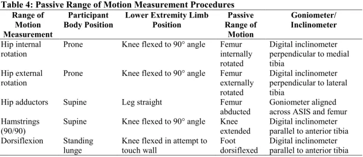
Table 4: Passive Range of Motion Procedures ………...…………. 45
Table 5: MVIC Procedures ………... 45
Table 5: EMG Descriptive Statistics ……… 46
LIST OF FIGURES
FIGURES………..……… 48
Figure 1: Control (CON) Group Subject ………...……….. 48
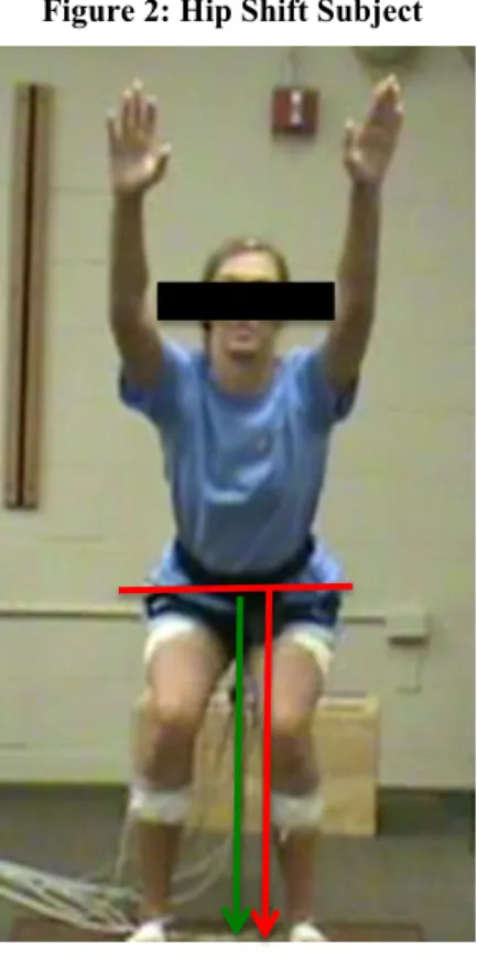
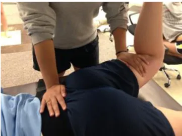
Figure 2: Hip Shift (HS) Group Subject ...………...……...………..48
Figure 3: EMG placement (GMAX, GMED)………..………...….. 48
Figure 4: EMG Placement (HADD) ………...……...….. 48
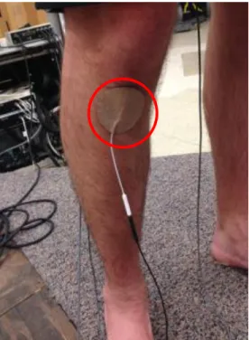
Figure 5: EMG placement (reference electrode)……….…….. 49
Figure 6: Flock of Birds placement (lower leg) …….………...…..……….. 49
Figure 7: Flock of Birds Placement (thigh) .……….…………...…..…….. 49
Figure 8: Flock of Birds Placement (sacrum)………..………...…….. 49
Figure 9: Passive range of motion (hip external rotation)………..……..………..…….. 50
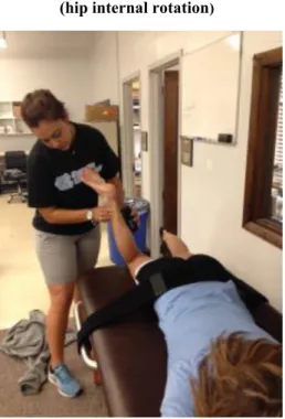
Figure 10: Passive range of motion (hip internal rotation)……….……….. 50
Figure 11: Passive range of motion (hip abduction)………...……….. 50
Figure 12: Passive range of motion (knee extension) ……….………...…….. 50
Figure 13: Passive range of motion (standing lunge) ...……….……….. 51
Figure 14: MVIC (hip extension) ……….………... 51
Figure 15: MVIC (hip abduction) ……….... 51
viii
LIST OF ABBREVIATIONS
ACL Anterior cruciate ligament LE Lower extremity
CHAPTER I
INTRODUCTION
Musculoskeletal injuries generate a large physical and financial toll.1 Collegiate sport related injuries occur at a rate of one injury every two games and one injury every five
practices;2 over 50% of these injuries affect the lower extremity.2 Forty percent of all collegiate injuries2 and 70% of anterior cruciate ligament (ACL) injuries are the result of non-contact mechanisms. Non-contact injuries may result from intrinsic factors including muscle strength, flexibility, and activation, and faulty biomechanics.2,3 Previous research has identified abnormal muscular activation patterns4-6 and lower extremity range of motion differences which contribute to faulty movement patterns that may increase injury risk.7-9 Greater hip adduction kinematics has been linked to ACL injuries, osteoarthritis, iliotibial band syndrome, and tibial stress
fractures.5,10-14 Similarly, greater hip adduction and internal rotation, and less dorsiflexion ranges of motion have been found in individuals with patellofemoral pain syndrome.9,15
Clinical movement screenings can identify individuals who display dysfunctional
movement patterns and are potentially at increased risk of injury. Clinical movement screenings include the overhead squat,16 single leg squat,17 single leg step-down,18 and jump-landing.18,19 Excessive hip adduction is a commonly observed dysfunctional movement pattern during
2
lateral hip shift. Hip adduction has also been linked to excessive knee valgus angle during the jump-landing task. 9 There are a number of factors that contribute to this and other dysfunctional movement patterns observed during movement screenings.
Previous research has established relationships between neuromuscular control, passive range of motion measurements, and dysfunctional movement patterns.4-6,21 Proximal lower extremity muscular activation patterns have been theorized to affect distal joint positioning. Individuals who display knee valgus during squatting tasks display smaller gluteal to hip adductor co-activation ratios compared to those who maintain a neutral knee alignment.4,5,10 Similarly, greater hip adductor activation has been linked to greater hip adduction motion.22 Hip adduction has also been linked to less dorsiflexion and greater hip internal rotation motion during squat and step-down tasks.5,10,11 However, additional research is needed to better understand the relationships between muscle activation patterns, lower extremity ranges of motion, and
dysfunctional movement patterns.
Therefore, the purpose of this study is to determine hip muscular activation and lower extremity range of motion patterns that contribute to lateral hip shift during the overhead squat. Once the contributing factors are identified, clinicians will be better able to develop intervention programs and improve movement quality. Correction of dysfunctional movement patterns will aid in reducing the risk of injuries.
Research Questions and Hypotheses:
RQ1: What are the differences in hip muscular activation patterns in individuals who display a
RQ1a: How does gluteus maximus muscle activation compare in individuals displaying a
lateral hip shift compared to individuals who maintain neutral pelvic alignment?
RH1a1: We hypothesize that individuals displaying a lateral hip shift will have greater
activation of the gluteus maximus on the ipsilateral limb compared to individuals who maintain neutral pelvic alignment.
RH1a2: We hypothesize that individuals displaying a lateral hip shift will have less
activation of the gluteus maximus on the contralateral limb compared to individuals who maintain neutral pelvic alignment.
RQ1b: How does gluteus medius activation compare in individuals displaying a lateral hip
shift compared to individuals who maintain neutral pelvic alignment?
RH1b1: We hypothesize that individuals displaying a lateral hip shift will have less
gluteus medius activation on the ipsilateral limb compared to individuals who maintain neutral pelvic alignment.
RH1b2: We hypothesize that individuals displaying a lateral hip shift will have less
gluteus medius activation on the contralateral limb compared to individuals who maintain neutral pelvic alignment.
RQ1c: How does hip adductor activation compare in individuals displaying a lateral hip shift
compared to individuals who maintain neutral pelvic alignment?
RH1c1: We hypothesize that individuals displaying a lateral hip shift will have greater
4
RH1c2: We hypothesize that individuals displaying a lateral hip shift will have less hip
adductor activation on the contralateral limb compared to individuals who maintain neutral pelvic alignment.
RQ2: What are the differences in lower extremity passive range of motion (flexibility) in
individuals who display a lateral hip shift compared to individuals who maintain neutral pelvic alignment?
RQ2a: What is the difference in hip internal rotation range of motion in individuals who
display a lateral hip shift compared to individuals who maintain neutral pelvic alignment? RH2a1: We hypothesize that individuals displaying a lateral hip shift will have greater hip internal rotation on the ipsilateral limb compared to individuals who maintain neutral pelvic alignment.
RH2a2: We hypothesize that individuals displaying a laterals hip shift will have less hip
internal rotation on the contralateral limb compared to individuals who maintain neutral pelvic alignment.
RQ2b: What are the differences in hip external rotation range of motion in individuals who
display a lateral hip shift compared to individuals who maintain neutral pelvic alignment? RH2b1: We hypothesize that individuals displaying a lateral hip shift will have less hip
external rotation on the ipsilateral limb compared to individuals who maintain neutral pelvic alignment.
RH2b2: We hypothesize that individuals displaying a lateral hip shift will have greater
RQ2c: What are the differences between hip abduction range of motion of the ipsilateral leg
in individuals displaying a lateral hip shift compared to individuals who maintain neutral pelvic alignment?
RH2c1: We hypothesize that individuals will have less hip abduction on the ipsilateral
limb compared to individuals who maintain neutral pelvic alignment.
RH2c2: We hypothesize that individuals will have greater hip abduction on the
contralateral side compared to individuals who maintain neutral pelvic alignment. RQ2d: What are the differences between ankle dorsiflexion range of motion on the
ipsilateral leg in individuals displaying a lateral hip shift compared the contralateral side? RH2d1: We hypothesize that individuals will have less range of motion on the ipsilateral
limb compared to individuals who maintain a neutral pelvic alignment.
RH2d2: We hypothesize that individuals will have less range of motion on the
contralateral limb compared to individuals who maintain a neutral pelvic alignment.
RQ3: What are the differences in hip muscular activation patterns on the ipsilateral limb
compared to the contralateral limb in individuals who display a lateral hip shift during an overhead squat?
RQ3a: How does gluteus maximus muscle activation compare in the ipsilateral limb
compared to the contralateral limb in individuals who display a lateral hip shift?
RH3a1: We hypothesize that individuals will have greater activation of the gluteus
maximus on the ipsilateral limb compared to the contralateral limb.
RQ3b: How does gluteus medius activation compare in the ipsilateral limb compared to the
6
RH3b1: We hypothesize that individuals will have less gluteus medius activation on the
ipsilateral limb compared to the contralateral limb.
RQ3c: How does hip adductor activation compare in the ipsilateral limb compared to the
contralateral limb in individuals who display a lateral hip shift?
RH3c1: We hypothesize that individuals will have greater hip adductor activation on the
ipsilateral limb compared to the contralateral limb.
RQ4: What are the differences in lower extremity passive range of motion (flexibility) on the
ipsilateral limb compared to the contralateral limb in individuals who display a lateral hip shift during an overhead squat?
RQ4a: What is the difference in hip internal rotation range of motion in the ipsilateral limb
compared to the contralateral limb in individuals who display a lateral hip shift?
RH4a1: We hypothesize that individuals will have greater hip internal rotation on the ipsilateral limb compared to the contralateral limb.
RQ4b: What is the difference in hip external rotation range in the ipsilateral limb compared
to the contralateral limb in individuals who display a lateral hip shift?
RH4b1: We hypothesize that individuals displaying a lateral hip shift will have less hip
external rotation on the ipsilateral limb compared to the contralateral limb.
RQ4c: What is the difference between hip abduction range of motion in the ipsilateral limb
compared to the contralateral limb in individuals who display a lateral hip shift?
RH4c1: We hypothesize that individuals will have less hip abduction on the ipsilateral
limb compared to the contralateral limb.
RQ4d: What is the difference between ankle dorsiflexion range of motion in the ipsilateral
RH4d1: We hypothesize that individuals will have less range of motion on the ipsilateral
8 CHAPTER II
REVIEW OF LITERATURE
Lower extremity injuries are common at the high school, recreation, collegiate, and professional levels of athletic competition.2,23 Therefore, identifying mechanisms resulting in increased injury risk becomes important. One well-established injury risk factor is dysfunctional movement patterns during activity.5,24,25 This review will discuss hip muscular activation and lower extremity range of motion patterns that contribute to dysfunctional movement patterns, specifically hip adduction resulting in a visually observed lateral hip shift. Primarily,
biomechanical risk factors (dysfunctional movement patterns) predisposing individuals to injury will be addressed. Additionally, functional movement screenings used to observe dysfunctional movement patterns will be compared and analyzed. Theorized neuromuscular characteristics contributing to dysfunctional movement patterns during functional tasks will be evaluated. Finally, this review will explore range of motion patterns contributing to dysfunctional movement patterns during functional tasks.
Epidemiology
syndrome, ilitobial band stress syndrome, anterior cruciate ligament (ACL), and medial collateral ligaments (MCL) sprains, and acute and chronic ankle sprains. Functionally, greater femoral rotation results in patellofemoral pain syndrome and MCL injuries.26,27 During functional tasks, ACL injuries have been associated with greater femoral internal rotation and increased hip adduction moment.28,29 Similarly, greater hip adduction has been found in individuals experiencing iliotibal stress syndrome. The dysfunctional movement patterns contributing to these injuries have been hypothesized to result from abnormal muscle activation patterns and range of motion abnormalities.
Patellofemoral pain syndrome (PFP) is a chronic knee injury that is commonly diagnosed in active populations. PFP results from abnormal patellar tracking and increased surface contact of the patella and femur. This malalignment can be caused by asymmetrical muscle activation and bony alignment. A greater lateral pull of the quadriceps on the patella, increases contact and patellofemoral stress.20 The lateral pull is increased by greater femoral adduction, internal rotation or external tibial rotation.20 Individuals suffering from PFP have greater hip adduction during movement and land in a more adducted position compared to matched control
individuals.30 Increased lateral stress on the knee is also a contributor to iliotibial band syndrome, another chronic knee injury.
Iliotibial band syndrome is common in sports with repetitive movement patterns of knee flexion and extension. Individuals with a previous history of iliotibial band syndrome
10
increased strain on the iliotibial band and iliotibial band syndrome.31 Excessive hip adduction and knee internal rotation is also known to contribute to acute lower extremity injuries such as ACL and MCL sprains.
Non-contact ACL injuries are highly prevalent, 70% of all ACL injuries, and commonly result from faulty biomechanical movement patterns.28,32 Several biomechanical risk factors have been identified as contributors of increased injury risk. Dynamic knee valgus, an inward
movement of the knee, has been established as one of the primarily identified faulty movement patterns.32,33 In addition, greater foot pronation, tibial internal rotation, and minimal hip and knee flexion are risk factors for injury during cutting tasks.32 Greater femoral adductor torque can increase knee abduction moment, which contributes to peak ground force reaction and increased joint load.29 MCL and medial meniscus injuries commonly occur concomitantly with ACL injuries, in what is known as the unhappy triad. 34
MCL injuries commonly occur during athletic activities, at a rate of approximately
74,000 annually in the United States.27 Injury to the MCL occurs when excessive valgus force or external rotation is applied to the knee.27 Tibial external rotation commonly occurs in
conjunction with femoral internal rotation, in an effort to maintain neutral knee alignment, and results in the MCL becoming taut.
instability demonstrated greater frontal plane knee displacement compared to a healthy
population.24 The greater frontal plane knee displacement may be caused by distal abnormalities at the ankle due to injury or by lumbo-pelvic hip dysfunction.25,36 Greater knee frontal plane displacement may be the result of less gluteal muscle activation.37
Anatomy
The study of human anatomy allows for an understanding of how structures within the human body function together. The pelvic girdle is comprised of paired hip bones connected anteriorly by the pubic symphysis and posteriorly by the sacrum. The hipbones are each comprised of three bones: the ilium, the ischium, and the pubis. These three bony components fuse together to form a suture in the acetabulum; the socket of the hip joint. The hip joint is comprised of the femoral head rotating inside of the acetabulum of the pelvis. The hip joint allows for motion to occur in all three planes of motion.
Hip transverse plane motion consists of femoral internal and external rotation. The hip external rotators rotate the femur away from the midline. The primary femoral external rotators are the piriformis, obturatus internus and externus, gemellus superior and inferior, and the quadratus femoris.20 The gluteus maximus also acts as a femoral external rotator. The adductor magnus and the posterior fibers of the gluteus medius can also serve as secondary external rotators. However, the anterior fibers of the gluteus medius act as a femoral internal rotator and the main roll of the gluteus medius is to assist with hip abduction.
12
adductors and abductors primarily control frontal plane hip motion. The hip adductors work antagonistically to the hip abductor muscle group. The hip adductor complex is comprised of the adductor longus, adductor magnus, adductor brevis, pectineus, and gracilis. These muscles produce hip adduction, which results in the femur moving toward the midline of the body. Around 30° of hip flexion the direction of pull of the adductor muscles change, placing them in a position to generate hip extension.40 The adductor magnus, adductor longus, and adductor brevis also act as internal rotators due to their medial attachment on the femur.40
epicondyle of the femur to the proximal portion of the medial border of the patella.45 It is the main passive restraint to lateral translation of the patella.45
Bony, muscular, and ligamentous anatomy are the mechanical contributors to bodily movements. Within the pelvo-femoral hip complex, the femoral head, acetabulum and pelvis provide bony support. The hip musculature and ligament structures provide additional support as well as trunk and lower extremity movement. Distally, passive and dynamic stabilizers support the knee. The knee is subjected to stress due to its placement on the lower extremity and is greatly affected by asymmetrical changes between limbs. Anatomical changes and dysfunctional movement patterns may cause muscle activation differences range of motion inequalities.
Muscle Activation and Dysfunctional Patterns
Atypical muscle activation patterns have been linked to abnormal movement patterns. Previously, distal muscular patterns involving the knee and ankle have been the focus of lower extremity research.46,47 Recently, proximal neuromuscular characteristics involving the pelvo-femoral-hip-complex have been studied in greater relation to injury predisposition.4,5,19
14
Previously, strength was thought to be a major contributor to pelvic and lower extremity alignment. However, research has demonstrated that strength is not a primary factor driving dysfunctional movement patterns.51 Instead, underactivity of the hip abductors5 may allow for hip adduction and knee valgus movement to occur. Evidence has shown gluteus medius activation is delayed and of shorter duration in individuals with PFP.50,52 It is theorized that individuals that display hip adduction during functional movements, have less hip abductor activation compared to those who do not display hip adduction.53 Less hip abductor activation may not be capable of balancing hip adductor activity, which is demonstrated by a smaller gluteal to hip adductor co-activation ratio.4,5 Postural alignments can also influence muscular activity during tasks. Postural hip adduction places the hip abductors in an elongated position, which alters length-tension relationships and may result in less or delayed hip abductor
activation.54 Altered hip abductor activity can result in abnormal lower extremity biomechanics. Multiple muscle activation imbalances have been associated with greater femoral internal rotation.25 The femoral external rotators can aid in limiting femoral internal rotation. For
Excessive femoral internal rotation has been identified as a lower extremity injury risk factor. Individuals with chronic knee pain exhibited greater femoral internal rotation. 15,57 Greater femoral internal rotation may lead to malalignment of the patella and increased contact surface on the lateral facets of the patella.58 Greater contact forces may lead to chondral degeneration and PFP symptoms.25 Greater femoral internal rotation may result from bony anatomical or neuromuscular factors. Neuromuscular factors include greater hip adductor15 and less gluteal activation57 during functional tasks.
Lower Extremity Functional Movement Screenings
Sports medicine clinicians utilize lower extremity functional movement screenings to visually observe lower extremity kinematics during athletic tasks. Depending on the demands of the physical activity, clinicians may use single-leg and double-leg cutting, squatting, or jumping tasks. Through observation of these tasks, clinicians are able to identify faulty movement
patterns that may increase an individual’s risk for injury. Once dysfunctional patterns are identified, flexibility and strengthening programs can be implemented to correct muscle imbalances and improve performance.
16
an asymmetrical (lateral) hip shift. The overhead squat allows for the observation of compensatory movements, which help identify abnormal muscle activation patterns.25,49
The single leg squat single leg squat is another common functional screening tool.4,18 The single leg squat requires greater neuromuscular control and muscle activation than a double-legged position due to decreased stability. The single leg squat may be affected by poor core control, hip musculature strength, range of motion, or muscle activation.4,56 Females who display hip adduction during the single leg squat demonstrate a loss of dynamic control, or ability to maintain a neutral pelvis, at the beginning and end of the squat.53 Excessive hip adduction may present with trunk movements toward the stationary leg in order to compensate for the adduction motion.59
The jump-landing jump-landing task requires participants to resist a downward
acceleration of the body and then immediately produce an upward force.46 This task is able to differentiate biomechanics in the frontal, transverse, and sagittal place, as well as ground reaction forces to determine individuals predisposed to knee injury.33 Faulty biomechanics observed during the jump-landing include less hip flexion angle, greater knee valgus, greater hip adduction angle, greater femoral and knee internal rotation, and greater hip extension force.33 Knee
abduction was more prevalently found during double leg functional screening tasks (jump-landing) as opposed to single-legged tasks.18 Previous research demonstrated that knee abduction counters adduction at the hip to achieve neutral alignment.5
The Trendelenburg Test was originally designed as a test for hip abductor strength during single leg stance. A positive sign consists of the non-stance ilium moving into a lower position than the stance ilium.62 Lowering of the stance limb ilium results in functional pelvis-on-femoral hip adduction. A positive Trendelenburg Test may also be indicative of underactivity of the gluteus medius of the stance leg.62 The gluteus medius underactivity may be indicative of an unequal hip adductor to gluteal ratio. Therefore, while the gluteus medius is underactive, simultaneously, the hip adductors may be overactive causing a greater adducted position during the single leg stance.
Functional Screenings and Biomechanical Risk Factors
Hip adduction is observed during functional screenings as a risk factor for injury. Greater hip adduction and internal rotation of the femur force the tibia to abduct and the foot to pronate resulting in dynamic knee valgus.63 Due to this position and movement compensations,
18
excessively adducted position results in increased joint forces throughout the knee.63 Clinically, hip adduction is described as a lateral hip shift or asymmetrical hip shift with movements in one lateral direction during an overhead squat. During a hip shift, the ipsilateral leg must allow for lateral movement of the pelvis to maintain alignment over the leg.
Hip adduction movement is associated with greater hip adductor activation.4,5,15,56 Furthermore, individuals who display hip adduction, contributing to medial knee displacement, during common clinical movement screenings display smaller gluteal to hip adductor co-activation ratios compared to those individuals who maintain a neutral knee alignment.4,5 Individuals displaying a lateral shift during an overhead squat may increase adductor activation on the ipsilateral leg and decrease activation on the contralateral leg to allow for the shift to occur. Hip adduction is also associated with greater femoral rotation in individuals with dynamic knee valgus, PFP15, and iliotibial band syndrome14.
Greater femoral internal rotation has been established as a factor contributing to chronic injuries.63 Similarly, it has been linked to acute injury risk factors, such as medial knee
displacement.48 Femoral internal rotation may occur as a compensatory movement to ensure normal knee mechanics when abnormal pronation and excessive tibial internal rotation are present.20 Similarly, external tibial rotation acts as a compensatory movement to increased femoral internal rotation.48 Excessive femoral internal rotation contributes to dynamic knee valgus motion.48 However, femoral internal rotation has not been researched as an isolated factor leading to injury.
During a step down task, hip adduction is found to strongly correlate with knee valgus.55 However, true knee valgus collapse is not typically seen unless the individual is injured. Therefore, researchers primarily focus on identifying excessive knee valgus during functional tasks because it is a well-established lower extremity injury risk factor.25,47,49 Medial knee displacement medial knee displacement is the observed visual appearance of knee valgus motion.5 Previous research has found that greater muscle activation of the hip adductors,5,6 gastrocnemius,4,5,and tibialis anterior5 occurs in participants displaying medial knee
displacement compared to the control group.5 More recent studies have identified hip adduction as a predisposing factor for lower extremity injury.4,5,48 However, hip adduction has only been established as an attribution to medial knee displacement.4,5,22 Similarly, decreased ankle
dorsiflexion is associated with medial knee displacement and has been identified as an important factor in proper kinematics during functional activities.46,49
Distally, ankle range of motion dynamically contributes to faulty movement patterns. Tightness of the plantar flexor muscles, the medial and lateral gastrocnemii and the soleus, are the primary restrictors to dorsiflexion, which has been linked to altered movement patterns.4,49,47 The primary limiting factor to normal ankle dorsiflexion is the eccentric restriction of the gastrocnemius.64 Overactivity of the gastrocnemius and soleus may present as calcaneal eversion, foot pronation, tibial internal rotation, and medial knee displacement.65 Decreased dorsiflexion during weight-bearing tasks results in pronation and tibial internal rotation to achieve additional stabilization and full body lowering.8
20
passive range of motion in individuals with medial knee displacement during an overhead squat task. In one study, individuals presenting with medial knee displacement, report with 42% greater gastrocnemius activation compared to the control group.5 When both the gastrocnemius and tibialis anterior have increased coactivation, restricted range of motion may occur, which can limit ankle dorsiflexion.5, 25
Previous research restricted ankle dorsiflexion through the use of a wedge under the forefoot during a double-legged squat. With restricted dorsiflexion, participants display a significant increase in knee valgus alignment compared to the same group squatting without a wedge.47 Similarly, individuals with medial knee displacement display approximately 20% less dorsiflexion range of motion with the knee in a flexed position.25 Greater dorsiflexion range of motion is associated with greater knee-flexion displacement and smaller ground reaction forces during landing activites.46 This landing position is one of decreased injury risk and reduces the forces absorbed through the lower extremities. Restricted dorsiflexion range of range of motion is linked to injuries to the ACL, MCL, meniscus66 and chronic knee injuries such as
patellofemoral pain syndrome.8
Conclusion
Greater femoral internal rotation has also be associated with increased risk of injury.48 Greater rotation may be due to bony alignment or decreased activation of the deep external rotator or the gluteal muscles. Abnormal distal kinematics may force increased internal femoral rotation in order to achieve proper mechanics.48 During squat screenings, greater femoral rotation may occur to allow for full range of motion at the knee.
Limited ankle dorsiflexion has found to be a contributor to faulty movements throughout proximal lower extremity portions. Similarly, decreased dorsiflexion has been observed in individuals with both acute and chronic injuries. Decreased dorsiflexion is associated with smaller knee flexion angle, increased knee valgus, and increased ground reaction forces.46
The majority of research has focused on knee valgus and muscular activation patterns at the knee. Hip kinematics need to be further studied and better understood. Research is needed for isolated hip adduction to establish what muscular activation patterns and range of motion
22 CHAPTER III
METHODOLOGY
Research Design
This study was conducted as a cross-sectional between groups comparison study. The subjects were separated into a control (group or hip shift group. Lower extremity muscle activation patterns and passive range of motion measurements were compared between individuals who display a lateral hip shift and those who maintain a neutral pelvic alignment during an overhead squat task.
Participants
Forty individuals (20 males, 20 females) healthy, physically active males and females aged 18-35 who were in good general health and participated in a minimum of 30 minutes of physical activity 3 days a week participated in this study. Participants were free of lower extremity or low back injury at the time of and for a minimum of 6 months prior to data
Instrumentation
The TrackStar electromagnetic motion-analysis system (Version 8.0; Ascension Technology Corporation, Burlington, VT) interfaced with two non-conductive force platforms was used to collect kinematic and kinetic data. A surface electromyography (EMG) system (model Bangoli-8; DelSys Incorporated, Boston, MA) with an interelectrode distance =10mm was used to sample muscle activity. Two two-dimensional (2D) video cameras (DCR-HC38 MiniDV Handycam Camcorder, Sony Electronics, San Diego, California) were used to capture subject motion and confirm group assignment during the screening protocol. Lower extremity passive range of motion was measured using a digital inclinometer (Saunders Group, Inc. Chaska, MN, USA) and standard 8-inch plastic goniometer.
Procedures
Participants reported to the Sports Medicine Research Laboratory for a screening session, and within one week returned for a single testing session wearing their own athletic shorts and shirt; participants were barefoot throughout the testing procedures.
Screening Protocol
Participants completed a health questionnaire to confirm inclusion in the study and subject demographics (eg. height and weight) were recorded. Participants completed a 5-minute warm-up on a stationary cycle ergometer at a self-selected pace. The screening protocol
consisted of 5 consecutive overhead squats to a squat depth comfortable to the participant, but a minimum of 60 of knee flexion. Participants stood with their feet shoulder width apart on two force platforms; tape was placed under the participant’s feet to serve as visual cues for
24
with their toes pointing straight ahead and arms extended overhead. Squat speed was controlled through the use a metronome set at 60 Hz;49 participants descended for two beats, ascended for two beats and paused for 1 beat between squats. Participants completed 5 practice repetitions of the overhead squat or until they felt comfortable with the task. A 1-minute rest period was allowed between completion of the practice trials and data collection.
Participants did not receive feedback other than what constituted a successful trial. A trial was deemed successful if: 1) the head remained facing forward, 2) the toes remained pointing forward, 3) the task was completed at the appropriate speed, and 5) the task was completed in a fluid motion. Participants were visually observed by the primary investigator so that group assignment could be determined.
Participants were placed in the control group (figure 1) if during at least 3 of the 5
repetitions the mid-sagittal line maintained neutral alignment. Participants were placed in the hip shift group (figure 2) if in at least 3 of the 5 repetitions the mid-sagittal line bisecting the body shifted laterally. Participants were not informed as to which group they were placed in, to avoid possibly influencing performance on future trials.
Experimental Protocol – Data Collection Session
Participants completed the data collection session within 1 week of the screening session. The experimental protocol consisted of passive range of motion measurements, maximum
were placed bilaterally over the muscle bellies of the 3 muscles of interest (gluteus maximus, gluteus medius, and hip adductors), as previously described.5,49,67 A reference electrode was placed bilaterally just medial to the tibial tuberosity of the ipsilateral limb. Electrode sites were identified, marked, shaved, abraded, and cleaned with 70% isopropyl alcohol. Electrodes and leads were secured with prewrap and athletic tape.
Passive Range of Motion
Passive range of motion was measured for hip internal rotation hip external rotation, hip abduction, knee extension, and standing weight bearing lunge ankle dorsiflexion. The following testing procedures were utilized for each range of motion measurement:
Hip Internal Rotation: The participant was placed in a prone position with the non-test
limb flat on the table. The test limb was flexed at the knee and the thigh was placed flat on the table. The participant’s hips were stabilized on the table with a strap placed over the sacrum and around the table. The participant’s hip was internally rotated to the point of first tissue resistance or the participant expressed discomfort (figure 10). A digital inclinometer was placed parallel to the length of the medial tibia; the measurement was taken with respect to the vertical axis.
Hip External Rotation: The participant was placed in a prone position with the non-test
limb flat on the table. The test limb was flexed at the knee and the thigh was placed flat on the table. The participant’s hips were stabilized on the table with a strap placed over the sacrum and around the table. The participant’s hip was externally rotated to the point of first tissue resistance or the participant expressed discomfort (figure 9). A digital inclinometer was placed parallel to the length of the fibula; the measurement was taken with respect to the vertical axis.
Hip Abduction: The participant was placed in a supine position with both the non-test
26
participant’s hips were stabilized on the table with a strap placed over the anterior superior iliac crests (ASIS) and around the table. The participant’s hip was abducted to the point of first tissue resistance or the participant expressed discomfort (figure 11). The stationary arm of a standard goniometer was placed across the two ASIS and the moving arm was placed in line with the midline of the femur of the test leg.
Knee Extension: The participant was placed in a supine position with the non-test limb
flat on the table. The test limb was flexed at the knee and the hip. The participant stabilized the test limb by holding the posterior thigh in this position. The hips were further stabilized on the table with a strap placed over the anterior superior iliac crests (ASIS) and around the table. The participant’s knee was extended to the point of first tissue resistance or the participant expressed discomfort (figure 12). An inclinometer was placed along the anterior aspect of the tibia of the test limb; the measurement was taken with respect to the horizontal axis.
Standing Weight Bearing Lunge: The participant was placed in a weight bearing lunge
position with the test limb in front of the non-test limb. The test limb knee and hip were flexed in an attempt to touch the test limb knee to the wall while maintaining heel contact with the ground (figure 13). The non-test limb was extended at the knee and hip. The inclinometer was placed along the anterior aspect of the tibia of the test limb; the measurement was taken with respect to vertical.
Maximal Voluntary Isometric Contractions
sampled at 1400 Hz. The following testing positions were utilized for testing of the gluteus maximus (GMAX), gluteus medius (GMED), and hip adductors (HADD):
Gluteus Maximus: The participant was placed in a prone position with the non-test limb
flat on the table. The test limb was flexed at the knee and the thigh was placed in extension, just past neutral alignment, so that the anterior thigh was not in contact with the table. The
participant’s hips were stabilized on the table with a strap placed over the sacrum and around the table. The participant contracted against gravity and manual resistance applied by the primary investigator, just proximal to the popliteal fossa (figure 14).
Gluteus Medius: The participant was placed in a side-lying position with the non-test
limb flat on the table. The test limb knee and the thigh were placed in extension, just past neutral alignment, so that the medial thigh was not in contact with the non-test limb. The participant’s hips were stabilized on the table with a strap placed over the iliac crest and around the table. The participant contracted against gravity and manual resistance applied by the primary investigator, just proximal to the lateral femoral epicondyle (figure 15).
Hip Adductors: The participant was placed in a side-lying position with the test limb flat
on the table. The non-test limb was placed in hip and knee flexion over the top of the test limb, so the sole of the foot was flat on the table. The test limb knee and thigh were placed in
extension, just past neutral alignment, so that the lateral thigh was not in contact with the table. The participant’s hips were stabilized on the table with a strap placed over the iliac crest and around the table. Participants contracted against gravity and manual resistance applied by the primary investigator, just proximal to the medial epicondyle (figure 16).
Overhead Squat Task
28
Kinematic and EMG data were collected and analyzed during the descent phase of the squat. The descent phase was defined as the time from initiation of knee flexion until peak knee flexion. Kinetic data was sampled at 140 Hz and kinematic data was sampled at 1400 Hz. The x-y-z global axes were established according to the right-hand 3-dimensional Cartesian coordinate system. The positive x-axis was designated forward, the positive y-axis to the left, and the positive z-axis upward relative to the participant. The pelvis and bilateral lower extremity were calculated as motion of the thigh relative to the pelvis, the shank relative to the thigh, and the foot relative to the shank. Hip joint centers were estimated using the Bell method.68 Knee and ankle joint centers were estimated as the midpoint between the medial and lateral femoral epicondyles and malleoli, respectively.4,5
Data Reduction
extremity bony landmarks were estimated using MotionMonitor software and established based on Euler angles.
Statistical Analyses
Separate mixed-model Analyses of Variance (ANOVAs) with 1-between subject factor (group: control and hip shift) and 1-within subject factor (limb: toward and away) were used to compare each of the dependent variables. Due to the directional hypotheses, the alpha level was set a priori at 0.10 for the omnibus ANOVA models. Post hoc analyses were performed using t-tests with a Bonferroni corrected alpha level (α≤0.025). See Table 1 for a breakdown of our statistical analyses.
Power analysis
A power analysis was conducted based off of previously published work by Bell, et al.49 Muscle activity and flexibility in individuals with medial knee displacement during the overhead
squat. The calculated sample size was tripled to allow for Bonferroni corrections and still ensure
30
CHAPTER IV
MANUSCRIPT
Introduction
Musculoskeletal injuries generate a large physical and financial toll.1 Collegiate sport related injuries occur at a rate of one injury every two games and one injury every five
practices;2 over 50% of these injuries affect the lower extremity.2 Forty percent of all collegiate injuries2 and 70% of anterior cruciate ligament (ACL) injuries result from non-contact
mechanisms, potentially resulting from intrinsic factors including strength, flexibility, muscle activation, and faulty biomechanics.2,3
Proximal hip muscular activation patterns and asymmetrical biomechanical patterns have been theorized to affect distal joint positioning and increase lower extremity injury risk.5
Specifically, greater hip adduction motion is associated with greater ACL injury, tibiofemoral osteoarthritis, iliotibial band syndrome, patellofemoral pain syndrome and tibial stress fracture risk.5,10-14 During functional movement tasks, greater hip adductor activation has been linked to greater hip adduction motion.22 During squat and step-down tasks, individuals exhibiting hip adduction also had less dorsiflexion and greater hip internal rotation motion.5,20,63
limb maintains a neutral position or is abducted away from the midline. Clinically, this is observed as a lateral hip shift. There are a number of factors that contribute to this and other dysfunctional movement patterns observed during movement screenings. In order to correct dysfunctional movement patterns, it is important to understand the underlying neuromuscular patterns associated with these movements.
Previous research has identified abnormal muscle activation patterns4-6 and lower extremity range of motion measures that contribute to dysfunctional movement patterns.7-9 Dynamic knee valgus motion is a commonly identified dysfunctional movement pattern4,5,10 and a primary predictor of lower extremity injuries.32,36115,10,11 Dynamic knee valgus angle
combines the motions of hip and knee rotation and hip adduction on a fixed foot.3 Individuals who display excessive knee valgus angle during squatting tasks display smaller gluteal to hip adductor co-activation ratios compared to those who maintain a neutral knee alignment.4,5,10 Limited ankle dorsiflexion has also been identified as a contributor to medial knee displacement, the visual observation of excessive knee valgus angle, during functional movement screens as well as patellofemoral pain syndrome.4,25,26 However, to our knowledge there is no research examining the underlying neuromuscular and range of motion characteristics associated with lateral hip shift.
32
limb shifted towards compared to individuals who maintain a neutral pelvic alignment. We also hypothesized that individuals who display a lateral hip shift would have greater hip internal rotation, less hip external rotation, less hip abduction, and less dorsiflexion ranges of motion on the toward limb of the observed hip shift compared to individuals who maintain neutral pelvic alignment throughout the squat. We hypothesized to observe similar differences between the limbs being shifted toward and away from of the hip shift group, but no difference between limbs for the control limb.
Methods
Participants
All study procedures were approved by the university’s Institutional Review Board and all participants read and signed an informed consent form prior to data collection. Forty healthy, physically active males (20) and females (20) aged 18-35 participated in this study. Study
directions were also disqualified to further isolate the hip shift movement. Sixty-seven individuals were screened for the study, 27 did not qualify due to the presence of medial knee displacement (11), toe-out gait (7), heel raise (3) during the overhead squat or having already filled a group of participants (6). One participant qualified for the hip shift group during screening, but then presented with no shift at data collection and was disqualified. Procedures
Participants reported to the research laboratory for a screening session where they completed a health history questionnaire to confirm inclusion in the study and participant
demographics (eg. height, age, and mass) were recorded, and group assignment was determined. Participants returned to the research laboratory within one week of the screening session for a single testing session. Participants wore their own athletic shorts and shirt and were barefoot throughout the screening and testing sessions.
Screening Protocol
34
collection. A 1-minute rest period was provided between completion of the practice trials and data collection.
Participants did not receive feedback on their squatting techniques other than what constituted a successful trial. A trial was deemed successful if: 1) the head remained facing forward, 2) the toes remained pointing forward, 3) the task was completed at the appropriate speed, and 4) the task was completed in a fluid motion. Participants were visually observed by the primary investigator so that group assignment could be determined.
Participants were placed in the hip shift group if in at least 3 of the 5 repetitions the mid-sagittal line bisecting the body shifted laterally (Figure 2). Participants were placed in the control group if during at least 3 of the 5 repetitions the mid-sagittal line maintained neutral alignment (Figure 1). Participants were not informed as to which group they were placed in, to avoid possibly influencing performance on future trials.
Experimental Protocol – Data Collection Session
medial to the tibial tuberosity on both limbs. Electrode sites were identified by the primary researcher, marked, shaved, abraded, and cleaned with 70% isopropyl alcohol. Electrodes and leads were secured with prewrap and athletic tape. EMG data were sampled at 1400Hz.
The x-y-z global axes were established according to the right-hand 3-dimensional Cartesian coordinate system. The positive x-axis was designated forward, the positive y-axis to the left, and the positive z-axis upward, relative to the participant. Lower extremity joint angles were calculated as the motion of the thigh relative to the pelvis (hip), the shank relative to the thigh (knee), and the foot relative to the shank (ankle). Hip joint centers were estimated using the Bell method.68 Knee and ankle joint centers were estimated as the midpoint between the medial and lateral femoral epicondyles and malleoli, respectively.4,5 Three-dimensional coordinates of lower extremity bony landmarks were estimated using MotionMonitor software and established based on Euler angles. Kinematic data were sampled at 140Hz.
Passive Range of Motion
Passive ranges of motion were measured for hip internal rotation, hip external rotation, hip abduction, knee extension, and standing weight bearing lunge ankle dorsiflexion. The testing procedures utilized for each range of motion measurement are described in table 4.
Maximal Voluntary Isometric Contractions
Maximal voluntary isometric contractions (MVIC) were completed bilaterally.
Participants completed 3 separate 5-second MVIC trials for each muscle group. EMG data was sampled at 1400Hz. The testing positions utilized for the gluteus maximus, gluteus medius, and hip adductors MVIC are described in Table 5.
Overhead Squat
36 Data Reduction
Kinematic data were filtered using a 4th order Butterworth filter. Kinematic and EMG data were sampled during the descent phase of the squats. The descent phase was defined as the time from initiation of knee flexion until peak knee flexion. Peak knee flexion angle was
identified so that the descent phase of each squat could be identified. EMG data were bandpass (20Hz-350Hz) and notch (59-61HZ) filtered; EMG data were rectified and smoothed with a 25 ms sliding window function. EMG data during the overhead were averaged across the middle 3 squats for each of the 5 overhead squats and across all trials. EMG data sampled during the overhead was normalized to the mean maximum 1-second interval during the muscle’s
respective MVIC trial. The average EMG amplitude during the descent phase of the overhead squat was divided by the average EMG during the MVICs. Muscular electrical activity was recorded during the MVICs for overhead squat EMG normalization. Gluteal to hip adductor co-activation ratios were calculated by dividing normalized values of the gluteus maximus
activation by hip adductor activation, normalized gluteus medius activation by hip adductor activation, and averaged gluteal activation (gluteus maximus activity + gluteus medius activity / 2) by hip adductor activation. Range of motion data were averaged across the 3 trials. The hip total arc range of motion was calculated by adding the average hip internal range of motion and average hip external range of motion for each limb.
Statistical Analyses
Separate mixed-model Analyses of Variance (ANOVAs) with 1-between subject factor (group: hip shift and control) and 1-within subject factor (limb: toward and away hip shift) were used to compare each of the dependent variables. Due to the directional hypotheses, the alpha level was set a priori at α≤0.10 for the omnibus ANOVA models. Post hoc analyses were performed using t-tests with a Bonferroni corrected alpha level (α≤0.05).
Results
Hip Muscle Activation
There was a significant between group main effect for gluteus medius activation (F(1,34)
=3.17, p=.084). There were no significant group-by-limb interactions for any of the muscle activation variables: gluteus maximus (F(1,37) =2.02, p=0.145), gluteus medius (F(1,37) =0.186,
p=0.669); hip adductors (F(1,38) =0.591, p=0.447). Similarly, we observed no significant
group-by-limb interactions for the co-activation ratios: gluteus maximus/ hip adductors (F(1,38) =2.387,
p=.131), gluteus medius/ hip adductors (F(1,34) =0.232, p=0.633), or gluteals/ hip adductors (F(1,33)
=1.422, p=0.242). The only significant main effect for group or limb observed for the muscle activation or co-activation measures was the gluteus medius (F(1,34) =3.17, p=.084)). Means,
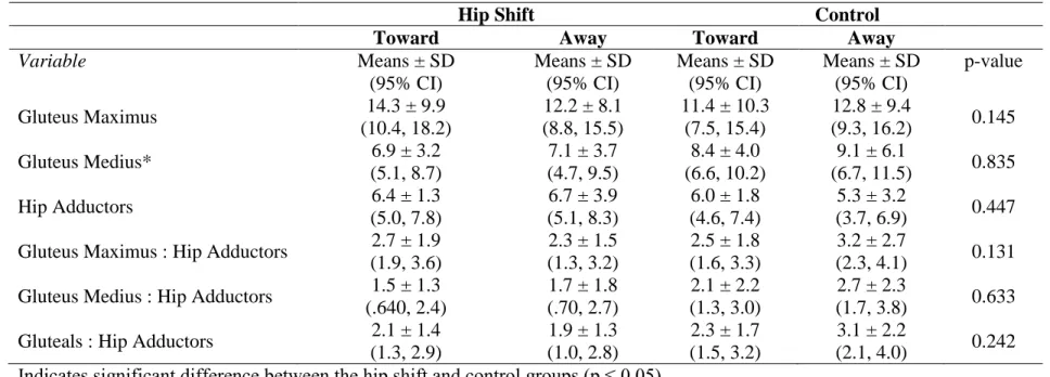
standard deviations, and 95% confidence intervals for all normalized EMG measures and co-activation ratios are presented in table 5.
.
Passive Range of Motion
There were significant group-by-limb interactions for hip abduction range of motion (F(1,38) =21.352, p=<.0005) as well as a significant main effect (F(1,38) =25.632, p<.0005).
38
F(1,38) =21.352, p=<.0005). No other significant group-by-limb interactions were found. A
significant main effect was found for dorsiflexion within the hip shift group (F(1,38) =4.703,
p=.036). Post-hoc testing revealed less dorsiflexion on the limb shifted towards compared to the limb shifted away from (p=.008). Femoral internal rotation range of motion was also statistically significant (F(1,38) =4.7888, p=.035). Specifically, individuals who presented with a hip shift had
greater internal rotation on the limb shifted toward compared to the limb shift away from. Similarly, a significant main effect was found for total arc ROM (F(1,38) =4.154, p=.049). The
limb being shifted toward presented with less total range of motion compared to the limb being shifted away from. No significant main effects for group or limb were observed for either hip external rotation or hamstring 90/90 range of motion measures (p>0.1). Means, standard
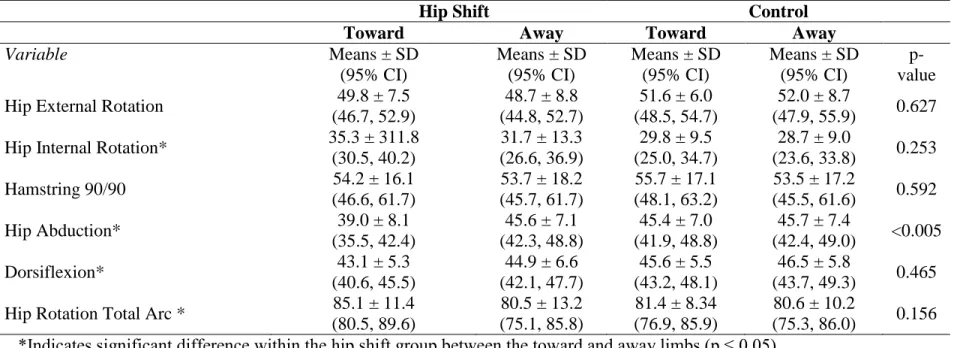
deviations, and 95% confidence intervals for all passive range of motion measures are presented in table 6.
Discussion
To the authors’ knowledge this is the first study to examine differences in hip muscle activation and passive ranges of motion measurements of individuals displaying a lateral hip shift during an overhead squat and those who do not. Individuals displaying a lateral hip shift presented with less hip abduction and decreased gluteus medius activation on the limb shifted toward compared to the control group. Within the hip shift group the limb shifted toward had less hip abduction and less dorsiflexion range of motion compared to the side being shifted away from (contralateral). The limb being shifted toward also had greater hip internal rotation and greater total hip arc ranges of motion compared to the side being shifted away from
We hypothesized that the limb being shifted toward would have more gluteus maximus activation overall; this was not supported by our data. The lateral hip shift results in femoral adduction and internal rotation, 20 this is similar to what occurs when an individual displays medial knee displacement.5 Therefore we assumed similar muscle activation patterns would be observed during the overhead squat. Previous research has shown that less gluteus maximus activation correlates to medial knee displacement during a single leg squat69 and single-limb step down.11 Similarly, previous research has demonstrated relationships between less gluteus
maximus activation and greater femoral internal rotation.48 Normalized gluteus maximus
activation may not have been statistically significant due to the large amount of variability in the data. We calculated coefficients of variation (CV = standard deviation / mean) for both the hip shift (CV = 0.68) and control groups (CV = 0.72). When compared to the coefficient of variation of the gluteus medius (hip shift = 0.52, control = .49) and hip adductors (hip shift = 0.53, control = 0.55), it is apparent that the muscle activation variability is greater in the gluteus maximus.
40
The muscle activation differences may be further amplified during more demanding functional testing such as jump landing tasks or single leg squatting. However, the effect sizes for all three muscle groups is low: gluteus maximus (0.167), gluteus medius (0.147), and hip adductors (0.06). Therefore, the muscle activation results may be not clinically significant.
Individuals who display a lateral hip shift have significantly less hip abduction range of motion on the limb being shifted toward compared to the limb being shifted away from. This finding may have important implications for injury prevention programs as a lack of hip abduction range of motion may be a predisposing factor for hip adductor injury. 70 Previous research has analyzed individuals with femoroacetabular impingement (FAI) kinematics through 3-dimensional models. The analysis was also able to identify the cause of limited range of motion as bone-to-bone impingement. 71 Similarly, individuals with FAI have less peak hip abduction and less total frontal plane hip range of motion during gait.72
We hypothesized individuals would have greater passive range of motion dorsiflexion in the limb shifted toward compared to the limb shifted away from, the opposite was found in our study. Previous research examining medial knee displacement concluded that individuals with restricted dorsiflexion motion had observable medial knee displacement during an overhead squat, a predisposing factor for injury.49 Our results support the continued research
demonstrating that less dorsiflexion range of motion contributes to dysfunctional movement patterns linked to injury predisposition.4,47,49 However, there was only a 2º difference between limbs within hip shift participants. Even though the dorsiflexion difference was statistically significant, it may not be a main factor contributing to a lateral hip shift during overhead squats.
hip shift group. This supports our hypothesis that individuals with a hip shift have more passive hip internal rotation range of motion on the limb that is shifted toward compared to the limb that is shifted away from. The combination of hip internal rotation and hip adduction has been identified as the main contributor to dynamic knee valgus.5 Researchers have also identified increased hip internal range of motion as a risk factor to patellofemoral pain.26 The bilateral analysis during this study allowed us to identify that the limb being shifted away from may have just as noteworthy predisposing risk factors for lower extremity injury.
The smaller ranges of hip motion observed in the limb being shifted away from may have negative implications along the lower extremity kinetic chain. Previous research demonstrated an inverse relationship between decreased femoral internal rotation and increased ACL strain.73 The less internal rotation range of motion, the greater the strain on the ACL during single leg landings.73 Similar research revealed that individuals with restricted femoral internal rotation had 4.0 and 5.29 greater odds of sustaining an ACL injury in the ipsilateral and contralateral limbs respectively.74 A related study found that 93% of their subjects with non-contact ACL ruptures had less than 80° of total hip rotation range of motion on the ipsilateral limb.75 Furthermore, limited total hip rotation arc (internal + external) has been linked to ACL injury risk.76 The limb shifted away from in the hip shift group in our study had a total hip rotation range of 80.5° ± 13.2. This further emphasizes the potential predisposition for lower extremity injury.
42
femoral internal rotation may predispose individuals to increased risk of labral and other soft tissue injury.
The potential exists that minimal differences were observed between groups because of a limitation in visually identifying group assignment. The primary researcher attempted to only include individuals who displayed substantial hip shifts; however, individuals conducted the hip shift during different phases of their squat. Two unique hip shift movement patterns were
observed: first, the individual who squatted with a neutral pelvis, and then shifted out and back to neutral near peak knee flexion; second, the individual who shifted earlier in the squat and then continued squatting in the hip shift position until peak knee flexion. We hypothesize that even though both of these squatting patterns met the criteria to be identified as lateral hip shift they different movement patterns displayed during both may have influenced the findings of our study. Future research should isolate a more specific hip shift pattern to identify between and within group differences.
Limitations
Practical Application
Sports medicine clinicians utilize clinical movement screenings to visually observe lower extremity kinematics during functional tasks. These screenings can identify individuals at high risk of non-contact injury and the underlying elements that contribute to the dysfunctional movement patterns. It can also detect asymmetrical imbalances specific to each athlete, which may be predisposing factors in their own right.
44 TABLES
Table 1: Statistical Analyses
Question Description Data Source Comparison Method
1
What are the differences in hip muscular activation patterns in individuals who display a lateral hip shift during
an overhead squat compared to individuals
who maintain neutral pelvic alignment?
Normalized Muscle Activation (EMG): - Gluteus Maximus - Gluteus Medius - Hip Adductors
Muscle activation of those with a
lateral hip shift compared to a control group.
Normalized EMG data. Mixed Model ANOVAs
were used to compare group
means. Bonferroni corrections were used for post-hoc.
2
What are the differences in lower extremity passive range of motion
(flexibility) in individuals who display
a lateral hip shift compared to individuals
who maintain neutral pelvic alignment?
Passive Range of Motion - Hip Internal Rotation - Hip External Rotation
- Hip Abduction - Ankle Dorsiflexion
Passive range of motion measurement
s of those with a lateral
hip shift compared to a control group.
Passive range of motion measurements.
Mixed Model ANOVAs were used to compare group means.
Bonferroni corrections were used for post-hoc.
Table 2: Power Analysis
Outcome Measure Effect Size Sample Size
Hip Adductor Activation 0.679 16 participants
Gluteus Maximus Activation 0.843 12 participants
* Bell DR, Vesci BJ, DiStefano LJ, Guskiewicz KM, Hirth CJ, Padua DA. Muscle activity and flexibility in individuals with medial knee displacement during the overhead squat. Athletic Training & Sports Health Care. 2012;4(3):117-125.
Table 3: Reliability
External Rotation
Internal Rotation
90-90 position Abduction Dorsiflexion
Table 4: Passive Range of Motion Measurement Procedures Range of Motion Measurement Participant Body Position
Lower Extremity Limb Position Passive Range of Motion Goniometer/ Inclinometer Hip internal rotation
Prone Knee flexed to 90° angle Femur internally rotated
Digital inclinometer perpendicular to medial tibia
Hip external rotation
Prone Knee flexed to 90° angle Femur externally rotated
Digital inclinometer perpendicular to lateral tibia
Hip adductors Supine Leg straight Femur abducted
Goniometer aligned across ASIS and femur Hamstrings
(90/90)
Supine Knee flexed to 90° angle Knee extended
Digital inclinometer parallel to anterior tibia Dorsiflexion Standing
lunge
Knee flexed in attempt to touch wall
Foot dorsiflexed
Digital inclinometer parallel to anterior tibia
Table 5: Maximum Voluntary Isometric Contraction Testing Procedures
Subject body position
Non-test limb position
Test limb position Researcher
position
Gluteus maximus
Prone Flat on table Knee flexed to 90° angle Resistance proximal to popliteal fossa Gluteus medius Side-lying, contralateral side
Flat on table Hip and knee in extension
Resistance
proximal to femoral epicondyle
Hip adductors
Side-lying, ipsilateral side
Hip and knee flexion over the top of the test limb
Flat on the table Resistance
Table 6: EMG Variables Presented as Normalized Means ± Standard Deviations and 95% Confidence Intervals
Indicates significant difference between the hip shift and control groups (p ≤ 0.05)
Hip Shift Control
Toward Away Toward Away
Variable Means ± SD
(95% CI)
Means ± SD (95% CI)
Means ± SD (95% CI)
Means ± SD (95% CI)
p-value
Gluteus Maximus 14.3 ± 9.9
(10.4, 18.2)
12.2 ± 8.1 (8.8, 15.5)
11.4 ± 10.3 (7.5, 15.4)
12.8 ± 9.4
(9.3, 16.2) 0.145
Gluteus Medius* 6.9 ± 3.2
(5.1, 8.7)
7.1 ± 3.7 (4.7, 9.5)
8.4 ± 4.0 (6.6, 10.2)
9.1 ± 6.1
(6.7, 11.5) 0.835
Hip Adductors 6.4 ± 1.3
(5.0, 7.8)
6.7 ± 3.9 (5.1, 8.3)
6.0 ± 1.8 (4.6, 7.4)
5.3 ± 3.2
(3.7, 6.9) 0.447 Gluteus Maximus : Hip Adductors 2.7 ± 1.9
(1.9, 3.6)
2.3 ± 1.5 (1.3, 3.2)
2.5 ± 1.8 (1.6, 3.3)
3.2 ± 2.7
(2.3, 4.1) 0.131 Gluteus Medius : Hip Adductors 1.5 ± 1.3
(.640, 2.4)
1.7 ± 1.8 (.70, 2.7)
2.1 ± 2.2 (1.3, 3.0)
2.7 ± 2.3
(1.7, 3.8) 0.633 Gluteals : Hip Adductors 2.1 ± 1.4
(1.3, 2.9)
1.9 ± 1.3 (1.0, 2.8)
2.3 ± 1.7 (1.5, 3.2)
3.1 ± 2.2
(2.1, 4.0) 0.242
Table 7: Passive Range of Motion Variables Presented as Means ± Standard Deviations and 95% Confidence Interval
*Indicates significant difference within the hip shift group between the toward and away limbs (p ≤ 0.05)
Hip Shift Control
Toward Away Toward Away
Variable Means ± SD
(95% CI)
Means ± SD (95% CI)
Means ± SD (95% CI)
Means ± SD (95% CI)
p-value Hip External Rotation 49.8 ± 7.5
(46.7, 52.9)
48.7 ± 8.8 (44.8, 52.7)
51.6 ± 6.0 (48.5, 54.7)
52.0 ± 8.7
(47.9, 55.9) 0.627 Hip Internal Rotation* 35.3 ± 311.8
(30.5, 40.2)
31.7 ± 13.3 (26.6, 36.9)
29.8 ± 9.5 (25.0, 34.7)
28.7 ± 9.0
(23.6, 33.8) 0.253
Hamstring 90/90 54.2 ± 16.1
(46.6, 61.7)
53.7 ± 18.2 (45.7, 61.7)
55.7 ± 17.1 (48.1, 63.2)
53.5 ± 17.2
(45.5, 61.6) 0.592
Hip Abduction* 39.0 ± 8.1
(35.5, 42.4)
45.6 ± 7.1 (42.3, 48.8)
45.4 ± 7.0 (41.9, 48.8)
45.7 ± 7.4
(42.4, 49.0) <0.005
Dorsiflexion* 43.1 ± 5.3
(40.6, 45.5)
44.9 ± 6.6 (42.1, 47.7)
45.6 ± 5.5 (43.2, 48.1)
46.5 ± 5.8
(43.7, 49.3) 0.465 Hip Rotation Total Arc * 85.1 ± 11.4
(80.5, 89.6)
80.5 ± 13.2 (75.1, 85.8)
81.4 ± 8.34 (76.9, 85.9)
80.6 ± 10.2
(75.3, 86.0) 0.156
48 FIGURES
Figure 1: Control Group Subject Figure 2: Hip Shift Subject
Figure 5: EMG placement (reference electrode) Figure 6: Flock of Birds placement (lower leg)
50
Figure 9: Passive range of motion Figure 10: Passive range of motion
(hip external rotation) (hip internal rotation)
Figure 11: Passive range of motion Figure 12: Passive range of motion