MECHANISMS UNDERLYING CO-CONTRACTION DURING DEVELOPMENT AND IN PATHOLOGY IN MAN
By
Margaret Joy Mayston
Department of Physiology, University College London.
A thesis submitted for the degree of Doctor of Philosophy, University of London.
ProQuest Number: 10016761
All rights reserved
INFORMATION TO ALL USERS
The quality of this reproduction is dependent upon the quality of the copy submitted. In the unlikely event that the author did not send a complete manuscript and there are missing pages, these will be noted. Also, if material had to be removed,
a note will indicate the deletion.
uest.
ProQuest 10016761
Published by ProQuest LLC(2016). Copyright of the Dissertation is held by the Author. All rights reserved.
This work is protected against unauthorized copying under Title 17, United States Code. Microform Edition © ProQuest LLC.
ProQuest LLC
789 East Eisenhower Parkway P.O. Box 1346
ACKNOWLEDGEMENTS
This thesis would not have been completed without the support, encouragement and
participation o f many people including family and friends.
I am particularly indebted to Professor John Stephens for giving me the opportunity to
spend three fascinating years in his laboratory. His tireless support and encouragement, and
entertaining stories at the moments when my courage failed, enabled me to complete this
thesis. I never cease to be amazed by his exciting and functional approach to the study of
motor control. I especially thank Dr. Linda Harrison for her support and for her help in
making the recordings. I am grateful to Dr. Philip Harrison for his support. Thanks to Louise Jobling and Michelle King for their help in collecting the data from the subjects with X-linked
Kallmann’s Syndrome and from the children at the All Souls School.
The section of the work on Kallmann’s Syndrome (XKS) arose from an invitation to
study mirror movements in the X-linked group by Dr Richard Quinton from the Royal Free Hospital. Following on from this, many evening hours were happily whiled away at the MRC
Cyclotron Unit during the PET scanning of the subjects with XKS. In connection with this I
thank Dr Mike Krams for his time and patience in teaching me how to read the MRI and PET
scans. Richard and Mike have been a great source of support, friendship and encouragement.
Finally, this work would not have been possible without the co-operation of the many
subjects who participated in the various studies: in particular the subjects with X-linked
Kallmann’s Syndrome, the staff and children of the All Souls School, and the staff, children
and their parents at the Bobath Centre for Children with Cerebral Palsy, London, who also
provided some o f the recording facilities. The MRC kindly sponsored me for three years,
enabling me to carry out this work.
MECHANISMS UNDERLYING CO-CONTRACTION DURING DEVELOPMENT AND IN PATHOLOGY IN MAN
M. J. Mayston, University College London Thesis submitted for Ph.D. University of London
1996 ABSTRACT
Postural and motor skills require the synergistic or co-ordinated action o f a number of different muscles. These muscle synergies change during childhood and are disordered in developmental abnormalities. The underlying mechanisms have been investigated using a number of neurophysiological techniques. Focal magnetic brain stimulation was used to investigate the integrity and distribution of the corticospinal pathways; cutaneous reflexes and phasic stretch reflexes to investigate spinal and transcortical reflex pathways; somatosensory evoked potentials to examine afferent pathways and cross-correlation analysis of multi-unit EMG signals to look for the presence of common synaptic drive to motoneurone pools.
Investigation of children performing a unimanual task showed the occurrence of contralateral involuntary activity (mirror movements). This decreased with age. A study of a group o f 4-11 year old children provided evidence to suggest that the mirror movements in children are due to a lack of interhemispheric inhibition by the corpus callosum. No bilateral activity was observed in adults unless there was contralateral background activity. In subjects with X-linked Kallmann’s Syndrome (XKS) who show obligatory mirror movements, it was concluded that there is an abnormal ipsilateral projection which accounts for the occurrence of common synaptic drive to left and right homologous motoneurone pools and underlies the mirror movements in XKS. In the group of children with cerebral palsy (CP) the associated activity was not due to a common synaptic drive although in some patients there was evidence of a novel ipsilateral projection.
CONTENTS
Page
Acknowledgements 1
Abstract 2
Contents 3
General introduction 6
Section one: Co-contraction of left and right homologous muscle pairs in normal adults
and during development 9
Chapter one: Bilateral EMG accompanies unilateral movements in man
Summary 10
Introduction 11
Methods 13
Results 18
Discussion 24
Chapter two: Mirror movements in children: a developmental study
Summary 33
Introduction 34
Methods 37
Results 41
Discussion 52
Chapter three: A study of interhemipsheric inhibition in adults and children using focal magnetic brain stimulation.
Summary 59
Introduction 61
Methods 63
Results 66
Section two: An investigation of mirror movements in X-linked Kallmann’s
syndrome 72
Chapter one: A neurophysiological study of mirror movements in X-linked Kallmann’s Syndrome
Summary 73
Introduction 75
Methods 77
Results 83
Discussion 102
T two: A study of somatosensory evoked potentials in X-linked Kallmann’s Syndrome
Summary 123
Introduction 124
Methods 125
Results 127
Discussion 132
Chapter three: A PET study of mirror movements in X-linked Kallmann’s Syndrome
Summary 134
Introduction 135
Methods 138
Results 145
Discussion 154
Section three: A neurophysiological study of associated reactions and associated
movements in man. 165
Summary 166
Introduction 168
Methods 171
Results 173
Section four: Co-contraction of antagonistic muscles in the lower limb during
development and in children with cerebral palsy 194
Summary 195
Introduction 197
Methods 201
Results 206
Discussion 217
Appendices 232
Appendix A: Modified version of Edinburgh Handedness Inventory (adults)
Appendix B: Modified version of Edinburgh Handedness Inventory (children)
Appendix C: Genotypic details: Subjects with X-linked Kallmann’s Syndrome
Appendix D: Clinical details: Children with spastic cerebral palsy
Appendix E: Copies of publications
GENERAL INTRODUCTION
Motor abilities change during the normal course of development in man and this has
been well documented in the clinical and behavioural literature (Illingworth, 1983). Relatively
little, however, is known about the underlying mechanisms involved. Studies in animal
models have been carried out by many authors and these have suggested the types of changing
anatomical and functional neuronal connectivity that might be involved. Studies of motor
development in man are less numerous, and have largely consisted of biomechanical and
EMG recordings from various muscle groups and joint complexes during voluntary, postural
and locomotor tasks (Berger et al, 1985 &1987; Forssberg, 1985; Leonard et al, 1991).
Changes in reflex function have been studied by a number o f groups, concentrating on
locomotor reflexes (Berger et al, 1982), H-reflexes (Leonard et al, 1990), stretch reflexes
(O’Sullivan et al, 1991; Myklebust & Gottlieb, 1993; Leonard & Hirschfeld, 1995) and
cutaneous reflexes (Issler & Stephens, 1983; Evans et al, 1990).
In this thesis the aim has been to use a combination of neurophysiological techniques
to investigate the mechanisms underlying the altering patterns of EMG activity that
accompany some simple voluntary and postural tasks during development. A novel approach
has been to apply cross-correlation analysis of EMG signals to look for changes in the
distribution of synaptic drive shared between motoneurone pools innervating synergistic and
antagonistic muscles.
A characteristic feature of the immature motor system in man, is that the distal
movements of the upper limb on one side of the body are accompanied by a similar but
involuntary movement on the contralateral side. Such movements are referred to as mirror
movements and result from the co-contraction of bilateral homologous muscle pairs. In the
underlying mechanisms investigated. An explanation is sought in terms of changing
transcallosal interhemispheric inhibition or changes in the distribution of shared synaptic
drive to bilateral homologous motoneurone pools.
Persistent mirror movements are a sign of motor disorder and can be observed in a
variety of pathologies such as Klippel-Feil Syndrome (Farmer et al, 1990) and congenital
hemiplegia (Carr et al, 1993). In these conditions, there is known to be a reorganisation of
corticospinal pathways. In Section two of the thesis, the origin o f the mirroring in subjects
with X-linked Kallmann’s Syndrome (XKS), a developmental disorder resulting from a
genetic defect, is investigated to determine whether the mechanism is the same in these
patients, or results from a persistence of the mechanisms underlying the mirror movements present in children as described in Section one.
In Section three, recordings have been made from children with cerebral palsy (CP) in
which the normal process of motor development has been interrupted due to neural damage.
In these individuals, the mirroring usually present during normal development is replaced by a
stereotyped pattern of motor activity. The mechanism underlying this activity is shown not to
be the result of changes in connectivity of the type seen in subjects with XKS, patients with
Klippel-Feil Syndrome or children with hemiplegia, and is different from the mechanism
producing mirror movements in normal children.
Another feature of the developing motor system is that rather stereotypical patterns of
co-contraction of antagonistic muscles in the lower limb present at birth are progressively
replaced by a more reciprocal pattern of activation later in childhood (Berger et al, 1985;
Leonard et al, 1988). In children with CP this pattern persists. In Section four of this thesis,
EMG recordings have been made from the lower limbs in normal children and children with
results from common excitatory input to the motoneurone pools innervating the antagonistic
SECTION 1
Co-contraction of left and right homologous muscle pairs in the
hands of normal adults and during development.
1. Bilateral EMG accompanies unilateral movements in man
page 10 - 32
2. Mirror movements in children : a developmental study
page 33 - 58
3. A study of interhemispheric inhibition in adults and children using focal
magnetic brain stimulation,
Section 1:Chapter one: Bilateral EMG accompanies unilateral movements in man
SUMMARY
1. Electromyographic recordings were taken from the upper limbs of seven healthy subjects while making unilateral forceful self-paced index finger abductions with the preferred hand.
2. Subjects were instructed to make forceful self-paced index finger abductions with their preferred hand while maintaining a sustained voluntary contraction of 10% MVC on the opposite side. In all subjects there was an increase in the rectified averaged EMG on the non preferred side, mean 1.4 ± 0.2% MVC (± SEM, n=7) above the 10% MVC background EMG. The time of onset of the involuntary EMG was not significantly different from the commencement of the EMG burst of the preferred side. The increase in EMG of the non preferred side was insufficient to cause any visible movement.
3. In a second series of experiments, recordings were made in a group of eight subjects during hand grip of a dynamometer maintained at 25%, 50% and 80% o f maximum voluntary force (MVF) with the preferred hand.
4. Involuntary contralateral EMG was observed in all o f the eight subjects performing the dynamometer experiment. When the grip force was increased this involuntary activity occurred earlier and was of greater amplitude than that recorded at 25% MVF.
INTRODUCTION
Independent finger movements are generally believed to be organised by direct mono
synaptic cortico-motoneuronal connections from the contralateral sensori-motor cortex, via
the lateral corticospinal tract (see Lemon, 1993 for a review). The discovery by Ferrier, and
Fritsch & Hitzig in the 1870’s that the cortex in the monkey and dogs was electrically
excitable, anticipated the detailed study in humans by Penfield & Boldrey (1937). They
showed that most movements were produced contralateral to the point of cortical stimulation,
with the exception of bilateral activation of various facial, masticatory and truncal muscles.
However, there have been several reports in the literature of the occurrence of
contralateral EMG accompanying unilateral movement in adults (Cemacek, 1961; Kristeva et
al, 1991; Durwen & Herzog, 1992), and it is well established that young children produce
bilateral activity when attempting to perform unilateral finger movements (Wolff et al, 1983; Lazarus & Todor, 1987). These involuntary movements are referred to as mirror movements
or associated movements and decrease with increasing maturity.
Mirror movements maybe defined as unintentional movements occurring in
homologous muscles which accompany movement in the contralateral hand. They were first
described by Erlenmeyer in 1879. Such movements are also referred to as synkinesis, motor
overflow and motor irradiation, and are considered to be a specific class of associated
movements. The particular characteristics of mirror movements are that they result from
activity in homologous muscle pairs, and the pattern of activity appears to be very similar in
both hands, although generally of smaller amplitude in the mirroring hand. If such
movements persist into adulthood they are considered to be pathological and have been
congenital hemiplegia (Carr et al, 1993). Mirror movements may familial (Conrad et al,
1978) or idiopathic (Cohen et al, 1991; Harrison et al, 1993). Nevertheless, mirrored EMG
activity is reported to occur in adults during complex or effortful tasks but this rarely
produces a visible movement (Cemacek, 1961; Durwen & Herzog, 1992). This activity
occurs predominantly during finger movements such as sequential finger-thumb opposition,
increases with effort and is thought to be related to the complexity of the task (Hermsdorfer et
al 1995). There are also suggestions that the strength of mirroring is influenced by the
handedness of the individual. Using functional magnetic resonance imaging, Kim et al
(1993a&b) have shown that subjects who are left handed show more ipsilateral cortical
activation during ipsilateral movements than those who are right handed.
The question arises as to how this bilateral muscle activation might be produced by the
central nervous system. Mass (1985) for example, suggested that it occurs as a result of activity in the ipsilateral uncrossed portion of the lateral corticospinal tract. Others have
suggested excitation or a lack of inhibition via the transcallosal pathway resulting in bilateral
cortical activation is responsible (Cohen et al, 1991; Danek et al, 1992).
It seems likely that the mechanism underlying the mirrored activity in adults differs
from that underlying persistent pathological mirror movements, rather than for example being
only quantitatively different along a continuum. The aim of this series of experiments was to
investigate the bilateral activity which accompanies unilateral hand movements in healthy
adults and to investigate the mechanisms underlying its occurrence. It is concluded that the
bilateral EMG which accompanies phasic and sustained hand activities is generated via a fast conducting pathway, probably from the contralateral cortex.
A preliminary account of these experiments has been presented to the Physiological
METHODS
Subjects
Recordings were made from two groups of healthy subjects aged (20-46 years) with informed
consent and local ethical committee approval.
Assessment o f handedness
Hand dominance of the subjects was assessed using a modified version o f the Edinburgh
Handedness Inventory (Oldfield 1971, see appendix A).
Assessment of the presence o f mirror movements
The presence of mirror movements in each individual was assessed according to the criteria o f Woods and Teuber (1978). Each subject was asked to oppose each finger to the thumb
from index to little and back, as neatly and as quickly as possible for 10 repetitions, with no
instruction given about the opposite side. Unintentional movements on the opposite side were
scored as follows:
0 = no mirror movements
1 = barely discernible but repetitive movement
2 = either slight but unsustained movement, or stronger, but briefer repetitive
movement.
3 = strong and sustained repetitive movement
EMG recording
EMG was recorded using pre-gelled surface electrodes (TECA disc/bar electrodes, Medelec,
Woking, Surrey, UK) attached to the skin overlying the left and right hand muscles as
described in each experiment, with a centre-to-centre distance of 20mm. The EMG was
amplified and filtered (-3dB at 20Hz and 5 kHz) using a 4 chaimel Medelec Sapphire clinical
EMG machine and stored on magnetic tape (Racal Store 4, Racal Ltd., Hythe, Southampton,
UK) for future analysis. In all the experiments the subjects were seated in a reclining chair
with the arms well supported.
EXPERIMENT ONE:
Recordings o f index finger abduction
EMG was recorded from surface electrodes attached to the skin overlying the left and right
first dorsal interosseous muscles of seven subjects (3 male; 4 left hand preference). Subjects
were seated at a table with their palms facing downwards and instructed to make about 120
self-paced forceful (“short and sharp”) index finger abductions with their preferred hand
whilst maintaining 10% of a maximum voluntary contraction (MVC) of the first dorsal
interosseous muscle in the non-preferred hand. Visual feedback of the EMG signal of the non-preferred hand was provided via a root mean square (RMS) voltmeter to enable the
subjects to maintain a steady contraction on that side. Verbal feedback was also given if
required. Any involuntary increase in EMG above the background 10% MVC was not
obvious in each sweep, therefore in order to reveal small changes the EMG signal from both
sides was rectified and averaged for 100 sweeps time-locked to the beginning of the EMG
The areas of the rectified-averaged EMG o f both sides were measured and the ratio of the
area of the burst of the involuntary EMG to the phasic voluntary EMG was calculated. In
addition, in two subjects recordings of the EMG activity of the wrist and finger extensors
were made while the subject maintained 20% MVC o f extension of the wrist and fingers
(Fext) while performing index finger abduction as described to determine if any contralateral
activity recorded was confined to homologous muscles or was more widespread.
Cross-correlation analysis
In two subjects cross-correlation analysis was performed using surface EMG recordings from
left and right first dorsal interossei muscles during i). phasic index abduction o f the preferred
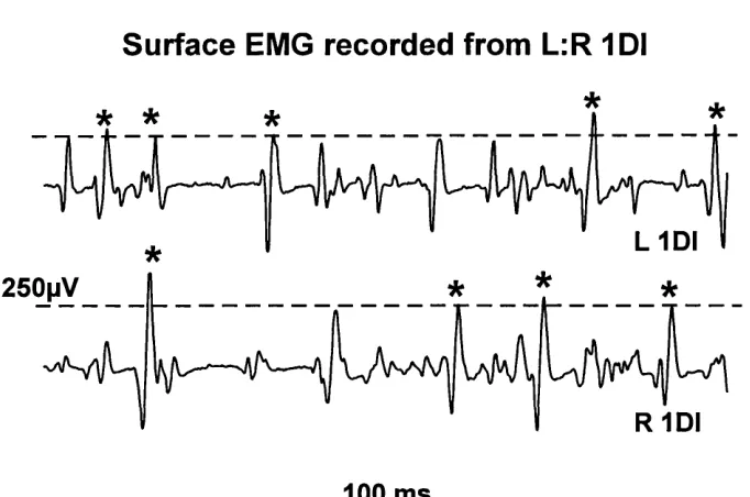
side with simultaneous contralateral activity of 10% MVC on the non-preferred side, and ii). a sustained co-contraction. As indicated in figure 1 (an example o f a sustained co
contraction), medium and large spikes were selected for analysis using a level detector
(Neurolog NL200, Neurolog, Hemel Hampstead, UK). The resulting trigger pulses were
passed into a microcomputer (Tandon 486) via a Cambridge Electronic Design 1401 interface
(CED 1401) for processing. Cross-correlograms were constructed between the times of
occurrence of motor unit spikes recorded from the left and right first dorsal interossei with a
bin width o f 1ms and a pre-and post trigger sweep of 100ms (Spike2 software, CED). At least
5000 spikes were used from each EMG signal; periods of silence between the phasic
abductions were excluded from the analysis. The size o f any central peak was estimated in
terms of E/M where E is the number of spikes in the peak in excess o f those expected by
chance and M is the mean bin count in a 1ms bin calculated from an area away from the
central peak, usually the first and last 75 bins. A central feature of less than 35ms was
Surface EMG recorded from L:R 1DI
L 1 D I
*
* *
100 ms
Figure 1: Surface EMG recorded from right and left IDI in a normal adult subject. The line indicates the set level of the level detector, and the asterisks denote the spikes used as triggers and events.
EXPERIMENT TWO:
Sustained hand grasp recordings
Eight subjects (3 male, 3 left hand preference) were seated well supported as in the previous
experiment. Surface EMG was recorded from the skin overlying the wrist and finger flexors
of the preferred hand; the wrist and finger flexors/extensors of the non-preferred (NP) hand.
The electrodes were placed proximal to the wrist, approximately a quarter o f the distance
between the wrist and elbow creases. Subjects were instructed to maintain a steady grip on a
hand-held dynamometer (University College London Mechanical/ Electronics workshops,
London, UK) for as long as possible using the preferred hand at 20%, 50% and 80% MVF.
The tasks were presented in randomly. No instruction was given about the NP hand. Visual
hand was rectified, and any EMG of the wrist and finger flexors and extensors of the opposite
side was integrated using a one second time constant (Neurolog, NL 703). The EMG and
force signals were passed into a microcomputer via a CED 1401. The average rectified EMG
of the wrist and finger flexors of the active side and the mean of the integrated EMG of the
wrist and finger flexors and extensors o f the opposite side was calculated for each 20 second
frame of EMG recorded for each task in each subject (SigAvg programme, CED). The
average EMG measured for each frame was expressed as a percentage of the maximum
EMG recorded during a maximal voluntary grip of the dynamometer.
The EMG of the wrist and finger muscles and the wrist and finger flexors and extensors of
both sides was recorded for each individual during three MVC’s and the means used as the
maximcv for that subject. In addition the force was recorded during the MVF of the
preferred side wrist and finger flexors, and the mean o f the force recorded in each of three
maximum grips used as the maximum force for that subject.
The subjects were given adequate rest between the sustained contractions to eliminate any
effects which might result from fatigue.
Statistical analysis
Various statisitical tests were used throughout this thesis; the chosen level o f significance was
P < 0.05 unless stated otherwise. When the data were normally distributed, matched and of
equal numbers of observations, the Student’s paired Mest was applied. But if the data were
matched for subject type but with an unequal number of observations the Student’s unpaired
^-test was applied. Linear regression was applied to the data recorded from the children who
participated in the study of mirror movements to determine if there was a significant
correlation between age and mirror movements for example, given by the correlation
RESULTS
Mirror movements
In all o f the subjects mirror movements were either absent or weak (grade 0-1).
EXPERIMENT ONE:
Index finger abduction
Figure 2a. shows the rectified average of the surface EMG recorded from the right and left
IDI from one subject during self paced phasic index abduction of the preferred side with the
contralateral side relaxed. No contralateral EMG was recorded. Figure 2b shows a single
sweep o f surface EMG recorded from the same muscles during the same task when 10% MVC was sustained on the contralateral side. An involuntary increase of EMG above the
10% background could occasionally be seen in a single sweep. This contralateral increase in
EMG was not obvious in every sweep of EMG and is more clearly seen in figure 2c. which
shows the EMG rectified and averaged for 100 sweeps. Taking the subjects as a group, the
average rectified EMG signal of the preferred side consisted of a burst o f EMG lasting
between 192 and 256 ms, mean duration 234±9ms (± SEM, n=7). The increase in EMG seen
on the contralateral non-preferred side was of similar duration and onset latency as that o f the
voluntary EMG burst of the preferred side. In all subjects there was an increase in the
averaged rectified EMG above the background of 10% MVC on the non-preferred side
lasting between 76 and 268 ms, mean 191dL28ms (± SEM, n=7). The time difference between
the onset of the EMG burst on the two sides was not significantly different from zero
(Student /-test, P>0.5). The mean amplitude of the averaged rectified EMG on the non
preferred side ranged from 10.7-12.3% MVC, mean 11.4d^0.2%MVC (± SEM, n=7). The
a. Phasic right index abduction: L1DI relaxed
1mV
R IDI (phasic)
L 10! (relaxed)
0 m s 200
b. S ingle p h asic b u rst: right index ab d u c tio n
Im V R 1DI (phasic)
jsoopv L
0 m s 200
L IDI (10% bgd
c. A verage 100 s w e e p s : rig h t index a b d u c tio n
200pV
R IDI (phasic)
L1DI (10% bgd)
Gain X4R1DI
L
0 ms 200
d. P h asic right index abduction:L F ext 10% bgd
Im V
RID! (phasic)
L Fext (20% bgd)
j
0 ms 200
EMG plus the increase associated with the voluntary burst) was 2.0-5.0% of that recorded on
the preferred side, mean 3.1±0.5% (± SEM, n=7), but was insufficient to cause any visible
movement on that side. Figure 2d. shows the rectified average of the recordings from one
subject during index finger abduction while maintaining a 20% contraction o f the wrist and
finger extensors on the contralateral side. No increase of rectified EMG was recorded on the
contralateral side.
Cross correlation analysis
Cross-correlograms constructed from the times of occurrence of spikes in the EMG signals
recorded from the two sides contained no short duration central peak. The absence of a
central feature in the correlogram indicates that there is no common synaptic drive to the motoneurones innervating the left and right first dorsal interossei which could account for the
bilateral activation of this muscle pair (see section four on mechanisms underlying co
contraction of antagonistic muscle pairs for a detailed description of the use and
interpretation of cross-correlation analysis for the detection of common synaptic input).
EXPERIMENT TWO:
Sustained grip recordings
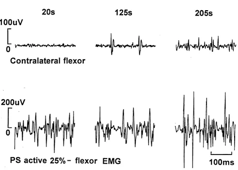
Figure 3 shows an example of raw EMG recorded from one subject at 125 and 205
seconds after the onset of the grip on the preferred side. The preferred side contraction
increased with time as the subject recruited an increasing number and amplitude of motor
units to maintain the required force, as seen in the lower panel of fig.3. AtSlOseconds after the
onset of the preferred side contraction at 25% MVF there was no involuntary activation of the
Contralateral EMG - preferred side active 25%
20s
125s
205s
lOOuV
[
Contralateral flexor
200uV
PS active 25% - flexor EMG
100ms
a single unit has become active, while at 205 seconds there was activity in several motor
units. All of the eight subjects showed contralateral EMG on the non-preferred side when the
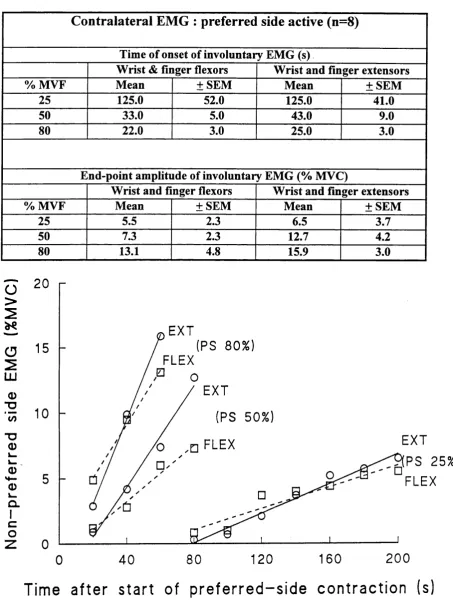
preferred side performed a sustained grip at 25%, 50% and 80% MVF. Involuntary EMG
activity was recorded in the non-preferred side wrist and finger flexors in all subjects but only
in 7 out of eight subjects in the wrist and finger extensors at 25% MVC; involuntary EMG
was recorded in both muscle groups on the non-preferred side at 50% and 80% MVF.
The results for the group of eight subjects are illustrated in fig.4, which shows the
time of onset and increase of the mean amplitude of the integrated EMG o f the non-preferred
side wrist and finger flexors and extensors at 25%, 50% and 80% MVF. The wrist and finger
flexors are represented by the dashed line and the wrist and finger extensors by the solid line.
On average the EMG appeared first in the non-preferred side wrist and finger flexors with a mean latency of 125±52 (± SEM, n=7) for 25% preferred side contraction, and 33.0 ±5 and
22.3 ±3 (±SEM, n=8) seconds respectively for the 50% and 80% preferred side contractions.
This involuntary activity gradually increased until the preferred side contraction ceased at
197.1 ± 11.1, 102.5 ± 7.7 and 95.0 ± 6.7 seconds respectively. At this point the mean
integrated EMG of the non-preferred side wrist and finger fiexor muscles had reached
5.5±2.3, 7.3±2.3 and 13.1±4.8 of MVC respectively. The mean integrated EMG of the non
preferred side wrist and finger extensors had reached 6.5±3.7, 12.7±4.2 and 15.9±3.0 of
MVC respectively. The time of onset and end-point amplitude of the involuntary EMG in the
Table 1:
C on tralateral E M G : preferred side active (n=8)
Time of onset of involuntary EMG (s)
Wrist & finger flexors Wrist and finger extensors
% MVF Mean ±SEM Mean ±SE M
25 125.0 52.0 125.0 41.0
50 33.0 5.0 43.0 9.0
80 22.0 3.0 25.0 3.0
End-point amplitude of involuntary EMG (% MVC)
Wrist and finger flexors Wrist and finger extensors
% MVF Mean ±SEM Mean ±SE M
25 5.5 2.3 6.5 3.7
50 7.3 2.3 12.7 4.2
80 13.1 4.8 15.9 3.0
o
>
s0
s
LU <D CO TD 0) k_ &- ..Q)
«4—
0) k. CL 1 C
o
20
EXT(PS 80%) 15
FLEX
EXT
10
(PS 50%)EXT ^ P S 25%)
FLEX FLEX 5
0
200
1601 2 0 80
4 0
0
DISCUSSION
In all of the subjects studied, involuntary contralateral EMG was recorded when
unilateral movements were made but this activity was only apparent in the rectified average
o f at least 100 phasic movements if the subjects maintained a background o f 10% MVC.
The record of involuntary EMG contralateral to the voluntarily active hand in this
group o f adults supports the findings from previous studies (Cemacek et al, 1961; Durwen &
Herzog, 1992). In those studies EMG was not recorded in all of the trials, but in the present
study the subjects maintained a background contraction of EMG contralaterally so that the
motoneurones were closer to firing threshold than in the case of the relaxed subjects and thus
were more readily activated.
Various mechanisms have been put forward to account for the production of bilateral
EMG activity by the CNS. Bilateral cortical activation has been observed in studies utilising
measures of regional cerebral blood flow using positron emission tomography (PET) (Shibasaki et al, 1993; Kawashima et al, 1993), magnetoencephalography (MEG) (Kristeva
et al, 1991) and functional magnetic resonance imaging (fMRI) (Kim et al, 1993 a&b),
although such activation was recorded while subjects performed more complex tasks than in
the present study. A lack of inhibition across the corpus callosum is another mechanism that
has been suggested to account for bilateral activation in cases of pathology such as callosal
agenesis and X-linked Kallmann’s syndrome (Dennis, 1976; Danek et al, 1992). A further
possibility to consider is that bilateral EMG could result from activity in the normally
occurring uncrossed ipsilateral portion of the corticospinal tract. Studies of stroke patients
find some support for this hypothesis. For example a study by Smutok et al, (1989), found
that reaction times and finger tapping ability were impaired in the “unaffected hand” and
branching of corticospinal tract fibres to supply bilateral motoneurone pools has been
proposed as the mechanism to account for the mirror movements seen in patients with
Klippel-Feil syndrome and children with congenital hemiplegia (Farmer et al, 1990; Carr et
al, 1993), such pathways have never been found in normal adults. Therefore, the following
discussion will focus on two possible mechanisms of mirrored activity in adults, firstly the
possibility o f activity being conducted via a normally occurring ipsilateral pathway, and
secondly bilateral cortical activation.
Activity in the normal ipsilateral corticospinal tract projection
Evidence from human studies
The ipsilateral corticospinal tract in humans accounts for approximately 15% of the
corticospinal projection, but once in the spinal cord further crossing occurs and there remains about 2% which is uncrossed (Wilkinson, 1992). This small portion of the tract is thought to
innervate axial and proximal muscles (Brinkman & Kuypers, 1973) and to be o f slow
conduction velocity. However, there is some evidence to suggest that fast conducting
ipsilateral pathways could exert some control over distal finger movements.
Most of this evidence is found in studies of acquired hemiplegia in adults, in which it
has been found that the function of the unaffected limb has been affected by the ipsilateral
cortical lesion. Colebatch & Gandevia (1989) in their study of stroke patients observed that
there was weakness of the muscles in the unaffected arm indicating that the ipsilateral tract
was functionally effective. Smutok et al, (1989) in their study found deficits in reaction times
and finger tapping function of the unaffected hand. Thus it is possible that the involuntary
activity recorded in the present study could have been produced by activity in the ipsilateral
which are influenced by the motor cortex.
One might anticipate that if the ipsilateral tract does exert some control over distal
movements that it should be able to be activated using focal magnetic brain stimulation. This has proved difficult. There are reports of an inhibition being demonstrated ipsilaterally (Carr
et aî, 1994; Meyer et al, 1995b), and reports of bilateral motor evoked potentials (MEP) in
proximal muscles (Basu et al, 1994). But Wassermann et al, (1994) recently demonstrated
that they could evoke ipsilateral responses using Magstim. They were able to record
bilateral EMG responses in the first dorsal interosseous muscle of the 6 subjects investigated,
although it occurred at later latency (4.5-6.5ms) than the contralateral response. The time of
onset of the involuntary activity recorded in the present study was not significantly different
from the commencement of the voluntary activity, therefore it is unlikely that the ipsilateral
projection or brainstem pathways are responsible for the involuntary activity recorded in the
present study. Wassermann et al, (1994) also noted that the optimal site for evoking the ipsilateral response was near the representation o f the contralateral face, suggesting that its
origin is from a different cortical site than the contralaterally projecting population of pyramidal cells. A high output of the stimulator was required (65-90%). Bilateral MEP’s in
the distal hand muscles have only been demonstrated in the presence of pathology (Farmer et
al, 1990 & 1991; Cohen et al, 1991; Carr et al, 1993). In the case of axial and truncal
muscles, and those usually acting symmetrically such as masseter and the diaphragm,
bilateral MEP’s were recorded (Carr et al, 1994), supporting the observation o f Kuypers
(1987) that the projection to those muscles is bilaterally organised.
Evidence from animal studies
An anatomical study using the macaque monkey (Galea & Darian-Smith, 1994) has shown
contralateral projection (frontal area 4, SMA, areas 3a, 3b, 2 & 5, insular and SII). They found that 8.1% of the tract projected ipsilaterally, but that further crossing occurred below
the level o f the decussation. Aizawa et al, (1990) in their study of the monkey recorded from
cells in the primary precentral cortex which exhibited activity before and during key pressing
movements of the ipsilateral and contralateral digits. Intracortical stimulation of cells in this
region elicited bilateral responses in the digits, suggesting that both ipsilateral and
contralateral projections to the fingers originated from that site, and thus could account for
bilateral activity in the distal muscles.
Taken together, evidence from animal and human studies suggest that it is possible
that the ipsilateral corticospinal projection could have some influence on the control of
ipsilateral hand movements. But this seems an unlikely mechanism to account for the
bilateral EMG activity recorded in the present study in which the task was simple and well practised by the subjects rather than a complex sequential finger task which produced the
bilateral motor cortical activity as described by Kim et al, (1993 a&b) and Rao et al, (1993).
Another factor to consider is the fast conduction of the signal producing the involuntary
activity such that it occurred at the same time as the voluntary activity. Ipsilateral
corticospinal axons do not normally make direct corticomotoneuronal connections (Kuypers,
1973) therefore it is unlikely that the activity recorded in the present study was conducted via
the normally present ipsilateral corticospinal tract projection, but rather by bilateral cortical
activity.
Bilateral cortical activation
Evidence from human studies
activity in the contralateral cortex is associated with this task as expected (Roland et al, 1982;
Deiber et al, 1991; Sabatini et al, 1993). However studies of more complex finger movement
sequences have resulted in significant bilateral activation of the sensorimotor cortex, in
particular the supplementary motor area (Kim et al, 1993 a&b; Shibasaki et al, 1993;
Kawashima et al, 1993; Rao et al, 1993).
In their study of six normal adults performing a sequential finger-thumb opposition
task, unilaterally and then bilaterally, using fMRI, Kim et al, (1993a) found bilateral
activation of the lateral motor cortex. When the right hand only was used the ipsilateral activation was 20 times smaller than when the left hand was moved. Whilst this small
ipsilateral activation could have been due to inadvertent movement on the contralateral side
the authors stress that the subjects were instructed to make only unilateral movements. A similar finding was obtained by Shibasaki et al, (1993) in their PET study of a complex
finger-thumb sequential task. A more consistent finding of functional brain scanning studies
is the bilateral activation of pre-motor areas such as the supplementary motor area (SMA). It
has been shown that there is significant SMA activation in tasks requiring planning, such as
an internally cued task, and when complex rather than simple tasks are performed (Rao et al,
1993; Shibasaki et al, 1993). In contrast to these studies, in the present study subjects were
instructed to maintain a 10% MVC background in the contralateral homologous muscle thus
raising the firing threshold of the motoneurone pool. It maybe possible that when an
individual makes unilateral movements a small command to the “inactive” side is normally
produced but is too small to be detected in relaxed muscle. Thus the use of background EMG
in this study enabled the small bilateral component of the motor command to be detected.
Further evidence for bilateral cortical activation o f premotor areas is found in the
electrodes placed on the scalp overlying the motor cortex. The resulting m ovement associated
potentials have been classified into four components: readiness potential, otherwise known as
the Bereitschaft potential (N l); premotion positivity (PI); motor potential (P2), and a
com plex post-movement potential (P2). N and P denote positive and negative deflections.
The readiness potential is usually seen 1-1.5 seconds prior to movement onset (Roland et al,
1980) and is thought to be associated with activity in the SMA. Shibasaki and Kato (1975)
studied the N l in 9 normal adult subjects during a unilateral movement but found no
significant difference between contralateral and ipsilateral responses, and concluded that the
readiness potential was bilateral. Tarkka and Hallett (1990) have also recorded a symmetrical
readiness potential in their study. Additional support for this finding is found in the study o f
normal adults performing unilateral and bilateral tasks using measurement o^m agneto
The study by Kristeva et al, (1991) also demonstrated bilateral premovement fields. In their
study o f 6 subjects, EMG was also recorded, and was never completely silent in the non
active side during unilateral hand movements. Whilst they found bilateral activation
preceding a voluntary unilateral movement, the movement evoked fields occurred only over
the contralateral fields during unilateral movements, although mirrored EMG activity was
recorded. They suggested that the mirrored motor activity was too small to produce a
movement evoked field. It would be interesting to apply the study o f movement related fields
to the current study. It would be anticipated that bilateral fields would be recorded during a
sustained contraction o f both sides, but it would be interesting to determine if a difference
could be found when the subject produced phasic activity rather than sustained activity as in
the current study.
Bilateral cortical activity could result from excitation via the corpus callosum. This
(Cracco et al, 1989), and the EMG recorded would be similarly delayed. In the present study
the contralateral involuntary EMG recorded was found to commence on average at the same
time as the onset of the voluntary EMG. In any case, the transcallosal pathway is thought to
be inhibitory in adults (Ferbert et al, 1992; chapter 3, this section). Further support for the
transcallosal inhibition hypothesis is suggested by the work on premovement potentials
reported by Shibasaki & Kato (1975). They found that the premovement positivity (PI) was
recorded in 6/9 normal adults during unilateral movements. This PI was recorded ipsilateral
to the moving hand, was not recorded in a bilateral task and thus they concluded that the PI
was associated with an inhibitory mechanism to prevent imitative movement on the opposite
side during a unilateral task.
Taken together, these reports of bilateral cortical activation are commensurate with
the findings o f the present study. The involuntary activity recorded must have been produced
by activity in a fast conducting pathway given that there was no significant time difference
between the start of the voluntary and involuntary activity. No peaks were observed in the
cross-correlograms constructed from the EMG recorded from the co-contraction of left and
right IDI. Thus it is unlikely that the involuntary activity was produced by activity in
descending axons branching to project bilaterally, or by activity in corticospinal axons whose
cell bodies share a common synaptic input to the bilateral homologous motoneurone pools.
Mirrored activity and effort
It has been suggested that mirrored activity is more likely to be seen in activities
requiring effort or repetition (Todor & Lazarus, 1986; Durwen & Herzog, 1992). In the
present study, involuntary EMG was recorded contralateral to the EMG recorded during grip
amplitude when the subject was instructed to maintain higher grip force and thus required
more effort. Similar results have been found in other studies (Cemacek, 1961; Armatas et al,
1994) and in agreement with the finding of the present study, suggest that the force requirement of the task influences the amplitude of the mirrored activity.
In their study of 8 normal subjects holding a dynamometer in one hand, Hopf et al,
(1974) recorded EMG contralateral to the voluntary activation when the subjects reached
maximal, or nearly maximal effort. In a similar experiment Armatas et al, (1994) studied the
mirrored activity associated with the exertion of two different finger pressures for the index and the little finger. They studied 22 subjects, finding that greater involuntary force was
recorded when a larger voluntary force was produced, and when the weaker finger (little vs
index finger) was used. They made no conclusion as to the underlying mechanism of the
mirrored activity, but suggested that it was most likely related to a lack of inhibition across
the corpus callosum. They further suggested that there are right-left differences in the cortical
control o f the two hands, such that the left hemisphere is more concerned with skilled
movements and the right hemisphere with holding/stabilising movements. This would result
in more mirrored activity during skills performed with the left hand because ftmctionally the
left has less practice of fine skilled movements than the right. Gandevia et al, (1993) in a
study o f lower limb muscles investigating the effect of a lack of afferent feedback, noted that
they recorded “inadvertent mirror contraction” in the contralateral unparalysed limb which
increased during the maximal effort on the paralysed side. They made no comment as to the
origin o f the activity. A similar observation was made by Dimitrejevic et al, (1992) in their
study of 17 subjects performing ankle dorsiflexion. They found co-contraction of
homologous and non-homologous muscles which increased with effort and fatigue. They
general increase in excitability of the motoneurone pools, such that previously subthreshold
input became effective. But in the current study the bilateral activity was only recorded in
homologous muscles and did not appear to occur as a result of a general spread of excitation.
As in the present study and that of Cemacek (1961), the excitation did not produce a visible
movement and was only detected by electromyography.
Excitation in other muscles
The results from the present study suggest that the bilateral EMG recorded is confined to
homologous muscles and does not spread to non-homologous muscles. This would appear to
support a hypothesis for a bilateral command which is only transmitted to the motoneurones
supplying the muscles required for the desired voluntary task. However, recordings were only made from the forearm extensors in 2 subjects. It will be necessary to repeat this section of
the study with a greater number of subjects and to investigate a larger number of non-
homologous muscles in order to be confident of the restriction of activity to homologous
muscle pairs.
Conclusion
This study has shown that phasic and maintained unilateral voluntary movements in man are
accompanied by a small activation of contralateral muscles. Whilst it is possible that a fast
conducting ipsilateral tract could be associated with the involuntary activity observed, it is
more likely that bilateral cortical activity underlies the bilateral EMG recorded in the present
Section 1: Chapter 2: Mirror movements in children : a developmental study SUMMARY
1. Electromyographic recordings were made in the upper limbs of forty-seven normal school
aged children while making self paced sequential finger-thumb opposition of the left and then
the right hand, with no instruction given about the contralateral side.
2. All o f the children aged 4-6 years had mirror movements. The intensity and frequency of
the mirror movements decreased with age until by age 11 years, 7% of the children had no
mirror movements, 79% had mild mirror (grade 1) movements and only 14 % had marked
mirror movements (grade 2).
3. The EMG recorded from the left and right first dorsal interosseous muscles (IDI) was
rectified and averaged for 25 sweeps, time locked to the start of the bursts of voluntary EMG.
The amount of involuntary EMG showed a significant decrease with age.
4. Cross-correlation analysis was performed in 5 children aged between 5 and 10 years to
determine if the bilateral EMG recorded during unilateral movements was the result of a
common synaptic drive to the homologous bilateral motoneurone pools. In all cases the
correlogram was flat, thus there was no evidence for a common synaptic drive that could
account for the bilateral activation of the homologous muscle pairs. The most likely
explanation for the mechanism underlying mirror movements in children, is that they are
produced by activity transmitted by the fast conducting contralateral projection of the
INTRODUCTION
Associated movements describe the unintended movements associated with voluntary
effort in another part of the body. They are also known as motor overflow, synkinesis and
motor irradiation. When these movements occur in homologous muscles of the hand, they are
known as mirror movements. Such movements are thought to be a characteristic of an
immature central nervous system (CNS) and while present interfere with the ability to
perform precise hand skills, particularly those requiring intermanual co-ordination. For
example, when a young child voluntarily uses one hand to pick up and manipulate an object
the same movement maybe observed in the opposite hand occurring involuntarily. The
presence o f such a movement makes the ability to grasp an object with one hand and
manipulate it with the other difficult or impossible. A typical example would be the ability to
cut using scissors in one hand, while holding an object in the other. In this case each hand has
a different task: one to hold, the other to make opening and closing movements which if mirrored in the other hand prevents the child from holding the object.
Such movements are known to decrease in intensity and occurrence as the child
matures (Fog and Fog, 1963; Abercrombie et al, 1964; Cohen et al, 1967, Connolly and
Stratton 1968; Wolff et al, 1983; van Sant and Williams, 1986; Lazarus and Todor 1987) .
They are often seen in young babies aged 6-8 months who at this stage exhibit strongly
symmetrical activity in all limbs and especially in the hands thus preventing unilateral hand
skills (personal observation). Associated movements have been observed to decrease
markedly after 8 years of age (Wolff et al, 1983, Lazarus and Todor, 1987), but may be seen
in older children and adults performing difficult or novel fine hand, skills, during effort or
under fatigue or stress (Podivinsky, 1964; Cemacek, 1961,). If significant mirror movements
(Fog and Fog 1963; Abercrombie 1964; Cohen et al, 1967). Specific tests for the presence of
these movements <VrL.used in the clinical assessment o f minimal neurological dysfunction
(Touwen, 1978).
Associated movements in children have been studied in a variety of ways (Connolly
and Stratton, 1968; Denckla, 1974; Woods and Teuber, 1978; Touwen, 1978; Van Sant and
Williams, 1986). Visual examination of the frequency and intensity of such movements
during repetitive finger-thumb opposition or clip-pinch task can be scored as present or
absent and a scale can be used to monitor changes in the intensity of mirror movements
during development and in pathology. The most common scoring system is that of Woods
and Teuber (1978), which scores the movements from 0-4; but that of Touwen is also used
and scores the associated movements from 0-2. In each case 0 denotes the absence of mirror
movements, the highest score in each case indicating symmetrical movements. Other tests
include timed finger-thumb opposition, hand pronation-supination, finger spreading, feet to
hands, stress gait (walking on heels, toes, lateral border of feet and medial border of feet) and
heel-toe alternation.
Denckla (1974), in her study of associated movements in children, used timing of
seven motor tasks to assess the co-ordination of a group of 156 normal right-handed children
aged between 5 and 11 years. She found that the older children performed the tasks more
rapidly, with significant differences between the age groups in the 5-8 year range, after which
there was a relative plateau. Another study of associated movements in a group of children
aged between 5 and 7 years at 6 month intervals over one year by Wolff et al (1983), showed
that the frequency of associated movements decreased over the 12 month period with
associated movements could be used to demonstrate the child’s developmental stage. The
study also demonstrated differences between children of the same chronological age.
To enable a more detailed investigation of the occurrence and intensity of associated
movements in children, measurements of force and/or EMG activity during the performance
of clip-pinch and finger lift tasks have been undertaken. In their study of a group of children
aged between 6 and 16 years, using a modified version of the clip-pinch task of Fog and Fog,
Lazarus and Todor (1987) demonstrated that the magnitude of associated movements
decreased with age dramatically between age 6.5 and 8.5 years, but that there was no
significant difference beyond the age of 8.5 years. They measured the occurrence of
associated movements as a percentage of maximum active hand force, making measurements of force bilaterally whilst the children performed the clip-pinch task at 50, 70 and 100%
maximum voluntary force (MVF) respectively.
The aim of the present study was to investigate the occurrence, intensity and origin of mirror movements during development. The presence of mirror movements was scored using
the criteria of Woods and Teuber (1978), and EMG was recorded simultaneously from left
and right first dorsal interosseous and abductor digiti minimi muscles while each child
performed a thumb-finger opposition task. Cross-correlation analysis of the EMG signals
recorded from left and right co-contracting muscle pairs was performed to determine whether
a common synaptic drive to homologous motoneurone pools was present.
A prelimimary account of these experiments has been presented to the Physiological
METHODS
Subjects
Recordings were made from forty-seven children (25 males) recruited from a local primary
school, with local ethical committee approval and written parental consent. In addition verbal
consent was sought from the children when appropriate. The children were aged between 4
and 11 years and had no known neurological abnormalities. Four of the children were left
handed and the subject group was of mixed ethnic origin.
Assessment o f handedness
All children were assessed using a modified version of the Edinburgh Handedness Inventory
(Oldfield, 1971). See Appendix B. In contrast to adults who are generally certain about handedness, the children when asked to indicate hand preference verbally could be unsure.
Therefore the children were asked to show the examiner how they performed activities such
as writing, using a spoon and cutting with scissors. Both hands were tested. They were also
asked to demonstrate how they kicked a ball as a further assessment of laterality (Peters
1988).
Assessment o f mirror movements
The presence and degree of mirror movements were assessed in each child and graded
according the criteria of Woods and Teuber (1978). The child was instructed to sit with their
hands in a supinated (palms facing up) relaxed posture on their knees. The child was
instructed to oppose each finger to their thumb sequentially. The instruction to the child was:
“Tap each finger to your thumb in turn, starting with your index (pointer) finger going down
verbal encouragement during the task was given when required. Both hands were tested. The
mirror movements were scored on a scale of 0-4 (see Chapter one, this section). It was noted
that some children held the index finger or all of the fingers of the contralateral hand in
extension instead of, or in addition to showing mirror movements; a record was made of the
occurrence of such activity.
Assessment o f bilateral integration o f hand activity
This was assessed in each child who was tested at the school. Each child seated on a chair
was asked to place their hands on their knees, one facing down (pronation) and the other
facing up (supination). They were then asked to simultaneously move them over and back as
quickly and as neatly as possible. Demonstration was given if required. The ability to perform
the task in this asymmetrical manner without reverting to performing it symmetrically or
clumsily was scored as follows (Njiokiktjien et al 1986):
Grade 0 = unable to perform the activity asymmetrically
Grade 1 = able but with some difficulty
Grade 2 = able but with slight hesitation
Grade 3 = able to perform the activity asymmetrically
Electromyographic recordings
Each child sat at a table with their forearms resting on it. The skin overlying the left and right
first dorsal interossei (IDI) and the left and right abductor digiti minimi (ADM) were
prepared using an alcohol wipe (Sterets). Bipolar surface electrodes (Teca, see Chapter one)
were attached to the skin overlying these muscles, and cut down for use with the smaller
children, the active electrode was placed over the muscle belly and the reference on an
adjacent area such as the metacarpophalangeal joint of the index finger. The electrodes were
held in place with micropore tape.
Each child was instructed to perform 25-40 sequential finger-thumb opposition movements as
they had done for the assessment of mirror movements before the electrodes were applied.
The child was given verbal instruction and encouragement with demonstration if required,
which was usually the case for the younger children. They were instructed to concentrate on
the active hand and to forget the other one, which was placed in a mid-pronated position. In some children in whom no contralateral EMG was recorded an attempt to provide
background EMG was made by asking the child to hold the roll of micropore tape between
the thumb and index finger. Both hands were tested.
The EMG recordings were made using a clinical EMG machine and stored on magnetic tape
as in previous experiments (see Chapter one). The signal was amplified and filtered as in
previous experiments, but in this case the low cut filter was set at lOOHz to reduce movement
artefact from the leads. All of the analysis was performed off-line.
The EMG signal of the left and right IDI was rectified and averaged time locked to the onset
of the voluntary EMG burst taking the most phasic 25 sweeps using averaging software
(SigAvg programme, see chapter one). The averaged rectified areas of the voluntary and any
involuntary occurring activity were measured and the ratio of involuntary to voluntary areas
calculated. The duration of the involuntary and voluntary busts were also measured. The data
from left and right ADM w tonot analysed because the ongoing EMG in the active side did
not enable the start of individual phasic bursts to be differentiated. When possible an average
was made of those bursts when there was a clear onset of contralateral EMG to enable the
of sweeps of bilateral EMG was calculated by noting the number of sweeps where a burst of
bilateral EMG similar to that on the active side could be clearly seen and expressing this as a
percentage of the total number of sweeps. In addition a calculation was made of the number
of sweeps where there was bilateral EMG. This was calculated in the following way:
when ongoing EMG or an observable burst was present contralaterally it was expressed as a
percentage of the total number of sweeps of EMG. In adults performing the same movement
with the contralateral hand relaxed, ongoing EMG or bursts of EMG were not observed
contralaterally during unilateral phasic finger movements.
Cross-correlation analysis
In five of the children cross correlograms were constructed from the multi-unit EMG recordings from left and right IDI (see chapter one). The children were instructed to maintain
a steady contraction in the left and right IDI muscles by gently pushing the left and right
index finger against resistance, or in the younger children holding a dowel with stickers on it
to maintain their interest. Only those parts of the record which contained spikes on both
RESULTS
In accordance with other studies wki6thave reported that the intensity and frequency o f mirror
movements decreases markedly after 8 years of age (Wolff et al, 1983)), the children were
grouped in age bands as follows:
4-6 years: n = 11
7-8 years: n = 1 9
9-10 years: n = 1 0
11 years: n = 7
Mirror movements
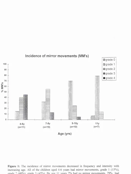
The mirror movements ranged from marked to absent (graded 0-3, n=88 hands) and were observed to decrease with age. Fig.l shows a bargraph of the incidence and grade of
mirroring in the children for both hands. The small size of the sample did not allow adequate
comparison to be made between the left and right sides, nor between the left and right handed
subjects. All children aged 4-6 years had mirror movements, 15% with grade 1, 40% with
grade 2 and 45% with grade 3. The intensity and frequency of occurrence of mirroring
decreased with age until by age 11, 7% had no mirror movements, 79% had grade 1 and 14%
grade 2.
It should be noted that the scoring of mirror movements was often made difficult by
the occurrence o f sustained involuntary extension rather than repetitive flexion movements of
the fingers. An example of this extension activity is seen in fig.2a & b. Figure 2a shows a
typical example of a 10 year old child with mirror movements. As the active hand (right)
moves the fingers of the left hand move into flexion. But in fig.2b, the active hand (left) of