Action potential in HH model
1Payal, 2Monika Choudhary 1Software Engineering, 2Computer Science 1NorthCap University, 2Guru Jambeshwar University
________________________________________________________________________________________________________
Abstract— In this paper we represent the main property of neuron i.e. action potential. In this we show how we can
generate and propagate the action potential using different voltage gated ion channels. To study the action potential we used different gated ion channels like Na+, K+, Ca2+ and Cl- . Calcium channels are mostly used for providing the
communication between neuron networks. We use some mathematical equations or circuit of HH model. Action potential is an important part which we can used to get complete model of neuron network.
Keywords— Action potential, mathematical, calcium channels, communication, neuron network, voltage gated ion channels.
________________________________________________________________________________________________________
I. INTRODUCTION
There are some unique cells called excitable cells have an ability to generate the electric signals. These excitable cells have some cells like muscle cell, neurons and receptors cells they used some ions channels receptors which helps to convert mechanical messages into some electrical signals. These ion channel receptors located in the plasma membrane and they form itself a
passageway from one side of membrane to another. These ion channels have an ability to open or close a chemical signals. When these channels are open then ions move into or out of the cells and allow one single ion to pass through them. In neurons and muscle cells there are some cells which open in stimuli rather than the chemical signals. After the few milliseconds of the opening if ion most of the ion channels are closed and entered in a resting state. The opening of ion channels gives the charge on the both side of the membrane. The resulting causes the adjacent voltage sensitive channel to open an electric signal that travels down towards the cell[5, 9].
Any ion channels that response open and close to change in electrical potential through the cell membrane which is situated in the cell. Ion channels act as either passive or active. In active channels the gates can be open or close the channel and in passive channel the gates are always open and ions can passes continuously. Passive channel are also known as leakage channels. Experimental research found four types of ion channels these ¬¬are Na+ , K+, Cl2+ and Cl-. In sodium channels (Na+) different cell have similar functional properties. In sodium channel it give raise the action potential for long period of time. On the other hand same way calcium channel (Cl2+) give rise to action potential. In other neurons some time calcium channel control the shape of action potential which is generated by sodium channel which affect the intercellular of Cl2+. Calcium channel is important to release the neurotransmitter at synapses. Like sodium channel in calcium channel there is the difference in the property of the activation and inactivation of the channel. On the other hand the most important function of K+ channel is to response changes in the membrane potential. The K2+ channels are the selective pores that allow only potassium ion to cross a cell membrane. There are as a minimum two families of potassium channels that are open at hyperpolarized membrane. It forms a curve shape of action potential and set the resting membrane potential [13,14].
II. ACTION POTENTIAL
Action potential is the wave like structure which shows the voltage signals that travels on the different types of cell membrane. Action potential in neurons is based on the voltage gated channels (Na+, K+). Some neurons used only Na+ channels or some used only Cl2+ channels. Action potential gives some threshold value and this value can be estimated by the constant value. In neuron this threshold value depends on many factors like temperature and other factors. In figure 1.4 it shows the movement if ions during the stages of action potential. Dash line (above -70) represents the threshold value (approx. -60mV).
In this figure we show the action potential between sodium channels and potassium channel how they activate and inactive using voltage gated ions.
It consists of two components voltage gated Na+ channels and voltage gated K+ channels. The simulations result of action potential is also driven between them.
IJEDR1703088
International Journal of Engineering Development and Research (www.ijedr.org)
596
Figure shows the ion flow in action potentialAfter this when ion flows from open to inactive phase then the channel switch to inactive state and never opened again. And when ion flows from inactive to closed phase the channel never switched back to closed state until membrane has repolarized. When it closed it never reopened. It increases since sodium ions are positively charged [6]. The depolarization phase is the fastest process in the generation of the action potential. In Eq. 2.1 we make the approximation value of sodium ion (𝑃𝑎∞) in the membrane potential.
𝐶𝑑𝑉
𝑑𝑡 = 𝑔𝑁𝑎𝑃𝑎
∞(𝑉)(𝐸
𝑁𝑎− 𝑉) + 𝑔𝐿(𝐸𝐿− 𝑉) 2.1
After depolarization the second phase is repolarization. In voltage gated K+ channel we consist of two states open and closed. In this when ion flow from closed to open state the K+ channel allows potassium ions from intracellular to extracellular environment. In this process it decreases the membrane potential in the cell which gives the negative charge. It brings the membrane to back towards the potential for K+ means it hyperpolarized the membrane. In Eq. 1.2 we make the approximation value of potassium ion
(𝑒𝑉−𝑉𝑇𝑘𝑎 ) in the membrane potential.
𝐶𝑑𝑉
𝑑𝑡 = 𝑔𝐿𝑘𝑎𝑒
𝑉−𝑉𝑇
𝑘𝑎 + 𝑔𝐿(𝐸𝐿− 𝑉) 2.2
Hyperpolarization is the third phase of action potential. In this phase the K+ ions flow from open to close state. In this intracellular environment is hyperpolarized. In other words, the -80mV decreases below the potential (-70mV). After hyperpolarization it channels itself shut down. This is the slowest process in the generation of action potential.
The curve shape of action potential depends on the different factors such as properties of cell membrane for example number of voltage gated ion channels, leak channels and membrane capacitance, temperature and other properties [6, 16].
III. Voltage - gated ion channels
Voltage-gated ions channels are controlled by the voltage gradient through the cell membrane. They are the selectively permeable to each of the major ions. These major ions like Na+, K+, Cl- and Ca2+ . Some where they function similar to each other. Voltage-gated ion channels are highly regulates by the voltage through the membrane. The equation of voltage gated ion channel is:
𝐼𝑚 = 𝐶𝑚
𝑑𝑉𝑚
𝑑𝑇 + g𝐾𝑛
4(V
𝑚− E𝑘) +
g𝑁𝑎m3h(V
𝑚 − E𝑁𝑎) + g𝑙(V𝑚 − E𝑙) + g𝐶𝑎m2
(𝑉𝑚−𝐸𝐶𝑎)
For the generation and the propagation of the nerve impulses they play a fundamental role. They also help to generate and propagate the action potential. Now we discuss about the different types of voltage-gated ion channels.
These gated ion channels are used in modeling channels. Alan Hodgkin and Andrew Huxleyused these gated channels for the activation of the action potential in different models.
Voltage gated Cl- channel Voltage-gated K+ channel Voltage-gated Ca2+ channel
Voltage sensors are those modules which are responsible for opening the action potential. And the membrane proteins are responsible for the change of synaptic input into physiologic stimuli. The model of voltage sensor function is the new way to generate the ion channels drugs rather than blocking the pore [2, 5, 16].
1) Voltage-gated sodium channel
Voltage-gated sodium channel is controlled by the sodium ion through the cell membrane. When Vm = VNa condition is satisfied then sodium ion is an equilibrium state [1]. There is no net movement in sodium ion. Sodium ion enters into the cell by diffusion and moves out by the electrostatic gradient. During this state the net value of sodium ion is 0 i.e. INa=0.
𝑔𝑁𝑎= g𝑁𝑎m
3h
𝐼𝑁𝑎= 𝑔𝑁𝑎∗ (𝑉 − 𝐸𝑁𝑎)
On the bases of the HH model the sodium ions have two states open and closed. And these states are worked according to the
m-particles. These m-particles are based on two states. These are permissive and non-permissive. When ions are open then
they are on permissive state and when ions are closed then they are in non-permissive state [17].
Figure Structure of sodium channel
Hodgkin and Huxley assumed that there are three m-particles to activate the sodium channels. And it represents as (1-m) and to inactivate the channels there are h-particles are presents. And these h-particles are represents as (1-h).
The sodium channel activation and inactivation is represented by the:
𝑑𝑚
𝑑𝑡 = 𝑎𝑚𝑉(1 − 𝑚) − 𝛽𝑚(𝑉)𝑚
𝑑ℎ
𝑑𝑡= 𝑎ℎ𝑉(1 − ℎ) − 𝛽ℎ(𝑉)ℎ
These are the transfer coefficient rates 𝛼h ; 𝛼m; 𝛽h ; 𝛽m. They are voltage dependents [12].
2) Voltage gated potassium channel
Voltage gated potassium channel is controlled by the potassium channels across through the cell membrane. When Vm = VK condition is satisfied then potassium ion is an equilibrium state. During this state the net value of potassium ion is 0 i.e. IK=0. When there is no flow of current in this channel then Vm ≠ VK.
𝑔𝐾= g𝐾𝑛4
𝐼𝐾= 𝑔𝐾∗ (𝑉 − 𝐸𝐾)
Figure Structure of potassium channel
Hodgkin and Huxley assumed that there are four n-particles to activate the potassium channels. And it represents as (1-n) [8, 10].
𝑔𝑙= g𝑙
𝑑𝑛
𝑑𝑡 = 𝑎𝑛𝑉(1 − 𝑛) − 𝛽𝑛(𝑉)𝑛
3) Voltage-gated leak channel
All different ion channels like sodium, potassium, calcium are flow these leak channels across the cell membrane.
When Vm = Vl condition is satisfied then the leak channels are in an equilibrium state. During this state the net value of leak channel is 0 i.e. IK=0. When there is no flow of current in this channel then Vm ≠ VL [11].
𝐼𝐿= 𝑔𝐿∗ (𝑉 − 𝐸𝐿)
4) Voltage-gated calcium channel
Voltage- gated calcium channel iscontrolled by the calcium channel across the cell membrane.
Ca2+ channel plays an important role in our cellular system likes muscular cell, neurotransmitter release and muscle contraction. And it helps to control calcium influx and helps to response the change of the intracellular calcium concentrations that occur in our cellular system.
The structure of Ca2+ channel model is based on the K+ and Na+ channel. In this channel there are four domains and each domain formed six transmembrane but still there are differences at atomic level. Non-identical Ca2+ channel is formed by the single polypeptide. When Vm = VCa condition is satisfied then calcium ion is an equilibrium state. During this state the net value of calcium ion is 0 i.e. ICa=0.
𝐼𝐶𝑎= 𝑔𝐶𝑎∗ (𝑉 − 𝐸𝐶𝑎)
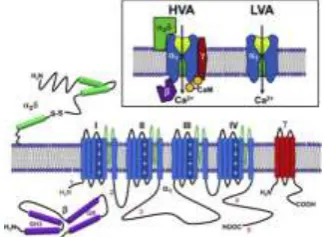
Figure Structure of Ca2+ likely α-helices are demonstrated as cylinders. The lengths of lines relate approximately to the lengths of the polypeptide segments represented.
In this figure α1 have four domains and each domain have six transmembrane. In the loop of domain I and II have high affinity using auxiliary β, α2δ and γ. In six transmembrane first 4 represent the voltage segment domain and last 2 represent the calcium selective domain pore. These domains segments contain positive charge and act as a voltage sensor controlling of calcium channel [3, 4].
IV. Activation and inactivation of voltage-gated Ca2+ channel
When voltage of calcium ion changes than the voltage gated calcium channels are in opened and closed state. At the closing state the ion cannot passes and the voltage gated channels are closed. In the depolarization state channels are activated on the open state and allow ions to pass. At this state membrae potential will be increased.
The probability of open channel is remained temporarily during depolarization and the voltage reduced by the inactivation of channel at closed state. They cannot reactivate immediately. It takes some time period for repolarizing of the membrane and for the recovery of inactivation [7, 16].
V. RESULT
Using the equation of HH model we create a code of these equations and show the action potential of the equation putting some values. This method involves some variable parameters of rate and time constants. It checks the conductance match the plots shown above.
A plot of the time versus voltage gated channels. It shows that 𝑔𝑁𝑎 is more dependent on m and h. It describes the peak level of action potential. In the figure m increases and n decreases.
In above figures we show the action potential of the different ion channels.
In fig (A) we show the action potential between the rate constant and time constant values like m, n, h using the above given equations of dy/dt.
In these equations we put the value of reversal potentials of different channels like ENa, EK, EL. In this figure we also put the value of conductance of ion channels (mS/cm2). Value of g
𝑁𝑎, g𝐾, g𝐿 and 𝑔𝑁𝑎, 𝑔𝐾, 𝑔𝐿.
Fig. A Fig. B
Fig. C
Figure Implementation of action potential
In fig (C) there is one difference we put the value of conductance of ion channels (mS/cm2). Value of 𝑔
𝑁𝑎, 𝑔𝐾, 𝑔𝐿. In this also we show the action potential between the rate constant values like n, m, h.
At last in fig (B) we show the current flow between these voltage gated ion channels. And we also show the simulation of the reversal potential, conductance and the current flow between them (INa, IK, IL).
Concentration of these ion channels (Na+, K+, Ca2+) change the peak value and velocity which is shown in fig A.
VI. CONCLUSION.
In this paper we show about the activation and in activation of calcium channel. In this we focused on the HH model for the numerical analysis of Ca2+ channel.
In this paper we also focused on different ionic channels which are used. We have represented the structure of calcium channel and how it works in neural networking. The numerical simulation represents the action potential of calcium channel. It also helps to understand the activation and inactivation of the calcium channel. This simulation represents the spontaneous sparks of the Ca2+ channel. The ability of this model describes the representative traveling of Ca2+ waves. Calcium channels
0 5 10 15 20 25
0 0.1 0.2 0.3 0.4 0.5 0.6 0.7 0.8 0.9 1
0 0.2 0.4 0.6 0.8 1 1.2 1.4 1.6 1.8 2 -60
-50 -40 -30 -20 -10 0 10
0 5 10 15 20 25
are also used for communication. The types of calcium channels are functioned in the different living organism. And these are helpful for convert the chemical signals into electrical signals. In different living cells they functioned like Excitation-contraction coupling, hearing, hormone release; regulation of transcription; synaptic integration, neurotransmission and muscle cardiac etc. There are many diseases due to the absence of calcium channels. So, calcium channels play an important role in neural networking. At the end we show the implementation of the action potential of the different ion channels which shows the generation and propagation of electric signals and which also shows the curve between the rate constants values.
REFERENCES
[1] Robert J. French and Gerald W. Zamponi “Voltage- Gated Sodium and Calcium Channels in Nerve, Muscle, and Heart” VOL. 4, No.1, March 2005.
[2] Vladimir Ruzov “Neuromodulation: Action Potential Modelling” Vol.4, March 2014.
[3] Jun He, Wei Zhou, Shaoqun Zeng, Qingming Luo “L-type calcium channels mediate synchronized spontaneous Ca2+ spikes in cultured cortical networks” 0-7803-8740-6, September 2005.
[4] Fei Liu, Monika Heiner “Multiscale modelling of coupled Ca2+ channels using coloured stochastic Petri nets” Vol. 7, No: 4, IEEE March 2013.
[5] M. D, Y. A and R. S, "Exploring Biological Neuron Models,[Accessed 21 11 2013]. [6] C. A. a. Physiology, "The Action Potential," 28 June 2013.
[7] M J Berridge. “Calcium oscillations”, Journal of Biological Chemistry, 265:9583{9586, 1990}.
[8] S. K. Aggarwal and R. MacKinnon, “Contribution of the S4 segment to gating charge in the Shaker K+ channel”, Neuron, 16(6):1169–1177, 1996.
[9] F. M. Ashcroft, “Ion Channels and Disease”, Academic Press, San Diego, California,2000. [10] J. R. Clay, “A simple model of K+ channel activation in nerve membrane”, J. Theor.Biol., 175.
[11] Martens, J.R., et al.,” Isoform-specific localization of voltage-gated K+ channels to distinct lipid raft populations”, J Biol Chem, 2001.276(11): p. 8409-14.
[12] Payandeh, J., et al., The crystal structure of a voltage-gated sodium channel. Nature, 2011. 475(7356): p. 353-8. [13] Kaufman, R. Tindjong, D. G. Luchinsky, P. V. E. McClintock, R. S. Eisenberg “Resonant Multi-Ion Conduction in
a Simple Model of Calcium Channels” ICNF2013 978-1-4799-0671, 2013 IEEE. [14] James Huettner “Ion channel”.
[15] Riza Erdem,Cesur Ekiz, “A Kinetic model for voltage-gated ion channels in cell membranes based on the path integral method,” Physica A , vol. 349, pp. 283-290, 2005.
[16] Tim G, St. Pierre, Jon Dobson, “Theoretical evaluation of cell membrane ion channel activation by applied magnetic field,” Eur Biophys J., vol. 29, pp. 455-456, 2000.