R E S E A R C H A R T I C L E
Open Access
What decides the suspicion of acute coronary
syndrome in acute chest pain patients?
Alexander Kamali
1,2*, Martin Söderholm
2and Ulf Ekelund
2Abstract
Background:Physicians assessing chest pain patients in the emergency department (ED) base the likelihood of acute coronary syndrome (ACS) mainly on ECG, symptom history and blood markers of myocardial injury. Among these, the ECG has been stated to be the most important diagnostic tool. We aimed to analyze the relative contributions of these three diagnostic modalities to the ED physicians’evaluation of ACS likelihood in clinical practice.
Methods:1151 consecutive ED chest pain patients were prospectively included. The ED physician’s subjective assessment of the patient’s likelihood of ACS (obvious ACS, strong, vague or no suspicion of ACS), the symptoms and the ECG were recorded on a special form. The ED TnT value was retrieved from the medical records. Frequency tables and logistic regression models were used to evaluate the contributions of the diagnostic tests to the level of ACS suspicion.
Results:Symptoms determined whether the physician had any suspicion of ACS (odds ratio, OR 526 for symptoms typical compared to not suspicious of ACS) since neither ECG nor TnT contributed significantly (ORs not
significantly different from 1) to this assessment. ACS was suspected in only one in ten patients with symptoms not suspicious of ACS. Symptoms were also more important (OR 620 for typical symptoms) than ECG (OR 31 for ischemic ECG) and TnT (OR 3.4 for a positive TnT) for the assessment of obvious ACS/strong suspicion versus vague/no suspicion. Of the patients with ST-elevation on ECG, 71% were considered to have an obvious ACS, as opposed to only 6% of those with symptoms typical of ACS and 10% of those with a positive TnT.
Conclusion:The ED physicians used symptoms as the most important assessment tool and applied primarily the symptoms to determine the level of ACS suspicion and to rule out ACS. The ECG was primarily used to rule in ACS. The TnT level played a minor role for the assessment of ACS likelihood. Further studies regarding ACS prediction based on symptoms may help improve decision-making in ED patients with possible ACS.
Background
Unstable angina pectoris (UA) or acute myocardial in-farction (AMI), i.e. acute coronary syndrome (ACS) is one of the main killers in the western world. In Sweden (population 9.5 million), chest pain with possible ACS is one of the leading causes of emergency care, with an es-timated 180,000 patients presenting to emergency de-partments (EDs) every year [1,2]. The treatment of ACS has improved dramatically over the last decades, but the diagnostic evaluation in the ED of patients with
suspected ACS has been almost unchanged. This evalu-ation thus remains difficult, especially in the face of an age-ing patient population with diverse symptoms and frequent comorbidities. Since clear diagnostic findings to rule ACS in or out are often lacking, patient management in the ED is normally based on the level of suspicion of ACS, i.e. the physician’s assessment of the patient’s likelihood of ACS.
Although the patient’s age, sex and the physical exam may play a role, the main tools to determine the likeli-hood of ACS are the symptom history, the ECG and blood markers of myocardial injury such as troponin T (TnT). The strengths, weaknesses and predictive values of these three diagnostic modalities have been exten-sively studied [3-19], and their theoretical importances analyzed. Based on these studies, the ECG has been
* Correspondence:alexander.kamali@regionhalland.se
1Department of Otolaryngology-Head and Neck Surgery, Halmstad Hospital, Halland, Sweden
2Section of Emergency Medicine, Department of Clinical Sciences at Lund, Lund University, Lund, Sweden
stated to be the most valuable test [4,5]. It is still unclear however, just how these three diagnostic tools are used by ED physicians in their clinical reasoning, and which of them is the most important when physicians decide the likelihood of ACS.
This study aimed to analyze, in routine ED care, the relative contributions of the symptoms, ECG and TnT to the physi-cian’s assessment of the patient’s overall likelihood of ACS.
Methods Setting
The Skåne University Hospital at Lund is a 900 bed in-stitution which serves as the primary hospital for some 290,000 inhabitants and has a cardiac intensive care unit with 19 beds. Percutaneous coronary intervention and coronary bypass surgery are available 24 hours a day. There is a traditional ED with approximately 65000 pa-tients per year with physician interns, residents and spe-cialists in internal and emergency medicine. During the study period, there were no standardized management protocols for patients with possible ACS, and no dedi-cated chest pain unit. Standard practice was however to admit patients at low risk to telemetry at the intermedi-ate care ward, and to admit those at high risk to the car-diac intensive care unit. A prehospital ECG system was in operation with ambulance ECGs sent to a cardiologist on call. If an ST elevation myocardial infarction was identified, the patient was transported directly to the angiography laboratory, bypassing the ED.
Patient inclusion and exclusion
All patients aged over 18 years presenting with non-traumatic chest pain as the chief complaint to the Lund ED at Skåne University Hospital between June 12th and
October 8th 2009 were prospectively screened for the study, and patients were included if the physician’s as-sessment verified that the patient’s chief compliant was chest pain. Ongoing chest pain was not required for in-clusion. Patients not following the physician’s recom-mendation of in-hospital care were excluded, as were patients unable to give a clear symptom history due to e.g. alcohol intoxication or dementia, and those trans-ferred to other hospitals for in-patient care. Patient numbers and causes of exclusion are shown in Figure 1. All included patients underwent a routine clinical evalu-ation in the ED including symptom history, physical exam, ECG and TnT.
All included patients gave informed consent, and the study was approved by the regional ethics committee in Lund (DNR2009/630).
Patient data collection
Shortly after the patient encounter, the responsible emergency physician made subjective assessments of the patient’s likelihood of ACS based on the symptoms, the ECG, and on the entire clinical picture. The assessments were noted on a study form either by the physician or by one of the authors (MS). During the assessments, all initial clinical data were available, including the first TnT. There was no retrospective review for quality or accuracy of the physicians’assessments in the study; they were used“as is”. The troponin T (TnT) result available to the ED physician was retrieved from the
electro-nic patient records. During the study period, values≥
0.05μg/L were being considered indicative of ACS [20]. Symptoms reported by the patients were classified by the physician on the form according to a predefined scale as a.) typical of AMI or b.) typical of UA, c.) not
1222 patients with non-traumatic chest pain
1151 patients with chest pain enrolled
530 discharged directly from the ED 621 admitted for in-hospital care
559 admitted with suspicion of ACS 140 (25,0%) with ACS
62 without suspicion of ACS 0 with ACS Excluded (n=71):
56 missing blood analyses or vital parameters 9 not following recommendation of in-hospital care
2 unclear anamnesis due to alcohol or dementia 4 moved to other hospital for in-hospital care
specific for ACS or d.) not suspicious of ACS. The defi-nitions of symptoms typical of AMI and UA were those generally used at the hospital during the study period, and followed the recommendations by the European So-ciety of Cardiology, the American College of Cardiology and the American Heart Association [21,22]. These defi-nitions were not provided on the study form. Where the physician noted two different degrees of suspicion, the strongest suspicion was registered for the study.
ECG changes recorded by the ED physicians were those defined in previous studies on risk scores in the ED [23-25]: ST-elevation or depression≥1 mm in at least two anatomically contiguous leads, pathological Q-waves (>0,04 seconds and/or > 1/3 of the R-wave amplitude), T-wave inversion≥1 mm in at least two leads. The physicians also recorded the presence of left bundle branch block (LBBB), atrial flutter (AFL) or fibrillation (AF) according to standard diagnostic criteria. In the present study, a normal ECG was defined as an ECG lacking all of the findings above. An ischemic ECG was defined as an ECG with ST elevation or depression or T-wave inversion. If the phys-ician noted more than one ECG finding, the one indicating the highest risk of ACS (ST elevation > ST depression > T inversion > Q wave) was registered for the study.
The physician classified his or her overall level of ACS suspicion, i.e. the assessment of the patient’s likelihood of ACS based on the entire clinical picture, as obvious ACS, strong, vague or no suspicion of ACS. In order to limit het-erogeneity of the physician assessments, suggested defini-tions of the different levels of suspicion [11] were given
(in Swedish) on the form:Obvious ACS,Typical symptoms
and ST-elevation with or without Q-waves on the ECG, or LBBB not known to be old;Strong suspicion of ACS,a.) Typ-ical symptoms or b.) ST-T changes or LBBB not previously observed, or c.) Acute heart failure or hypotension regard-less of ECG, or d.) Ventricular tachycardia or fibrillation or
AV-block III; Vague suspicion of ACS, Unclear symptoms
and history, non-ischemic ECG; No suspicion of ACS, No
suspicion of ischemic heart disease or stable angina pectoris. These definitions where non-controversial and reflected common clinical reasoning at the hospital during the study.
The diagnostic criteria for ACS generally applied at the hospital during the study period were those of the European Society of Cardiology, the American College of Cardiology and the American Heart Association [21,22]. In the study patients, discharge diagnoses were made by the responsible ED physician, or, if the patient was admitted to inpatient care, by the responsible specialist ward physician.
Statistical analysis
To get an overview of how the diagnostic tools were used to determine ACS suspicion, we present simple as-sociations between the physician’s ACS suspicion on one hand, and TnT levels, ECG changes and symptoms on the other (Tables 1 and 2).
To further evaluate how the diagnostic tools simultan-eously were used to determine the level of suspicion of ACS, two different logistic regression models were ap-plied (Table 3). In the first model the binary response was any suspicion of ACS compared to no suspicion, while in the second model we evaluated obvious/strong suspicion of ACS compared to vague/no suspicion. ECG
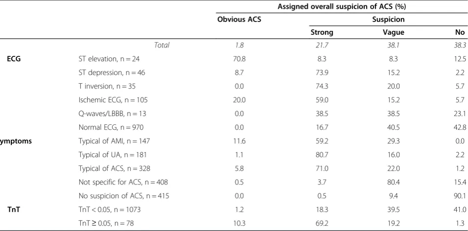
Table 1 The physician’s overall suspicion of ACS and the underlying assessments of the ECG, symptoms, and TnT Assigned overall suspicion of ACS (%)
Obvious ACS Suspicion
Strong Vague No
Total 1.8 21.7 38.1 38.3
ECG ST elevation, n = 24 70.8 8.3 8.3 12.5
ST depression, n = 46 8.7 73.9 15.2 2.2
T inversion, n = 35 0.0 74.3 20.0 5.7
Ischemic ECG, n = 105 20.0 59.0 15.2 5.7
Q-waves/LBBB, n = 13 0.0 38.5 38.5 23.1
Normal ECG, n = 970 0.0 16.7 40.5 42.8
Symptoms Typical of AMI, n = 147 11.6 59.2 29.3 0.0
Typical of UA, n = 181 1.1 80.7 16.0 2.2
Typical of ACS, n = 328 5.8 71.0 22.0 1.2
Not specific for ACS, n = 408 0.5 3.7 80.4 15.4
No suspicion of ACS, n = 415 0.0 0.5 9.4 90.1
TnT TnT < 0.05, n = 1073 1.2 18.3 39.5 41.0
TnT≥0.05, n = 78 10.3 69.2 19.2 1.3
changes (4 categories; normal, ischemic, with LBBB or Q-wave, or with AF, AFL or pacemaker), symptom category, TnT-level (≥0,05 or < 0.05 μg/L), sex and dichotomized age (≥65 or < 65 years) were included as covariates in both models. The reference categories were normal ECG, symptoms raising no suspicion of ACS, TnT < 0.05, male sex and age < 65 years, respectively. Factors were consid-ered significant if the P-value was below 0.05. Analyses were conducted with IBM SPSS Statistics 18 for Windows (IBM Corp., Somers NY, USA) software.
Results
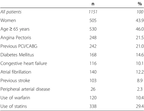
As shown in Figure 1, out of 1222 consecutive chest pain patients, a total of 1151 patients were included in the study. Fifty-six patients were excluded because of incomplete study data. Six-hundred and twenty-one (54.0%) were hospitalized and 140 of those (22.5%) proved to have ACS as the dis-charge diagnosis. Characteristics for the included patients are given in Table 4. Mean age was 60.7 ± 18.5 (SD) years.
Assessments of symptoms, ECG and TnT, and the overall likelihood of ACS
Table 1 shows the association between the designated likelihood of ACS and the underlying assessments of ECG, symptoms and TnT levels. Twenty-one (1.8%) of the 1151 patients were deemed as obvious ACS, 250 (21.7%) as strong suspicion of ACS, 439 (38.1%) as vague suspicion of ACS and 441 (38.3%) as no suspicion of ACS. Of the patients with ST-elevation, almost 71% were considered as obvious ACS. In contrast, only 5.8% of pa-tients with typical symptoms of ACS were assessed as obvious ACS, and only 10.3% of those with a positive TnT. Instead, both patients with typical symptoms of ACS and patients with positive TnT were in some 70% of the cases assigned a strong suspicion of ACS. ST de-pression or T inversion in the ECG were typically associ-ated with a strong overall suspicion of ACS (74% each). In patients with a normal ECG, ACS was still suspected in 57.2% of the cases. In contrast, when symptoms were
Table 2 Combinations of assessments of ECG findings, symptoms and TnT for cases with any suspicion of ACS
Assessments Isolated assessments Combinations of assessments
Ischemic ECG (ST elev or ST depr or T inv)
Symptoms typical of ACS
(AMI or UA)
TnT+ Only ischemic
ECG
Only symptoms typical of ACS
Only TnT+
Ischemic ECG + symptoms typical
of ACS
Ischemic ECG + TnT+
Symptoms typical of ACS + TnT+
All 3
All patients, %
10.1 28.5 6.8 0.4 16.9 0.0 6.8 2.7 5.4 2.1
Obvious ACS, n = 21
100 90.5 38.1 0.0 0.0 0.0 90.5 38.1 33.3 33.3
Strong suspicion, n = 250
26.8 93.2 21.6 0.0 51.6 0.0 22.4 7.6 20.4 6.8
Vague suspicion, n = 439
4.6 16.4 3.4 0.5 14.1 0.0 0.7 0.9 0.9 0.0
All figures percent. All three pathological = [ST elevation or ST depression or T inversion] and [typical symptoms of AMI or UA] and [positive TnT].
Table 3 Logistic regression analysis
Assignment of [obvious ACS] or [strong suspicion of ACS] or [vague suspicion of ACS] versus
[no suspicion of ACS]
Assignment of [obvious ACS] or [strong suspicion of ACS] versus [vague suspicion of ACS] or
[no suspicion of ACS]
P-value Odds ratio (95% CI) P-value Odds ratio (95% CI)
ECG Ischemic ECG 0.127 2.68 (0.76-9.50) < 0.001 30.6 (11.7-80.2)
Q-wave or LBBB 0.154 4.38 (0.57-33.4) 0.027 11.1 (1.32-94.0)
AF, AFL or PM 0.526 1.37 (0.52-3.60) 0.048 3.04 (1.01-9.15)
Symptoms Typical of ACS < 0.001 526 (185–1500) < 0.001 620 (138–2780)
Not specific for ACS < 0.001 48.7 (31.6-75.1) 0.043 4.95 (1.05-23.3)
TnT TNT+ 0.112 6.55 (0.65-66.3) 0.007 3.35 (1.39-8.09)
Age≥65 years 0.001 2.16 (1.40-3.35) 0.014 1.90 (1.14-3.17)
Female 0.913 1.02 (0.68-1.55) 0.043 0.59 (0.36-0.98)
Intercept < 0.001 0.074 < 0.001 0.003
non-suspicious of ACS, there was no overall suspicion of ACS in 90% of the cases.
There were associations between the assessments of the ECG, symptoms and the TnT level. Among patients with an ischemic ECG, 27.6% had a pathological initial TnT, compared to only 4.2% among those with a normal ECG. Similarly, TnT was positive in 18.9% of the pa-tients with symptoms typical of ACS, compared to 0.5% in those with non-suspicious symptoms. Among patients with ST-elevation, 79.2% had symptoms typical of ACS. In patients with a normal ECG, a majority had symp-toms not specific (36.6%) for, or not suspicious (39.9%) of, ACS.
Combinations of assessments of symptoms, ECG, TnT versus overall likelihood of ACS
In Table 2 it can be seen that all patients assessed as ob-vious ACS had ischemic ECG changes, and that 90.5% had both ischemic ECG changes and symptoms typical of ACS. Among patients with a strong suspicion of ACS, typical symptoms were considered present in 93.2%. In patients with a vague suspicion of ACS, there was almost never any combination of assessments clearly indicative of ACS.
Logistic regression analysis
In Table 3, the associations between the physicians’level of ACS suspicion and covariates included in the logistic regression models are described. In the physicians’ as-signment of any versus no suspicion of ACS, symptoms typical of and not specific for ACS contributed strongly and significantly (odds ratio OR 526 and 48.7, respect-ively, compared to no symptoms of ACS), but an ische-mic ECG or a positive TnT did not.
In the assignment of an obvious/strong versus vague/ no suspicion, symptoms typical of ACS was the most important factor (OR 620), but nonspecific symptoms
(OR 4.95), ischemic ECG (OR 30.6) and a positive TnT (OR 3.35) also contributed significantly to this assess-ment. Since no patient with obvious ACS had a normal ECG or symptoms not suspicious of ACS, it was not possible to create a model to analyze the assignment of obvious ACS versus strong/vague/no suspicion.
Discussion
The present results indicate that the ED physician uses the symptoms as the most important diagnostic tool when deciding the level of suspicion of ACS in chest pain patients, and that the ECG is considered more im-portant than TnT. To the best of our knowledge, this study is the first to evaluate the relative importances of the symptoms, ECG and TnT in routine care. This study did not, however, analyze the optimal use of these diag-nostic tools, i.e. their predictive values for the diagnosis of ACS. The results therefore do not show whether or not the physicians were correct in their use of the three different diagnostic modalities.
The predictive value of symptoms of ACS has been ex-tensively studied, and although the symptom history is a cornerstone in the ED assessment, the limitations are well known [3-5]. No single symptom makes ACS highly likely or unlikely. For instance, the likelihood ratio (LR) for ACS/AMI of chest pain radiating to both arms or
shoulders is only approximately 4–7, the LR of
exer-tional chest pain 2.5, nausea and vomiting 2 and of
pos-itional chest pain 0.3 [6-9]. Some 30–50% of AMI
patients lack chest pain [26], and among those with chest pain typical of AMI or ACS, 50% or less have it [10,11]. The chest pain quality, duration and severity are all suboptimal predictors of ACS [5,12,13]. Despite this, the ED physicians in the present study used the symp-toms as the most important factor to determine the ACS likelihood. When ACS was ruled out, the symptoms pro-vided the decisive information - neither the ECG nor TnT contributed significantly to the assignment of any versus no suspicion of ACS (Table 3). When symptoms were non-suspicious of ACS, the physician suspected ACS in less than one out of ten cases (Table 1). In addition, suspicions of ACS were sometimes based on symptoms alone, but almost never on ECG or TnT alone (Table 2).
When the physician could not rule out (i.e. assign no suspicion of ) ACS, he or she also seemed to use symp-toms as the most important diagnostic modality to grade the suspicion. In the regression model comparing obvi-ous/strong with vague/no suspicion of ACS (Table 3), the odds ratio for symptoms typical of ACS was consid-erably higher than for ischemic ECG and positive TnT. Further, symptoms typical of ACS were clearly more often associated with a strong suspicion of ACS than were an ischemic ECG (Tables 1 and 2), and nonspecific
Table 4 Characteristics of the included patients
n %
All patients 1151 100
Women 505 43.9
Age≥65 years 530 46.0
Angina Pectoris 248 21.5
Previous PCI/CABG 242 21.0
Diabetes Mellitus 168 14.6
Congestive heart failure 116 10.1
Atrial fibrillation 140 12.2
Previous stroke 103 8.9
Peripheral arterial disease 26 2.3
Use of warfarin 120 10.4
symptoms were in >80% of the cases associated with a vague suspicion of ACS (Table 1).
The ECG has been considered to be the most valuable ED test in patients with possible ACS, providing almost as much information as all other information combined [4,5]. This view is supported by published statistical de-cision support models, where ECG data have invariably been found to be crucial for the prediction of ACS in the ED, as opposed to data on symptoms and blood markers of myocardial injury [27]. In some models with ECG variables only, adding symptoms and other clinical variables did not improve ACS prediction [28]. In the present study, the ECG was indeed the most important factor when the ED physicians identified a case of obvi-ous ACS (Tables 1 and 2), i.e. when ACS was ruled in. However, the ECG was not considered as valuable for grading the ACS suspicion, and for ruling out ACS. A majority of patients with a normal ECG were still sus-pected to have ACS (Table 1), and the ECG did not con-tribute significantly to the assessment of any versus no suspicion of ACS (Table 3). A possible cause of this is that the shortcomings of the ECG for ACS prediction were recognized by the physicians in this study. Some 20% of AMI patients and 40% of UA patients have nor-mal ECGs in the ED [14]. Only about one in four of those with significant ST depression prove to have ACS [15], and only 5% with T-wave changes meeting ACS cri-teria have AMI [16]. Further, the ECG often does not de-tect transient myocardial ischemia [17], ischemia in patients with prior AMI [18], or ischemia in the area of the left circumflex coronary artery [19]. These limita-tions may be even more clinically relevant in EDs with a prehospital ECG system, such as in Lund, where patients with marked and clear-cut ECG changes (i.e. ST eleva-tion myocardial infarceleva-tion) usually bypass the ED on the way to the angiography suite.
Perhaps as expected, TnT was the least valuable diag-nostic tool to the ED physicians. TnT was not a signifi-cant factor in the assignment of any versus no suspicion of ACS, and had a markedly lower odds ratio than ische-mic ECG and typical symptoms in the assessment of obvious/strong versus vague/no suspicion of ACS (Table 3). In six patients out of ten with a normal TnT, the physician still suspected ACS (Table 1), and in only 10% of the patients with a positive TnT, the physician
noted an obvious ACS. TnT’s small role for the ACS
suspicion was probably due to its limited sensitivity and specificity for ACS in the ED [29], and it remains to been seen if newer high-sensitivity assays [30-32] will in-crease the importance of TnT in the assessment of pa-tients with a possible ACS.
Efforts to improve ED decisions are best based on an understanding of the practical decision-making in rou-tine care. Although the ECG might theoretically be
superior to symptoms when predicting ACS, it may not be surprising to the practicing ED physician that symp-toms emerged as a more important method to decide ACS suspicion than ECG and TnT in this study. The
patient’s description of his or her symptoms includes
a multitude of information (ranging from the pain localization to concurrent symptoms and perhaps even the clarity of the description) that physicians integrate when assessing the patient. Much of this information is difficult to quantify and study with traditional research protocols, and hence also to include in decision support models. Further, combinations of symptoms are very common and are even more difficult to study. We there-fore believe that the practical importance of symptoms for ACS prediction, and especially the combination of symptoms, is larger in routine care than suggested by published studies on predictive values [6-9]. Further in-vestigation of ACS prediction based on symptoms is needed, and also of the incorporation of symptom infor-mation in decision support models. For the time being, optimal decision-making in cases of possible ACS may involve physician interpretation of the symptoms and computerized ECG interpretation, since modern com-puter models are generally superior to physicians in de-tecting ACS on the ECG alone [33-35].
Limitations of the study
This study was performed at one university hospital ED only, and the results are not necessarily generalizable to other physician or patient populations. Definitions of the different levels of overall suspicion of ACS were given on the study forms. Although these definitions were non-controversial and did not specify which diagnostic modality is the most important, they most likely influ-enced the physicians’ assessments. Different definitions (or no definitions) may therefore have led to somewhat different results. Changing assessment scales for the symptoms and the overall likelihood of ACS might also have changed the results.
The study was designed so that the physician’s inter-pretations of the ECG, symptoms and TnT were not iso-lated from each other. The physician was thus aware of the ECG when assessing the symptoms and vice versa. This is not always the case in routine care, and our re-sults may therefore not be applicable to the ED physi-cian’s clinical reasoning in each individual patient case. Larger studies at other centers are needed to confirm our findings, perhaps also with different definitions of the levels of ACS suspicion.
Conclusion
not use the ECG in this way. Instead, the physicians used symptoms as the most important assessment tool, and ap-plied primarily the symptoms to determine the level of suspicion of ACS and to rule out ACS. The ECG was only primarily used to rule in ACS, whereas the TnT level in general played a minor role for the ACS likelihood. To our knowledge, this study is the first to evaluate the rela-tive importances of these diagnostic tools in routine care. Further studies of ACS prediction based on symptoms may help improve ED decision-making in patients with possible ACS.
Competing interests
The authors declare that they have no competing interests.
Authors’contributions
AK analyzed and interpreted the data, wrote and critically revised the manuscript. MS collected and interpreted the data and critically revised the manuscript. UE conceived and designed the study, interpreted the data and wrote and critically revised the manuscript. All authors read and approved the final version of the manuscript.
Acknowledgements
We are very much indebted to Susann Ullén for expert statistical advice.
Sources of funding
This work was supported by an ALF grant at Skåne University Hospital, Lund, Sweden, by the Region Skåne, The Swedish Heart and Lung foundation and the Laerdal foundation for Acute Medicine.
Received: 9 January 2013 Accepted: 8 April 2014 Published: 17 April 2014
References
1. Ekelund U, Nilsson HJ, Frigyesi A, Torffvit O:Patients with suspected acute coronary syndrome in a university hospital emergency department: an observational study.BMC Emerg Med2002,2(1):1.
2. The National board of health and Welfare (Socialstyrelsen):
Kranskärlssjukdom: (Guideline) Vetenskapligt Underlag för Nationella Riktlinjer för Hjärtsjukvård; 2008:42.
3. Ernfridsson M, Ekelund U:ABC om misstänkt akut koronart syndrom. Lakartidningen2005,102:28–29.
4. Lee TH, Goldman L:Evaluation of the patient with acute chest pain. N Engl J Med2000,342(16):1187–1195.
5. Goldman L, Kirtane AJ:Triage of patients with acute chest pain and possible cardiac ischemia: the elusive search for diagnostic perfection. Ann Intern Med2003,139(12):987–995.
6. Goodacre S, Locker T, Morris F, Campbell S:How useful are clinical features in the diagnosis of acute, undifferentiated chest pain? Acad Emerg Med2002,9(3):203–208.
7. Panju AA1, Hemmelgarn BR, Guyatt GH, Simel DL:The rational clinical examination. Is this patient having a myocardial infarction?JAMA1998, 280(14):1256–1263.
8. Bruyninckx R, Aertgeerts B, Bruyninckx P, Buntinx F:Signs and symptoms in diagnosing acute myocardial infarction and acute coronary syndrome: a diagnostic meta-analysis.Br J Gen Pract2008,58(547):105–111.
9. Swap CJ, Nagurney JT:Value and limitations of chest pain history in the evaluation of patients with suspected acute coronary syndromes. Jama2005,294(20):2623–2629.
10. Bassan R, Bassan R, Scofano M, Gamarski R, Fernando H, Pimenta L, Volschan A, Araujo M, Clare C, Fabrício M, Sanmartin CH, Mohallem K, Gaspar S, Macaciel R:Chest pain in the emergency room. Importance of a systematic approach.Arq Bras Cardiol2000,74(1):13–29.
11. Swap C, Nagurney J:Patients with suspected acute coronary syndrome in a university hospital emergency department: an observational study. American Medical Association2002,2(1):1–7.
12. Kontos MC, Jesse RL:Evaluation of the emergency department chest pain patient.Am J Cardiol2000,85(5A):32B–39B.
13. Edwards M, Chang AM, Matsuura AC, Green M, Robey JM, Hollander JE: Relationship between pain severity and outcomes in patients presenting with potential acute coronary syndromes.Ann Emerg Med2011, 58(6):501–507.
14. Pope JH, Selker HP:Diagnosis of acute cardiac ischemia.Emerg Med Clin North Am2003,21(1):27–59.
15. Green GB, Hill PM:Approach to Chest Pain. InEmergency Medicine. A Comprehensive Study Guide.Edited by Tintinalli JE, Kelen GD, Stapczynski JS. New York, NY: McGraw-Hill; 2004:333–343.
16. Lin KB, Kathy B, Lin BA, Shofer F, McCusker C, Meshberg E, Hollander J: Predictive value of T-wave abnormalities at the time of emergency department presentation in patients with potential acute coronary syndromes.Acad Emerg Med2008,15(6):537–543.
17. Sharkey S, Berger C, Brunette D, Henry T:Impact of the electrocardiogram on the delivery of thrombolytic therapy for acute myocardial infarction. Am J Cardiol1994,73(8):550–553.
18. Wagner G:Marriott’s Practical Electrocardiography.11th edition. Edited by Wilkins LW. Philadephia: 2008:145–162.
19. Zhou S:An automated algorithm to improve ECG detection of posterior STEMI associated with left circumflex coronary artery occlusion. Comput Cardiol2006,33:33–36.
20. Thygesen K, Alpert J, White H:Universal definition of myocardial infarction.Circulation2007,116(22):2634–2653.
21. Alpert JS, Antman E, Bassand JP, Klein W, Ohman M, Lopes Sendon J, Rydén L, Simoons M, Tendera M:Myocardial infarction redefined–a consensus document of the joint European society of cardiology/American college of cardiology committee for the redefinition of myocardial infarction. J Am Coll Cardiol2000,36(3):959–969.
22. Luepker RV, Luepker R, Apple F, Christenson R, Crow R, Fortmann S, Goff D, Goldberg R, Hand M, Jaffe A, Julian D, Levy D, Manolio T, Mendis S, Mensah G, Pajak A, Prineas R, Reddy S, Roger V, Rosamond W, Shahar E, Sharrett R, Sorlie P, Tunstall-Pedoe H:Case definitions for acute coronary heart disease in epidemiology and clinical research studies: a statement from the AHA council on epidemiology and prevention; AHA statistics committee; world heart federation council on epidemiology and prevention; the European society of cardiology working group on epidemiology and prevention; centers for disease control and prevention; and the national heart, lung, and blood institute. Circulation2003,108(20):2543–2549.
23. Granger CB, Granger C, Goldberg J, Dabbous O, Pieper K, Eagle K, Cannon C, Van de Werf F, Avezum A, Goodman S, Flather M, Keith A:Predictors of hospital mortality in the global registry of acute coronary events. Arch Intern Med2003,163(19):2345–2353.
24. Brieger D, Fox K, FitzGerald G, Eagle KA, Budaj A, Avezum A, Granger CB, Costa B, Anderson FA Jr, Steg PG:Predicting freedom from clinical events in non-ST-elevation acute coronary syndromes: the global registry of acute coronary events.Heart2009,95(11):888–894.
25. Soderholm M, Malekian Deligani M, Choudhary M, Bjork J, Ekelund U:Ability of risk scores to predict a low complication risk in patients admitted for suspected acute coronary syndrome.Emerg Med J2011, Epub ahead of print. 26. Gupta M, Tabas JA, Kohn MA:Presenting complaint among patients with
myocardial infarction who present to an urban, public hospital emergency department.Ann Emerg Med2002,40(2):180–186. 27. Steurer J, Held U, Schmid D, Ruckstuhl J, Bachmann L:Clinical value of
diagnostic instruments for ruling out acute coronary syndrome in patients with chest pain: a systematic review.Emerg Med J2010, 27(12):896–902.
28. Kennedy RL, Fraser H, McStay L, Harrison R:Early diagnosis of acute myocardial infarction using clinical and electrocardiographic data at presentation: derivation and evaluation of logistic regression models. Eur Heart J1996,17(8):1181–1191.
29. Clinical policy: critical issues in the evaluation and management of adult patients presenting with suspected acute myocardial infarction or unstable angina:American college of emergency physicians.Ann Emerg Med2000, 35(5):521–525.
30. Hollander JE:Highly sensitive troponins the answer or just more questions?J Am Coll Cardiol2009,54(13):1173–1175.
31. Peacock WF:The value of nothing: the consequence of a negative troponin test.J Am Coll Cardiol58(13):1340–1342.
elevation myocardial infarction in patients with suspected acute coronary syndrome and negative troponin results on admission. Clin Chem56(4):642–650.
33. Olsson S-E, Olsson SE, Ohlsson M, Ohlin H, Dzaferagic S, Nilsson ML, Sandkull P, Edebnbrandt L, Olsson SE, Ohlsson M, Ohlin H, Dzaferagic S, Nilsson ML, Sandkull P, Edebnbrandt L:Decision support for the initial triage of patients with acute coronary syndromes.Clin Physiol Funct Imaging2006, 26:151–156.
34. Forberg JL, Forberg J, Green M, Bjork J, Ohlsson M, Edenbrandt L, Ohlin H, Ekelund U:In search of the best method to predict acute coronary syndrome using only the electrocardiogram from the emergency department.J Electrocardiol2009,42(1):58–63.
35. Heden B, Hedén B, Ohlin H, Rittner R, Edenbrandt L:Acute myocardial infarction detected in the 12-lead ECG by artificial neural networks. Circulation1997,96(6):1798–1802.
doi:10.1186/1471-227X-14-9
Cite this article as:Kamaliet al.:What decides the suspicion of acute coronary syndrome in acute chest pain patients?BMC Emergency Medicine
201414:9.
Submit your next manuscript to BioMed Central and take full advantage of:
• Convenient online submission
• Thorough peer review
• No space constraints or color figure charges
• Immediate publication on acceptance
• Inclusion in PubMed, CAS, Scopus and Google Scholar
• Research which is freely available for redistribution