R E S E A R C H A R T I C L E
Open Access
Feasibility of a 20-gauge ProCore needle in
EUS-guided subepithelial tumor sampling:
a prospective multicenter study
Do Hoon Kim
1, Gwang Ha Kim
2*, Chang Min Cho
3, Chang Hwan Park
4, Soo-Young Na
5, Tae Hyeon Kim
6,
Yu Kyung Cho
7, Ji Hyun Kim
8, Dong-Wan Seo
1and Korean EUS Study Group
Abstract
Background:Endoscopic ultrasonography-guided fine-needle biopsy (EUS-FNB) may facilitate tissue acquisition for a definitive diagnosis of gastrointestinal subepithelial tumors (SETs). This study aimed to determine the diagnostic yield of EUS-FNB using a novel 20-gauge ProCore needle with a coiled sheath in tissue sampling of gastrointestinal SETs. Methods:Between July 2016 and February 2017, 39 patients with gastrointestinal SETs were prospectively recruited from six university hospitals in Korea. Hypoechoic SETs≥2 cm in size and originating from the submucosal and/or muscularis propria layer under EUS were eligible. This study was registered onClinicalTrials.gov(NCT02884154). Results:A total of 36 patients were included in the final analyses. EUS-FNB was diagnostic in 88.9% of SETs. Tissue adequacy was judged as optimal in 97.2% of FNB specimens according to on-site visual evaluation by endosonographers, and in 88.9% of specimens according to pathologists. A macroscopically optimal core sample was obtained with two needle passes in 94.4% of cases. Technical failure rate was encountered in two cases (5.6%) after two needle passes. There were two cases (5.6%) of bleeding, which was managed endoscopically.
Conclusions:EUS-FNB using a 20-gauge ProCore needle is a technically feasible and effective modality for histopathologic diagnosis of gastrointestinal SETs, providing adequate core samples with fewer needle passes;ClinicalTrials.govnumber, NCT02884154.
Keywords:Biopsy, Endoscopic ultrasonography, Gastrointestinal tract, Subepithelial tumor
Background
Gastrointestinal subepithelial tumors (SETs) include benign, potentially malignant, and malignant lesions. Endoscopic ultrasonography (EUS) is a useful tool for the characterization of SETs, providing details on the gastro-intestinal wall structures in addition to morphologic features [1]. However, the differential diagnosis of hypoe-choic SETs remains challenging, and a definitive diagnosis can rarely be established on imaging modalities alone [2]. As the treatment decision for patients with SETs largely depends on the histopathological diagnosis, effective and high-quality tissue acquisition is required.
In this context, EUS-guided sampling methods, including fine-needle aspiration (FNA), Trucut biopsy (EUS-TCB), and fine-needle biopsy (EUS-FNB) have been introduced [3]. EUS-FNA with a 22-gauge or 25-gauge needle provides cytological aspirates, with a diagnostic yield of 70% to 79% [4,5]. Alternatively, tissue cores can be procured with EUS-TCB, which enables immunohisto-chemical analysis and thus facilitates a definitive diagnosis [6]. However, the diagnostic yield of EUS-TCB is not higher than that of EUS-FNA, and technical failures fre-quently occur [5,7]. The Trucut needle is relatively stiff, and the firing mechanism produced by the torqued echoendoscope limits its maneuverability and accessibility in certain locations within the gastrointestinal tract, such as the gastric antrum or duodenum [8].
Recently, a novel EchoTip ProCore high definition ultrasound biopsy needle with a coiled sheath has
* Correspondence:[email protected]
2Department of Internal Medicine, Pusan National University School of
Medicine and Biomedical Research Institute, Pusan National University Hospital, 179 Gudeok-ro, Seo-gu, Busan 49241, South Korea Full list of author information is available at the end of the article
become available. This needle, which has a unique reverse bevel technology, allows for a simultaneous core biopsy to be obtained along with aspirated material. The 19-gauge ProCore needle was shown to obtain histologically ad-equate samples in nearly 90% of cases, with excellent tech-nical feasibility in both intra-intestinal and extra-intestinal mass lesions [9]. However, as this 19-gauge needle is still stiff, its maneuverability is not satisfactory in the gastric antrum or duodenum. Furthermore, the diagnostic yield of the 22-gauge ProCore needle was found to be 81.8% to 86.0% in gastric SETs [10–12]. The novel 20-gauge needle offers improved flexibility for those more difficult EUS-FNA biopsy approaches, with easy to-and-fro pas-sage of the needle, along with the benefits of the larger 20-gauge needle to yield histologic grade tissue.
Although there is evidence supporting the utility of these procedures, diagnostic yields have varied according to the location in the gastrointestinal tract and specific needle types. The aim of this prospective multicenter study was to investigate the feasibility and diagnostic yield of EUS-FNB with a 20-gauge ProCore biopsy nee-dle in the diagnosis of gastrointestinal SETs.
Methods
Patients
Consecutive patients with gastrointestinal SETs were prospectively enrolled at six university hospitals in Korea (Asan Medical Center, Pusan National University Hos-pital, Kyungpook National University HosHos-pital, Chonnam National University Hospital, Jeju National University Hospital, and Wonkwang University Hospital) from July 2016 to February 2017. Those with a hypoechoic SET ≥2 cm in size and located in the submucosa and/or mus-cularis propria layer under EUS examination were eligible. Exclusion criteria were as follows: (i) anechoic or hypere-choic SETs on EUS that suggested cyst, vessel, or lipoma; (ii) those with a history of coagulopathy, presenting as a platelet count < 50,000/mm3or prothrombin time < 50%. All enrolled patients provided written informed consent for their participation in the study. The study protocol was approved by the institutional review board of each hospital and was conducted in accordance with the Declaration of Helsinki and its amendments, and the Good Clinical Practice guidelines. This study was also reg-istered onClinicalTrials.gov (NCT02884154). All authors had access to the study data and reviewed and approved the final manuscript.
Procedure technique
All procedures were performed using a linear array echoendoscope (GF-UCT 140, UCT 240 or UCT 260; Olympus, Tokyo, Japan) with the patient under con-scious sedation. After the target lesion was endosonogra-phically visualized, a 20-gauge ProCore needle (EchoTip
ProCore; Wilson-Cook Medical Inc., USA) was advanced into the target tissue. The key features for 20-gauge Pro-Core needle are (1) the core trap designed for receiving tissue into needle, (2) Menghini bevel for obtaining sam-ple, (3) the coiled sheath to facilitate needle flexibility and (4) the ReCoil stylet aids stylet management minim-izing the risk of contamination. After successful punc-ture, the endosonographer moved the needle to and fro within the lesion for more than 10 to 15 times, while an assistant simultaneously slowly pulled out the stylet over 30 to 60 s without suction to achieve minimal negative pressure within the needle.
At least three needle passes were performed using the designated needle, and the number of required needle passes to obtain sufficient core tissue was recorded. If such samples were not obtained after three needle passes, the number of required passes was considered to be four. Technical failure was defined as malfunction of the needle before three needle passes. Diagnostic failure was defined as failure to obtain sufficient core samples after three passes. If a diagnostic or technical failure was encoun-tered, an alternative needle was used, according to the judgment of the endosonographers. In the rescue cohort, a maximum of three passes were attempted using the al-ternative needle, until either sufficient core samples were obtained or technical failure was encountered.
Histological analysis
Cytological material was sent to the cytologists as a fixed or an air-dried slide.
A pathologic diagnosis was based on hematoxylin and eosin with or without immunohistochemical staining, such as CD34, CD117, S100, and smooth muscle actin. Because the morphological characteristics of mesenchy-mal tumors are nonspecific, a positive diagnosis by EUS-FNB was only considered true positive when im-munohistochemical analysis was conclusive. In patients who underwent endoscopic or surgical resection of the tumors, the final diagnosis was based on histopatho-logical assessment of the resected specimen. Otherwise, histopathological assessment of the FNB samples was deemed to be the gold standard.
Outcome parameters
The primary objective was to determine diagnostic accur-acy. Diagnostic accuracy was defined that the sufficient samples were obtained for satisfactory assessment of histo-logic architecture and immunohistochemical evaluation within three needle passes. Because the morphological characteristics of mesenchymal tumors are nonspecific, a positive diagnosis by EUS-FNB was only considered true positive when immunohistochemical analysis was conclu-sive. The secondary outcome measures were the tissue ad-equacy, the number of needle passes required to obtain a sufficient tissue core, and the rates of diagnostic failure, technical failure, and adverse events. Tissue adequacy was defined as macroscopically and histologically optimal core samples. Adverse events were defined as any deviation from the clinical course after EUS-guided sampling, such as ex-cessive bleeding at the site of puncture or perforation.
Statistical analysis
The required sample size was estimated to be a minimum of 32, based on a two-tailed 95% confidence interval with
a width equal to 0.298 and a diagnostic accuracy of 80%. Assuming a dropout rate of 20%, the final required sample size was calculated to be 38 patients.
Descriptive statistics were used to document the char-acteristics of SETs and procedure-related outcomes. Cat-egorical variables were presented as frequencies and proportions. Continuous variables were summarized as median values with range or means with standard devi-ation. All statistical analyses were performed using SPSS version 21.0 (SPSS Inc., Chicago, IL).
Results
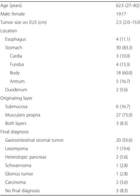
Of the 39 enrolled patients, three were excluded from the final analyses: two had lesions that were suspected to be larger than 2 cm, but were found to be < 2 cm on EUS examination, and one lesion was revealed to originate from the muscularis mucosa layer. Ultimately, 36 patients were included in the analysis (Fig. 2). Demographic and clinical characteristics of the SETs are shown in Table1.
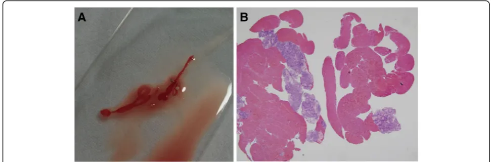
(Fig. 3). Three cases were considered as optimal by the endosonographer but were revealed to be suboptimal on histopathological examination: there was insufficient sub-mucosal tissue in two cases and only fibrotic tissue was found in one case.
Technical failure was encountered in two cases (5.6%) after two needle passes in the fundus and duodenal bulb. However, optimal tissue samples were obtained before the technical failure in both cases. In terms of adverse events, procedure-related bleeding occurred in two cases (5.6%). The bleeding was associated with needle punc-ture and was managed endoscopically with hemoclips.
Finally, 10 patients with diagnostic results avoided surgery: seven patients with leiomyoma, two with heterotopic pancreas, and one with glomus tumor. Fig. 2Flowchart of the study
Table 1Baseline characteristics of the patients and subepithelial lesions
Age (years) 62.5 (27–82)
Male: female 19:17
Tumor size on EUS (cm) 2.5 (2.0–15.0) Location
Esophagus 4 (11.1)
Stomach 30 (83.3)
Cardia 3 (10.0)
Fundus 4 (13.3)
Body 18 (60.0)
Antrum 5 (16.7)
Duodenum 2 (5.6)
Originating layer
Submucosa 6 (16.7)
Muscularis propria 27 (75.0)
Both layers 3 (8.3)
Final diagnosis
Gastrointestinal stromal tumor 20 (55.6)
Leiomyoma 7 (19.4)
Heterotopic pancreas 2 (5.6)
Schwannoma 1 (2.8)
Glomus tumor 1 (2.8)
Carcinoma 2 (5.6)
No final diagnosis 3 (8.3)
Variables are presented as number (%) or median (range) EUS, endoscopic ultrasonography
Table 2Procedural characteristics and outcomes of the EUS-guided fine-needle biopsies
Diagnosis achieved 32 (88.9) Sample adequacy during three needle passes
Macroscopic adequacy 35 (97.2) Definite tissue core 32 (88.9) Suspicious tissue core 3 (8.3) Histological adequacy 32 (88.9)
Number of required needle passes to obtain optimal tissue core samplea
Macroscopic 1.2 ± 0.6
Histological 1.5 ± 1.0
Technical failure 2 (5.6)
Adverse events 2 (5.6)
Variables are presented as number (%) or mean ± standard deviation
a
The diagnostic yield of EUS-FNB was 95.0% (19/20) in patients with a GIST. One lesion, which was not diag-nosed on EUS-FNB, was surgically resected and con-firmed as a GIST. Of 19 patients with a GIST, 13 had undergone surgical resection by the time of the ana-lyses, and the histopathological diagnoses were consist-ent with the EUS-FNB diagnoses.
Discussion
We investigated the diagnostic yield of EUS-FNB using a novel 20-gauge ProCore needle with a coiled sheath in the diagnosis of gastrointestinal SETs. Needle punctures were successful in all cases, irrespective of the location, and the diagnostic yield was 88.9%. The rates for obtain-ing macroscopically and histologically optimal core sam-ples with EUS-FNB were 97.2% and 88.9%, respectively. The median number of needle passes for both macro-scopic and histological adequacy was one. Our findings suggest that the 20-gauge ProCore needle may yield op-timal core tissue samples with fewer needle passes com-pared with 22-gauge ProCore needle.
EUS-guided samplings are pivotal methods for tissue ac-quisition in gastrointestinal SETs, and a number of studies have evaluated the feasibility of EUS-guided sampling techniques in the diagnosis of SETs. EUS-FNA usually yields small sample volumes that are mainly processed for cytological evaluation [4, 5]. However, the cytological as-pirate obtained by EUS-FNA is quantitatively low, and is often insufficient for differential diagnosis, especially in cases of gastric mesenchymal tumors that mandate an immunohistochemical assay. Samples with preserved tis-sue architecture are necessary to make a definitive diagno-sis of hypoechoic SETs, especially when they are located in the muscularis propria layer. Although EUS-TCB pro-vides large core tissue samples allowing histological
examination as well as immunohistochemical staining, the Trucut needle is associated with technical difficulties be-cause of its inherent stiffness, which results in a high tech-nical failure rate [5,6,13]. Furthermore, the Trucut needle allows only one pass in a single axis, which thereby results in a limited diagnostic yield.
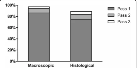
Recently, EUS-FNB technique has been developed, allowing core biopsy samples to be attained along with as-pirated material. The ProCore needle is made of stainless steel, with a nitinol stylet and there is a reverse bevel to hook and cut tissue. Studies suggested that EUS-FNB may be advantageous for optimizing specimen adequacy and diagnostic accuracy. The 19-gauge ProCore needle showed histologic adequacy of 89.5% and diagnostic ac-curacy of 86.0% in the diagnosis of intra-intestinal and extra-intestinal mass lesions [9]. The diagnostic accuracy was 81.8–86.0% for gastric SETs when the 22-gauge nee-dle was used [10–12]. In the present study, optimal macroscopic and histological core samples were procured in 97.2% and 88.9% of cases with three needle passes, which resulted in a high diagnostic histologic accuracy rate. Furthermore, adequate tissue core was obtained within two needle passes in most cases, with only 5.6% of cases requiring three needle passes to achieve a diagnosis.
The diagnostic yield of EUS-guided sampling depends on a variety of factors, such as the nature of the target lesion, site of the puncture, the availability of a cytopa-thologist, the experience of the endosonographer, and the type and size of the needle used [3]. Regarding the needle size, the large-caliber needles seem to have the advantage of acquiring more tissue, which enables the assessment of architectural features. However, a larger needle is prone to have technical difficulties with respect to its maneuverability and accessibility, whereas a smaller needle is flexible and can be fanned in multiple directions within the target lesion. Indeed, studies com-paring the diagnostic performance for SETs did not demonstrate any significant advantage of EUS-TCB or EUS-FNB over a standard FNA needle in terms of cyto-logic parameters, amount of diagnostic cell block mater-ial, adequacy, and accuracy [4, 5, 14]. Of note, the number of needle passes required for diagnosis was sig-nificantly lower when using the ProCore needle, suggest-ing that a better quality sample was obtained in each pass [11,14–16]. In the present study, the median num-ber of needle passes to achieve both macroscopic and histological adequacy was one. The first pass of the 20-gauge ProCore needle yielded a histologically optimal
Table 3Sample adequacy during three needle passes
Pass 1 (n= 36) Pass 2 (n= 36) Pass 3 (n= 34) Total (n= 36)
Macroscopic adequacy 31 (86.1%) 33 (91.7%) 32 (94.1%) 35 (97.2%)
Histological adequacy 27 (75.0%) 29 (80.6%) 27 (79.4%) 32 (88.9%)
tissue core in 75.0% of cases, and the histological tissue adequacy on each pass is over 75% throughout the proce-dures. These results demonstrate the high quality of the tissue obtained by a single pass of the ProCore needle.
It is sometimes difficult to determine whether sufficient core tissues are obtained during EUS-guided sampling, as indicated by the discrepancy between macroscopically and histologically assessed tissue adequacy. One possibility is that the visible materials do not consist of a tissue core, while another is that the materials acquired are not repre-sentative of the target lesion. The latter can be more chal-lenging in clinical practice, because a tissue core may well be acquired but then revealed to give little information in respect to the diagnosis. In this present study, three cases were considered suboptimal for histological evaluation despite being macroscopically assessed as optimal core samples. All three cases contained visible tissue materials that seemed to be core samples, but were later revealed to be non-diagnostic. As most institutions do not have on-site pathologists, certain criteria for the macroscopic visual assessment of a specimen by the endosonographer can be helpful to ensure the adequacy of tissue cores and to reduce unnecessary punctures.
Although bleeding and perforation are potentially life-threatening adverse events of EUS and EUS-guided procedures, the incidence of adverse events has been re-ported as being low [17, 18]. In addition, most adverse events were caused by 19-gauge needles [7,19]. Previous report using 22-gauge ProCore needles showed either no or low adverse event rates [10–12, 20]. In the present study, minor bleeding occurred during the procedure in two cases (5.6%), and was controlled endoscopically, thereby supporting the safety of EUS-FNB procedures.
Our study had several limitations of note. First, we in-cluded only academic centers with highly experienced endosonographers. Second, the size of all SETs included in our study was≥2 cm. According to recent guidelines for SETs, when neoplastic SETs are 2–5 cm in diameter or when SET < 2 cm have clinically malignant features on endoscopy, the guidelines recommend detailed exam-ination with EUS, computed tomography with contrast enhancement, and/or EUS-FNA. Clinically malignant features means irregular borders, ulceration, and/or growth during endoscopic follow-up. When there are no clinically malignant features, gastric SETs < 2 cm could be followed up by endoscopy or EUS once or twice a year until the tumors increase in size or become symp-tomatic, even if they are diagnosed as GISTs later on [21]. Therefore, we included only SETs with ≥2 cm in size. Third, in cases which do not need treatment surgi-cally or endoscopisurgi-cally, the FNB results were determined as final diagnosis. There is a very rare possibility of ma-lignant transformation in benign tumors, but it is a problem related to the natural history of tumors, not a
wrong diagnosis. Fourth, cytological aspirates and histo-logical core samples were not interpreted separately. In previous studies, the amount of diagnostic cell block material did not vary according to the use of either a beveled or standard needle, and the use of a beveled needle provided no benefit in terms of the diagnostic cell block [14]. Another possible limitation of our study is that the mitotic count and Ki-67 labeling index of the GIST were not determined. Although the diagnosis of GIST was successfully made before surgical resection, there may be considerable discrepancy in the mitotic count or Ki-67 index of the tumors between the EUS-FNB and surgical specimens [12].
Conclusions
In conclusion, EUS-FNB using a 20G ProCore needle is a technically feasible, safe, and effective modality for his-topathologic diagnosis of gastrointestinal SETs larger than 2 cm, providing adequate tissues core with fewer needle passes.
Abbreviations
EUS:Endoscopic ultrasonography; EUS-FNB: Endoscopic ultrasonography-guided fine-needle biopsy; FNA: Fine-needle aspiration; GIST: Gastrointestinal stromal tumor; SET: Subepithelial tumor; TCB: Trucut biopsy
Availability of data and materials
The datasets used and analyzed during the current study will be available from the corresponding author on reasonable request.
Authors’contributions
DHK and GHK study concept and design, analysis and interpretation of data, statistical analysis; DHK, GHK, CMC, CHP, SYN, THL, YKC, and JHK acquisition of data, administrative, technical, or material support; DHK drafting of the manuscript; GHK, CMC, CHP, SYN, THL, YKC, and JHK critical revision of the manuscript for important intellectual content; GHK and D-WS study supervi-sion. All of the authors read and approved the final manuscript.
Ethics approval and consent to participate
All enrolled patients provided written informed consent for their participation in the study. The study protocol was approved by the institutional review board of each hospital (Asan Medical Center, Pusan National University Hospital, Kyungpook National University School of Medicine, Chonnam National University Medical school, Jeju National University School of Medicine, Wonkwang University School of Medicine, Seoul St. Mary’s Hospital, and Inje University Pusan Paik Hospital) and was conducted in accordance with the Declaration of Helsinki and its amendments, and the Good Clinical Practice guidelines. This study was also registered onClinicalTrials.gov(NCT02884154). All authors had access to the study data and reviewed and approved the final manuscript.
Consent for publication
Not applicable
Competing interests
The authors declare that they have no competing interests.
Publisher’s Note
Springer Nature remains neutral with regard to jurisdictional claims in published maps and institutional affiliations.
Author details
1Department of Gastroenterology, University of Ulsan College of Medicine,
Medicine and Biomedical Research Institute, Pusan National University Hospital, 179 Gudeok-ro, Seo-gu, Busan 49241, South Korea.3Department of
Internal Medicine, Kyungpook National University School of Medicine, 130 Dongdeok-ro, Jung-gu, Daegu 41944, South Korea.4Department of Internal Medicine, Chonnam National University Medical school, 42 Jebong-ro, Donggu, Gwangju 61469, South Korea.5Department of Internal Medicine,
Jeju National University School of Medicine, 15, Aran 13-gil, Jeju-si, Jeju, Jeju-do 63241, South Korea.6Department of Gastroenterology, Wonkwang University School of Medicine, 895 Muwang-Ro, Iksan, Jeonlabuk-do, Iksan 54538, South Korea.7Department of Gastroenterology, The Catholic
University of Korea, Seoul St. Mary’s Hospital, 222 Banpo-daero, Seocho-gu, Seoul 06591, South Korea.8Department of Gastroenterology, Inje University Pusan Paik Hospital, Bokji-ro 75, Busangjin-gu, Busan 47392, South Korea.
Received: 3 May 2018 Accepted: 8 October 2018
References
1. Hwang JH, Saunders MD, Rulyak SJ, Shaw S, Nietsch H, Kimmey MB. A prospective study comparing endoscopy and EUS in the evaluation of GI subepithelial masses. Gastrointest Endosc. 2005;62:202–8.
2. Moon JS. Role of endoscopic ultrasonography in guiding treatment plans for upper gastrointestinal subepithelial tumors. Clin Endosc. 2016;49:220–5. 3. Polkowski M, Bergman JJ. Endoscopic ultrasonography-guided biopsy for
submucosal tumors: needless needling? Endoscopy. 2010;42:324–6. 4. Philipper M, Hollerbach S, Gabbert HE, Heikaus S, Bocking A, Pomjanski N,
et al. Prospective comparison of endoscopic ultrasound-guided fine-needle aspiration and surgical histology in upper gastrointestinal submucosal tumors. Endoscopy. 2010;42:300–5.
5. Fernandez-Esparrach G, Sendino O, Sole M, Pellise M, Colomo L, Pardo A, et al. Endoscopic ultrasound-guided fine-needle aspiration and trucut biopsy in the diagnosis of gastric stromal tumors: a randomized crossover study. Endoscopy. 2010;42:292–9.
6. Levy MJ, Wiersema MJ. EUS-guided Trucut biopsy. Gastrointest Endosc. 2005;62:417–26.
7. Polkowski M, Gerke W, Jarosz D, Nasierowska-Guttmejer A, Rutkowski P, Nowecki ZI, et al. Diagnostic yield and safety of endoscopic ultrasound-guided trucut [corrected] biopsy in patients with gastric submucosal tumors: a prospective study. Endoscopy. 2009;41:329–34.
8. Varadarajulu S, Fraig M, Schmulewitz N, Roberts S, Wildi S, Hawes RH, et al. Comparison of guided 19-gauge Trucut needle biopsy with EUS-guided fine-needle aspiration. Endoscopy. 2004;36:397–401.
9. Iglesias-Garcia J, Poley JW, Larghi A, Giovannini M, Petrone MC, Abdulkader I, et al. Feasibility and yield of a new EUS histology needle: results from a multicenter, pooled, cohort study. Gastrointest Endosc. 2011;73:1189–96. 10. Lee JH, Cho CJ, Park YS, Ahn JY, Kim DH, Na HK, et al. EUS-guided 22-gauge
fine needle biopsy for the diagnosis of gastric subepithelial tumors larger than 2 cm. Scand J Gastroenterol. 2016;51:486–93.
11. Han JP, Lee TH, Hong SJ, Kim HK, Noh HM, Lee YN, et al. EUS-guided FNA and FNB after on-site cytological evaluation in gastric subepithelial tumors. J Dig Dis. 2016;17:582–7.
12. Lee M, Min BH, Lee H, Ahn S, Lee JH, Rhee PL, et al. Feasibility and diagnostic yield of endoscopic ultrasonography-guided fine needle biopsy with a new core biopsy needle device in patients with gastric subepithelial tumors. Medicine (Baltimore). 2015;94:e1622.
13. Na HK, Lee JH, Park YS, Ahn JY, Choi KS, Kim DH, et al. Yields and utility of endoscopic ultrasonography-guided 19-gauge trucut biopsy versus 22-gauge fine needle aspiration for diagnosing gastric subepithelial tumors. Clin Endosc. 2015;48:152–7.
14. Witt BL, Adler DG, Hilden K, Layfield LJ. A comparative needle study: EUS-FNA procedures using the HD ProCore(™) and EchoTip(®) 22-gauge needle types. Diagn Cytopathol. 2013;41:1069–74.
15. Lee BS, Cho CM, Jung MK, Jang JS, Bae HI. Comparison of histologic core portions acquired from a core biopsy needle and a conventional needle in solid mass lesions: a prospective randomized trial. Gut Liver. 2017;11:559–66. 16. Bang JY, Hawes R, Varadarajulu S. A meta-analysis comparing ProCore and
standard fine-needle aspiration needles for endoscopic ultrasound-guided tissue acquisition. Endoscopy. 2016;48:339–49.
17. Hamada T, Yasunaga H, Nakai Y, Isayama H, Horiguchi H, Matsuda S, et al. Rarity of severe bleeding and perforation in endoscopic ultrasound-guided fine needle aspiration for submucosal tumors. Dig Dis Sci. 2013;58:2634–8.
18. Zhang XC, Li QL, Yu YF, Yao LQ, Xu MD, Zhang YQ, et al. Diagnostic efficacy of endoscopic ultrasound-guided needle sampling for upper
gastrointestinal subepithelial lesions: a meta-analysis. Surg Endosc. 2016;30: 2431–41.
19. Eckardt AJ, Adler A, Gomes EM, Jenssen C, Siebert C, Gottschalk U, et al. Endosonographic large-bore biopsy of gastric subepithelial tumors: a prospective multicenter study. Eur J Gastroenterol Hepatol. 2012;24:1135–44. 20. Kim GH, Cho YK, Kim EY, Kim HK, Cho JW, Lee TH, et al. Comparison of
22-gauge aspiration needle with 22-22-gauge biopsy needle in endoscopic ultrasonography-guided subepithelial tumor sampling. Scand J Gastroenterol. 2014;49:347–54.