R E S E A R C H
Open Access
Differential expression, molecular cloning,
and characterization of porcine beta
defensin 114
Guoqi Su
1,2, Kunhong Xie
1,2, Daiwen Chen
1,2, Bing Yu
1,2, Zhiqing Huang
1,2, Yuheng Luo
1,2, Xiangbing Mao
1,2,
Ping Zheng
1,2, Jie Yu
1,2, Junqiu Luo
1,2and Jun He
1,2*Abstract
Background:β-defensins have attracted considerable research interest because of their roles in protecting hosts from various pathogens. This study was conducted to investigate the expression profiles of the porcineβ-defensin 114 (PBD114) in different breeds and in response to infections. Moreover, the function of PBD114 protein was partially investigated.
Methods:Six Tibetan pigs (TP) and six DLY (Duroc×Landrace×Yorkshire) pigs were slaughtered to explore the expression profiles ofPBD114in different breeds and tissues. For infection models, sixteen DLY pigs were divided into two groups and challenged either with sterile saline orE. coliK88. The recombinant protein PBD114 (rPBD114) was obtained by using a heterologous expression system inE. coli.
Results:PBD114gene was highly expressed in tissues such as the intestine, liver, spleen, and thymus. Interestingly, the expression level ofPBD114gene was higher in the TP pigs than in the DLY pigs (P< 0.05), and was significantly elevated uponE. coliK88 challenge (P< 0.05). The nucleotide sequences ofPBD114from Tibetan and DLY pigs was identical, and both showed a 210-bp open reading frame encoding a 69-amino acid mature peptide. To explaore the function of PBD114 protein,PBD114gene was successfully expressed inE. coliOrigami B (DE3) and the molecular weight of the rPBD114 was estimated by SDS-PAGE to be 25 kDa. The rPBD114 was purified and mass spectrometry verified the protein as PBD114. Importantly, rPBD114 showed antimicrobial activities againstE. coliDH5αandE. coliK88, and the minimal inhibitory concentrations (MICs) were 64 and 128μg/mL, respectively. Hemolytic and cytotoxicity assays showed that rPBD114 did not affect cell viability under physiological concentrations.
Conclusions:PBD114is an infection response gene that is differentially-expressed between different porcine breeds and tissues. The antimicrobial activity of PBD114 protein, against pathogens such as theE. coliK88, suggested that it may serve as a candidate for the substitution of conventionally used antibiotics.
Keywords:Antimicrobial activity, DLY,E. coli, Porcine beta defensin 114, Tibetan
Introduction
The availability of antibiotics for treating bacterial infec-tions has significantly improved the health of animals and humans. In addition, sub-therapeutic levels of anti-biotics have been added to pig feed as growth promoters in many countries [1]. However, liberal antibiotic use
combined with the ability of microorganisms to develop antibiotic resistance has led to multi-drug resistant, which has fuelled a potential global public health crisis [2]. To reduce the emergence of antimicrobial resistance, approaches to reduce the utilization of antibiotics in ani-mal production are needed.
Innate immunity is the first line of host defense against invading pathogens such as bacteria, virus, and fungus. Previous studies have indicated that one of the primary innate immune responses are the secretion of
antimicrobial peptides (AMPs) [3,4]. To date, thousands
© The Author(s). 2019Open AccessThis article is distributed under the terms of the Creative Commons Attribution 4.0 International License (http://creativecommons.org/licenses/by/4.0/), which permits unrestricted use, distribution, and reproduction in any medium, provided you give appropriate credit to the original author(s) and the source, provide a link to the Creative Commons license, and indicate if changes were made. The Creative Commons Public Domain Dedication waiver (http://creativecommons.org/publicdomain/zero/1.0/) applies to the data made available in this article, unless otherwise stated.
* Correspondence:hejun8067@163.com
1
Animal Nutrition Institute, Sichuan Agricultural University, Chengdu 611130, People’s Republic of China
2Key Laboratory for Animal Disease-Resistance Nutrition of China Ministry of
of antimicrobial peptides (AMPs) have already been identified in vertebrates, invertebrates, plants and fungi (http://aps.unmc.edu/AP/main.php). AMPs are classified based on structural and sequence homology, with host defence peptides (HDPs) comprising one of the major subclasses of the family of AMPs [5]. HDPs are a family of low molecular weight peptides secreted by organisms, and can protect hosts from a broad range of pathogens
including bacteria, virus and fungus [3–5]. Upon
micro-bial invasion, mature active HDPs are released quickly by proteolytic processing from precursor peptides either in cytoplasmic granules of most mammalian neutrophils and macrophages or in epithelial cells of the respiratory,
gastroenteric, and urogenital tracts [6, 7]. HDPs disrupt
the bacterial membrane by forming non-specifc electro-static interactions with the membrane lipid components [8]. So bacteria are less able to develop resistance to HDPs than to traditional antibiotics. Therefore, the ad-ministration of HDPs is a potentially novel therapeutic strategy for inflammatory and infectious diseases of the
gastrointestinal tract [7,9], and may present a promising
alternative to the traditional antibiotic feed additives used in the livestock industry.
The first identified porcine beta defensin was porcine beta defensin 1 (PBD1), which can be detected through-out the respiratory and digestive tracts [6]. Further stud-ies found that PBD1 displayed strong antimicrobial
activity against Bordetella pertussis, Staphylococcus
aur-eus and multi-resistant E. coli[10–12]. In addition, pre-viously reported that porcine beta defensin (PBD2) was expressed in the intestine, and mature PBD2 synthetic peptide exhibited high antimicrobial activity against a broad range of pathogenic bacteria, but only limited hemolytic activity against porcine red blood cells [1,12]. A recent study reported that there were at least 29
dif-ferent β-defensins expressed in porcine tissues. The
average nucleotide sequence identity from the 27 pairs
of orthologous β-defensins between humans and pigs
was 84.38%, and this probably indicated thatβ-defensins
have multiple biological functions [13]. Although the porcine beta defensins may function as an important regulator of host defense against exogenous pathogens, the expression profile and biological functions of PBD114 are still unclear. The present study was
con-ducted to explore the expression profiles of thePBD114
in different breeds and in response to infections. More-over, the function of PBD114 protein has also been investigated.
Materials and methods Strains and vectors
E. coli DH5α and E. coli Origami B (DE3) strains were
purchased from TIANGEN (Beijing, China). E. coli K88
was kindly provided by Professor Lianqiang Che, Institute
of Animal Nutrition, Sichuan Agricultural University.
Sal-monella typhimurium (S. typhimurium) ATCC14028,
Staphylococcus aureus(S. aureus) CICC10384, Streptococ-cus, Pichia pastorisX33 and Bacillus subtilis (B. subtilis) were kindly provided by Professor Qigui Yan, College of Animal Science and Technology, Sichuan Agricultural University. The plasmids pMD19-T Simple and pET32a (+) were purchased from Takara (Dalian, China) and Merck KGaA (Darmstadt, Germany) respectively.
Animals
Six 60 days old TP (6.63 ± 0.85 kg) and six 60 days old DLY (21.72 ± 0.36 kg) pigs were purchased from the ani-mal husbandry science research institute, Ganzi Tibetan Autonomous Prefecture in Sichuan Province. All pigs were immediately euthanized to collect liver, spleen, kid-ney, lung, thymus, duodenum, jejunum, ileum, cecum and colon, then collected tissues were immediately snap-frozen in liquid nitrogen for quantitative real-time PCR.
Sixteen 7 days old DLY pigs (3.14 ± 0.07 kg) were pur-chased from the Sichuan Giastar Group. All pigs were randomly divided into two groups: CON and K88. Pigs of K88 group and CON group were given a gavage of 80
mL 1 × 10⁹CFU/mL E. coli K88 and the same volume
normal saline respectively. Diarrhoea scores were re-corded every 4 h for a total 24 h after challenge. Artificial
milk (Table1) was fed every 3 h during the study period.
After the experiment, all pigs were euthanized to collect duodenum, jejunum, ileum, cecum and colon, and then immediately snap-frozen in liquid nitrogen for quantita-tive real-time PCR.
Cell culture
Intestinal porcine epithelial cells (IPEC-J2) were cultured
in 75 cm2 cell culture flask in DMEM-F12 with 10%
FBS, 100 U/mL penicillin, and 100μg/mL streptomycin.
1 × 10⁵ cells/well were seeded in 12-well plates and
grown to ~ 80% confluence at 37 °C in a CO2incubator
(5%v/v), then cultured for 12 h in culture medium with-out FBS and penicillin-streptomycin. Cells were
chal-lenged with 1 × 10⁵/6/7 CFU/well E. coli K88 for 1.5 h,
control cells were not challenged. Total cellular RNA was collected using RNAiso Plus (Takara, Dalian, China).
Total RNA isolation and PCR
Total RNA was isolated from pigs using RNAiso Plus
(Takara, Dalian, China) according to the manufacturer’s
primer F (5'-CCGGAATTCACCTTGGTGGATCCT-GAACG-3') and reverse primer R (5'-TTGCGGCCGCA-GATAAATCTTCTTCAAACGC-3') supplemented with EcoR I/NotI restriction sites. The PCR reaction was
per-formed according the manufacturers’instructions of 2 ×
PCR Solution Premix Taq™ (Takara, Japan). The
ampli-fied PCR products were collected and puriampli-fied by a DNA Fragment Purification Kit (Takara, Dalian, China). Then
the amplified products ofPBD114 by PCR were directly
cloned into the pMD19-T Simple Vector, positive clones were identified by colony PCR and sequenced by Sangon
Biotech (Shanghai, China). Sequence alignment of
PBD114 from TP and DLY were performed by
DNA-MAN 8.0, and phylogenetic tree of beta defensin 114
among human, Sus scrofa, Bos indicus and Pan
troglo-dytes was analyzed with DNAMAN 8.0. Quantitative
real-time PCR was performed on the ABI PRISM 7500 Fast Sequence Detection System for ninety-six well plates (Applied Biosystems) using the SYBR green 2×
RT-PCR mix (Takara, Dalian, China). The primers of β
-actin,TNFαand PBD114 are shown in Table2, and the geneβ-actinwas used as a housekeeping gene. The rela-tive gene expressions compared with the housekeeping geneβ-actinwere calculated by 2-CT[15].
Preparation of rPBD114
GenePBD114(without signal peptide) was synthesized by TsingKe (Beijng, China) and inserted into expression vec-tor pET32a(+) to construct recombinant expression vecvec-tor pET32a(+)-PBD114. The construction of the engineered
strainE. coliOrigami B (DE3)-pET32a(+)-PBD114,
induc-tion of recombinant engineered strain and SDS-PAGE were performed according previous study [16], the
engi-neered strainE. coliOrigami B (DE3)-pET32a(+) was used
as negative control. Identification was carried out through mass spectrometry (MALDI-TOF/TOF) by Beijing Gen-omics Institute. Purification of recombinant PBD114 pro-tein (rPBD114) referred to our previous study [16]. Briefly,
the supernatant with crude rPBD114 was applied to Ni2+
-IDA column (Sangon Biotech, Shanghai, China) and puri-fied according specification. Ten resin volumes of Binding Buffer (50 mmol/L NaH2PO4, 300 mmol/L NaCl, pH 8.0) were added to wash way the impure protein, and then five resin volumes of Elution Buffer (50 mmol/L NaH2PO4, 300 mmol/L NaCl, 150 mmol/L imidazole, pH 8.0) were added to eluted the rPBD114 from the column. The pro-tein concentration was determined by the BCA assay (Beyotime, Shanghai, China). The purifed rPBD114 was electrophoresed on a 12% SDS-PAGE gel. The rest was stored at−80 °C to analyze biological activities.
Structure homology-modelling and physical and chemical parameters of PBD114
The automated protein structure homology-modelling of PBD114 was performed by SWISS-MODEL online (https://swissmodel.expasy.org/) to predict the structure of PBD114. The computation of various physical and chemical parameters for PBD114 carried out by ExPASy online (https://web.expasy.org/protparam/) to predict theoretical pI, net charged residues and aliphatic index of PBD114.
Minimal inhibitory concentration
The minimal inhibitory concentration (MIC) for antimicro-bial activity analysis of purified rPBD114 was measured by Table 1Composition and nutrient content of artificial milk
(87.5% DM basis)
Items Control
Ingredients, %
Whole milk powder, 24% CP 58.00
Whey protein concentrate, 34% CP 25.00
Casein 2.00
Coconut oil 12.40
CaH2PO4,22%P 0.10
Choline chloride, 50% 0.10
Vitamin mixturea 0.10
Mineral mixtureb 0.50
L-Alanine, 95% 0.85
L-Leu, 95% ─
L-Arg, 98.5% 0.06
DL-Met, 98.5% 0.06
L-Lys.HCl, 78.5% 0.75
L-Thr, 98% 0.03
L-Trp, 98% 0.05
Nutrient contentc
Crude protein, % 26.13
Digestible energy, MJ/kg 19.21
Calcium, % 1.02
Total phosphorus, % 0.81
Available phosphorus, % 0.67
Total isoleucine, % 1.24
Total leucine, % 2.40
Total valine, % 1.36
Total lysine, % 2.19
Total methionine, % 0.79
Total arginine, % 0.77
CPcrude protein
a
Vitamin premix provided per kg powder diet: vitamin A, 0.94 mg; vitamin D3,
0.01 mg; vitamin E, 20 mg; vitamin K3, 1 mg; vitamin B12, 0.04 mg; riboflavin, 5
mg; niacin, 20 mg; pantothenic acid, 15 mg; folic acid, 1.5 mg; thiamin, 1.5 mg; pyridoxine, 2 mg; biotin, 0.1 mg
b
Mineral premix provided per kg powder diet: Zn, 90 mg; Mn, 4.0 mg; Fe, 90 mg; Cu, 6.0 mg; I, 0.2 mg; Se, 0.3 mg
c
the microtiter broth dilution method [17]. E. coli DH5α, pathogenicE. coliK88+,S. typhimurium,S. aureus,B. sub-tiliswere grown to 0.4 OD600at 37 °C in LB,Streptococcus
was grown to 0.4 OD600 at 37 °C in THY
(Todd-Hewit-t+yeast extract), Pichia pastoris X33 was grown to 0.4
OD600at 37 °C in YPD. The target cell culture was diluted
to 1 × 10⁵CFU/mL with same media respectively. A total
of 100μL of rPBD114 and 100μL of cell suspension were
added into each well. The activity of rPBD114 was tested over a concentration range of 256, 128, 64, 32, 16, 8, 4, 2, 1,
0.5 and 0.25μg/mL, and all assays were tested in
tripli-cated. Bacterial plates were incubated at 37 °C for 16 h, fun-gal plate was incubated at 30 °C for 24 h, and the absorption of cell culture was recorded at 600 nm. MIC was defined as the lowest concentration of peptide at which there was no change in optical density.
Hemolytic activity assay
The hemolytic activity of rPBD114 was measured spec-trophotometrically using a hemoglobin release assay [18]. Fresh pig blood was collected to prepare erythro-cytes by centrifugation at 1500 r/min for 10 min at room temperature. The erythrocytes were gently washed three times with PBS (pH 7.2) and resuspended to 4% (v/v) with PBS (pH 7.2). 4% erythrocytes were incubated with
different concentrations (0.5~256μg/mL, double
gradi-ent) of rPBD114 for 1 h at 37 °C, and then centrifuged at 1000 r/min for 5 min. Three mL supernatant were then added to a 2-cm quartz cuvette, and the absorbance was measured at 414 nm with UV-1100 spectrophotometer (ShangHai, China). No hemolysis and 100% hemolysis were determined in PBS and Triton X-100, respectively.
Cytotoxicity assay
The cytotoxicity of rBPD114 was measured according to previously a study [19]. Briefly, IPEC-J2 cells were cul-tured in DMEM-F12 with 10% FBS, 100 U/mL penicillin,
and 100μg/mL streptomycin for 48 h and then
resus-pended to 105cells/mL in FBS free DMEM-F12 media.
A volume of 100μL of cells was aliquoted into sterile
flat-bottomed 96-well plates (Corning, USA). Final
con-centration of 0, 4, 16, 64 and 256μg/mL rPBD114 were
added to the cells and incubated at 37 °C/5% CO2for 24
h. Cell viability was evaluated with the CCK-8 assay (Beyotime, Shanghai, China) according to the manufac-turer’s instructions.
Statistical analysis
All of the statistical analyses were performed using
Graphpad Prism 7.0. Differences in PBD114 expression
among breeds were investigated using Student’s t-test
(and nonparametric tests). Treatment differences of PBD114expression of IPEC-J2 and cytotoxicity were
sta-tistically analyzed by One-way ANOVA, Tukey’s test was
applied post hoc. Data were expressed as the mean ± standard error of the mean. Values in the same row with different superscripts are significant (P< 0.05), while values with same superscripts are not significant differ-ent (P> 0.05).
Results
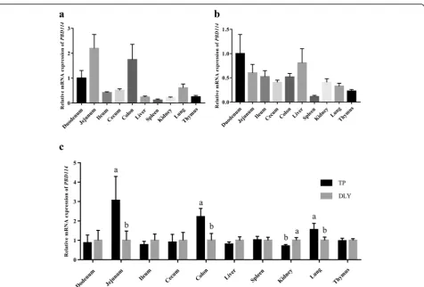
Expression profiles ofPBD114gene in different pig breeds and tissues
To evaluate expression profiles of PBD114, mRNA
ex-pressions in various tissues among TP (Fig.1a) and DLY
(Fig. 1b) were measured. PBD114 widely expressed in
duodenum, jejunum, ileum, cecum, colon, liver, spleen, kidney, lung and thymus, while the tissue with the high-est expression abundance was jejunum in TP and duo-denum of DLY. The different expression levels of PBD114 between TP pigs and DLY pigs were display in Fig.1c. The results showed that the expression levels of PBD114 of TP pigs in jejunum, colon and lung were
higher than DLY pigs (P< 0.05). However, the abundance
of PBD114 of TP pigs in kidney were lower than DLY pigs (P< 0.05).
Expression profiles ofPBD114gene uponE. coliK88 challenge
The effects ofE. coli K88 on the expression of PBD114
were displayed in Fig. 2. Diarrhea scores of K88 group
were higher than CON group over 24 h except at 20 h (P< 0.05), and the results indicated thatE. coliK88
chal-lenged model was successful. In vivo, the expressions of
Table 2Primers sequences used for quantitative RT-PCR
Gene Accession No. Primer sequences (5′→3′) Product length, bp
β-actin XM_003124280.5 TGGAACGGTGAAGGTGACAGC 177
GCTTTTGGGAAGGCAGGGACT
TNFα NM_214022.1 GCATCGCCGTCTCCTACCAG 173
GGGCAGGTTGATCTCGGCAC
PBD114 NM_001129973.1 TTGGTGGATCCTGAACGATGCT 130
PBD114 in duodenum, jejunum and ileum were signifi-cantly induced byE. coliK88 (P< 0.05).In vitro, the
ex-pressions of TNFα in IPEC-J2 were markedly increased
by 1 × 10⁵and 1 × 10⁶CFU/mL E. coliK88, and 1 × 10⁵
CFU/mL E. coli K88 also obviously induced the
expres-sion ofPBD114(P< 0.05).
Cloning and sequence analysis of thePBD114gene
In order to compare the homology of PBD114 between
TP pigs and DLY pigs, the 210-bp open reading frame of PBD114 from TP pigs and DLY pigs were successfully
amplified (Additional file 1: Figure S1). Then PBD114
amplicons from TP and DLY pigs were cloned and se-quenced, and the results of sequence alignment indicated
that DNA sequence identity ofPBD114 between TP and
DLY pigs was identical with NM_001129973.1 (Fig. 3a).
Amino acid sequence identity and phylogenetic tree of
beta defensin 114 among Homo sapiens, Sus scrofa, Bos
indicus and Pan troglodytes was compared. Phylogenetic tree analysis results indicated that beta defensin 114 ofSus scrofawas close toHomo sapiens(Fig.3b). Moreover, with 45.83% amino acid sequence identity of beta defensin 114
from human andSus scrofa(Fig.3c).
Expression and purification of rPBD114
The recombinant strain E. coli Origami B
(DE3)-pET32a(+)-PBD114 and E. coli Origami B
(DE3)-pET32a(+) were constructed, and positive colonies were identified by colony PCR (data were not showed). And then the identified positive colony was cultured and
in-duced by 1 mmol/L isopropyl-β-d-thiogalactoside (IPTG)
at 28 °C for 9 h. Then SDS-PAGE analysis showed that the target protein with the Trx tag conformed to the
theoret-ical molecular weight (Fig. 4), of 25.18 kDa. The pellets
were disrupted with sonication and dissolved in lysis buf-fer. The crude soluble recombinant protein was purified
by Ni2+-IDA affinity chromatography. SDS-PAGE was
performed and the purity of rPBD114 was analyzed with Image Lab (Bio-Rad). The result showed that the purity of rPBD114 was 95% and the expression level of rPBD114 was 5.0 mg/L. The target band was collected and amino acid sequence of rPBD114 was identified by mass
spec-trometry (MALDI-TOF/TOF) (Fig. 5a). Through
search-ing uniprot-Sus-scrofa (68,152 sequences, 25,615,784 residues), rPBD114 sequence had a 100% math with NP_
001123445 (Fig.5b), more MS information was shown in
Additional file2data.
Fig. 1The mRNA level ofPBD114in Tibetan and DLY pigs.a, the mRNA level ofPBD114in tissues of Tibetan pigs;b, the mRNA level ofPBD114
Protein structure, physical and chemical parameters of PBD114
Protein structure prediction was performed online to ex-plore the biological function of PBD114. SWISS-MODEL analysis found that the structure of PBD114 was similar to human beta defensin 2 with 35.14%
se-quence identity (Additional file 1: Figure S2). The
pre-dicted structure of PBD114 indicated that PBD114 was
consisted of an alpha helix fragment and a β-pleated
sheet fragment (Additional file 1: Figure S2), the detail
predicted results were provided in Additional file 1:
Figure S3. Physical and chemical parameters of PBD114 was analyzed by ProtParam online, theoretical pI, net charged residues and aliphatic index of PBD114 was 7.46, + 1 and 66.38 respectively, and more information
was shown in Additional file3data.
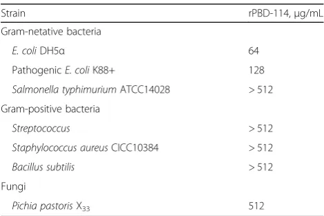
Antibacterial activities
The antimicrobial activity of rPBD114 was explored by
determining its MIC against E. coli DH5α, pathogenic
E. coli K88+, S. typhimurium, S. aureus, B. subtilis and Pichia pastoris X33 (Table 3). The results showed that rPBD114 exhibited strong antimicrobial activity against E. coli DH5α and pathogenic E. coli K88+, The MIC
value ofE. coliDH5αand pathogenicE. coli K88+ were
64 and 128μg/mL respectively. However, much higher
concentration rPBD114 (more than 256μg/mL) were
needed to against S. typhimurium, S. aureus, B. subtilis
and Pichia pastorisX33. The tolerance of these four
or-ganisms to rPBD114 indicate that Pichia pastoris X33
would be more tolerant to expression of rPBD114.
Hemolytic activity and cytotoxicity assays
Erythrocytes were collected from fresh porcine blood and incubated with different concentrations of rPBD114 for 1 h. Less than 3.9% hemolysis was found at all
con-centrations (0–256μg/mL). The results showed that
rPBD114 had slight hemolyticin vitro (Fig. 6a). IPEC-J2
was also used to examine cytotoxicity of rPBD114, cell viability was measured by CCK-8 after treating with dif-ferent concentrations of rPBD114. There was little
cyto-toxicity (Fig. 6b) when the concentration of rPBD114
was less than 256μg/mL. However, the cytotoxicity was
significant when the concentration of rPBD114 was
256μg/mL (P< 0.05). These results suggested that the
safe concentration of rPBD114 should be lower than
256μg/mL.
Fig. 2Escherichia coliK88 induced the expression ofPBD114in 6 days old pigs and IPEC-J2.a, diarrhea scores of 7 days old piglets challenged byE.
coliK88 or not;b, effects ofE. coliK88 challenge on the mRNA level ofPBD114in intestinal tissues; Effects ofE. coliK88 challenge on the mRNA level of
Discussion
The innate and adaptive immunity are two essential ele-ments of host defense. Innate immunity is highly con-served from fruit flies to human and is the first line of defense against invading pathogens. One mechanism of
the innate immunity is the secretion of broad-spectrum antimicrobial substances, such as cathelicidins and small cationic polypeptides named defensins [20]. Previous
studies reported that porcine β-defensin 1 and β
-defensin 2 existed broad-spectrum antimicrobial activ-ities and immune modulating function [20]. Human beta defensin 114 exhibited strong antimicrobial activity
against E. coli, C. albicans and S. aureus, and
anti-inflammatory function [21]. Choi et al. [13] found PBD114 by a BLAST analysis in 2012. However, the ex-pression profile of PBD114 in various tissues of pigs and biological function of PBD114 have not been studied. Therefore, the present study was conducted to explore
the expression profiles of thePBD114in different breeds
and in response to infections. Moreover, the function of PBD114 protein has also been investigated.
Our results showed that PBD114 widely expressed in
duodenum, jejunum, ileum, cecum, colon, liver, spleen, lung, kidney and thymus, in both TP and DLY pigs. Similar expression patterns also have been demonstrated by previous study on porcine beta defensins [13]. The
extensive expression of PBD114 suggested that this
en-dogenous peptide antibiotic may contribute to both mu-cosal and systemic host defenses in pigs. Chinese TP is a special Chinese indigenous pig breed, which are distrib-uted in high-altitude areas of Qinghai-Tibet Plateau which has cold climate. They are raised in pollution-free, purely natural alpine and cold mountainous areas all
Fig. 3Sequence alignment of PBD114 and phylogenetic analysis of beta defensin 114.a, DNA sequences cloned from TP and DLY pigs were
aligned by DNAMAN 8.0;b, amino acid sequences of beta defensin 114 in sus scorfa,Homo sapiens,Pan troglodytesandBos indicuswere aligned by DNAMAN 8.0;c, phylogenetic analysis of beta defensin 114 in sus scorfa,Homo sapiens,Pan troglodytesandBos indicuswere performed by DNAMAN 8.0
Fig. 4SDS-PAGE of recombinant PBD114 protein. M, 150 kDa
year round. And their immunity and disease resistance
are stronger than Duroc pigs [22, 23]. The higher level
mRNA of PBD114 was detected in intestine of TP and
DLY pigs, furthermore, the results of comparison among
breeds showed that the abundance ofPBD114mRNA of
TP pigs in jejunum, colon and lung were higher than DLY pigs. As well, defensins are a family of endogenous cationic antimicrobial peptides that play an important role in the innate immune system of mammals and pro-vide protection against bacterial infections in the
intes-tine [24, 25], Hence, the high expression of PBD114 in
TP pigs may have implications for the contribution of PBD114on high disease resistance of TP, and which was
the same as PBD1, 2 and 3 [22,26].
Due to the impact of thousands of years of artificial se-lection for survival of these breeds of different signatures of selection in TP and domestic pigs [23], we chose to investigate whether there were some variants in the
PBD114 gene. So the DNA sequence of PBD114 was
cloned from TP and DLY pigs respectively. Agarose gel electrophoresis of cloning PCR showed that the size of PBD114from TP and DLY pigs was same to theoretical value (210 bp). Further DNA sequence alignment of PBD114 between TP and DLY pigs indicated that the identity was 100%, and exactly consistent with NM_
001129973.1 in NCBI. The sequence of gene PBD114
did not mutated during thousands evolutionary history,
and this hinted that PBD114was very important to
sur-vive and breed of pigs. Moreover, previous study reported that human defensin 114 exhibited a broad
spectrum of antimicrobial activity with Escherichia coli,
Staphylococcus aureus and Candida albicans [27].
Amino acid sequence alignment of beta defensin 114 be-tween pig and human showed that the identity was 45.83%, and phylogenetic analysis was performed with
amino acid of Sus scrofa, Homo sapiens, Pan troglodytes
andBos indicus, and showed that PBD114 was closely re-lated to DEFB114. These results suggested that PBD114 may possess antimicrobial activity like DEFB114.
To explore whetherPBD114 take part in resistance to
pathogens, we carried out experiments in vitro and in
vivo. The results showed thatE. coliK88 significantly
in-duced the expression ofPBD114 in vitroandin vivo. On
the one hand, the results indicated that PBD114was an
inducible defensin. According previous studies, PBD114
was not only induced by E. coli K88, but also could be
promoted by nutrients and probiotics [27, 28]. On the
other hand,PBD114may play an important role in
kill-ing E. coli K88 or immune modulating function to
alleviate the damage of E. coli K88. Because previous
study reported that DEFB114 (a human homologous protein of PBD114) not only exhibited antimicrobial
ac-tivity but also could inhibit RAW264.7 release TNFα
Fig. 5Mass spectrometry identification of rPBD114.a, peak figure of amino acid fragments;b, through searching uniprot-Sus-scrofa(68152
sequences, 25615784 residues), rPBD114 sequence had a 100% math with NP_001123445 (show in red)
Table 3MIC of rPBD114
Strain rPBD-114,μg/mL
Gram-netative bacteria
E. coliDH5α 64
PathogenicE. coliK88+ 128
Salmonella typhimuriumATCC14028 > 512
Gram-positive bacteria
Streptococcus > 512
Staphylococcus aureusCICC10384 > 512
Bacillus subtilis > 512
Fungi
after stimulation with LPS [26]. In addition, PBD2 pro-tected intestinal health via modulating of TJ proteins in intestine and inhibiting the production of inflammatory mediators [29].
To explore the antimicrobial activity of PBD114, PBD114 was expressed in anEscherichia coliexpression system.
Be-cause Escherichia coli expression system technology was
mature and simple, still the first choice as host for AMPs
production [16, 30]. In addition, PBD114 possessed three
disulfide bonds, so we selected pET32a(+) andE. coli
Ori-gami B (DE3) to construct recombinant expression bacteria. pET32a(+) vector possessed thioredoxin tag which can in-crease the activity and amount of target protein present in the soluble fraction.E. coliOrigami B (DE3) with glutathi-one reductase (gor) and/or thioredoxin reductase (trxB) mutations enhance the formation of disulfide bonds in the
E. coli cytoplasm [31]. We successfully constructed
recombinant bacteria, E. coli Origami B (DE3)-pET32a
(+)-PBD114 and expressed the rPBD114 protein. rPBD114 was identified by mass spectrometry and the sequence coverage 100% was identical to NP_001123445. Physical and chemical parameters showed that rPBD114 possessed positive net charge (+ 1), aliphatic index (66.38) and grand
average of hydropathicity (−0.530), and suggested that
PBD114 protein has the biochemical properties of anti-microbial peptides [32]. As we all know, protein structure determines protein function. To learn more about the func-tion of PBD114 protein, we predicted the protein structure of PBD114 protein on SWISS-MODEL and the results showed that the structure of PBD114 protein was similar with human defensin 2. Human beta defensin 2 is produced by a number of epithelial cells and exhibits potent anti-microbial activity against Gram-negative bacteria and
Can-dida, but not Gram-positive Staphylococcus aureus [33].
Similarly, MIC of rPBD114 protein was carried out and rPBD114 protein exhibited antimicrobial activity against E. colibut not Gram-positive bacteria and fungus. However, a great number of studies have shown that defensins have a wide range of antimicrobial activities [34–37]. The anti-microbial activity of rPBD114 protein in our study
sug-gested thatPichia pastorisX33would be more tolerant to
expression of rPBD114. The methylotrophic yeast Pichia
pastoriscan grow to extremely high cell densities, enabling
efficient protein production and secretion [38, 39]. The
most attractive feature of this system is that the recombin-ant protein could be exactly folded, fully decorated and eas-ily purified [40]. However, the yeast may have been more resistant to the defensins if it were not properly processed.
Therefore, further study in Pichia pastoriswas needed. In
addition, hemolytic and cytotoxicity of rPBD114 protein in-dicated that rPBD114 was safe to mammals. These results suggested that rPBD114 may serve as a candidate for the replacement of conventionally used antibiotics. However, further researches are needed to investigate such as the structural and functional analysis, immunogenicity, and biologic function of rPBD114 proteinin vitroandin vivo.
Conclusions
PBD114is an infection response gene that is differentially-expressed between different porcine breeds and tissues. The antimicrobial activity of PBD114 protein, against
pathogens such as the E. coliK88, few hemolytic activity
and cytotoxicity suggested that it may serve as a candidate for the substitution for conventionally used antibiotics.
Additional files
Additional file 1: PBD114 cloning PCR and predictive spatial structure of rPBD114. (DOCX 840 kb)
Fig. 6Hemolytic and cytotoxicity of recombinant PBD114 protein.
Additional file 2: Detail information of MS. (XLSX 9 kb)
Additional file 3: Detail information of physicochemical. (DOCX 19 kb)
Abbreviations
AMPs:Antimicrobial peptides; DEFB114: Human beta defensin 114; DLY: Duroc×Landrance×Yorkshire; IPTG: Isopropyl-β-d-thiogalactoside; LB: Lysogeny broth; LPS: Lipopolysaccharide; MALDI-TOF/TOF: Matrix-assisted laser desorption/ionization time of flight mass spectrometry; MIC: Minimal inhibitory concentration; PBD114: Porcine beta defensin 114;
rPBD114: Recombinant porcine beta defensin 114; SDS-PAGE: Sodium dodecyl sulfate polyacrylamide gel electrophoresis; THY: Todd-Hewitt+yeast extract; TJ: Tight junction; TP: Tibetan pig; YPD: Yeast Extract Peptone Dextrose Medium
Acknowledgements
We thank Honglin Yan, Weikang Wang, Qian Lin and Yaqiang Dai for assistance in collecting samples of TP pigs, and thank Huifen Wang and Quyuan Wang for purchasing consumables and reagents.
Authors’contributions
JH, JL, JY, PZ, XM, YL, ZH, BY and DC participated in the design of the study. QS, KX and QL collected the experiments data. GS analyzed the data and wrote the first draft of the manuscript. All authors read and approved the final manuscript.
Funding
This work was supported by the Key Research and Development Program of Sichuan Province (2018NZDZX0005), and the Youth Innovation Teams of Animal Feed Biotechnology of Sichuan Province (2016TD0028).
Availability of data and materials
The datasets during and/or analyzed during the current study are available from the corresponding authors on reasonable request.
Ethics approval and consent to participate
The experimental procedures followed the actual law of animal protection that were approved by the Animal Care Advisory Committee of Sichuan Agricultural University (No. 20160709) and were performed in accordance with the National Research Council’s Guide for the Care and Use of Laboratory Animals.
Consent for publication
All authors read and approved the final manuscript.
Competing interests
The authors declare that they have no competing interests.
Received: 1 February 2019 Accepted: 22 May 2019
References
1. Veldhuizen EJ, Rijnders M, Claassen EA, van Dijk A, Haagsman HP. Porcine beta-defensin 2 displays broad antimicrobial activity against pathogenic intestinal bacteria. Mol Immunol. 2008;45:386–94.
2. Zhu YG, Johnson TA, Su JQ, Qiao M, Guo GX, Stedtfeld RD, et al. Diverse and abundant antibiotic resistance genes in Chinese swine farms. Proc Natl Acad Sci U S A. 2013;201222743.
3. Auvynet C, Rosenstein Y. Multifunctional host defense peptides: antimicrobial peptides, the small yet big players in innate and adaptive immunity. FEBS J. 2009;276:6497–508.
4. Yang D, Liu ZH, Tewary P, Chen Q, de la Rosa G, Oppenheim JJ. Defensin participation in innate and adaptive immunity. Curr Pharm Des. 2007;13:3131–9. 5. Selsted ME, Ouellette AJ. Mammalian defensins in the antimicrobial immune
response. Nature Immunol. 2005;6:551.
6. Zhang G, Wu H, Shi J, Ganz T, Ross CR, Blecha F. Molecular cloning and tissue expression of porcine beta-defensin-1. FEBS Lett. 1998;424:37–40. 7. Salzman NH, Hung K, Haribhai D, Chu H, Karlsson-Sjöberg J, Amir E, et al.
Enteric defensins are essential regulators of intestinal microbial ecology. Nature Immunol. 2009;11:76.
8. Zhang Y, Teng D, Mao R, Wang X, Xi D, Hu X, et al. High expression of a plectasin-derived peptide NZ2114 in Pichia pastoris and its
pharmacodynamics, postantibiotic and synergy against Staphylococcus aureus. Appl Microbiol Biotechnol. 2014;98:681–94.
9. Cohen ML. Changing patterns of infectious disease. Nature. 2000;406:762–7. 10. Elahi S, Buchanan RM, Attah-Poku S, Townsend HG, Babiuk LA, Gerdts V. The
host defense peptide beta-defensin 1 confers protection against Bordetella pertussis in newborn piglets. Infect Immun. 2006;74:2338–52.
11. Li CL, Xu TT, Chen RB, Huang XX, Zhao YC, Bao YY, et al. Cloning, expression and characterization of antimicrobial porcine beta defensin 1 in Escherichia coli. Protein Expr Purif. 2013;88:47–53.
12. Veldhuizen EJA, Dijk AV, Tersteeg MHG, Kalkhove SIC, Meulen JVD, Niewold TA, et al. Expression ofβ-defensins pBD-1 and pBD-2 along the small intestinal tract of the pig: lack of upregulation in vivo upon Salmonella typhimurium infection. Mol Immunol. 2007;44:276–83.
13. Choi MK, Le MT, Nguyen DT, Choi H, Kim W, Kim JH, et al. Genome-level identification, gene expression, and comparative analysis of porcine ß-defensin genes. BMC Genet. 2012;13:98.
14. Kusminski CM, Park J, Scherer PE. MitoNEET-mediated effects on browning of white adipose tissue. Nat Commun. 2014;5:3962.
15. Livak KJ, Schmittgen TD. Analysis of relative gene expression data using real-time quantitative PCR and the 2(−Delta Delta C(T)) method. Methods. 2001;25:402–8. 16. Su G, Tang F, Chen D, Yu B, Huang Z, Luo Y, et al. Expression, purification
and characterization of a novel antimicrobial peptide: Gloverin A2 from Bombyx mori. Int J Pept Res Ther. 2018:1–7.
17. Zhang J, Yang YL, Teng D, Tian ZG, Wang SR, Wang JH. Expression of plectasin in Pichia pastoris and its characterization as a new antimicrobial peptide against Staphyloccocus and Streptococcus. Protein Expres Purif. 2011;78:189–96.
18. Feng X, Liu C, Guo J, Song X, Li J, Xu W, et al. Recombinant expression, purification, and antimicrobial activity of a novel hybrid antimicrobial peptide LFT33. Appl Microbiol Biotechnol. 2012;95:1191–8.
19. Deng X, Cao M, Zhang J, Hu K, Yin Z, Zhou Z, et al. Hyaluronic acid-chitosan nanoparticles for co-delivery of MiR-34a and doxorubicin in therapy against triple negative breast cancer. Biomaterials. 2014;35:4333–44.
20. Qi S, Chen J, Guo R, Yu B, Chen D.β-Defensins gene expression in tissues of the crossbred and Tibetan pigs. Livest Sci. 2009;123:161–8.
21. Yu H, Dong J, Gu Y, Liu H, Xin A, Shi H, et al. The novel humanβ-defensin 114 regulates lipopolysaccharide (LPS)-mediated inflammation and protects sperm from motility loss. J Biol Chem. 2013;288:12270–82.
22. Zhao Y, Yu B, Mao XB, Han GQ, Mao Q, Huang ZQ, et al. Molecular cloning and expression analysis of IFN-βpromoter stimulator 1 in Tibetan pigs. Mol Biol Rep. 2012;39(6):7011–7.
23. Li M, Tian S, Jin L, Zhou G, Li Y, Zhang Y, et al. Genomic analyses identify distinct patterns of selection in domesticated pigs and Tibetan wild boars. Nat Genet. 2013;45:1431.
24. Veldhuizen EJA, Koomen I, Ultee T, van Dijk A, Haagsman HP. Salmonella serovar specific upregulation of porcine defensins 1 and 2 in a jejunal epithelial cell line. Vet Microbiol. 2009;136:69–75.
25. Ganz T. Defensins and other antimicrobial peptides: a historical perspective and an update. Comb Chem High T Scr. 2005;8:209–17.
26. Chen J, Qi S, Guo R, Yu B, Chen D. Different messenger RNA expression for the antimicrobial peptides beta-defensins between Meishan and crossbred pigs. Mol Biol Rep. 2010;37:1633.
27. Ren M, Zhang SH, Zeng XF, Liu H, Qiao SY. Branched-chain amino acids are beneficial to maintain growth performance and intestinal immune-related function in weaned piglets fed protein restricted diet. Asian-Australas J Anim Sci. 2015;28:1742–50.
28. Liu H, Hou C, Wang G, Jia H, Yu H, Zeng X, et al. Lactobacillus reuteri I5007 modulates intestinal host defense peptide expression in the model of IPEC-J2 cells and neonatal piglets. Nutrients. 2017;9.
29. Han F, Zhang H, Xia X, Xiong H, Song D, Zong X, et al. Porcine beta-defensin 2 attenuates inflammation and mucosal lesions in dextran sodium sulfate-induced colitis. J Immunol. 2015;194:1882–93.
30. Parachin NS, Mulder KC, Viana AAB, Dias SC, Franco OL. Expression systems for heterologous production of antimicrobial peptides. Peptides. 2012;38:446–56. 31. Prinz WA, Åslund F, Holmgren A, Beckwith J. The role of the Thioredoxin
and Glutaredoxin pathways in reducing protein disulfide bonds in the Escherichia coli cytoplasm. J Biol Chem. 1997;272:15661–7.
33. Schröder JM, Harder J. Human beta-defensin-2. Int J Biochem Cell Biol. 1999; 31:645–51.
34. Kahlon AK, Tripathi S, Sharma A. Chapter 8 - recent developments and future prospects of natural and synthetic Antitubercular peptide drugs. In: Amandeep KK, Shubhandra T, Ashok S, editors. Applied microbiology and bioengineering: Academic Press; 2019. p. 121–59.
35. Yang M, Zhang C, Zhang MZ, Zhang S. Beta-defensin derived cationic antimicrobial peptides with potent killing activity against gram negative and gram positive bacteria. BMC Microbiol. 2018;18:54.
36. Sun E, Belanger CR, Haney EF, Hancock REW. Host defense (antimicrobial) peptides. In: Koutsopoulos S, editor. Peptide applications in biomedicine, biotechnology and bioengineering: Woodhead Publishing; 2018. p. 253–85. 37. Chen RB, Zhang K, Zhang H, Gao CY, Li CL. Analysis of the antimicrobial
mechanism of porcine beta defensin 2 against E. coli by electron microscopy and differentially expressed genes. Sci Rep. 2018;8:14711. 38. Corrales-Garcia LL, Possani LD, Corzo G. Expression systems of humanβ
-defensins: vectors, purification and biological activities. Amino Acids. 2011; 40:5–13.
39. De Schutter K, Lin Y C, Tiels P, Van Hecke A, Glinka S, Weber-Lehmann J, et al. Genome sequence of the recombinant protein production host pichia pastoris. Nat Biotechnol. 2009;27(6):561–66.