Copyright © 2002, American Society for Microbiology. All Rights Reserved.
Mating-Type Locus of
Cryptococcus neoformans
: a Step in the
Evolution of Sex Chromosomes
Klaus B. Lengeler, Deborah S. Fox, James A. Fraser, Andria Allen, Keri Forrester,
Fred S. Dietrich, and Joseph Heitman*
Department of Molecular Genetics and Microbiology, Howard Hughes Medical Institute, Duke University, Durham, North Carolina 27710
Received 27 June 2002/Accepted 11 July 2002
The sexual development and virulence of the fungal pathogen Cryptococcus neoformansis controlled by a bipolar mating system determined by a single locus that exists in two alleles,␣and a. The␣and a mating-type alleles from two divergent varieties were cloned and sequenced. TheC. neoformansmating-type locus is unique, spans >100 kb, and contains more than 20 genes.MAT-encoded products include homologs of regulators of sexual development in other fungi, pheromone and pheromone receptors, divergent components of a MAP kinase cascade, and other proteins with no obvious function in mating. The␣and a alleles of the mating-type locus have extensively rearranged during evolution and strain divergence but are stable during genetic crosses and in the population. TheC. neoformansmating-type locus is strikingly different from the other known fungal mating-type loci, sharing features with the self-incompatibility systems and sex chromosomes of algae, plants, and animals. Our study establishes a new paradigm for mating-type loci in fungi with implications for the evolution of cell identity and self/nonself recognition.
Self/nonself recognition events underlie the function of the major histocompatibility locus in defense against infection and organ transplant rejection, the self-incompatibility systems that prevent inbreeding in plants, and the production of offspring by sexual reproduction. During sexual reproduction, special-ized genomic regions promote self/nonself interactions. Sex-determining regions include the mating-type loci in fungi and the sex chromosomes in plants and animals. Dimorphic sex chromosome systems independently evolved in animals, mosses, and dioecious plants. A related but distinct sexual incompatibility system is found in many lower eukaryotes, in-cluding algae, protozoans, monoecious plants, and fungi. In these organisms, multiallelic mating-type (MAT) loci monitor cell interactions for sexual compatibility, and if inbreeding is detected, mating is aborted (17, 18, 50, 56).
A common theme of sex determinants is the need to be transmitted as a single unit, and recombination within sex-determining regions is suppressed to avoid generating self-fertile or sterile offspring. Several mechanisms operate to sup-press recombination. In the fungal MAT loci, extensive sequence divergence prevents recombination between differ-ent alleles. In the case of sex chromosomes, both sequence divergence and chromosomal rearrangements suppress recom-bination. These rearrangements affect nearly the entire sex chromosome in humans or mice, whereas in lower vertebrates and certain dipterous insects, only a limited region of the sex chromosomes is rearranged. These findings suggest that the dimorphic sex chromosomes evolved via accumulation of chro-mosomal aberrations.
Fungal mating-type loci serve as paradigms for
understand-ing gene regulation durunderstand-ing sexual development and the deter-mination of cell fate and identity (7, 16, 32, 34, 37, 40, 54). Sexual development of ascomycetous fungi is commonly con-trolled by a bipolar mating system involving a single mating-type locus. In these cases, theMAT locus spans only a few thousand base pairs and exists in two unrelated alleles that control cell identity by encoding transcription factors that act on distant target genes (16, 40, 54).
In contrast to mating in ascomycetes, mating in basidiomy-cetes is commonly regulated by two independent, unlinked loci, resulting in tetrapolar mating systems (7, 37, 40). Both mating-type loci can be multiallelic, giving rise to thousands of different mating types in some mushroom fungi (38). The structure of mating-type loci in basidiomycetes has been de-termined for several model systems, including the mushrooms Coprinus cinereus (41, 52, 53) and Schizophyllum commune (66–68, 70) and the maize pathogenUstilago maydis(4, 27, 39, 62). Similar to ascomycete mating-type loci, one locus encodes a pair of homeodomain transcription factors that controls a subset of developmental processes involved in sexual repro-duction (A locus inC. cinereusandS. communeand b locus in U. maydis). The second locus (B inC. cinereusandS. commune and a inU. maydis) encodes a G protein-coupled pheromone receptor linked to one or more pheromone genes.
The opportunistic human fungal pathogenCryptococcus neo-formansis an encapsulated yeast that is distributed worldwide in association with pigeon guano and trees (6). This pathogen has increased in medical importance over the past several decades because of its ability to cause fatal meningioencepha-litis in immunocompromised hosts. In contrast to many basid-iomycetes,C. neoformanshas a bipolar mating system with two opposite mating types,MAT␣andMATa.
A portion of the C. neoformans MAT␣ locus was initially identified by a difference cloning approach and was found to contain the MF␣1 pheromone gene (49). Subsequent work * Corresponding author. Mailing address: Department of Molecular
Genetics and Microbiology, Howard Hughes Medical Institute, Duke University, Durham, NC 27710. Phone: (919) 684-2824. Fax: (919) 684-5458. E-mail: [email protected].
704
on September 8, 2020 by guest
http://ec.asm.org/
revealed that the C. neoformans MAT locus is unusual in size and gene composition, spanning an⬃55-kb region (35) and containing several additional␣-specific genes, including STE12␣andSTE20␣(10, 46, 69, 71, 73). Recent studies have revealed the following: (i) theMATlocus is larger than previ-ously suspected (C. M. Hull, R. C. Davidson, and J. Heitman, submitted for publication), (ii) the ␣ and a alleles encode divergent alleles of related genes (9, 46), and (iii) the archi-tectures of the twoMATalleles may differ. Here we present our study on theC. neoformansmating-type locus that estab-lishes a novel paradigm for the structure of mating-type loci with implications for fungal evolution and the evolution of specialized sex chromosomes.
MATERIALS AND METHODS
Strains.The strains used for construction of bacterial artificial chromosome (BAC) libraries and analysis of the mating-type loci wereC. neoformansserotype A var.grubiistrains H99 (MAT␣) and 125.91 (MATa) andC. neoformans sero-type D var.neoformanscongenic strains JEC21 (MAT␣) and JEC20 (MATa) (31, 42, 46). The serotype D strains used to analyze recombination in theMATregion (Fig. 6) were derived from crosses betweenMAT␣strain DSF51 (znf1␣::NAT1
ste12␣::URA5 ade2) or RDC20-5 (ste12␣::URA5 ade2) andMATastrain JEC53 (ura5 lys1). The stability of theMATloci through several crosses (see Fig. 4) was analyzed using the serotype D strains NIH12, NIH433, B3501, B3502, JEC20, and JEC21 (31, 42). Structural analysis ofMATloci in the population ofC. neoformans var.neoformans was conducted using the unrelated serotype D strains CDC92-18, CDC92-27, MMRL760 (allMAT␣), and #11 (MATa).
BAC and subgenomic libraries.To obtain high-quality chromosomal DNA fromC. neoformans, protoplasts were isolated as described previously (46). In the present study, the lysis of protoplasts was prolonged to 48 h and proteinase K was added to the lysis buffer at a final concentration of 4 mg/ml. Fresh buffer was added after a 24-h incubation. Plugs containing lysed protoplasts were washed twice with ice-cold Tris-EDTA (TE) buffer containing 0.2 mM phenyl-methylsulfonyl fluoride and subsequently washed four to five times with ice-cold TE buffer for 1 h each. Plugs could be stored indefinitely at 4°C in 50 mM EDTA. In collaboration with Research Genetics (Huntsville, Ala.), the chromosomal DNA was partially digested withHindIII, and⬃100-kb fragments were isolated after separation of digested DNA via pulsed-field gel electrophoresis. Genomic fragments were cloned into the BAC vector pBeloBAC11, and clones with inserts were identified using standard blue/white screening techniques. BAC clones of interest were identified by Southern hybridization with mating-type-specific gene probes by using colony lift membranes (Research Genetics). To close the re-maining gaps in strains H99 and 125.91 not covered by the analyzed BAC clones (Fig. 1), desired fragments were identified by Southern blotting and isolated from subgenomic libraries or generated by PCR.
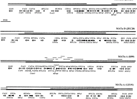
FIG. 1. Structures of the serotype D (MATaof JEC20 andMAT␣of JEC21) and serotype A (MATaof 125.91 andMAT␣of H99)␣anda mating-type alleles and adjacent genomic regions. The mating-type-specific regions are shown as thick bold lines, and flanking regions are shown as thinner black lines. Sequences were analyzed using BLASTX, and identified genes are shown as arrows in the direction of transcription. Genes encoding pheromone response pathway elements are shown as black arrows, locus-specific genes are shown as white arrows, and all other genes are shown as grey arrows. Bars above the mating-type alleles represent the BAC clones, genomic fragments, and PCR products analyzed.
on September 8, 2020 by guest
http://ec.asm.org/
BAC sequencing strategy.To generate high-quality BAC DNA, plasmid DNA was subjected to cesium chloride equilibrium centrifugation. The plasmid DNA of individual BAC clones was isolated from 500 ml of cultures by using alkaline lysis. The DNA was resuspended in 15 ml of TE buffer containing ethidium bromide (400l of a 10-mg/ml stock), and cesium chloride was added to a final density of 1.4 mg/ml. The DNA-CsCl solution was transferred into a 15-ml ultracentrifuge tube, and the sample was centrifuged in an NVT65.1 rotor at 65,000 rpm for⬃24 h at room temperature. The lower, plasmid-containing DNA band was removed using a 5-ml syringe with a 20-gauge needle and introduced into a 4-ml ultracentrifuge tube, which was then filled with CsCl solution to a final density of 1.4 mg/ml. The sample was centrifuged for an additional 24 h at 70,000 rpm. The DNA was removed from the second gradient, ethidium bromide was extracted several times with salt-saturated isopropanol, and the DNA was dialyzed against 5 liters of TE buffer for several hours. After adding 1/10 volume of 3 M sodium acetate, BAC DNA was precipitated with 0.6 volume of isopro-panol (⫺20°C), washed with 70% EtOH, and resuspended in TE buffer.
Three to five micrograms of BAC DNA was sheared with a Hydroshear device (Gene Machines) to generate⬃1.5- to 3-kb DNA fragments. Sheared fragments were subsequently subjected to standard blunting and fill-in reactions, and dou-ble-stranded adapters, provided by the Duke Center for Genome Technology (CGT), were ligated in 100-fold excess to the blunted DNA fragments. Frag-ments were separated from free adapters by agarose gel separation, and the DNA was cloned into a special, pUC18-based linearized vector provided by the CGT containing ends compatible to the adapters ligated onto the BAC DNA fragments (51). Before large-scale sequencing, the percentage of clones with inserts and the average insert size were carefully checked. Clones were picked automatically into 384-well plates containing Luria-Bertani Hogness medium by using a Genomic Solutions Flexis robot, and the plates were heat sealed and stored at⫺80°C. H99 genomic shotgun libraries were prepared accordingly, starting with CsCl-purified genomic DNA. For sequencing, clones were grown in 96-well plates containing Terrific broth medium in a Higro orbital shaker (Gene Machines), and DNA was isolated using a RevPrep robot (Gene Machines). Sequencing reactions were performed with a Hydra workstation (Robbins) and an MJ Research thermal cycler using standard BigDye chemistry (Applied Bio-systems). After removal of unincorporated dye, samples were analyzed on a PE3700 96-capillary sequencer, and sequence data were automatically trans-ferred to the UNIX-based CGT server. The resulting sequence data were ana-lyzed and assembled with the Pare/Phrased sequence package (19, 20), and assemblies were examined using Consed (29). Sequences from theC. neoformans
Genome Project that were added during the assembly of the serotype DMAT␣
mating-type locus were provided by the Stanford Genome Technology Center and The Institute for Genomic Research, funded by the National Institute of Allergy and Infectious Diseases and the National Institutes of Health under cooperative agreements U01 AI47087 and U01 AI48594, respectively. The genes were identified by comparing BAC sequences to sequences in the GenBank database by using the BLASTX algorithm (1).
PCR.Recombination within the mating-type region of the serotype DMAT␣
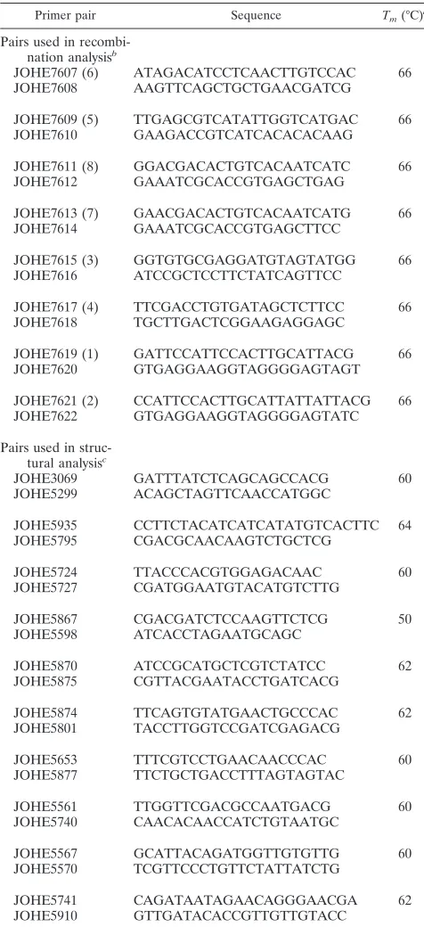
andMATaloci was analyzed using mating-type and strain-specific primers de-signed for the corresponding sequences generated in this study (see Fig. 6). The primer sequences and primer combinations are listed in Table 1. Fragments of
⬃500 bp were amplified using a synthesis time of 30 s and an annealing temper-ature of 66°C. Primers used in the structural analysis of mating-type loci from unrelated serotype D strains (Fig. 7) were initially designed for sequence analysis of the serotype D MAT locus from strain JEC21 and are also listed in Table 1. Depending on the primer combination, an annealing temperature between 50 and 64°C was used, whereas the synthesis time was 5 min for each reaction.
Nucleotide sequence accession numbers.GenBank accession numbers for the sequences reported here are as follows: JEC21 serotype D MAT␣AF542531; JEC20 serotype D MATa, AF542530; H99 serotype A MAT␣, AF542529; 125.91 serotype A MATa, AF542528; Tcn760, transposable element, AF542532.
RESULTS
Cloning and sequencing the mating-type locus ofC.
neofor-mans.We set out to clone and sequence the mating-type locus
ofC. neoformansto test the following hypotheses. First, does theMATlocus encode homeodomain transcription factors that govern cell identity as in other fungi? Second, did the␣anda alleles of the MATlocus diverge from a common ancestral region of DNA, which we proposed earlier based on the iden-tification of the divergentSTE20␣andSTE20agenes (46, 69)?
Third, has theMATlocus been conserved or rearranged during the evolution of this pathogen?
To determine the complete structure of the␣anda mating-type alleles of C. neoformans, genomic BAC libraries were generated from a congenic pair of serotype D␣andastrains
TABLE 1. Primer pairs used in recombination and structural analysis of theC. neoformans MATlocus
Primer pair Sequence Tm(°C)a
Pairs used in recombi-nation analysisb
JOHE7607 (6) ATAGACATCCTCAACTTGTCCAC 66 JOHE7608 AAGTTCAGCTGCTGAACGATCG
JOHE7609 (5) TTGAGCGTCATATTGGTCATGAC 66 JOHE7610 GAAGACCGTCATCACACACAAG
JOHE7611 (8) GGACGACACTGTCACAATCATC 66 JOHE7612 GAAATCGCACCGTGAGCTGAG
JOHE7613 (7) GAACGACACTGTCACAATCATG 66 JOHE7614 GAAATCGCACCGTGAGCTTCC
JOHE7615 (3) GGTGTGCGAGGATGTAGTATGG 66 JOHE7616 ATCCGCTCCTTCTATCAGTTCC
JOHE7617 (4) TTCGACCTGTGATAGCTCTTCC 66 JOHE7618 TGCTTGACTCGGAAGAGGAGC
JOHE7619 (1) GATTCCATTCCACTTGCATTACG 66 JOHE7620 GTGAGGAAGGTAGGGGAGTAGT
JOHE7621 (2) CCATTCCACTTGCATTATTATTACG 66 JOHE7622 GTGAGGAAGGTAGGGGAGTATC
Pairs used in struc-tural analysisc
JOHE3069 GATTTATCTCAGCAGCCACG 60 JOHE5299 ACAGCTAGTTCAACCATGGC
JOHE5935 CCTTCTACATCATCATATGTCACTTC 64 JOHE5795 CGACGCAACAAGTCTGCTCG
JOHE5724 TTACCCACGTGGAGACAAC 60 JOHE5727 CGATGGAATGTACATGTCTTG
JOHE5867 CGACGATCTCCAAGTTCTCG 50 JOHE5598 ATCACCTAGAATGCAGC
JOHE5870 ATCCGCATGCTCGTCTATCC 62 JOHE5875 CGTTACGAATACCTGATCACG
JOHE5874 TTCAGTGTATGAACTGCCCAC 62 JOHE5801 TACCTTGGTCCGATCGAGACG
JOHE5653 TTTCGTCCTGAACAACCCAC 60 JOHE5877 TTCTGCTGACCTTTAGTAGTAC
JOHE5561 TTGGTTCGACGCCAATGACG 60 JOHE5740 CAACACAACCATCTGTAATGC
JOHE5567 GCATTACAGATGGTTGTGTTG 60 JOHE5570 TCGTTCCCTGTTCTATTATCTG
JOHE5741 CAGATAATAGAACAGGGAACGA 62 JOHE5910 GTTGATACACCGTTGTTGTACC
aT
m,annealing temperature.
bNumbers in parentheses correspond to those shown for the primer combi-nations in Fig. 6.
cListed according to their position spanning theMATlocus from left to right (Fig. 7).
on September 8, 2020 by guest
http://ec.asm.org/
(JEC21 and JEC20) as well as from the serotype A␣and a strains H99 and 125.91. Probes to known mating-type-specific genes were used to identify BAC clones spanning each allele of the mating-type locus. Shotgun libraries were produced from two or three BAC clones encompassing each allele and se-quenced (Fig. 1). In the case of the␣locus from serotype D, the⬃38-kb mating-type region defined by Moore and Edman
was analyzed using PCR products and existing cosmid clones (49).
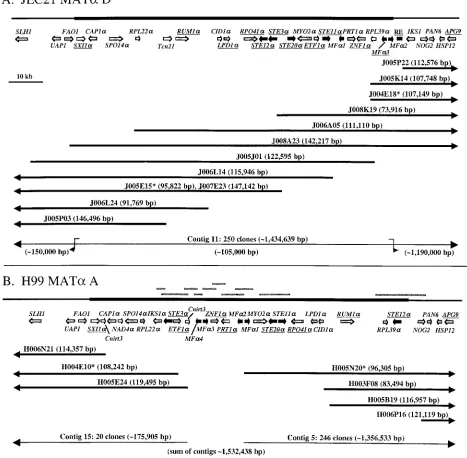
To establish gene order in this region and to rule out pos-sible rearrangements of the BAC clones chosen for sequenc-ing, hybridization-based BAC maps were generated (Fig. 2). An initial screen was conducted using probes to theSTE12␣ andSTE20␣genes and sequences flanking the previously iden-FIG. 2. Mapping of the serotype D (A) and serotype A (B)MAT␣loci by hybridization. The serotype D and serotype AMAT␣BAC libraries (strains JEC21 and H99) were screened with probes specific for severalMAT-specific genes, the right end (RE) of the locus, and the flanking gene
APG9. BAC clones that hybridized to these probes were analyzed by dot blot hybridizations using probes to the underlined genes. Additional hybridizations were conducted with a high-density BAC clone filter. These hybridization data were used to generate a linkage map and establish the gene order of theMATlocus. For all BAC clones depicted here, end sequences from the University of British Columbia database were incorporated to define endpoints that lie between hybridization probes. BACs sequenced to completion are marked with asterisks. The sizes of the BAC clones and BAC contigs depicted were determined at the University of British Columbia Genome Center. Clones used to span a gap in the H99MATlocus are depicted as short grey lines above the locus. For additional details, see the legend to Fig. 1.
on September 8, 2020 by guest
http://ec.asm.org/
tified right end of the MAT locus. Hybridizing BAC clones were subjected to further analysis with additional probes, in-cluding theSXI1␣ andRUM1␣genes (Fig. 2). The resulting BAC map confirmed the gene order for the initial⬃38- and ⬃55-kb mating-type regions previously analyzed (35, 49) and extended this map to include numerous additional genes. No aberrant recombination events appear to have occurred during the construction of the BAC library. This was further con-firmed by comparison of the sequences of the MAT␣ loci generated in this study with BAC fingerprint maps for the␣ strains JEC21 and H99 generated at the Genome Center of the University of British Columbia (61).
Data generated during both mapping processes revealed that theMAT␣locus of strain H99 was not completely covered in a library of⬃6,000 available BAC clones. This finding was confirmed during our sequencing efforts. The⬃10-kb gap in the H99MATlocus was closed by identifying and sequencing the following: (i) genomic clones spanningMAT-specific genes, (ii) MAT-specific plasmid clones from the H99 shotgun se-quencing project, and (iii) a 1.6-kb PCR product spanning the final remaining gap that proved recalcitrant to recovery in Escherichia coli (Fig. 2B). This ⬃10-kb region spans three pheromone genes and theZNF1␣and PRT1␣ genes and in-cludes several large inverted repeats that may render this re-gion difficult to clone. To provide deeper sequence coverage, sequences for strains H99 (serotype A) and JEC21 (serotype
D) that were available from GenBank and public genome sequencing projects were entered in the assembly. The overall region of double-stranded DNA that was bidirectionally se-quenced was 245 and 210 kb for the serotype D␣andaalleles, respectively, and 225 and 150 kb for the serotype A␣and a alleles, respectively, for a total of 830 kb of genomic sequence. Mapping the borders of the mating-type locus.A portion of the serotype D␣mating-type allele was identified by Moore and Edman in 1993, and subsequent work in several labs has contributed to further define the structure of theMATlocus (46, 49, 69, 71). Karos and coworkers reported a map of the serotype DMAT␣locus that spans an⬃55-kb region between theRPO41andNOG2genes (35). Here we present evidence that redefines the left border of the mating-type locus, dem-onstrating that the MAT locus spans an additional ⬃50-kb region upstream of theRPO41gene and defining the authentic left junction between genomic DNA and theMATlocus.
The junctions between the MAT locus and neighboring genomic DNA were identified by comparing the serotype D␣ and a allele sequences. The serotype D ␣ strain analyzed (JEC21) was generated by backcrossing an␣strain 10 times to an astrain (JEC20), resulting in a congenic strain pair that should differ only at theMATlocus (31, 42). Hence, sequences bordering the MAT locus should be identical or nearly so, whereas sequences within MAT should be distinct. A DNA sequence comparison of the␣andaalleles by dot plot analysis FIG. 3. Mapping of the ends of the mating-type locus by sequence comparison. Twenty kilobases of the sequences surrounding the proposed junctions between theMAT␣andMATamating-type alleles and flanking DNA and surrounding theRPO41mitochondrial RNA polymerase genes of the serotype DMAT␣andMATastrains JEC21 and JEC20 were compared using the DNA Strider program. Corresponding sequences were subjected to a pairwise comparison using a window size of 11 bp. In the graphical outputs, sequence identity is indicated by dots and stretches of sequence identity appear as diagonal lines. For additional details, see the legend to Fig. 1.
on September 8, 2020 by guest
http://ec.asm.org/
revealed that flanking sequences on one side and within the UAP1-FAO1 and IKS1-NOG2 genes are nearly identical, whereas on the other side, the sequences diverge, defining the left and right junctions between theMATlocus and surround-ing genomic DNA (Fig. 3, left and right panels). In addition, the order of genes surrounding the UAP1-FAO1and IKS1-NOG2genes in the flanking regions is identical between the␣ andaalleles up to the proposed junctions and then diverges in the opposite mating-type alleles (Fig. 1). In Southern analysis, probes specific to the sequences outside the predicted junc-tions yielded identical restriction patterns in␣ and astrains (Fig. 4), whereasMAT-specific probes yielded mating-type spe-cific patterns (Fig. 4). Our findings indicate that the mitochon-drial RNA polymerase geneRPO41originally reported to de-fine the left border of the MATlocus is in fact part of the mating-type locus and not a flanking gene. The finding that the gene order and sequence both diverge on either side of the RPO41 gene further supports this conclusion (Fig. 1 and 3, middle panel). AnRPO41-specific probe also yielded different restriction patterns for the␣andastrains JEC21 and JEC20 (Fig. 4).
The sequences flanking theMATloci in the␣andastrains JEC21 and JEC20 share⬃99% identity for several kilobases before reaching 100% identity, reflecting the position of the most recent recombination between the mating-type junctions and surrounding genomic DNA. A small⬃100-bp region just upstream of the leftMATlocus junction shares limited simi-larity between the␣andastrains and may reflect an ancient recombination event between the alleles.
Gene order is nearly identical in the regions flanking the mating-type locus in the serotype A and D strains, whereas the central region spanning the MAT locus itself is extensively rearranged. In the left flanking regions, all four mating-type alleles share synteny. In contrast, in the right flanking region, theNOG2,PAN6,HSP12, andAPG9genes all share synteny but the gene that immediately flanks theMATlocus in serotype D (IKS1) is an integral component of the mating-type locus in both the␣andaalleles in serotype A (Fig. 1). TheIKS1gene does not appear to be a component of the MAT locus in serotype D, as it is embedded in sequences that share⬃99% identity between strains JEC21 and JEC20. As discussed fur-ther below, theIKS1gene may have entered theMATlocus in serotype A (gene capture model) or exited the locus in sero-type D (gene egress model). Comparison of the IKS1 gene sequences reveals that the serotype AIKS1␣andIKS1aalleles are dramatically divergent (52% identity), whereas the IKS1 genes flanking the serotype DMATalleles share 99% identity with each other and significant identity (85%) with the sero-type AIKS1␣allele. These findings support a model in which theIKS1gene was lost from the locus and fixed in the flanking region by inversion and recombination events, with concomi-tant loss of theIKS1agene in the serotype D lineage.
In conclusion, based on synteny and sequence comparisons, our study demonstrates that the mating-type locus ofC. neo-formansis significantly larger than previously proposed, span-ning an⬃105- to 130-kb region that lies between theFAO1and IKS1-NOG2genes in serotype D and the FAO1and NOG2 genes in serotype A. The serotype D ␣ and a alleles span 105,656 and⬃117,308 bp, respectively, whereas the serotype A ␣andaalleles span⬃102,764 and⬃127,082 bp, respectively.
Thus, thea alleles of the MATlocus are larger than the ␣ alleles.
Genes contained in the mating-type locus.Approximately 20 genes contained in theMATlocus were identified when the BLASTX algorithm was used to compare theMATlocus se-quence with sese-quences in GenBank (Fig. 1). Table 2 summa-rizes the genes identified within the mating-type alleles and flanking sequences. Transcripts corresponding to several of these genes are present in expressed sequence tags derived from cDNA from strain H99 and the serotype D strain B3501, a precursor to strain JEC21 (University of Oklahoma Health Science Center [http://www.genome.ou.edu/cneo.html]). The four alleles of the mating-type locus have been annotated with respect to the exon-intron structure of the genes contained and expressed sequence tags corresponding to genes within the MAT locus. This information is available electronically (http://cneo.genetics.duke.edu/mating-type/). Repetitive se-quences and transposon remnants have also been annotated (Fig. 5).
Previous studies on theC. neoformansmating-type locus had suggested that key regulators of sexual differentiation found in other basidiomycetous fungi might be missing. However, in the newly definedMATlocus, we identified homologs of both key mating-type components from model basidiomycetes. First, a pheromone receptor (STE3␣/a) and several pheromone pre-cursor genes (MF␣/a) were identified in the locus, similar to those present in theamating-type loci ofU. maydisand Usti-lago hordeiand the B loci ofC. cinereusandS. communeand in accord with several recent reports (14, 47, 63). In contrast to those in the model basidiomycetes, the genes for the phero-mones and pheromone receptor are not tightly linked to one another but are instead dispersed throughout theC. neofor-mans MATlocus (Fig. 1). Second, a gene encoding a novel homeodomain protein (SXI1␣) was identified in both the se-rotype A and D ␣ mating-type alleles but not within the a mating-type alleles. This homeodomain homolog is analogous to the components found in the b mating-type loci ofU. maydis andU. hordeiand the A loci ofC. cinereusandS. commune. The pheromones, pheromone receptor, and Sxi1␣ transcrip-tion factor have all been linked to roles in the sexual develop-ment ofC. neoformans(14, 63; Hull et al., submitted).
The sexual development of fungi is regulated by a phero-mone-activated mitogen-activated protein (MAP) kinase sig-naling cascade. InC. neoformans, several elements of the MAP kinase pathway are encoded by genes in theMATlocus and exist in divergent forms in the␣andaalleles. These include homologs of the p21-activated protein kinase Ste20, the MEK kinase Ste11, and the transcription factors Ste12 and Znf1, which function in the sexual development and virulence of this organism (9, 10, 14, 15, 69, 73). The link between the compo-nents of the pheromone response pathway and theMATlocus is novel, and the biological importance of this unusual gene clustering for the organism is unknown but may involve the unique properties associated with the MAT␣ locus that pro-mote haploid fruiting and virulence.
In addition to mating-specific genes, several other genes are contained within theMATlocus that have no obvious role in sexual development (Table 2). However, based on their simi-larity to genes identified in other organisms, the functions of the products of a few of these genes can be predicted. For
on September 8, 2020 by guest
http://ec.asm.org/
FIG. 4. Mating-type locus is stable through several genetic crosses. The strains and backcrossing scheme used during the construction of the congenic pair of serotype D strains JEC21 and JEC20 are shown on the left. The strains used in the analysis are indicated in boldface type. Mating type is indicated as␣or a. Strains in boldface type were subjected to restriction enzyme digestion (BamHI,HindIII, andPstI) and Southern blotting using the mating-type-specific and nonspecific probes indicated. No differences were apparent between JEC21 and JEC20 and the ancestral strains NIH12, NIH433, B3501, and B3502. The relative positions of the probes used are indicated below in the corresponding mating-type locus.
on September 8, 2020 by guest
http://ec.asm.org/
example, Rum1 is a retinoblastoma binding protein 2-like co-regulator that corepresses genes regulated by theMAT locus-encoded homeodomain transcription factors bE and bW inU. maydis(55). By analogy, the Rum1␣and Rum1aproteins may play similar roles in sexual differentiation in C. neoformans. Other proteins that might be involved in differentiation are the phospholipase D homolog Spo14, which is involved in meiosis and sporulation inSaccharomyces cerevisiae(59, 60), and the class V myosin heavy-chain homolog Myo2, which plays an important role in polarized growth and secretion inS. cerevi-siae (3, 36, 72). Another interesting protein is Cap1, which shares amino acid identity (163 of 574 [28%] amino acids) with the product of a previously characterized gene ofC. neofor-mans, CAP10, which is involved in capsule biosynthesis (8). Cap1 might therefore play a role in the synthesis of the cap-sular polysaccharide that is essential for virulence.
The MAT locus contains transposon remnants and many repetitive sequences. Multiple transposon-related sequences were identified in each allele of theMATlocus (Fig. 5). Most of these sequences represent decayed versions of long terminal repeats associated with a ubiquitous family of retrotranspos-able elements that inhabits the genome ofC. neoformans(28). These include several copies of LTR11, LTR14, Cnirt3, and Cnirt4. In general, the position of these elements was not conserved with respect to the locus borders or neighboring genes, suggesting that these elements were recently acquired
by each allele. There are three examples of particular interest. First, the region between theSXI1␣andCAP1␣genes in strain JEC21 differs from that in the serotype A strain H99 in which a Cnirt3 element has inserted and replaced intervening se-quences. A second complete copy of the Cnirt3 element is also present between theSTE3␣andMF␣4genes. The two Cnirt3 elements are in a direct orientation, but because each is flanked by⬃50-bp inverted repeats, homologous recombina-tion events could occur between the distal or internal ends of the two elements and transpose the intervening genes. A sec-ond interesting case is the serotype Da-specific geneNCP1a, which shares homology with a Neurospora crassa protein of unknown function but is missing sequences homologous to the N-terminal region and instead contains a fragment of the Cnirt4 transposable element. Finally, several different trans-posase-related genes were identified in the␣alleles, implying that one or more copies of Tc1/mariner-type transposons were present in the locus and might have contributed to structural rearrangements during the evolution of theMATlocus. For example, local transposition of an inserted element could cre-ate inverted sequence repeats and promote inversions by ho-mologous recombination.
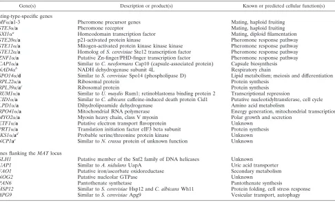
The mating-type locus was also found to contain a surprising number of repetitive sequences, including simple sequence repeats. As shown in Fig. 5, all four alleles contained multiple copies of several different simple tri- and tetranucleotide re-TABLE 2. Genes within and flanking the mating-type alleles ofC. neoformans
Gene(s) Description or product(s) Known or predicted cellular function(s)
Mating-type-specific genes
MF␣/a1-3 Pheromone precursor genes Mating, haploid fruiting
STE3␣/a Pheromone receptor Mating, haploid fruiting
SXI1␣a Homeodomain transcription factor Mating, diploid filamentation
STE20␣/a p21-activated protein kinase Pheromone response pathway
STE11␣/a Mitogen-activated protein kinase kinase kinase Pheromone response pathway STE12␣/a Homolog ofS. cerevisiaeSte12 transcription factor Pheromone response pathway ZNF1␣/a Putative Zn-finger/PHD-finger transcription factor Pheromone response pathway CAP1␣/a Similar toC. neoformansCap10 (capsule-associated protein) Capsule biosynthesis NAD4␣b NADH dehydrogenase subunit 4L Respiratory chain
SPO14␣/d Similar toS. cerevisiaeSpo14 (phospholipase D) Lipid metabolism; meiosis and differentiation
RPL22␣/a Ribosomal protein Protein synthesis
RPL39␣/ac Ribosomal protein Protein synthesis
RUM1␣/a Similar toU. maydisRum1; retinoblastoma binding protein 2 Transcriptional repression
CID1␣/a Similar toC. albicanscaffeine-induced death protein Cid1 Putative nucleotidyltransferase, cell cycle LPD1␣/a Dihydrolipoamide dehydrogenase Amino acid metabolism
RPO41␣/a Mitochondrial RNA polymerase Energy generation, mitochondrial transcription MYO2␣/a Myosin heavy chain, class V myosin Polar growth and secretion
ETF1␣/a Putative electron transport flavoprotein Unknown PRT1␣/a Translation initiation factor eIF3 beta subunit Protein synthesis IKS1␣/ad Probable serine/threonine protein kinase Unknown NCP1ae Similar toN. crassaprotein of unknown function Unknown
Genes flanking theMATlocus
SLH1 Putative member of the Snf2 family of DNA helicases Unknown
UAP1 Similar toA. nidulansUapA Uric acid transporter
FAO1 Putative iron/ascorbate oxidoreductase Secondary metabolism
NOG2 Putative nucleolar GTPase Unknown
PAN6 Pantothenate synthetase Pantothenate synthesis
HSP12 Similar toS. cerevisiaeHsp12 andC. albicansWh11 Protein folding, cell stress response APG9 Similar toS. cerevisiaeApg9 Vesicular transport, autophagy aPresent only inMAT␣mating-type alleles.
bPresent only in the serotype AMAT␣strain H99. cMissing in the serotype DMATastrain JEC20.
dWithin theMATlocus in serotype A; flanking theMATlocus in serotype D. ePresent only in the serotype DMATastrain JEC20.
on September 8, 2020 by guest
http://ec.asm.org/
peats. A particularly notable example was 71 imperfect copies of a tetranucleotide repeat contained within the serotype D STE11agene that were not present in any of the otherSTE11 genes. In addition, the pheromone precursor genes are often encoded by divergent pairs of genes that were embedded in regions that constitute large inverted repeats. This may give rise to unique mechanisms by which the genes are duplicated, rearranged, and lost as the alleles of the mating-type locus diverged from their common ancestors. For example, inver-sions between the identical MF␣1 and MF␣2 genes in the serotype D␣allele would transpose the order and direction of the intervening genes (PRT1␣,ZNF1␣,RPL39␣, andMF␣3). Recombination is suppressed in the mating-type region.An important feature of mating type is stable inheritance as a single unit, and recombination is suppressed in these regions to avoid generation of sterile or self-fertile offspring. We used PCR analysis with ␣ and a allele-specific primers to test whether the sequences we defined as theMATlocus faithfully cosegregate with mating type and whether recombination oc-curs in this locus (Fig. 6). Twenty-four progeny derived from
two defined crosses between multiply marked strains were tested by PCR to test whether recombination occurred be-tween the ends of theMATlocus. In addition, mating type was scored by genetic backcrosses. No recombination was observed in the mating-type region, and mating-type-specific sequences faithfully cosegregated with the corresponding mating types as determined by mating assays with tester strains. Forche and coworkers recently reported an amplified fragment length polymorphism-based physical map forC. neoformansand es-tablished the recombination frequency for the mating-type chromosome at ⬃24 kb/centimorgan, demonstrating that re-combination readily occurs elsewhere on this chromosome (24). In addition, theCNB1gene resides on the mating-type chromosome but is completely unlinked to theMATlocus in genetic crosses (data not shown) (25), providing additional evidence for recombination events distal toMAT.
Sequences flanking the mating-type locus are nearly identi-cal, but a few sequence polymorphisms between the␣strain JEC21 and thea strain JEC20 are present immediately up-stream and downup-stream of theMATlocus. We used primers FIG. 5. Multiple transposon remnants and repetitive sequences are embedded in theMATlocus. Transposable element-related sequences are depicted for the four alleles of theMATlocus. Complete element copies are indicated in a larger font size and boldface type. In addition, local sequence repeats were identified and annotated for each allele.
on September 8, 2020 by guest
http://ec.asm.org/
designed for these sequences and the same set of meiotic progeny to test whether recombination occurred just outside of the borders of theMATlocus. No recombination events were observed, providing additional evidence for the integrity of the locus and its correct assignment (Fig. 6).
The␣and a alleles of theMATlocus are stably inherited. The␣and acongenic pair of serotype D strains JEC21 and JEC20 was generated by a series of 10 backcrosses (Fig. 4) (31, 42). One concern was whether the mating-type alleles might have rearranged during the process of strain construction, pos-sibly as a result of increased recombination during meiosis. Southern analysis was used to compare the genomic structure of the␣andaalleles of strains JEC21 and JEC20 with those of their ancestors by using probes to sequences within and flanking theMATlocus (Fig. 4). No differences in restriction patterns were observed for any of the genes analyzed. Thus, the structure of the mating-type locus has been stably inherited through multiple generations, providing additional evidence that recombination is suppressed in this genomic region.
Structure of the serotype D MAT␣ mating-type locus is conserved in nature.The serotype D ␣and astrains JEC21 and JEC20 and their derivatives are widely used because of their congenic background and because of the ability to con-duct classical genetic experiments with them (31). This was one of the major reasons why these strains were chosen to deter-mine the structure of the mating-type locus in serotype D. However, an important issue is whether the structure of the mating-type locus of these lab strains is representative of un-related serotype D strains. We addressed this by a PCR-based approach using primers that amplify overlapping fragments spanning the original⬃38-kb serotype DMAT␣mating-type locus proposed by Moore and Edman. Fragments of identical sizes were obtained with 9 of the 10 primer pairs using as templates DNA from the unrelated serotype D strains JEC21, CDC92-18, CDC92-27, and MMRL760 (Fig. 7). Only one primer combination produced a larger,⬃14-kb PCR product from strain MMRL760 (Fig. 7), compared with an ⬃10-kb PCR product from JEC21 and the two CDC strains. Further PCR analysis revealed that an insertion of⬃4 kb had occurred between theSTE11␣andMF␣1genes of theMATlocus of this atypical yet still fertile strain (Fig. 7).
FIG. 6. Recombination between the MAT alleles is suppressed. Twenty-four progeny from two defined crosses were tested by PCR for recombination events between mating-type alleles. Primer pairs were either mating-type specific (pairs 3, 4, 5, and 6) or strain specific (pairs 1, 2, 7, and 8). Fragments amplified within the mating-type region by the different primer pairs are indicated as thick black bars. No recom-bination events were observed, and the PCR-amplified fragments all cosegregated with the corresponding mating types (␣or a), as deter-mined by backcrosses. The control strains were the serotype D strains JEC20 (MATa) and JEC21 (MAT␣), indicated by a* and␣*, respec-tively. The meiotic progeny were also tested for segregation of parental markers. Ten of 24 strains showed a recombinant pattern (r), whereas the remaining 14 strains exhibited a parental genotype (p), which is indicated only for the control strains.
FIG. 7. TheMAT␣ mating-type locus is conserved in the population. Using one primer combination, overlapping fragments (3 to 10 kb) spanning part of the mating-type locus were PCR amplified from (lanes from left to right in panel 1) the control strain JEC21 and the unrelated serotype D clinical isolates CDC92-18, CDC92-27, and MMRL760. Fragments of identical sizes were amplified from all four strains with nine primer combinations (panels 1 to 4 and 6 to 10). For descriptions of the 10 primer pairs, which are also represented by numbered bars indicating their positions on theMATlocus, see Table 1. One primer pair yielded a larger PCR product for strain MMRL760 (panel 5, lane 4), indicative of an⬃4-kb insertion between theSTE11␣ andMF␣1genes. DNA sequence analysis revealed that the insertion of a novel mariner-related transposable element resulted in the addition of 4,006 bp relative to the JEC21 serotype D␣allele and the creation of a TA target site duplication.
on September 8, 2020 by guest
http://ec.asm.org/
Sequence analysis of this region of theMATlocus of strain MMRL760 revealed that a novel mariner-related transposable element had inserted into the locus. Compared with that of strain JEC21, an additional 3,906 bp are present in theMAT locus of strain MMRL760, and this novel sequence is flanked by 136-bp inverted repeats that are identical at 135 of 136 positions. In addition, the element is inserted at a TA sequence and created a TA-TA duplication at the insertion site. The right half of this element encodes an open reading frame that might represent a transposase gene. Importantly, by compari-son with the results of the ongoing genome project, this region of the element was found to share significant sequence identity with five distinct regions of the genome of serotype D strain JEC21. Curiously, theMATlocus of strain JEC21 contains a region of several hundred base pairs that shares identity with the left end of this element. Thus, this element may have either transposed into a remnant of itself, or strain JEC21 contains a fragment of the element as a result of a previous excision event.
Structural rearrangements during evolution and divergence of theMATalleles.Our findings reveal that theC. neoformans mating-type locus is significantly larger than previously sus-pected. In serotype D, the␣allele spans⬃105 kb and thea allele spans⬃117 kb, whereas in serotype A, the␣andaalleles span⬃103 and⬃127 kb, respectively. Thus, in both serotypes, theaallele is larger than the␣ allele. The number of genes identified within the locus ranges from 19 (MATain serotypes A and D) to 23 (MAT␣ in serotype A). While some of the genes encoded by theMATlocus have already been shown or predicted to function in the pheromone response pathway that regulates mating (Fig. 1, black arrows), other genes have no obvious function with respect to sexual development.
The gene order is strikingly different between different al-leles of the mating-type locus. Genes outside the mating-type locus exhibit synteny in both serotype A and serotype D (Fig. 1 and 8A), whereas gene order inside the mating-type locus has been dramatically remodeled (Fig. 8A and data not shown). In addition, a few genes are present in either the ␣ or the a mating-type allele but not in both, includingSXI1␣,RPL39␣, andNCP1ain serotype D andSXI1␣andNAD4␣in serotype A (Fig. 1 and 8A) (Table 2). When the␣ ora mating-type alleles were compared between serotypes A and D, the rear-rangement of the locus was even more striking (Fig. 8B). Fur-thermore, several genes identified are unique to the mating-type allele of only one seromating-type (Table 2). Interestingly, the IKS1gene flanks the mating-type locus in serotype D but is located within the mating-type locus in serotype A, possibly as the result of a DNA inversion. With this exception, the order of the genes outside theMATlocus is conserved between the two mating types and varieties. Interestingly, the orders of the genes just inside the left ends of theMAT␣mating-type loci of serotype A strain H99 and serotype D strain JEC21 are similar, with the exception of one inversion (SPO14␣) and two small insertions (Cnirt3andNAD4␣) (Fig. 8B).
In summary, our findings reveal that the␣andamating-type alleles diverged from a common ancestral region of DNA by a process involving rearrangements, inversions, and nucleotide substitutions. Moreover, the␣andaalleles have both under-gone extensive rearrangements as the serotype A and serotype D strains evolved into varieties or even distinct species.
DISCUSSION
We have analyzed the structure of four mating-type alleles of the fungal pathogenC. neoformans, including the␣and a alleles of the serotype A and D varietiesgrubiiandneoformans. TheMATlocus ofC. neoformansis considerably larger than previously reported (35) and spans⬃105 to 130 kb. While the gene order outside the locus is largely conserved, even between the two serotypes, genes inside the mating-type locus have been subject to extensive rearrangements. This is true not only for the two opposite mating-type alleles in a given serotype but also for a singleMATallele compared between serotypes. In addition, a few genes were identified that are present in only one or the other allele. Recombination in the mating-type locus and the surrounding genomic region is suppressed, and the mating-type alleles are stable through multiple genetic crosses without rearrangement. The basic structure of the mat-ing-type locus inC. neoformansis largely conserved within the population of this organism, but the detection of a transpos-able element in the␣locus of an atypical serotype D strain reveals that genetic alterations can occur in the population.
When compared to those of other model ascomycetes or basidiomycetes, the mating-type locus ofC. neoformanshas a unique structure in terms of both size and gene composition. The MAT locus in most ascomycetes is limited in size and encodes transcription factors that determine mating type and cell identity. In basidiomycetes with tetrapolar mating systems, one mating-type locus resembles ascomycete mating-type loci in size and gene composition. The second locus encodes pher-omone and pherpher-omone receptor systems and can extend up to 20 kb via gene duplications. Only a few other fungal mating-type loci have been found to contain genes lacking an obvious function in mating (33, 54, 64). The mating-type locus ofC. neoformansis the largest single-copyMATlocus known, and the locus contains a striking number of genes, including ones that function in mating and others with no predicted role in sexual differentiation.
In contrast to what occurs in other basidiomycete mating-type loci, the C. neoformans genes encoding pheromones (MF␣1-3andMFa1-3) and pheromone receptors (STE3␣and STE3a) are not adjacent to each other but rather dispersed throughout the locus (Fig. 1). The MF␣1-3 pheromone and STE3␣pheromone receptor genes were identified in the pre-viously publishedC. neoformans MATlocus (35), but no tran-scriptional regulators of the homeodomain or HMG domain family were previously known. In our studies, BLASTX anal-ysis of the complete␣andamating-type sequences identified a gene close to the left end of the␣mating-type alleles that exhibited weak similarity to other homeodomain transcription factors involved in mating and cell identity in other fungi (Fig. 1). As will be presented elsewhere, deletion analysis of the SXI1␣(sex inducer 1␣) gene reveals a role for Sxi1␣in sexual development (Hull et al., submitted). NoSXI1␣-related gene is present in either a allele studied, and no cross-hybridizing genes are present ina-specific DNA by Southern analysis (Hull et al., submitted). The identification of the mating-type-specific homeodomain transcription factor Sxi1␣ brings the mating-type system ofC. neoformanscloser to those of other basidio-mycetes with respect to the main regulators involved in sexual development than previously suspected. Our findings reveal
on September 8, 2020 by guest
http://ec.asm.org/
that both transcriptional regulators and a pheromone and pheromone receptor system are present in theC. neoformans MATlocus, but the arrangement of the locus is distinct com-pared to those of other model basidiomycetes in which the two regulators are unlinked.
In addition to these regulatory genes,⬃15 other genes were identified in the different mating-type alleles (Table 2). Some encode components of the pheromone response pathway that regulates mating, fruiting, or virulence ofC. neoformans(14,
15, 35, 45–47, 49, 63, 69, 71, 73). Interestingly, a similar unusual cluster of genes that may be involved in pheromone signaling was recently reported in another opportunistic human fungal pathogen, Pneumocystis carinii (65). Genome sequencing re-vealed a locus that shares similarities with the mating-type locus of C. neoformans and contains genes encoding com-ponents of a putative pheromone response pathway (65). Whether this region represents a true mating-type locus ofP. cariniiis not known, and no sexual cycle has been described for FIG. 8. Structural comparison of theC. neoformansmating-type alleles. (A) Comparison of the␣andamating-type alleles of serotype D for analysis of the relative positions of the genes found within and adjacent to theMATlocus. Vertical and diagonal lines connect diverged gene alleles present in both alleles and illustrate substantial gene rearrangements within theMATlocus, whereas gene order outside theMATlocus has been conserved. In addition, a few genes that are present in only one of the twoMATalleles were identified (white arrows). (B) Comparison of the␣ mating-type alleles between seromating-types A and D. Similar to what occurred in the␣andaalleles from serotype D, significant rearrangements of gene order have occurred in the␣allele during strain divergence (vertical and diagonal lines), and some genes are unique to one serotype (white arrows). Interestingly, theIKS1gene flanks theMATlocus in serotype D but is located within the locus in serotype A. For additional details, see the legend to Fig. 1.
on September 8, 2020 by guest
http://ec.asm.org/
this pathogenic fungus. The finding that the basidiomyceteC. neoformansand the ascomycete P. cariniishare similarly ar-ranged mating-type loci raises the question of whether the MATlocus plays a role in the virulence of P. carinii, as has already been established for theMAT␣locus ofC. neoformans. Our studies reveal that three different types of mating-type loci exist in fungi. The first comprises the classicalMATloci of ascomycetes, in which mating type is determined by specialized transcription factors encoded by a single, compact locus. The second comprises the tetrapolar mating systems of the basid-iomycetes, in which mating type is determined by two distinct, unlinked loci encoding transcriptional regulators and phero-mone and pherophero-mone receptor systems. The third is the novel mating-type locus inC. neoformansand a related region inP. carinii, in which mating-specific transcription factors, a phero-mone and pherophero-mone receptor system, and elements of the pheromone-activated MAP kinase cascade are part of a single, contiguous multigene locus. BecauseC. neoformansis a basid-iomycete and P. cariniiis an ascomycete, this class of MAT locus either evolved prior to the divergence of the two major fungal phyla or resulted from convergent evolution.
Unlike most model basidiomycetes but similar toC. neofor-mans,U. hordeihas a bipolar mating system and recombination in the type region is suppressed. Two opposite mating-type and pathogenicity alleles,MAT-1 andMAT-2, have been identified and have been found to span 500- and 460-kb re-gions, respectively (44).MAT-1 andMAT-2 include one locus encoding mating-type-specific transcription factors and a sec-ond locus containing tightly linked pheromone and receptor genes. Both loci reside on the same chromosome and are separated by 450 to 500 kb of intervening DNA in which recombination is suppressed. These findings explain at a mo-lecular level how a tetrapolar mating system can be converted into a bipolar system by linking of the commonly found mating-type loci on a single chromosome and the involvement of mechanisms that suppress recombination across the interven-ing sequences (2, 44).
An interesting question is how recombination is suppressed across the MAT loci of C. neoformans and U. hordei. The sequence of the interval between the two loci ofU. hordeiis being determined and contains many repetitive sequences and transposable elements that may contribute to the suppression of recombination (J. Kronstad, personal communication). Our analysis of theC. neoformansmating-type alleles reveals two factors that may also play a role. First, mating-type-specific alleles of several genes vary from 5 to⬃50% in sequence (46), and some genes are unique to one or the other allele. Second, gene positions in the mating-type alleles are extensively rear-ranged (including inversions). For example, theRPO41␣and RPO41agenes are almost identical (97%) but are oriented in opposite directions in serotype D. Thus, crossover events be-tween these two alleles would result in one acentric and one dicentric chromosome, both of which would be unstable. These sequence and structural differences likely prevent proper align-ment of this chromosomal region during meiosis and thereby suppress recombination.
Interestingly, for the ascomyceteNeurospora tetrasperma, ge-netic and cytological studies have shown that during meiosis the chromosomes containing the mating-type locus are un-paired over a large interval that includes theMATlocus, and
recombination is suppressed in this region. In addition, specific sites flanking this region trigger recombination events that may function to ensure proper chromosome segregation during meiosis (26, 48). These observations suggest that the chromo-somes containing the fungal MAT loci share features with mammalian sex chromosomes.
The mating-type-determining region of C. neoformans shares features with both the self-incompatibility locus that governs pollen recognition in species of the crucifer plant Bras-sica(50) and the mating-type locus of the green alga Chlamy-domonas reinhardtii.For example, the multiallelic S locus in Brassica spp. is composed of divergent and rearranged se-quences linking theSRKand SCR genes involved in pollen-stigmata interactions (5). InChlamydomonas, the mating-type locus is located in a region of⬃830 kb in which recombination is suppressed (23). In addition, a 190-kb core region thought to contain the mating-type determining factors is highly rear-ranged via several translocations, inversions, duplications, and deletions (21–23). These chromosomal aberrations are thought to be responsible for suppressing recombination in the core region and the flanking 640 kb of genomic DNA. The mating-type region in C. reinhardtii is located close to one end of linkage group VI (23). This is similar toC. neoformansbecause analysis from the ongoing genome project reveals that the MATlocus resides⬃170 kb from one telomere of this 1.8-Mb chromosome. Whether chromosomal location has any impact on the function of the mating-type loci in these organisms is not known, but it is interesting that the HML and HMR silent mating-type cassettes inS. cerevisiaeare also located near the ends of yeast chromosome III.
Sex determination in higher eukaryotes is often accompa-nied by the presence of dimorphic sex chromosomes. An in-teresting model that explains the evolution of sex chromo-somes is based on the initial requirement for genetic differences in multiple loci for the definition of sexual identity. Since the generation of self-fertile or sterile progeny is unfa-vorable, mechanisms had to evolve to ensure tight linkage between the genes involved (11, 12), possibly including the evolution of nonhomologous genes and chromosomal rear-rangements. Once established, these mechanisms suppress the exchange of genetic material in these regions, and genetic divergence between genomic regions results in the evolution of a “diallelic” sex chromosome system. It has been proposed that animal sex chromosomes evolved from autosomes that were initially homologous except for a small sex-determining region (30). Following suppression of recombination in this region, subsequent divergence between the two “autosomes” resulted in the evolution of the sex chromosomes responsible for the hetero- (XY) and homogametic (XX) sexes in mammals (13, 43, 57, 58). The pseudoautosomal region on the mammalian Y chromosome may reflect its ancestral autosomal origin.
The ⬃1.8-Mb mating-type chromosome in C. neoformans shares features with mammalian sex chromosomes. Although the sex-determining region comprises only⬃7% of this fungal chromosome, recombination is suppressed in the mating-type region but does occur in more distal regions of the chromo-some. While recombination is suppressed between most of the sex chromosomes of mammals, recombination does occur in the pseudoautosomal region and is thought to be essential for proper chromosome segregation. Similar to mammalian sex
on September 8, 2020 by guest
http://ec.asm.org/
chromosomes, theMATlocus ofC. neoformansis character-ized by nonhomologous genes and extensive rearrangements. In addition, the lack of genetic exchange favors the accumula-tion of repetitive sequences and transposable elements within the sex-determining region, which favors intrachromosomal rearrangements and drives divergence. TheMATloci in the green algaC. reinhardtiiand the fungiN. tetrasperma,P. carinii, andU. hordeiall share similar features with sex chromosomes. Since sex is thought to have originally evolved in lower eu-karyotes, such as yeasts and algae, it is intriguing that the sex-determining systems of several unicellular eukaryotes share features resembling an early step in the evolutionary pathway to the dimorphic sex chromosomes of multicellular eukaryotes.
ACKNOWLEDGMENTS
We thank Christina Hull, Robin Wharton, and John Perfect for advice and comments and Jim Kronstad for providing high-density BAC filter arrays and BAC data.
This study was supported by R01 grant AI50113 and P01 grant AI44975 (NIAID) to the Duke mycology research unit. Joseph Heit-man is a Burroughs Welcome Scholar in molecular pathogenic mycol-ogy and an associate investigator of the Howard Hughes Medical Institute.
REFERENCES
1.Altschul, S. F., T. L. Madden, A. A. Schaffer, J. Zhang, Z. Zhang, W. Miller, and D. J. Lipman.1997. Gapped BLAST and PSI-BLAST: a new generation of protein database search programs. Nucleic Acids Res.25:3389–3402. 2.Bakkeren, G., and J. W. Kronstad.1994. Linkage of mating-type loci
distin-guishes bipolar from tetrapolar mating in basidiomycetous smut fungi. Proc. Natl. Acad. Sci. USA91:7085–7089.
3.Beach, D. L., J. Thibodeaux, P. Maddox, E. Yeh, and K. Bloom.2000. The role of the proteins Kar9 and Myo2 in orienting the mitotic spindle of budding yeast. Curr. Biol.10:1497–1506.
4.Bo¨lker, M., M. Urban, and R. Kahmann.1992. Theamating type locus of
U. maydisspecifies cell signaling components. Cell68:441–450.
5.Boyes, D. C., M. E. Nasrallah, J. Vrebalov, and J. B. Nasrallah.1997. The self-incompatibility (S) haplotypes ofBrassicacontain highly divergent and rearranged sequences of ancient origin. Plant Cell9:237–247.
6.Casadevall, A., and J. R. Perfect.1998.Cryptococcus neoformans. ASM Press, Washington, D.C.
7.Casselton, L. A., and N. S. Olesnicky.1998. Molecular genetics of mating recognition in basidiomycete fungi. Microbiol. Mol. Biol. Rev.62:55–70. 8.Chang, Y. C., and K. J. Kwon-Chung.1999. Isolation, characterization, and
localization of a capsule-associated gene,CAP10, ofCryptococcus neofor-mans. J. Bacteriol.181:5636–5643.
9.Chang, Y. C., L. A. Penoyer, and K. J. Kwon-Chung.2001. The second STE12 homologue ofCryptococcus neoformansisMATa-specific and plays an important role in virulence. Proc. Nat. Acad. Sci. USA98:3258–3263. 10.Chang, Y. C., B. L. Wickes, G. F. Miller, L. A. Penoyer, and K. J.
Kwon-Chung.2000.Cryptococcus neoformans STE12␣regulates virulence but is not essential for mating. J. Exp. Med.191:871–882.
11.Charlesworth, B.1991. The evolution of sex chromosomes. Science251:
1030–1033.
12.Charlesworth, B. 1994. Evolutionary genetics. The nature and origin of mating types. Curr. Biol.4:739–741.
13.Charlesworth, B.1978. Model for evolution of Y chromosomes and dosage compensation. Proc. Natl. Acad. Sci. USA75:5618–5622.
14.Chung, S., M. Karos, Y. C. Chang, J. Lukszo, B. L. Wickes, and K. J. Kwon-Chung.2002. Molecular analysis ofCPR␣, aMAT␣-specific phero-mone receptor gene ofCryptococcus neoformans.Eukaryot. Cell1:432–439. 15.Clarke, D. L., G. L. Woodlee, C. M. McClelland, T. S. Seymour, and B. L. Wickes.2001. TheCryptococcus neoformans STE11␣gene is similar to other fungal mitogen-activated protein kinase kinase kinase (MAPKKK) genes but is mating type specific. Mol. Microbiol.40:200–213.
16.Coppin, E., R. Debuchy, S. Arnaise, and M. Picard.1997. Mating types and sexual development in filamentous ascomycetes. Microbiol. Mol. Biol. Rev.
61:411–428.
17.Dee, J.1982. Genetics ofPhysarum polycephalum, p. 211–251.InH. C. Aldrich and J. W. Daniel (ed.), Cell biology ofPhysarumandDidymium, vol. 1. Academic Press, New York, N.Y.
18.Dzelzkalns, V. A., J. B. Nasrallah, and M. E. Nasrallah.1992. Cell-cell communication in plants: self-incompatibility in flower development. Dev. Biol.153:70–82.
19.Ewing, B., and P. Green.1998. Base-calling of automated sequencer traces using phred. II. Error probabilities. Genome Res.8:186–194.
20.Ewing, B., L. Hillier, M. C. Wendl, and P. Green.1998. Base-calling of automated sequencer traces using phred. I. Accuracy assessment. Genome Res.8:175–185.
21.Ferris, P. J., E. V. Armbrust, and U. W. Goodenough.2002. Genetic structure of the mating-type locus ofChlamydomonas reinhardtii. Genetics160:181– 200.
22.Ferris, P. J., and U. W. Goodenough.1997. Mating type in chlamydomonas is specified bymid, the minus-dominance gene. Genetics146:859–869. 23.Ferris, P. J., and U. W. Goodenough.1994. The mating-type locus of
Chlamy-domonas reinhardtiicontains highly rearranged DNA sequences. Cell76:
1135–1145.
24.Forche, A., J. Xu, R. Vilgalys, and T. G. Mitchell.2000. Development and characterization of a genetic linkage map ofCryptococcus neoformansvar.
neoformansusing amplified fragment length polymorphisms and other mark-ers. Fungal Genet. Biol.31:189–203.
25.Fox, D. S., M. C. Cruz, R. A. L. Sia, H. Ke, G. M. Cox, M. E. Cardenas, and J. Heitman.2001. Calcineurin regulatory subunit is essential for virulence and mediates interactions with FKBP12-FK506 inCryptococcus neoformans. Mol. Microbiol.39:835–849.
26.Gallegos, A., D. J. Jacobson, N. B. Raju, M. P. Skupski, and D. O. Natvig.
2000. Suppressed recombination and a pairing anomaly on the mating-type chromosome ofNeurospora tetrasperma. Genetics154:623–633.
27.Gillissen, B., J. Borgemann, C. Sandmann, B. Schroeer, M. Bolker, and R. Kahmann.1992. A two-component regulatory system for self/non-self rec-ognition inUstilago maydis. Cell68:647–657.
28.Goodwin, T. J. D., and R. T. M. Poulter.2001. The diversity of retrotrans-posons in the yeastCryptococcus neoformans. Yeast18:865–880.
29.Gordon, D., C. Abajian, and P. Green.1998. Consed: a graphical tool for sequence finishing. Genome Res.8:195–202.
30.Graves, J. A., and J. W. Foster.1994. Evolution of mammalian sex chromo-somes and sex-determining genes. Int. Rev. Cytol.154:191–259.
31.Heitman, J., B. Allen, J. A. Alspaugh, and K. J. Kwon-Chung.1999. On the origins of the congenic MAT␣and MATastrains of pathogenic yeast Cryp-tococcus neoformans. Fungal Genet. Biol.28:1–5.
32.Herskowitz, I.1989. A regulatory hierarchy for cell specialization in yeast. Nature342:749–757.
33.Hull, C. M., and A. D. Johnson.1999. Identification of a mating type-like locus in the asexual pathogenic yeastCandida albicans. Science285:1271– 1275.
34.Johnson, A. D.1995. Molecular mechanisms of cell-type determination in budding yeast. Curr. Opin. Genet. Dev.5:552–558.
35.Karos, M., Y. C. Chang, C. M. McClelland, D. L. Clarke, J. Fu, B. L. Wickes, and K. J. Kwon-Chung.2000. Mapping of theCryptococcus neoformans MAT␣ locus: presence of mating type-specific mitogen-activated protein kinase cascade homologs. J. Bacteriol.182:6222–6227.
36.Karpova, T. S., S. L. Reck-Peterson, N. B. Elkind, M. S. Mooseker, P. J. Novick, and J. A. Cooper.2000. Role of actin and Myo2p in polarized secretion and growth ofSaccharomyces cerevisiae. Mol. Biol. Cell11:1727– 1737.
37.Kothe, E.2001. Mating-type genes for basidiomycete strain improvement in mushroom farming. Appl. Microbiol. Biotechnol.56:602–612.
38.Kothe, E.1996. Tetrapolar fungal mating types: sexes by the thousands. FEMS Microbiol. Rev.18:65–87.
39.Kronstad, J. W., and S. A. Leong.1990. The b mating-type locus ofUstilago maydiscontains variable and constant regions. Genes Dev.4:1384–1395. 40.Kronstad, J. W., and C. Staben.1997. Mating type in filamentous fungi.
Annu. Rev. Genet.31:245–276.
41.Ku¨es, U., W. V. J. Richardson, A. M. Tymon, E. S. Mutasa, B. Gottgens, S. Gaubatz, A. Gregoriades, and L. A. Casselton.1992. The combination of dissimilar alleles of the A␣and Agene complexes, whose proteins contain homeo domain motifs, determines sexual development in the mushroom
Coprinus cinereus. Genes Dev.6:568–577.
42.Kwon-Chung, K. J., J. C. Edman, and B. L. Wickes.1992. Genetic associa-tion of mating types and virulence inCryptococcus neoformans. Infect. Im-mun.60:602–605.
43.Lahn, B. T., and D. C. Page.1999. Four evolutionary strata on the human X chromosome. Science286:964–967.
44.Lee, N., G. Bakkeren, K. Wong, J. E. Sherwood, and J. W. Kronstad.1999. The mating-type and pathogenicity locus of the fungusUstilago hordeispans a 500-kb region. Proc. Natl. Acad. Sci. USA96:15026–15031.
45.Lengeler, K. B., R. C. Davidson, C. D’Souza, T. Harashima, W.-C. Shen, P. Wang, X. Pan, M. Waugh, and J. Heitman.2000. Signal transduction cas-cades regulating fungal development and virulence. Microbiol. Mol. Biol. Rev.64:746–785.
46.Lengeler, K. B., P. Wang, G. M. Cox, J. R. Perfect, and J. Heitman.2000. Identification of the MATamating-type locus ofCryptococcus neoformans
reveals a serotype A MATastrain thought to have been extinct. Proc. Natl. Acad. Sci. USA97:14455–14460.
47.McClelland, C. M., J. Fu, G. L. Woodlee, T. S. Seymour, and B. L. Wickes.