Iran J Public Health, Vol. 47, No.10, Oct 2018, pp.1567-1574
Original Article
Genetic Polymorphisms of Catalase and Glutathione
Peroxidase-1 in Keratoconus
Davood YARI
1,2,*Ramin SARAVANI
1,2, Samira SARAVANI
3, Kamran EBRAHIMIAN
4,
Hamid Reza GALAVI
21. Cellular and Molecular Research Center, Zahedan University of Medical Sciences, Zahedan, Iran 2. Dept. of Clinical Biochemistry, School of Medicine, Zahedan University of Medical Sciences, Zahedan, Iran
3. Dept. of Biology, School of Fundamental Science, University of Zabol, Zabol, Iran 4. Dept. of Molecular Biotechnology, Faculty of Pharmacy, University of Barcelona, Barcelona, Spain
*Corresponding Author: Email: saravaniramin@yahoo.com
(Received 06 May 2017; accepted 13 Jul 2017)
Introduction
Keratoconus (KC) is a degenerative eye disease which results from thinning of the cornea and causes vision distortion (1).This condition is characterized by the stromal thinning of the cor-nea which often results in bilateral and asymmet-ric corneal distortion and anterior corneal protru-sion (2).Symptoms are highly variable and depend
on the stage of the progression of the disorder. In the initial stage of the disease, there may be no symptoms; in the advanced stage, there is significant distortion of vision accompanied by serious visual loss. Patients with KC never be-come completely blind from their disease (3).KC is a major suggestion for corneal transplantation
Abstract
Background: Keratoconus (KC) is a degenerative eye disease which results from thinning of the cornea and causes vision distortion. Oxidative stress damage to KC corneas may be because of the failure of corneas to process reactive oxygen species which leads to corneal thinning and loss of vision. Genetic variants in antioxi-dant defense genes such as catalase (CAT) and glutathione peroxidase (GPX) can decrease antioxiantioxi-dant capacity or increase oxidative stress and alter the risk of KC in patients. We investigated and evaluated the effects of single nucleotide polymorphisms in CAT, GPX-1 on the risk of KC in an Iranian population sample.
Methods: This case-control study was performed on 140 patients with KC and 150 healthy control subjects in a sample of Iranian population from Zahedan, southern Iran in 2015. Genotyping of CAT rs7943316 and GPX-1 rs1050450 polymorphisms was done using polymerase chain reaction and restriction fragment length polymorphism (PCR-RFLP) method.
Results: CAT rs7943316 A/T, AA genotype and A allele have a protective role against disease (OR =0.28, 95% CI =0.13-0.61, P=0.001 and OR = 0.50, 95% CI =0.35-0.72, P=0.0001, respectively) and decreased the risk of KC. Moreover, GPX-1 rs1050450 T allele increased the risk of KC in comparison with C allele (OR = 1.42, 95% CI = 1.01-2.03, P=0.03).
Conclusion: CAT rs7943316 A/T, AA genotype, and A allele decreased the risk of KC. Moreover, in GPX-1 rs1050450 C/T polymorphism, T allele was associated with an increased risk of KC in our population.
in developed countries(4).KC usually occurs in the second decade of life with progress in the next two decades and affects both genders and all ethnicities. The expected prevalence in the gen-eral population is 54 per 100000(5, 6).KC is most commonly an isolated disease, while several re-ports describe an association with Down syn-drome, monosomy X (Turner syndrome), Leber’s congenital Ehlers-Danlos syndrome, neurocuta-neousangiomatosis, neurofibromatosis, xeroder-mapigmentosa, collagenosis, retinitis pigmentosa and Marfan syndrome is described (7).The cause and original pathological mechanism are un-known but biochemical, genetic, and environ-mental factors are possible causes of KC and dif-ferent etiologies may play a role in the develop-ment of this disease(8, 9).
Environmental factors which can influence on eye consist of rub eyeballs, allergic reaction, and solarization (9). An excess of environmental fac-tors, particularly UV exposure, causes oxidative damage to KC corneas because of the failure of KC corneas to process reactive oxygen species (ROS) which leads to corneal thinning and loss of vision (10). The accumulation of ROS can damage cells by reacting with proteins, DNA, and membrane phospholipids (11). The normal cor-nea’s antioxidant enzymes eliminate the ROS be-fore they damage cells; these consist of superox-ide dismutase, glutathione reductase, catalase, and glutathione peroxidase, but in the disease condi-tion, ROS can devastate cellular defenses and promote cell damage (12).
Catalase (CAT) is a common enzyme that found in the peroxisome of many aerobic organisms. This enzyme is a tetramer with molecular weight of about 240000 with four heme groups per te-tramer (13, 14).Hydrogen peroxide (H2O2) is
converted to H2O and O2 by CAT and thereby
mitigates the toxic effects of hydrogen peroxide (15). Location of CAT gene is on chromosome 11p13 and comprise 13 exons; rs7943316 (-21 A/T) settled in nearly to the start site at promot-er region (16). Deficiency of catalase may cause elevated concentrations of hydrogen peroxide and increase the risk of the progress of patholo-gies (17).Glutathione Peroxidase-1(GPX-1)
en-codes a member of the glutathione peroxidase family. GPX-1 is an intracellular antioxidant en-zyme that converts H2O2 to water and protects
the organism from oxidative damage(18, 19).GPx-1 is selenium-containing Enzyme, locat-ed at chromosome 3p21.3(20).GPx-1 has four SNPs that alter the amino acid produced but only one has been studied extensively in human dis-ease, including rs1050450 (or GPx1 Pro197Leu). This C>T variation changes the amino acid from proline (Pro) to leucine (Leu) at position 197 (21).The GPx-1 variant may be associated with a decreased ability to scavenge ROS (22).
To the best of our knowledge, no studies have investigated the role of antioxidant gene CAT and GPX polymorphisms for KC patients. The present study aimed to evaluate the impact of CAT rs7943316 A/T and GPX-1 rs1050450 C/T polymorphisms on KC patients in a sample of Iranian population. These polymorphisms may alter the enzymes’ antioxidant capacity and may be associated with the risk of KC induced by ox-idative damage.
Materials and Methods
Patients
This case-control study was done on 140 patients with KC (61 men and 79 women), and 150 healthy individuals as the control group (65 men and 85 women), who were enrolled from Alzahra Eye Hospital, Zahedan, southern Iran in 2015. Ethical approvals for recruitment were obtained from the local Ethics Committee of Zahedan University of Medical Sciences and informed consents were obtained from all patients and healthy individuals. Detection of KC was done under criteria previously mentioned (23).
Sampling and extraction of DNA
Genotyping of CAT rs7943316 A/T Polymor-phism
The restriction fragment length polymorphism, polymerase chain reaction (RFLP -PCR),was used for genotyping CAT rs7943316 polymorphism us-ing forward and reverse primer sequences as fol-lows: CTTCCAATCTTGGCCT-GCCTAG -3´ and
5´-CCGCTTTCTAAACGGACCTTCG-3´,
respective-ly(25).PCR was directed using PCR master mix (AmpliqonTaq 2x mastermix, Denmark) according to the manufacturer's instructions. The following conditions were involved for amplification of SNPs in (20 μL volume): 1 μL template DNA (~100 ng/ μL), 1 μL of each primer (10 pmol/μL), 10 μL mastermix and 7 μLDNase free water were add-ed.PCR product size was 312 bp. The PCR cycling conditions consisted of an initial denaturing step for 5min at 95°C followed by 30 cycles for 30 sec at 95°C, 40 sec at 60°C, and 30 sec at 72°C, anda final extension step for 5 min at 72°C. The PCR prod-ucts were visualized on a 2% agarose gel containing 0.5 𝜇g/mL of ethidium bromide. Amplified DNA was digested with restriction enzyme HinfI
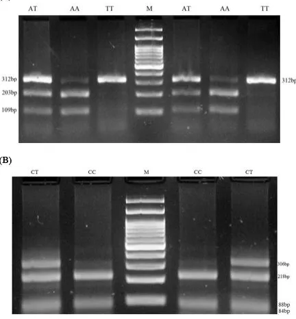
(BIORON, Germany) at 37 °C for 3 h according to the manufacturer's instructions. HinfI digestion resulted in two 109bp and 203bp fragments for AA genotype, three fragments of 312, 203 and 109bp for AT genotype, and no digested product is a 312bp for TT genotype(Fig. 1A).
Genotyping of GPX-1 rs1050450 C/T Poly-morphism
Genotyping of GPX-1 rs1050450 was done by RFLP-PCR method. Briefly, forward and reverse primers were 5´-TTATGACCGACCCCAAGCTC-3´
and 5´-GACACCCGGCACTTTATTAGTG -3, respectively
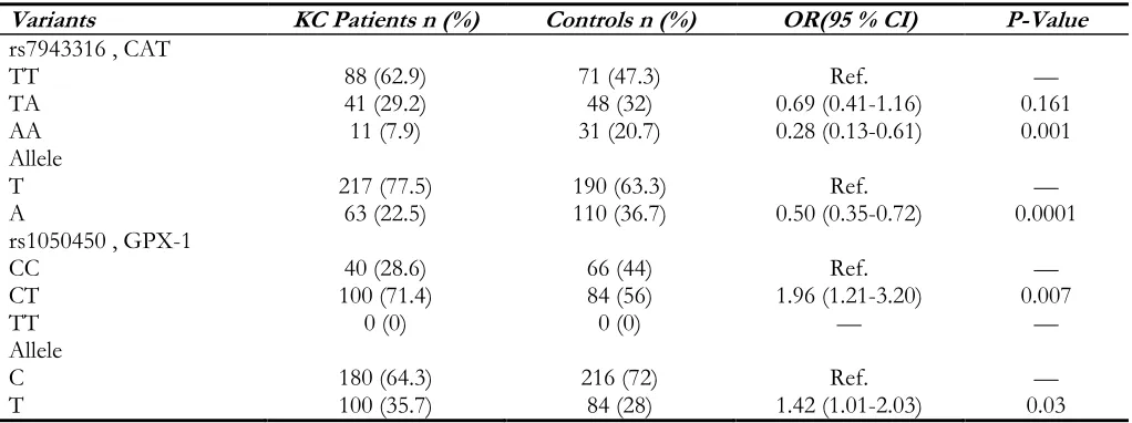
(25). The PCR conditions were justified as fol-lows: 5 min preheating at 95 °C, 30 cycles of 95 °C for 30 sec, 61 °C for 25 sec, and 72 °C for 28 sec followed by a final extension step for 5 min at 72 °C. PCR product size was 312 bp.Ten microliters of PCR product were later digested by ApaI restriction enzyme (Thermo Scientific, Lithuania) at 37 °C for 3 h. C allele produced 84, 88 and 218bp patterns, while T allele produced 84 and 306bp (Fig. 1B).
Fig. 1: Electrophoresis pattern for detection of SNP in CAT rs7943316 A/T and GPX-1 rs1050450 C/T
(A) RFLP -PCR products of CAT rs7943316 A/T, M: DNA marker (100 bp), product size (109, 203, 312bp),
Statistical analysis
The statistical analysis was performed using SPSS ver. 19.0 software (Chicago IL, USA). Compari-son of quantitative variants between two groups was assessed by Student’s t-test. The frequencies of the alleles and genotypes were analyzed using theχ2-test or Fisher’s exact test. The odds
ra-tio(OR) and 95% confidence intervals (CI) were also estimated. P-value < 0.05 was considered statistically significant.
Results
No significant differences were observed be-tween the patients with KC and healthy individu-al concerning gender and age (patients: 140, aged 10 to 80 yr and mean ± SD = 28 ± 12.5, P-value: 0.2 and controls: 150, aged 8 to 83 yr and mean ± SD = 29.9 ± 15.6, P-value: 0.2).
Table 1, containing the genotypes and allele distri-bution of CAT rs7943316 and GPX-1 rs1050450 variants in KC patients and control group. In fact, Table 1 shows the relationship between gene vari-ants (SNP) and number of patients with KC and healthy controls was tested by computing the odds ratio (OR) and 95% confidence intervals (95% CI) from logistic regression analyses.
The frequency distributions of CAT rs7943316 A/T genotypes in KC patients were: TT, 62.9%, TA, 29.2% and AA, 7.9%; and the distribution in
healthy controls were: TT, 47.3%, TA, 32%, and AA, 20.7%. This SNP was associated with KC. The AA and A allele decreased the risk of KC (OR = 0.28, 95% CI = 0.13-0.61, P-value=0.001 and OR =0.50, 95% CI =0.35-0.72, P-value =0.0001, respectively) (Table 1).
The frequencies distributions of CC and CT gen-otypes of GPX-1 rs1050450 C/T gene polymor-phism in KC patients were: 28.6% and 71.4% and in healthy controls were: 44% and56%, re-spectively. The T allele was associated with KC. Thers1050450 T allele increased the risk of KC in comparison with C allele (OR =1.42, 95% CI =1.01-2.03, P-value= 0.03) (Table 1).
The possible association between polymorphisms and clinical and pathological characteristics were analyzed. Significant association was observed between GPX-1 rs1050450 polymorphism with level of KC and cross-linking (CXL) (P-value= 0.04 and 0.04, respectively) (Table 2).
We analyzed the possible association between polymorphisms and clinical and pathological characteristics (Table 2). KC ocular showed the existence of keratoconus in a patient's eye and relation with SNPs. Level of KC represents the level of disease and evaluating these criteria with polymorphisms from patients and our findings were not associated with KC.CXL represents number of persons has cross-linking surgery and relation with SNPS.
Table 1: The genotypes and allele distribution of CAT and GPX-1 variants in keratoconus (KC) patients and control group
Variants KC Patients n (%) Controls n (%) OR(95 % CI) P-Value
rs7943316 , CAT
TT 88 (62.9) 71 (47.3) Ref. —
TA 41 (29.2) 48 (32) 0.69 (0.41-1.16) 0.161
AA 11 (7.9) 31 (20.7) 0.28 (0.13-0.61) 0.001
Allele
T 217 (77.5) 190 (63.3) Ref. —
A 63 (22.5) 110 (36.7) 0.50 (0.35-0.72) 0.0001
rs1050450 , GPX-1
CC 40 (28.6) 66 (44) Ref. —
CT 100 (71.4) 84 (56) 1.96 (1.21-3.20) 0.007
TT 0 (0) 0 (0) — —
Allele
C 180 (64.3) 216 (72) Ref. —
Table 2: Association between the CAT rs7943316 and GPX-1 rs1050450 polymorphisms and clinicopathological
characteristics
Parameters evaluated Patients n (%) (rs7943316) P-value (rs1050450) P-value
KC ocular
OD 42(30.0)
OS 36(25.7) 0.95 0.05
OU 62(44.3)
Level of KC
KK 1 33(23.6)
KK 2 45(32.1) 0.82 0.04
KK 3 62(44.3)
CXL
OD 39(27.9)
OS 40(28.6) 0.45 0.04
OU 42(30.0)
Candidate 19(13.6)
KC: keratoconus, OD (right eye), OS (left eye), OU (both eyes), CXL (cross linking surgery)
Discussion
We investigated the impact of CAT rs7943316 A/T and GPX-1 rs1050450C/T polymorphisms on KC risk in a sample of Iranian population be-cause these polymorphisms may alter the en-zymes’ antioxidant capacity, leading to synergistic effects with KC induced by oxidative damage. Our results indicated that CAT rs7943316 A/T, AA genotype, and A allele significantly decreased the risk of KC. Moreover, GPX-1 rs1050450 C/T, T allele was associated with an increased risk of KC in our population. Genetic variations in antioxidant defense may decrease antioxidant capacity or increase oxidative stress and alter the risk of KC in patients. SNPs and gene variants suggest an intricate aetiology or convergence of multiple disease pathways. Elevation of oxidative stress from SNPs in specific antioxidant enzymes maybe associated with disease (21).
Inflammation plays a crucial role in the patho-genesis of KC. Corneal thinning and distortion in KC associated with some features of inflamma-tion in ocular rosacea (26). Further evidence em-phasizes the reduced levels of antioxidant defense enzymes in KC to remove ROS associated with inflammatory reactions (27).Oxidative stress
re-sults from increased ROS or decreased levels of antioxidants (28). Ultraviolet light (UV) is a source of ROS, and excessive exposure to sun-light leads to oxidative damage to KC corneas, where there is a reduced amount of the enzymes including CAT and GPX-1 necessary to remove the ROS (29). UV-B is damaging to the cornea, and UV-A damages both the cornea and lens (30). The effects of sunlight on the eye may be acute after a latent period of several hours. UV-B can foundation homolytic fission of H2O2 to generate hydroxyl radical (31). Moreover, there is an accumulation of oxidized mitochondrial DNA in KC relative to control corneas (32).The normal cornea’s antioxidant enzymes such as CAT and GPX eliminate the ROS before they damage cells, but in the disease condition, ROS can dev-astate cellular defenses and promote cell damage (12).KC corneas do not eliminate ROS in a nor-mal way, which may play a major role in the pathogenesis of this disease(33).
2.2-fold increase of catalase mRNA level and 1.8-fold of enzyme activity. Since H2O2 induces cat-alase expression, KC corneas had high levels of this ROS, explaining some oxidative damages related with KC corneas (36).The elevated levels of oxidative stress products and decreased anti-oxidant capacity and antianti-oxidant defenses in KC corneas indicate that oxidative stress may be in-volved in the progress of this pathology (35). CAT rs7943316 and GPX-1 rs1050450 polymor-phisms alter their antioxidant capacity and associ-ated with different kinds of diseases such as Kashin-Beck (37), prostate cancer (38), peripheral neuropathy (39), diabetes mellitus (16) recurrent miscarriage (40), and brain tumors (41). In oppo-site to our findings, CAT rs7943316 genotype (TT) was associated with an increased risk of T2DM and no evidence was found to support an association between GPx-1 (198 C/T) polymor-phism and patients with T2DM (25). Allele and genotype frequencies of the CAT–21A/T and GPX1–198C/T, variations on patients with cata-ract and significant differences were not observed compare controls (41); both in terms of disease and result obtained did not establish relationship with our results.
The variability of the XRCC1, POLG and FEN1 genes in the base excision repair pathway may play a role in KC pathogenesis and increase the risk of this disease (42, 43). Patients with KC corneas that have lower paraoxonase activity there were sensitive to oxidative stress (44). The current report is the first to suggest an asso-ciation between the polymorphisms of antioxi-dant enzyme genes with KC in the Iranian popu-lation. However, the present study has some limi-tations. For example, we examined just one polymorphism from each gene associated with KC and we did not consider different ethnics.
Conclusion
CAT rs7943316 decreased the risk of KC. More-over, GPX-1 rs1050450 T allele increased the risk of KC in the study population. Further studies with different ethnicities and nationalities are re-quired to confirm our findings.
Ethical considerations
Ethical issues (Including plagiarism, informed consent, misconduct, data fabrication and/or fal-sification, double publication and/or submission, redundancy, etc.) have been completely observed by the authors.
Acknowledgements
This paper was funded as research Grant (no.6878) from the Deputy for Research, Zahedan University of Medical Sciences, Zahedan, Iran.
Conflict of interest
The authors declare that there is no conflict of interests regarding the publication of this paper.
References
1. Cuellar-Partida G, Springelkamp H, Lucas SE et al (2015). WNT10A exonic variant increases the risk of keratoconus by decreasing corneal thickness. Hum Mol Genet,24(17):5060-8. 2. Wheeler J, Hauser MA, Afshari NA et al (2012).
The Genetics of Keratoconus: A Review.
Reprod Syst Sex Disord, (Suppl 6):001.
3. Rabinowitz YS (1998). Keratoconus. Surv
Ophthalmol,42(4):297-319.
4. Kang PC, Klintworth GK, Kim T et al (2005). Trends in the indications for penetrating keratoplasty, 1980-2001. Cornea, 24(7):801-3. 5. Romero-Jimenez M, Santodomingo-Rubido J,
Wolffsohn JS (2010). Keratoconus: a review.
Cont Lens Anterior Eye,33(4):157-66.
6. Saee-Rad S, Hashemi H, Miraftab M et al (2011). Mutation analysis of VSX1 and SOD1 in Iranian patients with keratoconus. Mol
Vis,17:3128-36.
7. Stabuc-Silih M, Strazisar M, Ravnik-Glavac M et al (2010). Genetics and clinical characteristics of keratoconus. Acta Dermatovenerol Alp
Pannonica Adriat, 19(2):3-10.
8. Sugar J, Macsai MS (2012). What causes keratoconus? Cornea, 31(6):716-9.
9. Davidson AE, Hayes S, Hardcastle AJ et al (2014). The pathogenesis of keratoconus. Eye
10. Cristina Kenney M, Brown DJ (2003). The cascade hypothesis of keratoconus. Cont Lens
Anterior Eye,26(3):139-46.
11. Esterbauer H, Schaur RJ, Zollner H (1991). Chemistry and biochemistry of 4-hydroxynonenal, malonaldehyde and related aldehydes. Free Radic Biol Med,11(1):81-128. 12. Squadrito GL, Pryor WA (1998). Oxidative
chemistry of nitric oxide: the roles of superoxide, peroxynitrite, and carbon dioxide.
Free Radic Biol Med,25(4-5):392-403.
13. Kirkman HN, Gaetani GF (1984). Catalase: a tetrameric enzyme with four tightly bound molecules of NADPH. Proc Natl Acad Sci U S
A,81(14):4343-7.
14. Sabet EE, Salehi Z, Khodayari S et al (2014). Polymorphisms of glutathione peroxidase 1 (GPX1 Pro198Leu) and catalase (CAT C-262T) in women with spontaneous abortion.
Syst Biol Reprod Med, 60(5):304-7.
15. Yung LM, Leung FP, Yao X et al (2006). Reactive oxygen species in vascular wall.
Cardiovasc Hematol Disord Drug
Targets,6(1):1-19.
16. Flekac M, Skrha J, Hilgertova J et al (2008). Gene polymorphisms of superoxide dismutases and catalase in diabetes mellitus. BMC Med Genet, 9:30.
17. Vitai M, Fatrai S, Rass P et al (2005). Simple PCR heteroduplex, SSCP mutation screening methods for the detection of novel catalase mutations in Hungarian patients with type 2 diabetes mellitus. Clin Chem Lab Med,43(12):1346-50.
18. Bera S, Weinberg F, Ekoue DN et al (2014). Natural allelic variations in glutathione peroxidase-1 affect its subcellular localization and function. Cancer Res,74(18):5118-26. 19. Lubos E, Loscalzo J, Handy DE (2011).
Glutathione peroxidase-1 in health and disease: from molecular mechanisms to therapeutic opportunities. Antioxid Redox
Signal,15(7):1957-97.
20. Goldberg M, Alberts DS, Buckmeier JA et al (2011). Loss of heterozygosity at the glutathione peroxidase 1 locus is not an early event in colon carcinogenesis. Genes Cancer, 2(9):910-913.
21. Crawford A, Fassett RG, Geraghty DP et al (2012). Relationships between single
nucleotide polymorphisms of antioxidant enzymes and disease. Gene, 501(2):89-103. 22. Nemoto M, Nishimura R, Sasaki T et al (2007).
Genetic association of glutathione peroxidase-1 with coronary artery calcification in type 2 diabetes: a case control study with multi-slice computed tomography. Cardiovasc
Diabetol, 6:23.
23. Saravani R, Hasanian-Langroudi F, Validad MH et al (2015). Evaluation of possible relationship between COL4A4 gene polymorphisms and risk of keratoconus.
Cornea, 34(3):318-22.
24. Hashemi M, Yousefi J, Hashemi SM et al (2015). Association between Programmed Cell Death 6 Interacting Protein Insertion/Deletion Polymorphism and the Risk of Breast Cancer in a Sample of Iranian Population. Dis
Markers, 2015:854621.
25. Saravani S, Miri HR, Saravani R et al (2015). Association of catalase (rs7943316) and glutathione peroxidase-1 (rs1050450) polymorphisms with the risk of type 2 diabetes (T2DM). Mol Gen Mikrobiol Virusol, 30(4):216-20.
26. McMonnies CW (2015). Inflammation and keratoconus. Optom Vis Sci, 92(2):e35-41. 27. Behndig A, Karlsson K, Johansson BO et al
(2001). Superoxide dismutase isoenzymes in the normal and diseased human cornea. Invest
Ophthalmol Vis Sci, 42(10):2293-6.
28. Matough FA, Budin SB, Hamid ZA et al (2012). The role of oxidative stress and antioxidants in diabetic complications. Sultan Qaboos Univ
Med J, 12(1):5-18.
29. Gordon-Shaag A, Millodot M, Shneor E et al (2015). The Genetic and Environmental Factors for Keratoconus. Biomed Res Int, 2015:795738.
30. Mihail S (1989). [The eye and ultraviolet radiation]. Rev Chir Oncol Radiol O R L Oftalmol
Stomatol Ser Oftalmol,33(4):241-4.
31. Shoham A, Hadziahmetovic M, Dunaief JL et al (2008). Oxidative stress in diseases of the human cornea. Free Radic Biol Med,45(8):1047-55.
32. Atilano SR, Coskun P, Chwa M et al (2005). Accumulation of mitochondrial DNA damage in keratoconus corneas. Invest
33. Buddi R, Lin B, Atilano SR et al (2002). Evidence of oxidative stress in human corneal diseases. J Histochem Cytochem,50(3):341-51. 34. Gondhowiardjo TD, van Haeringen NJ (1993).
Corneal aldehyde dehydrogenase, glutathione reductase, and glutathione S-transferase in pathologic corneas. Cornea,12(4):310-4. 35. Arnal E, Peris-Martinez C, Menezo JL et al
(2011). Oxidative stress in keratoconus? Invest
Ophthalmol Vis Sci,52(12):8592-7.
36. Kenney MC, Chwa M, Atilano SR et al (2005). Increased levels of catalase and cathepsin V/L2 but decreased TIMP-1 in keratoconus corneas: evidence that oxidative stress plays a role in this disorder. Invest Ophthalmol Vis
Sci,46(3):823-32.
37. Xiong YM, Mo XY, Zou XZ et al (2010). Association study between polymorphisms in selenoprotein genes and susceptibility to Kashin-Beck disease. Osteoarthritis Cartilage,18(6):817-24.
38. Steinbrecher A, Meplan C, Hesketh J et al (2010). Effects of selenium status and polymorphisms in selenoprotein genes on prostate cancer risk in a prospective study of European men. Cancer Epidemiol Biomarkers
Prev,19(11):2958-68.
39. Tang TS, Prior SL, Li KW et al (2012). Association between the rs1050450
glutathione peroxidase-1 (C > T) gene variant and peripheral neuropathy in two independent samples of subjects with diabetes mellitus. Nutr Metab Cardiovasc Dis,22(5):417-25.
40. Khadzhieva MB, Lutcenko NN, Volodin IV et al (2014). Association of oxidative stress-related genes with idiopathic recurrent miscarriage. Free Radic Res,48(5):534-41. 41. Zhang Y, Zhang L, Sun D et al (2011). Genetic
polymorphisms of superoxide dismutases, catalase, and glutathione peroxidase in age-related cataract. Mol Vis,17:2325-32.
42. Wojcik KA, Synowiec E, Sobierajczyk K et al (2014). Polymorphism of the DNA base excision repair genes in keratoconus. Int J Mol
Sci,15(11):19682-99.
43. Wojcik KA, Synowiec E, Polakowski P et al (2014). Polymorphism of the flap endonuclease 1 gene in keratoconus and Fuchs endothelial corneal dystrophy. Int J Mol
Sci,15(8):14786-802.