1535-9778/06/$08.00⫹0 doi:10.1128/EC.00385-05
Copyright © 2006, American Society for Microbiology. All Rights Reserved.
Transcript Profiles of
Candida albicans
Cortical Actin Patch Mutants
Reflect Their Cellular Defects: Contribution of the Hog1p
and Mkc1p Signaling Pathways†
Ursula Oberholzer,
1Andre
´ Nantel,
1Judith Berman,
2and Malcolm Whiteway
1*
Biotechnology Research Institute, National Research Council of Canada, 6100 Royalmount, Montreal H4P 2R2, Quebec, Canada,1and Department of Genetics, Cell Biology & Development, University of Minnesota,
6-160 Jackson Hall, 321 Church St. SE, Minneapolis, Minnesota 554552
Received 23 December 2005/Accepted 14 June 2006
InCandida albicans, Myo5p and Sla2p are required for the polarized localization and function of cortical
actin patches, for hyphal formation, and for endocytosis. Deletion of either the MYO5 or the SLA2 gene
generated a common transcriptional response that involved changes in the transcript levels of cell wall protein-and membrane protein-encoding genes. However, these profiles were distinct from those observed for a mutant with specific deletions of the actin-organizing domains of Myo5p or for wild-type cells treated with cytochalasin A, both of which also generate defects in the organization of cortical actin patches. The profiles observed for
themyo5⌬andsla2⌬mutants had similarities to those of wild-type cells subjected to an osmotic shock, and
the defects in cortical patch function found with myo5⌬ and sla2⌬ mutants, but not cortical actin patch
distribution per se, affected sensitivity to various stresses, including heat and osmotic shocks and cell wall damage. Secondary effects coupled with defective endocytosis, such as lack of polarized lipid rafts and associated protein Rvs167-GFP (where GFP is green fluorescent protein) and lack of polarized wall remodeling
protein GFP-Gsc1, were also observed for the myo5⌬ and sla2⌬ mutants. The mitogen-activated protein
kinases Hog1p and Mkc1p, which mediate signaling in response to osmotic stress and cell wall damage, do not
play a major role in regulating the transcript level changes in themyo5⌬andsla2⌬mutants. Hog1p was not
hyperphosphorylated in the myo5⌬and sla2⌬mutants, and the transcript levels of only a subset of genes
affected in themyo5⌬mutant were dependent upon the presence of Hog1p and Mkc1p. However, it appears that
Hog1p and Mkc1p play important roles in themyo5⌬mutant cells because double deletion of myosin I and
either Hog1p or Mkc1p resulted in very-slow-growing cells.
Organisms are capable of responding to a variety of envi-ronmental stresses. In the budding yeastSaccharomyces cerevi-siaeand the related pathogenic yeastCandida albicans, activa-tion of the stress response pathways involves mechanisms for sensing a particular stress and conveying the signal via mito-gen-activated protein kinase (MAPK) modules to key effec-tors. Effectors include transcription factors that regulate the expression of specific genes in response to stress. The proteins encoded by these genes help cells repair the damage inflicted and generally increase resistance to stress (43, 48, 49). Stresses include rapid changes in external osmolarity and temperature and insults to the cell wall, to DNA, and to the actin cyto-skeleton. Some stresses have pleiotropic effects; for example, hyper- and hypoosmotic stresses cause not only ion imbalance and cell shrinkage or swelling but also a rapid although tran-sient depolarization of the actin cytoskeleton (7, 23, 26, 60). Perturbations of the actin cytoskeleton are known to arrest dividing cells until the damage is repaired (25). Repolarization of the actin cytoskeleton appears to be a critical step in the recovery response and required for cells to resume cellular division.
Mechanisms that trigger repolarization of the actin cytoskel-eton are poorly understood but may involve the very same signaling components involved in responding to stress in the first place. For example, repolarization of the actin cytoskele-ton following a hypoosmotic shock involves the cell wall sen-sors Wcs1p and Mid2p, which are both potential activators of the Pkc1p-Slt2p cell wall integrity pathway (23). Repolariza-tion of the actin cytoskeleton following a hyperosmotic shock requires the activity and proper polarized localization of the MEKKK Ssk2p of the Sln1p branch of the Hog1-dependent pathway (60). It is postulated that Ssk2p may control the ac-tivity of Bni1p and other proteins of the actin cytoskeleton to promote actin repolarization. Rvs161p, organizer of the actin cytoskeleton and component of lipid rafts, has also been shown to play a role in the repolarization of actin following a hyper-osmotic shock (7). Finally, Ras2p is necessary for actin repo-larization following a mild heat shock (26).
The actin cytoskeleton of the fungal pathogen C. albicans
plays a key role in morphogenesis and hyphal formation (1, 4, 5, 32, 41, 55, 58). In yeasts, this cytoskeleton is comprised of cortical actin patches found at sites of polarized growth and actin cables that serve as tracks for secretion of vesicles to sites of polarized growth (11, 45, 46). Components of the cortical actin patches are also required for endocytosis inS. cerevisiae
(15, 45). Indeed, cortical actin patches are the actual sites of endocytosis (29).S. cerevisiaemyosin I (Myo3/5p) and Sla2p have been shown to play important roles in organizing the * Corresponding author. Mailing address: Biotechnology Research
Institute, National Research Council of Canada, 6100 Royalmount, Montreal H4P 2R2, Quebec, Canada. Phone: (514) 496-6146. Fax: (514) 496-6213. E-mail: [email protected].
† Supplemental material for this article may be found at http://ec .asm.org/.
1252
on September 8, 2020 by guest
http://ec.asm.org/
actin cytoskeleton and mediating endocytosis by the cortical actin patches (18, 28, 33, 34). In addition,C. albicans SLA2
andMYO5are required for hyphal formation,MYO5being the unique gene encoding myosin I, hereby designated Myo5p (5, 41).
The C. albicans sla2⌬ and myo5⌬ mutants suffer similar related complications, including a disorganized actin cytoskel-eton and endocytic defects. These defects may trigger cellular responses, such as constitutive activation of the Hog1p and Pkc1p-Slt2p stress response pathways, to compensate for per-manent cortical actin patch depolarization and malfunction. Understanding the extent of these defects will shed light on the physiological role of Sla2p and Myo5p and may reveal how wild-type cells normally respond to direct perturbations of the actin cytoskeleton. In the present study, we have used genome-wide transcript profiling as a tool for analyzing complex phe-notypes, with a goal of understanding the physiological roles of
C. albicans Myo5p and Sla2p during vegetative and hyphal growth. We have found that mutations in these proteins affect endocytic-related functions in membrane and cell wall biogen-esis and play an important role in tolerance to stress. Unex-pectedly, these defects are reflected in the transcript profiles obtained, validating the use of this genome-wide approach in uncovering the physiological roles of cellular components. However, neither the Hog1p nor the Pkc1p-Slt2p pathway appears to be hyperactivated in these mutants, suggesting that these pathways do not play a central role in response to per-manent perturbations of the actin cytoskeleton inC. albicans.
MATERIALS AND METHODS
DNA manipulations.TheRVS167-GFPconstruct was made by PCR amplifi-cation using primers UO87 and UO114 and genomic DNA as a template. The 2.6-kb PCR product was cloned into pVEC as a BamHI-XbaI fragment (pU158).
The XhoI and NsiI restriction sites were added 3⬘ to the BAR sequence at
nucleotide 1076 relative to the start codon in pU158 by PCR using UO121 and UO122 (see Table S1 at http://candida.bri.nrc.ca/papers/myo/). The green fluo-rescent protein (GFP) sequence was PCR amplified with UO105 and UO127, and to generate pU165, the 700-bp PCR product was cloned as a SalI-NsiI
fragment into the 8.9-kb PCR product consisting ofRVS167in pVEC. Several
clones were verified by sequencing, and one difference was observed between our
clonedRVS167sequence and the one available in the Stanford sequence
data-base (http://www-sequence.stanford.edu/group/candida): K87T.
TheGFP-GSC1construct was made by PCR amplification using UO133 and UO134 with genomic DNA as a template. The 1.4-kb PCR product comprising
the promoter and 5⬘coding region was cloned into pBluescript SK as a SacI-XbaI
fragment (pU181). The BamHI site was removed by filling in with Klenow followed by religation (pU184). New NotI and BamHI sites were introduced right after the ATG in pU184 by PCR using UO141 and UO142. The GFP sequence was PCR amplified with UO132 and UO143, and to generate pU187, the 700-bp PCR product was cloned as a NotI-BamHI fragment into the 4.4-kb
PCR product consisting of the 5⬘ end ofGSC1in pBS. Several clones were
verified by sequencing. Finally, the 2.1-kb SacI-XbaI fragment from pU187 was subcloned into pVEC to generate pU191.
Phenotypic analyses.Strains were tested for calcofluor white (50g/ml) and
zymolyase (100g/ml) sensitivity as described by Harcus et al. (24). Cells were
also tested for heat shock resistance at 48°C for 10, 20, 30, and 60 min, as well as for osmotic sensitivity on 1.0 M NaCl, 1.5 M NaCl, and 1.2 M sorbitol yeast extract-peptone-dextrose (YPD) plates (24).
Fluorescence microscopy.Cultures of the various strains grown overnight in YPD were diluted 1:20 in YPD supplemented with 10% fetal bovine serum and grown for 1 to 2 h at 37°C. Rhodamine-phalloidin (Molecular Probes, Eugene, Oregon) and calcofluor white (Sigma) staining was done as described by Oberholzer et al. (41). Cytochalasin A was added where indicated to a final
concentration of 5M. Staining of lipid rafts with filipin III (Sigma) was done
as described previously (36), as was the procedure for visualizing FM4-64 uptake (54).
To obtain mutant and wild-type strains expressingRVS167-GFPand
GFP-GSC1, Ura⫺strains were transformed with 10g of pU165 linearized with BglII
and with an 8.4-kb fragment obtained by PCR amplification of pU191 using UO156 and UO157. Transformants were screened for the expression of Rvs167-GFP and Rvs167-GFP-Gsc1 by epifluorescence and/or Western blot analysis. Cells ex-pressing Rvs167-GFP and GFP-Gsc1 were grown to saturation in synthetic dextrose (SD)-Ura medium and diluted 1:20 in SD-Ura medium supplemented with 10% fetal bovine serum. Cells were then incubated for 90 min to 2 h at 37°C before being mounted directly on slides and visualized by epifluorescence
mi-croscopy using a Leica-DM-IRB inverted microscope with a 63⫻objective and
a 10⫻projection lens. Pictures were acquired with a Sensys
charge-coupled-device camera by use of Openlab 3.1 software.
Disruption of HOG1 and MKC1.TheHOG1andMCK1genes were deleted
in themyo5⌬and/or CAI4 backgrounds by use of PCR-amplified disruption
cassettes. For disruption ofHOG1, PCR was performed using oligonucleotides
UO186 (5⬘ATTTTAAACAAGTTATAGAAAGAAAATTTTTACAAAGATA
AAGCATATAAGAAAATGTCTGCAGATGGAGAATTTACAAGAACCG
TAAAACGACGGCCAG3⬘) and UO187 (5⬘CTTTTAAATTTATTTCTATAA
TTGCTAGCTTGTATTTTTGAAGATTAAGCTCCGTTGGCGGAATCCAA
GTTGTTTTGCTCCGGAAACAGCTATGACCATG3⬘) to amplify a 2.0-kb
fragment from pBS-URA3, a kind gift from Catherine Bachewich, and
oligonu-cleotides UO210 (5⬘TTCAAGTCGTCTTTGAAAACATACACCGTGGAATA
ATAACAACAACATTTTAAACAAGTTATAGAAAGAAAATTTTTACAA
AGATAAAGCATATAAGAAACCCGGGATCGATAGAGCT3⬘) and UO211
(5⬘ATGCTCCCATTCCCACGGGATTTAGCTCAGTGTATCTATTGGTGA
TTTCAAAAACAGTCCCAAATATCTGGGTTCTTGTAAATTCTCCATCT
GCAGACATATCCGGTAATTTAGTGTG3⬘) to amplify a 2.4-kb fragment
from pSAT-tet (47). PCR products were purified using a QIAquick PCR kit. Four PCRs were pooled, ethanol precipitated, and used for transformation of strains COU46 and CAI4. Transformants for the deletion of the first allele were selected on SD-Ura plates and screened by colony PCR using oligonucleotides
UO194 (5⬘CGGACTAGTGGCACTAAACATCAATTTCC3⬘) and UO196 (5⬘
ATAATCGCTGTGCTACTGGTGAG3⬘). Transformants for both alleles
de-leted were grown in YPD for 1 day before selection on SD-Ura plates
supple-mented with nourseothricin, 200g/ml, and screened by colony PCR using
UO194 and UO205 (5⬘CACCGAAATTTTCATGGATCC3⬘).
For disruption ofMKC1, PCR was performed using oligonucleotides UO214
(5⬘AACCTGAAACCCAAAAAAAAAAATTTTTTTTTGCTCACTACTAGT
TGTCCTTTTTAAACTTTCTCTTGAACAGCAGTTTTATAAAGAACCAA
TTTCCATAGGAAACAGCTATGACCATG3⬘) and UO187 (5⬘AAAAGGAG
GTACTAAAGGTCAATATATATAATAACCACCACTAATGGATAGACT AATTCGAGAGTAACATACCCCGGGATAACGTGGTTGTGTGTTTCAA
GTAAAACGACGGCCAGT3⬘) to amplify a 2.0-kb fragment from pBS-URA3
and oligonucleotides UO225 (5⬘AACTGAAACCCAAAAAAAAAAATTTTTT
TTTGCTCACTACTAGTTGTCCTTTTTAAACTTTCTCTTGAACAGCAGT
TTTATAAAGAACCAATTTCCATACCCGGGATCGATAGAGC3⬘) and
UO226 (5⬘ACTCCTTGACTATTTTGAATCGACTATCAATGATAAATTCC
TGGTTGTAGACCTTATTTACGGATCTGCCATAATATATGGGTGCTTC
TTGTTGATCCATATCCGGTAATTTAGTGTGTG3⬘) to amplify a 2.4-kb
fragment from pSAT-tet (47). PCR products were purified using a QIAquick PCR kit. Four PCRs were pooled, ethanol precipitated, and used for
transfor-mation of strain COU46. Themkc1⌬mutant strain was kindly provided by J. Pla`.
Transformants for the deletion of the first allele were selected on SD-Ura plates
and screened by colony PCR using oligonucleotides UO217 (5⬘CTTCACGAG
CATACACAAAATCAG3⬘) and UO196. Transformants for both alleles deleted
were grown in YPD for 1 day before selection on SD-Ura plates supplemented
with nourseothricin, 200g/ml, and screened by colony PCR using UO217 and
UO205.
All potential candidates were further tested for the correct integration events
by Southern blotting. The absence ofMKC1was confirmed by Southern blotting
in the following way. Ten micrograms of genomic DNA for each strain was digested with HindIII, separated on a 1% agarose gel, and transferred to Zeta-Probe nylon membranes (Bio-Rad). The DNA was then probed using
radioac-tively32
P-labeled PCR product amplified from genomic DNA from SC5314
using UO217 and UO228. This fragment corresponds to the 5⬘region of the
MKC1open reading frame (nucleotide⫺191 to nucleotide⫹350). In addition,
the absence of Hog1p was confirmed by Western blotting using␣-p38 antibodies
at a 1:1,000 dilution (Cell Signaling Technology, MA). Blots were also probed
with␣-actin MAB150 (Chemicon International, Temecula, CA) at a 1:1,000
dilution to control for loading.
Western blot analysis.Wild-type and mutant strains were grown in YPD to an optical density at 600 nm of 1.0 and treated with 0.5 M NaCl for 3 min. Cells were rapidly collected, washed with phosphate-buffered saline, and frozen. Whole-cell
on September 8, 2020 by guest
http://ec.asm.org/
extracts were obtained by glass bead beating the cells 10 times for 15 s each in 50 mM Tris-HCl, pH 7.5, 100 mM NaCl, 1 mM EDTA, 0.1% Triton X-100, and 25 mM NaF supplemented with protease inhibitors. Extracts were cleared by
cen-trifugation at 9,800⫻gfor 2 min, and 10l was loaded on 8% sodium dodecyl
sulfate (SDS)-polyacrylamide gels. Phosphorylation levels of Hog1 were detected by Western blotting using anti-phospho p38 (Cell Signaling Technology, MA) at a 1:1,000 dilution.
Probes, chip hybridization, and quantification.Most of the transcript profiling in this study used Cy3- and Cy5-labeled cDNA probes that were produced from
approximately 3 to 5g of poly(A)(⫹) RNA and hybridized, as described
previously, to microarrays spotted with amplicons from 6,002 putative open reading frames (39). For each condition, at least three independent experiments with reciprocal labeling were done, for a total of six individual hybridizations,
unless otherwise stated. Transcript profiles of themyo5⌬vs. wild type (Wt),
mkc1⌬vs. Wt, andmyo5 mkc1⌬vs.myo5⌬mutants, described in the latter part
of this report, were produced with Cy3- and Cy5-labeled cDNA probes prepared
from 40g of total RNA and hybridized to a new generation of long
oligonu-cleotide microarrays. These were spotted with 6,263 70-mer oligonuoligonu-cleotides that
are specific for genes recently identified as part of a reannotation of theC.
albicansgenome (10). More details about these microarrays are available on our web page at http://www.bri.nrc.gc.ca/services/microarray/scanning_e .html. The latter profiling data are the result of four hybridizations of inde-pendently produced RNA preparations.
All microarrays were washed in 1⫻SSC (1⫻SSC is 0.15 M NaCl plus 0.015 M
sodium citrate) and 0.2% SDS at 42°C followed by two washes in 0.1⫻SSC and
0.2% SDS at 42°C and finally three quick consecutive washes in 0.1⫻SSC. Chips
were air dried before being scanned with a ScanArray Lite microarray scanner (Packard Bioscience). QuantArray was used to quantify fluorescence intensities, and Lowess normalization and statistical analysis were performed using Gene-spring v.7 (Agilent Technologies, CA). The microarray data produced in this study are available on our web page at http://candida.bri.nrc.ca/papers/myo/. The data can also be found in the supplemental material on the Eukaryotic Cell web page (http://ec.asm.org/).
Northern blot analysis.Total RNA was extracted with phenol and glass beads from wild-type and myosin I deletion strains grown to early log phase in YPD at an optical density at 600 nm of 0.8. Twenty micrograms of total RNA per sample was separated on a 7.5% formaldehyde, 1% agarose gel, blotted onto Zeta-Probe
nylon membrane (Bio-Rad, Ontario, Canada), and probed with32P-labeled
DNA specific forACT1 (orf19.5007),orf19.5302, CRH1(orf19.2706), AGP2
(orf19.4679), orf19.7296, SOD5 (orf19.2060), EBP1 (orf19.125), PHR1
(orf19.3829), andTOS2(orf19.1911) as described previously (42). All Northern probes were PCR products subsequently labeled by random priming (Amersham
Biosciences, NJ). Oligonucleotides used were UO106 and UO107 (ACT1),
UO238 and UO239 (orf19.5302), UO240 and UO241 (orf19.7296), UO242
and UO243 (EBP1), UO244 and UO245 (CRH1), UO246 and UO247 (PHR1),
UO248 and UO249 (AGP2), and UO252 and UO253 (TOS2) (see Table S1 at
http://candida.bri.nrc.ca/papers/myo/). TheSOD5PCR product was kindly
pro-vided by M. Martchenko.
RESULTS
myo5⌬andsla2⌬mutants display endocytic defects.InC.
albicans, perturbations in the actin cytoskeleton are coupled to defects in hyphal development. For example, myosin I function is important for proper cortical actin patch distribution and for endocytosis and is critical for the formation of true hyphae (41, 42). The sla2⌬ mutant also exhibits defects in both hyphal formation and the organization of cortical actin patches (5) (see Fig. S1A at http://candida.bri.nrc.ca/papers/myo/). Chem-ical treatments that disrupt the actin cytoskeleton, such as cytochalasin A treatment, can also modify hyphal development and cortical actin patch distribution (1) (see Fig. S1A at http: //candida.bri.nrc.ca/papers/myo/). Correlating with these actin cytoskeleton defects, themyo5⌬and sla2⌬mutations, as well as cytochalasin A treatment, also negatively affect fluid-phase endocytosis (see Fig. S1B at http://candida.bri.nrc.ca/papers /myo/).
However, not all defects in cortical actin distribution cause
such pleiotropic effects. Deletion of the Src homology 3 (SH3) and A domains of myosin I (the⌬SH3⌬A mutant) affects the organization of the actin cytoskeleton but neither fluid-phase endocytosis (see Fig. S1A and S1B at http://candida.bri.nrc.ca /papers/myo/) nor hyphal development. The⌬SH3⌬A mutant is particularly interesting because it is able to form apparently wild-type hyphae despite the highly depolarized distribution of cortical actin patches.
Clustering of themyo5⌬andsla2⌬transcript profiles.
Un-like theirS. cerevisiaecounterparts, themyo5⌬andsla2⌬ mu-tants ofC. albicans are viable and exhibit only slight growth defects, indicating that theMYO5andSLA2genes do not play an essential role inC. albicans. Transcript profiles ofmyo5⌬
and sla2⌬ mutant cells grown under yeast (myo5⌬-Y and
sla2⌬-Y, respectively)- or hypha-inducing conditions were ob-tained using whole-genome DNA microarrays and compared to transcript profiles of the wild type to gain insight into the physiological roles of Myo5p and Sla2p during vegetative and hyphal growth. Transcript profiles of ⌬SH3⌬A mutant cells grown under yeast and hyphal growth conditions as well as profiles of wild-type yeast and hyphal cells treated for 10 and 30 min with 5M cytochalasin A were also obtained. Overall, transcript profiling was used as a tool to assess the similarities and differences among the different conditions and mutants.
Some 2,500 genes for which the transcript levels vary signif-icantly (P ⬍ 0.05) under at least one condition tested were selected for further analysis. Principal component analysis (PCA) and hierarchical clustering show that, of all the condi-tions, the myo5⌬ and sla2⌬ transcriptional profiles of cells grown as yeast or hyphae are most similar (Fig. 1) (also see Fig. S2 at http://candida.bri.nrc.ca/papers/myo/). In contrast, treat-ment of cells with cytochalasin A or deletion of the SH3 and A regions of myosin I produce transcript profiles that do not cluster with each other or with any of the other profiles ob-tained. Thus, the common modified profiles represent those of themyo5⌬andsla2⌬endocytosis-defective mutants.
Even though themyo5⌬andsla2⌬mutants are defective in hyphal formation, the yeast-to-hypha transcript profiles of wild-type and mutant strains clustered together on the PCA graph (Fig. 1) because the changes in the yeast-to-hypha tran-script profiles are only minimally altered in the myo5⌬ and
sla2⌬ hyphal-formation-defective mutants. Thus, it appears that the physical state of the cells (pseudohyphae) does not preclude expression of hyphal-specific genes. Apparently the signal transduction pathways relaying the hypha-inducing en-vironment to the transcriptional machinery were unaffected by the altered state of themyo5⌬andsla2⌬mutant cells. How-ever, some hyphal-specific genes are not induced to the same level in themyo5⌬mutant as in the wild type under conditions that normally induce hyphae; these genes include ECE1,
GPX1, andYHB1 (data not shown). As well, the hyphal-spe-cific RNR3 gene was repressed in the sla2⌬ mutants grown under hypha-inducing conditions (data not shown).
We observed a significant number of genes commonly af-fected in strains deleted forSLA2orMYO5by comparing the fluorescence ratios of significantly modulated transcripts in the
myo5⌬andsla2⌬mutants relative to that of the wild type (Fig. 2) (P⬍1e-86). Importantly, the changes (n-fold) of commonly modulated genes and even those that passed the significance test (P⬍0.05) in one mutant but not the other were similar in
on September 8, 2020 by guest
http://ec.asm.org/
both mutants relative to the wild type. Many transcript levels that were increased in common also correspond to genes in-duced when wild-type cells are stressed osmotically (32%;P⬍
1e-15) (16) (Table 1). Among common transcripts with levels in the mutants less than 0.6⫻or greater than 1.4⫻the levels in the wild type, many encode cell wall components (12%), mem-brane proteins involved in protein sorting, trafficking, trans-port, and other functions (11%), and proteins involved in membrane biogenesis (6%) (Table 2). As well, many of these transcripts were stress-induced genes and genes involved in protein folding and degradation (9%). Table S2 at http: //candida.bri.nrc.ca/papers/myo/ shows transcript levels for genes that were significantly modulated in themyo5⌬mutant in two different types of DNA microarrays that were spotted with either PCR amplicons or 70-mer oligonucleotides, with the addition of some genes that appeared to be significant on only one type of chip, as indicated (see below). Importantly, none of these genes were modulated in control experiments comparing transcript profiles of MYO5 and SLA2 revertant strains with that of wild-type SC5314. The only exception is
IRO1, which is found adjacent to the URA3 gene that was deleted in the making of CAI4 (19). Accordingly, the transcript levels ofIRO1are lower in all of the CAI4-derived strains than in SC5314 (data not shown). Table S1 at http://candida.bri.nrc .ca/papers/myo/ also highlights the similar transcript patterns ofmyo5⌬andsla2⌬under yeast growth conditions (myo5⌬-Y andsla2⌬-Y) and especially those ofmyo5⌬-Y andsla2⌬under hyphal growth conditions.
Genes involved in membrane biogenesis and function are
affected in myo5⌬ and sla2⌬ mutants. Transcript levels of
several genes involved in membrane biogenesis, including FIG. 1. PCA of the transcript profiles. This data reduction
algo-rithm organizes the experimental conditions in a three-dimensional space based on the similarity between their respective transcrip-tional profiles. The conditions include comparingmyo5⌬,sla2⌬, and ⌬SH3⌬A mutants against Wt cells under yeast (Y) or hyphal (H) growth conditions, comparing Wt and mutant strains grown under hyphal growth conditions to the same strains grown under yeast growth conditions (HY), treatment of Wt yeast cells with cytochalasin A for 10 or 30 min (cyt 10 and cyt 30, respectively), or treatment of hyphal cells with cytochalasin A for 30 min (cyt 30 H). White ovals denote clus-tering of conditions that are discussed in the text.
FIG. 2. Similarity between the transcript profiles of themyo5⌬andsla2⌬mutants. Shown is a scatter plot of the average fluorescence ratios observed under yeast growth conditions formyo5⌬versussla2⌬. Transcripts with a statistically significant change in abundance (ttest,P⬍0.05, coupled to the Benjamini and Hochberg false discovery rate) are colored as indicated in the Venn diagram (red,myo5⌬transcripts; green,sla2⌬
transcripts; yellow, overlap). The numbers in the Venn diagram indicate numbers of genes.
on September 8, 2020 by guest
http://ec.asm.org/
CHO1and SFK1 (membrane biogenesis),RTA2, RTA3, and
RTA4(putative flippases), andPLC2,PLC3, andPLB4 (phos-pholipases), were significantly modulated in both mutants (see Table S2 at http://candida.bri.nrc.ca/papers/myo/). Phospho-lipases and Sfk1p play an important role in intracellular sig-naling (6, 56). It is intriguing that three independent flippase genes are overexpressed in the mutants. Flippases are known to maintain an asymmetric distribution of sphingolipids in the extracellular leaflet of the lipid bilayer and may therefore play an important role in regulating endocytosis (31).
Because lipid rafts are membranous structures that may anchor and localize proteins in a polarized manner duringC. albicanshyphal morphogenesis (36, 38), we assessed whether the endocytic mutant strains exhibited defects in lipid raft formation. We visualized the wild-type and mutant strains grown under hypha-inducing conditions stained with filipin III
for ergosterol, a component of lipid rafts. The hyphal tips of wild-type and⌬SH3⌬A cells were heavily stained with filipin III, indicating that lipid rafts are polarized (Fig. 3). In contrast, the endocyticmyo5⌬andsla2⌬mutants clearly lacked polar-ized lipid rafts under these conditions.
InS. cerevisiae, Rvs167p is a cortical actin patch component that interacts through its SH3 domain with multiple proteins, including Las17p/Bee1p (8, 52), which recently was found to be associated with lipid rafts (22).C. albicans RVS167was fused withGFP, immediately 3⬘ of the BAR sequence, and intro-TABLE 1. Comparison of gene lists obtained under various conditionsa
Condition
No. (%) ofmyo5⌬
mutant transcripts
inducedb
Pvalue
No. (%) ofmyo5⌬-Y and
sla2⌬-Y mutant
transcripts inducedc
Pvalue
No. (%) of Cyt
Ad-treated gene
transcripts induced
Pvalue
No. (%) of⌬SH3⌬A
mutant transcripts induced
Osmotic stress 47 (33) 1.28e-18 45 (32) 2.85e-16 8 (15) 4 (14)
Oxidative stress 13 (9) 1.64e-4 11 (8) 1.35e-3 20 (36) 6.87e-8 5 (17)
Heat shock 13 (9) 8.17e-6 13 (9) 13 (24) 1.49e-4 3 (10)
Totale 143 141 55 29
a
Only lists of genes for which transcript levels were greater than 1.3 andPvalues were⬍0.05 under each of the conditions were compared.
b
Commonly identified transcripts with higher levels in themyo5⌬mutant, obtained using amplicon and oligonucleotide chips.
c
Commonly identified transcripts with higher levels in themyo5⌬-Y andsla2⌬-Y mutants.
d
Cyt A, cytochalasin A.
e
Total number of induced transcripts for each condition.
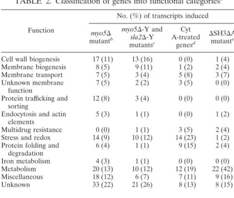
TABLE 2. Classification of genes into functional categoriesa
Function
No. (%) of transcripts induced
myo5⌬
mutantb
myo5⌬-Y and
sla2⌬-Y
mutantsc
Cyt A-treated
genesd
⌬SH3⌬A
mutante
Cell wall biogenesis 17 (11) 13 (16) 0 (0) 1 (4)
Membrane biogenesis 8 (5) 9 (11) 1 (2) 2 (4)
Membrane transport 7 (5) 3 (4) 5 (8) 3 (7)
Unknown membrane function
7 (5) 2 (2) 3 (5) 0 (0)
Protein trafficking and sorting
12 (8) 3 (4) 0 (0) 0 (0)
Endocytosis and actin elements
5 (3) 1 (1) 0 (0) 1 (2)
Multidrug resistance 0 (0) 1 (1) 3 (5) 2 (4)
Stress and redox 14 (9) 10 (12) 14 (23) 1 (2)
Protein folding and degradation
6 (4) 1 (1) 9 (15) 2 (4)
Iron metabolism 4 (3) 1 (1) 0 (0) 0 (0)
Metabolism 20 (13) 10 (12) 12 (19) 22 (42)
Miscellaneous 18 (12) 6 (7) 7 (11) 9 (16)
Unknown 33 (22) 21 (26) 8 (13) 8 (15)
Total 151 81 62 51
aTable 2 is based on data presented in Tables S1, S2, and S3 at http://candida
.bri.nrc.ca/papers/myo/.
bCommonly identified transcripts in themyo5⌬mutant, obtained using
am-plicon and oligonucleotide microarrays.
cCommonly identified transcripts in themyo5⌬-Y andsla2⌬-Y mutants.
dTranscripts unique to cytochalasin A (Cyt A)-treated genes and not
identi-fied in themyo5⌬andsla2⌬mutants.
eTranscripts unique to the⌬SH3⌬A mutant and not identified in themyo5⌬
andsla2⌬mutants; these may also have been identified under cytochalasin A
treatment conditions.
FIG. 3. The myo5⌬and sla2⌬ mutants do not exhibit polarized lipid rafts. The wild-type and mutant cells grown under hypha-inducing conditions were stained with filipin III and visualized by epifluores-cence microscopy. Bar⫽5m.
on September 8, 2020 by guest
http://ec.asm.org/
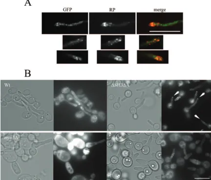
duced into wild-type and mutant strains. The Rvs167p-GFP levels expressed in these strains were examined by immunoblot analysis (data not shown) and localized by epifluorescence microscopy. Rvs167p-GFP localized in patches at hyphal tips of wild-type cells, and these Rvs167p-GFP patches partially overlapped with cortical actin patches (Fig. 4A). Non-actin-overlapping Rvs167p-GFP patches may colocalize with lipid rafts. In the hyphal-formation-defectivemyo5⌬andsla2⌬ mu-tants, Rvs167-GFP was mainly cytoplasmic, with some patches visible in the sla2⌬ mutant (Fig. 4B). However, a distinct Rvs167p-GFP signal could be observed at hyphal tips of the
⌬SH3⌬A mutant cells (Fig. 4B). The majority of these mutant hyphae had Rvs167p-GFP signal at the tip (77% [n⫽151] of hyphal cells of the⌬SH3⌬A mutant, compared to 80% [n ⫽
97] in the wild type). It is possible that Rvs167p-GFP localiza-tion at hyphal tips corresponds to polarized lipid rafts.
Genes involved in cell wall remodeling are affected inmyo5⌬
andsla2⌬mutants.Transcript levels of several genes encoding
cell wall remodeling enzymes were elevated in themyo5⌬and
sla2⌬mutants. These included genes involved in chitin
synthe-sis (CHS2andCHS7), in-1,6-glucan assembly (KRE1,KRE6,
KRE7, KRE9, and KRE13), and in -1,3-glucan assembly (orf19.7214,BGL2,EXG1, andXOG1). In addition, transcript levels of several cell wall proteins that may have a role in cell wall assembly, such as glycosylphosphatidylinositol-anchored proteins (PHR1andDCW2), cell wall mannoproteinCCW14, and others (CRH1andECM4), were increased in themyo5⌬
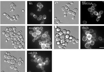
and sla2⌬ mutants. In contrast, CHT2 and CHT3 transcript levels were decreased in the myo5⌬ mutant. This changed transcript pattern may be linked to thickening of the cell wall observed as an aberrant calcofluor staining of the cell wall of the myo5⌬ and sla2⌬ mutants but not of the wild-type and
⌬SH3⌬A strains (Fig. 5) (41). We also assessed cell wall de-fects by monitoring resistance to 50 g/ml calcofluor and 0.05% SDS (see below). Consistent with a cell wall defect, we found that themyo5⌬andsla2⌬mutants were more sensitive to calcofluor and SDS than either the wild-type or the
⌬SH3⌬A strain (Fig. 6).
Elevated levels of glucan and chitin synthases may cause a thickening of the cell wall in themyo5⌬andsla2⌬mutants. In FIG. 4. Rvs167-GFP does not localize in a polarized manner in themyo5⌬andsla2⌬mutants. (A) Partial colocalization of Rvs167p-GFP with cortical actin patches in wild-type hyphal tips. Hyphal cells expressing Rvs167p-GFP were fixed, stained with rhodamine-phalloidin (RP), and visualized by epifluorescence microscopy. (B) Wild-type and mutant hyphal forms expressing Rvs167p-GFP were visualized by epifluorescence microscopy. Arrowheads point to mutant hyphal tips showing Rvs167-GFP signal. Bar⫽10m.
on September 8, 2020 by guest
http://ec.asm.org/
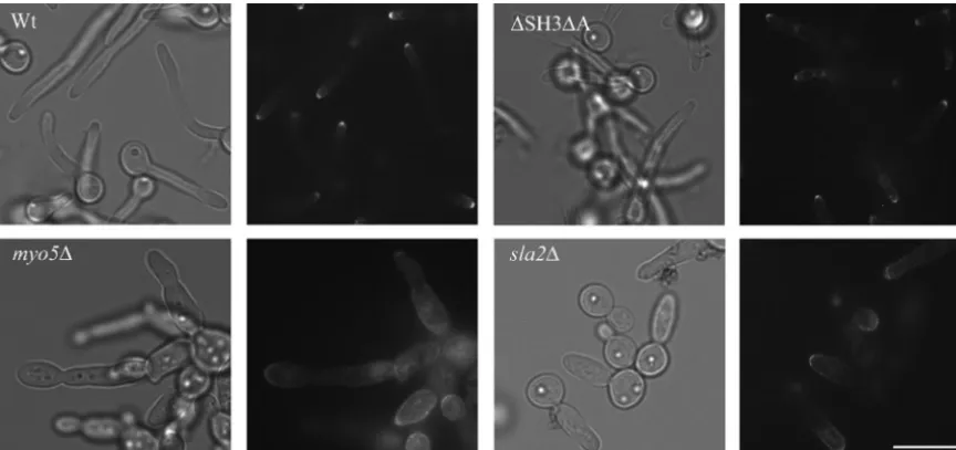
addition, a thicker cell wall could arise because of inefficient recycling of cell wall synthesizing proteins at sites of polarized growth, resulting in uniform localization around the cell wall. To assess such a defect, we introduced GFP-tagged  -1,3-glucan synthase Gsc1p (37) and visualized its localization in all strains. We found that GFP-Gsc1p was polarized to the hyphal tips of wild-type and⌬SH3⌬A cells (Fig. 7). In contrast, the GFP-Gsc1p signal was more diffuse in thesla2⌬mutants and greatly dispersed around the cell periphery in themyo5⌬ mu-tant. Thus, these results show that these mutants exhibit sec-ondary defects in cell wall assembly, possibly as a consequence of defective endocytosis and elevated transcript levels of cell wall remodeling genes.
The myo5⌬ and sla2⌬ mutants are more stress sensitive
than the wild-type and⌬SH3⌬A strains.Analysis of the
tran-script profiles showed that themyo5⌬ andsla2⌬mutants ex-pressed high transcript levels for stress genes such asDDR48,
HSP12, and SOD5, suggesting that the mutants are stressed. We determined whether these mutants had modified stress responses by performing spot test assays on media containing different salts and 0.05% SDS, on 50g/ml calcofluor-contain-ing plates, or followcalcofluor-contain-ing heat shock treatment of differcalcofluor-contain-ing times at 48°C (Fig. 6). We observed that themyo5⌬andsla2⌬null mutants exhibited increased salt, SDS, calcofluor, and heat shock sensitivities compared to the wild-type and ⌬SH3⌬A strains, suggesting that the form of constitutive stress they FIG. 5. Aberrant chitin depositions in the cell walls ofmyo5⌬andsla2⌬mutant cells. Wild-type and mutant cells were fixed, stained with calcofluor white, and visualized by epifluorescence microscopy. Bar⫽5m.
FIG. 6. Themyo5⌬andsla2⌬mutants exhibit increased sensitivity to various stresses. Tenfold dilutions of overnight cultures of wild-type and mutant strains were spotted on various solid media as indicated and grown for 3 days at 30°C.
on September 8, 2020 by guest
http://ec.asm.org/
experience does not cross-protect them against additional exo-genous stress. In addition, these results suggest that misorga-nization of the cortical actin cytoskeleton per se, as in the
⌬SH3⌬A mutant, does not disturb resistance to stress.
Transcript profiles for the⌬SH3⌬A mutant and wild-type
cells exposed to cytochalasin A treatment were different from
those of themyo5⌬andsla2⌬mutants.The transcript profiles
of wild-type cells treated with cytochalasin A, a drug that inhibits polymerization of actin filaments, or of cells expressing a mutant form of Myo5p (⌬SH3⌬A mutant) were different both from those of themyo5⌬andsla2⌬null mutants and from each other (Fig. 1) (compare transcript levels for these condi-tions in Table S2 at http://candida.bri.nrc.ca/papers/myo/ with those ofmyo5⌬-Y). Importantly, 36% of genes uniquely up-regulated by cytochalasin A treatment were oxidative stress genes (Table 1) (16). Among the genes up- or downregulated by cytochalasin A, 5% encode multidrug resistance pumps, 23% encode stress genes, and 15% encode protein folding and degradation genes (Table 2). More specifically, cytochalasin A-treated cells had elevated transcript levels for genes encod-ing multidrug resistance pumps Mdr1p, Cdr1p, and Cdr2p and stress proteins Hsp70p, Sba1p, and Grp3p (see Table S3 at http://candida.bri.nrc.ca/papers/myo/). Of interest, we noted that the transcript levels for the Cap1p transcription factor in-volved in the oxidative stress response (17, 61) were up by two-fold. Transcript levels for a number of genes encoding oxi-doreductases and dehydrogenases, including OYE2, OYE32,
GPX2,EBP1, and putative dehydrogenase orf19.3139, were up accordingly in these cells. Transcript profiling of wild-type cells treated with cytochalasin A for 150 min shows that most elevated transcript levels at the 30-min condition returned to basal levels (data not shown). This indicates that even longer treatment of wild-type cells with cytochalasin A, which disorganizes the actin cytoskeleton and inhibits endocytosis more dramatically than a 30-min treatment, does not mimic an endocytosis defect gener-ated by deletion of cortical actin patch components.
The ⌬SH3⌬A mutant had elevated or reduced transcript
levels for some genes commonly modulated in the myosin I null mutant. However, the transcript levels for common genes were clearly reduced overall and the number of genes uniquely modulated in this mutant was limited to a few (see Table S3 at http://candida.bri.nrc.ca/papers/myo/). These observations, to-gether with the actin patterns observed for these cells, suggest that misorganization of the cortical actin patches alone is not sufficient to produce the specific transcriptional response ob-served in the case of thesla2⌬andmyo5⌬endocytic mutants.
Phosphorylation of Hog1 is not involved in regulating
tran-script levels in themyo5⌬andsla2⌬mutants.The induction of
genes by osmotic stress (and general stress) in themyo5⌬and
sla2⌬mutants suggests that the osmotic stress response path-way is activated in these mutants (Table 1). In fact, there is considerable overlap between the lists of significantly modu-lated transcripts in myo5⌬ orsla2⌬ cells and wild-type cells treated with an osmotic shock (P ⬍ 1e-30 and P ⬍ 1e-15, respectively). TheC. albicans Hog1 protein kinase is part of the p38 family and mediates signaling in response to a wide variety of stresses, including high salt and H2O2 concentra-tions, but not in response to heat shock (50). InS. cerevisiae, Hog1p mediates signaling in response to hyperosmotic stress. Hog1p receives signals from two independent branches that sense hyperosmotic changes in the environment. Activation of either the Sho1p-Ste11p or the Sln1p-Ssk1p branch or both leads to phosphorylation and activation of scaffold protein kinase Pbs2p, followed by phosphorylation, activation, and nu-clear translocation of Hog1p (43).C. albicansHog1p is simi-larly phosphorylated and translocated to the nucleus as part of the response (50). The long-term adaptation to hyperosmotic stress involves a change in transcriptional expression mediated by Hog1p phosphorylation of several transcription factors, in-cluding Sko1p, Hot1p, and Smp1p (2, 3, 12, 44).
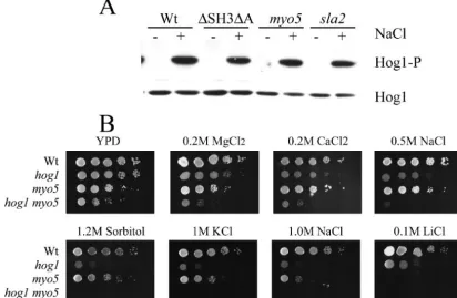
We therefore determined whether Hog1p is constitutively phosphorylated in themyo5⌬andsla2⌬mutants in the absence of salt stress. Figure 8A shows that Hog1p became phosphor-ylated in wild-type cells exposed to 0.5 M NaCl for 3 min but FIG. 7. Depolarized localization of GFP-Gsc1p in cell walls ofmyo5⌬andsla2⌬mutants. Wild-type and mutant cells grown under hypha-inducing conditions expressing GFP-Gsc1p were visualized by epifluorescence microscopy. Bar⫽10m.
on September 8, 2020 by guest
http://ec.asm.org/
that Hog1p phosphorylation was not detected in the myo5⌬
andsla2⌬mutants grown in YPD, suggesting that Hog1p is not constitutively activated in these mutants. Moreover, the salt sensitivity observed with themyo5⌬andsla2⌬mutants cannot be due to improper activation of the high-osmolarity pathway because phosphorylation of Hog1p occurred normally in these mutants when exposed to salt and because the salt sensitivities of thehog1⌬andmyo5⌬mutants were different as determined by spot test assays (Fig. 8B).
To further address the requirement for the high-osmolarity stress response MAPK pathway in the transcriptional response of themyo5⌬mutant, we deleted theHOG1gene in themyo5⌬
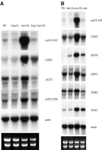
mutant background. Surprisingly, themyo5 hog1⌬double mu-tant grew poorly compared to single mumu-tants and the wild type, suggesting that Hog1p is required when Myo5p is absent (Fig. 9A). Examination ofmyo5 hog1⌬cells revealed that many were abnormally enlarged and formed pseudohyphae despite being cultured in non-hypha-inducing conditions (data not shown). Transcript profiling of themyo5 hog1⌬ double mutant com-pared to themyo5⌬single mutant revealed that the induction of 12 genes in themyo5⌬mutant was dependent on the pres-ence of Hog1. These genes include AGP2, CRH1, SOU1,
orf19.5302, andorf19.7296 (see Table S2 at http://candida.bri .nrc.ca/papers/myo/). Northern blot analyses confirmed that elevated transcript levels for AGP2, CRH1, orf19.5302, and
orf19.7296in themyo5⌬mutant are dependent on Hog1p (Fig. 10, left panel).
The transcript levels of a distinct set of genes affected by the
deletion ofMYO5are dependent on the Mkc1 protein kinase.
As described above, the transcript levels of genes encoding cell wall organization and biogenesis proteins are affected by de-letion of MYO5 orSLA2. The transcript levels for many of these are induced whenS. cerevisiaewild-type cells are exposed to chemical challenges to the cell wall (9, 20). These observa-tions, together with the observed sensitivity of themyo5⌬and
sla2⌬mutants to SDS and calcofluor, suggest that the cell wall integrity pathway is differentially modulated in these mutants. The elevatedPLC2andPLC3transcript levels and repressed
PLB4transcript levels observed for these mutants also suggest that intracellular signaling via the phosphoinositol secondary messenger may be activated. In mammalian cells this typically leads to the activation of Pkc1p, although this has not been demonstrated to occur in yeast (56). Activation of the Pkc1p cell wall integrity pathway in S. cerevisiaeinvolves the phos-phorylation of the MAPK Slt2p (13, 35).
To address the requirement of the Pkc1p cell wall integrity pathway for the mRNA transcript profiles observed for the
myo5⌬andsla2⌬mutants, we deleted theC. albicans homo-logue for SLT2, MKC1, in the myo5⌬ mutant. The double mutant had a clear growth defect (Fig. 9B). For the data generated in this section, we used newly designed long oligo-nucleotide microarrays (see Materials and Methods). The tran-script profiles for themyo5⌬mutant and the wild type under yeast growth conditions generated with the cDNA and oligo-FIG. 8. (A) Hog1p is not phosphorylated in themyo5⌬andsla2⌬mutants grown in YPD but becomes phosphorylated when cells are treated with NaCl. Phospho-Hog1p was detected by Western blot analysis in wild-type and mutant strains grown in YPD (⫺) and stressed with 0.5 M NaCl for 3 min (⫹), using anti-phospho p38 antibodies. Hog1p protein levels were also detected using anti-p38 antibodies. (B) Thehog1⌬andmyo5⌬
strains exhibit different salt sensitivities. Tenfold dilutions of overnight cultures of wild-type,hog1⌬,myo5⌬, andhog1 myo5⌬strains were spotted on solid medium containing various salts.
on September 8, 2020 by guest
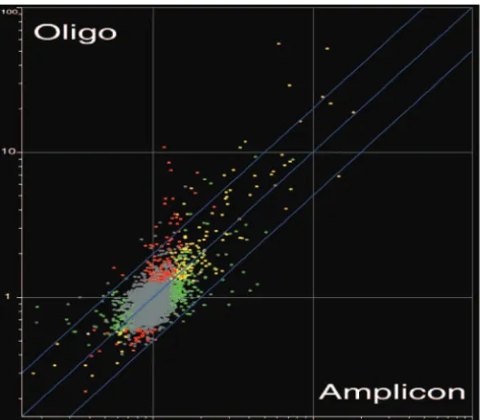
http://ec.asm.org/
nucleotide chips were compared (Fig. 11) (also see Table S4 at http://candida.bri.nrc.ca/papers/myo/). The profiles were markably similar, suggesting that the data sets are highly re-producible (P⬍0.05). Analysis of the transcript profile of the
myo5 mkc1⌬double mutant compared to that of the myo5⌬
single mutant showed 21 genes whose elevated transcript levels inmyo5⌬were dependent on the presence of MKC1. These included genes encoding cell wall proteins (seven genes), detoxification proteins (three genes: EBP1, SOD5, and
orf19.251), and unknown proteins (six genes, including
orf19.5302). We confirmed by Northern blot analysis that the transcript levels of six genes (orf19.5302,CRH1,EBP1,SOD5,
TOS2, and PHR1) were entirely or partially dependent on
MKC1(Fig. 10, right panel).TOS2(targetofSBF-2) transcript levels in themyo5 mkc1⌬double mutant were examined de-spite the absence of chip data because SBF is a known target of the Mkc1p protein kinase. Surprisingly, very few
homo-logues of these genes are modulated whenS. cerevisiaeis chal-lenged with calcofluor white or zymolyase, causing cell wall perturbations known to activate the Pkc1p-Slt2p signaling pathway (9, 20).
The transcript levels of a significant number of genes were upregulated in both themyo5⌬and themkc1⌬strain but were not increased in the myo5 mkc1⌬double mutant, suggesting that a common pathway can be activated independently in both single mutants. These included genes whose transcript levels were dramatically upregulated in themyo5⌬andsla2⌬single mutants, such asHSP12,RNH2,PRY4,RTA2,PLC2/3,DDR48,
XOG1, andPST.
DISCUSSION
In this study, we undertook a genome-wide analysis of the transcript profiles of theC. albicans myo5⌬andsla2⌬mutants FIG. 9. Growth defect ofhog1 myo5⌬andmkc1 myo5⌬double mutants compared to growth of single mutants. (A) The left panel shows the absence of Hog1p in thehog1⌬andhog1 myo5⌬strains by Western blotting using anti-p38 antibodies. The right panel shows colonies of the corresponding strains grown for 3 days on YPD at 30°C. (B) The left panel shows the absence of theMKC1gene in themkc1⌬andmkc1 myo5⌬strains by Southern blotting (see Materials and Methods). The right panel shows colonies of the corresponding strains grown for 3 days on YPD at 30°C.
on September 8, 2020 by guest
http://ec.asm.org/
compared to that of the wild type under conditions promoting budding and hyphal growth to understand the physiological roles of the Myo5p and Sla2p proteins. Myo5p and Sla2p are involved in cortical actin patch function, and deletion of either gene caused very similar transcript profiles that included a dramatic and unexpected upregulation of stress-related genes and genes involved in cell wall remodeling and membrane biogenesis. This is the first time that an altered transcript profile has been observed for cortical actin skeleton mutants,
highlighting the complex adaptive response of mutants to their respective genetic defects. This altered transcript profile ob-served for the myo5⌬ and sla2⌬ mutants may be part of a response to compensate for endocytic-related defects, support-ing the idea that endocytosis is critical for appropriate and timely expression of membrane and cell wall proteins during polarized growth. We interpret the cellular localization and distribution of the GFP-tagged glucan synthase (GFP-Gsc1p) in themyo5⌬andsla2⌬mutants, as discussed by Pruyne and FIG. 10. The Hog1p and Mkc1p kinases are required for the elevated transcript levels of selected genes in themyo5⌬mutant strain. (Left panel) Northern blot analysis showing transcript levels fororf19.5302,CRH1,AGP2,orf19.7296, andACT1in the strains shown at the top. (Right panel) Northern blot analysis showing transcript levels fororf19.5302,CRH1,SOD5,EBP1,PHR1,TOS2, andACT1in the strains shown at the top. mm,mkc1 myo5⌬double mutant.
on September 8, 2020 by guest
http://ec.asm.org/
Bretscher (45), to result from inadequate recycling of cell wall remodeling proteins, due to defective fluid-phase endocytosis. Similarly, defective fluid-phase endocytosis could result in the absence of polarized lipid rafts at hyphal tips and of raft-associated proteins such as Rvs167p-GFP.
Surprisingly, we found that disruption of the actin cytoskel-eton with cytochalasin A or by deleting actin-organizing do-mains of Myo5p does not trigger a similar transcriptional response. Although transcript levels of some of the stress-induced genes were elevated in the⌬SH3⌬A mutant, the in-crease (n-fold) in transcript levels was lower. Furthermore, cytochalasin A treatment induced transcript levels for several genes involved in the oxidative stress response and these were not modulated under any other condition, including in our mutants grown in YPD. Thus, specific functions of Myo5p and Sla2p, and not simply generic disturbance of the actin cytoskel-eton, are necessary for the observed transcriptional response. The signal that triggers the modulated gene transcript pro-file in thesla2⌬and myo5⌬mutant cells remains unclear. A key membrane or cell wall protein inappropriately localized at the cell surface due to defective endocytosis could be part of a sensing mechanism leading to the response observed. Many of the significantly overexpressed genes inmyo5⌬andsla2⌬are also overexpressed in a strain that lacks the adenylate cyclase Cdc35p (overlap of significant genes in themyo5⌬andcdc35⌬
mutants has aP of⬍1e-60). Thecdc35⌬mutant displays no apparent cortical actin patch and associated endocytic defect (data not shown), suggesting that a secondary cell wall defect may underlie the particular transcriptional response observed with themyo5⌬andsla2⌬mutants. Although we did not detect abnormal chitin deposition in the cell wall of thecdc35⌬ mu-tant, this strain has been reported to exhibit aberrant cell walls
(Fig. 5) (24). It is known that cell wall defects are sensed by proteins such as Mid2p, Mtl1p, and Wscp1-4 (30), and these proteins may also be involved in sensing and transducing sig-nals to the transcriptional machinery in response to cell wall defects in thesla2⌬andmyo5⌬cortical actin patch mutants.
Contribution of the osmotic stress response and cell wall
integrity pathways.We analyzed the contribution of two
sig-naling pathways known to mediate stress responses to the transcriptional changes found in thesla2⌬andmyo5⌬mutant cells. Hog1p is not required for many of the changes in tran-script levels observed with themyo5⌬orsla2⌬mutants. Hog1p was not hyperphosphorylated in mutants as would be expected if the Hog1p pathway were required for the transcript profile observed. As well, very few of the modulated genes showed Hog1p-dependent changes when HOG1 was deleted in the
myo5⌬mutant. Finally, the distinct salt sensitivity of themyo5⌬
and sla2⌬ mutants compared to that of the hog1⌬ mutant suggests that a Hog1p-independent signaling pathway is af-fected and responsible for this sensitivity.
The Slt2p (Mkc1p) MAPK, part of the cell wall integrity signaling pathway, is also not significantly required for the modulation of modified transcript levels observed for the
myo5⌬mutant. Transcript levels of many genes encoding cell wall, secreted, and stress proteins identified in thesla2⌬and
myo5⌬mutant transcript profiles were also upregulated inS. cerevisiaecells challenged with cell wall perturbations that ac-tivate the cell wall integrity pathway (9, 20). Because the ele-vated transcript levels of most of these genes were not depen-dent on the presence of Mkc1p in themyo5⌬mutant, this also indicates that if cell wall defects acted as a signal to promote upregulation of key genes then signaling would not involve activation of the Pkc1p-Mkc1p pathway.
FIG. 11. Similarity between themyo5⌬-Y profiles obtained with amplicon and 70-mer oligonucleotide (oligo) DNA microarrays. Shown is a scatter plot of the average fluorescence ratios observed under yeast growth conditions formyo5⌬by use of amplicon microarrays versus 70-mer oligonucleotide microarrays. Transcripts with a statistically significant change in abundance (ttest, P⬍ 0.05, coupled to the Benjamini and Hochberg false discovery rate) are colored as indicated in the Venn diagram (red,myo5⌬oligonucleotide transcripts; green,myo5⌬amplicon transcripts; yellow, overlap). The numbers in the Venn diagram indicate numbers of genes.
on September 8, 2020 by guest
http://ec.asm.org/
It remains possible that the Hog1p and Mkc1p pathways regulate a redundant set of genes whose expression is unaf-fected when only one pathway is inactivated. This does not appear to be the case for the CRH1 and orf19.5302 genes, whose elevated transcript levels in themyo5⌬mutant are dra-matically reduced when either HOG1 or MKC1 is deleted. Alternatively, a different pathway altogether may control the expression of the genes listed in Table S2 at http://candida.bri .nrc.ca/papers/myo/. For example, the calcineurin pathway, which regulates ion homeostasis and is activated by hyperos-motic stress, could be a candidate signaling pathway. A tran-sient increase in Ca2⫹ intracellular levels in response to a
hyperosmotic stress activates calcineurin, a Ca2⫹
/calmodulin-dependent type 2B phosphatase (21, 57). This in turn leads to dephosphorylation of transcription factor Crz1p and its relo-cation to the nucleus (51). Consistent with this hypothesis, we observed that CRZ1 and CRZ2 (homologue ofCRZ1) tran-script levels were 5- and 10-fold upregulated, respectively, in the myo5⌬ mutant. Examination of gene transcript patterns dependent on Crz1 inS. cerevisiae exposed to high levels of Ca2⫹or Na⫹(59) revealed that the transcript levels of only
CRH1, PHO842,SAC6, andRVS161 appeared to be affected also in the myo5⌬ and sla2⌬ mutants, suggesting that this pathway is not significantly involved in mediating the transcript profile observed. Future studies will help uncover the regula-tory circuit(s) involved in controlling the genes described in Table S2 at http://candida.bri.nrc.ca/papers/myo/.
Although the Hog1p and Mkc1p pathways do not play a major role in the response described herein, we have shown that they are required for the transcript levels of a specific set of genes affected in themyo5⌬mutant (see Tables S2 and S4 at http://candida.bri.nrc.ca/papers/myo/) as well as a set of genes not affected in themyo5⌬mutant (data not shown). The elevated transcript levels observed have also been reported in a similar study (17). Hog1p and Mkc1p may still play an im-portant role in these endocytic mutants, since deletion of
HOG1orMKC1in themyo5⌬mutant caused severe growth defects. Synthetic genetic interaction between HOG1 and
MYO5or any other genes encoding components of the actin cytoskeleton has not been reported forS. cerevisiaeor related yeasts. However, theS. cerevisiae mkc1/(slt2) mutant displays synthetic lethal interactions between mutations in several genes encoding components of the cortical actin cytoskeleton, such asbni1,tpm1,rvs161, andrvs167(53).
Avirulence of the myosin I mutant cannot be explained by
the altered transcript profile observed.It is believed that
vir-ulence ofC. albicansnot only depends on its ability to switch between different morphogenic forms but also requires the coexpression of virulence traits (24, 27, 40). We have shown previously that myosin I, a regulatory component of the actin cytoskeleton, is required for virulence in a systemic infection model system usingGalleria mellonellaor mice (14; data not shown). The avirulent myosin I mutant is unable to form hy-phae, and it is possible that this morphogenesis defect ac-counted for the failure to infect model hosts. Most yeast-to-hypha induced or repressed genes, including SOD5, PHR1,
PRY4,PST1,DDR48,OPT1, andOPT9, were properly induced or repressed in themyo5⌬andsla2⌬mutants (see Table S2 at http://candida.bri.nrc.ca/papers/myo/), supporting the hypoth-esis that the ability to generate the hyphal form is required for
virulence. However, the reduced viability of our mutants under stressful conditions is likely to diminish survival in the host and therefore reduce virulence.
ACKNOWLEDGMENTS
We thank Herve´ Hogues for designing the 70-mer oligonucleotide microarrays. We are grateful to members of the laboratory, especially Doreen Harcus and Hugo Lavoie, for fruitful discussions.
This work is NRCC publication number 46204, and it was supported by CIHR grant number MOP-42516 and by the National Institute of Dental and Craniofacial Research grant DE144666 to J.B.
REFERENCES
1.Akashi, T., T. Kanbe, and K. Tanaka.1994. The role of the cytoskeleton in the polarized growth of the germ tube in Candida albicans. Microbiology
140:271–280.
2.Alepuz, P. M., E. de Nadal, M. Zapater, G. Ammerer, and F. Posas.2003. Osmostress-induced transcription by Hot1 depends on a Hog1-mediated
recruitment of the RNA Pol II. EMBO J.22:2433–2442.
3.Alepuz, P. M., A. Jovanovic, V. Reiser, and G. Ammerer. 2001. Stress-induced MAP kinase Hog1 is part of transcription activation complexes.
Mol. Cell7:767–777.
4.Anderson, J. M., and D. R. Soll.1986. Differences in actin localization during bud and hypha formation in the yeast Candida albicans. J. Gen. Microbiol.
132:2035–2047.
5.Asleson, C. M., E. S. Bensen, C. A. Gale, A.-S. Melms, C. Kurischko, and J. Berman.2001.Candida albicans INT1-induced filamentation in Saccharomy-ces cerevisiaedepends on Sla2p. Mol. Cell. Biol.21:1272–1284.
6.Audhya, A., and S. D. Emr.2002. Stt4 PI 4-kinase localizes to the plasma membrane and functions in the Pkc1-mediated MAP kinase cascade. Dev.
Cell2:593–605.
7.Balguerie, A., M. Bagnat, M. Bonneu, M. Aigle, and A. M. Breton.2002. Rvs161p and sphingolipids are required for actin repolarization following
salt stress. Eukaryot. Cell1:1021–1031.
8.Balguerie, A., P. Sivadon, M. Bonneu, and M. Aigle.1999. Rvs167p, the budding yeast homolog of amphiphysin, colocalizes with actin patches. J. Cell
Sci.112:2529–2537.
9.Boorsma, A., H. de Nobel, B. ter Riet, B. Bargmann, S. Brul, K. J. Hellingwerf, and F. M. Klis.2004. Characterization of the transcriptional response to cell
wall stress in Saccharomyces cerevisiae. Yeast21:413–427.
10.Braun, B. R., M. van Het Hoog, C. d’Enfert, M. Martchenko, J. Dungan, A. Kuo, D. O. Inglis, M. A. Uhl, H. Hogues, M. Berriman, M. Lorenz, A. Levitin, U. Oberholzer, C. Bachewich, D. Harcus, A. Marcil, D. Dignard, T. Iouk, R. Zito, L. Frangeul, F. Tekaia, K. Rutherford, E. Wang, C. A. Munro, S. Bates, N. A. Gow, L. L. Hoyer, G. Kohler, J. Morschhauser, G. Newport, S. Znaidi, M. Raymond, B. Turcotte, G. Sherlock, M. Costanzo, J. Ihmels, J. Berman, D. Sanglard, N. Agabian, A. P. Mitchell, A. D. Johnson, M. Whiteway, and A. Nantel.2005. A human-curated annotation of the Candida albicans
genome. PLoS Genet.1:36–57.
11.Bretscher, A.2003. Polarized growth and organelle segregation in yeast: the
tracks, motors, and receptors. J. Cell Biol.160:811–816.
12.de Nadal, E., L. Casadome, and F. Posas.2003. Targeting the MEF2-like transcription factor Smp1 by the stress-activated Hog1 mitogen-activated
protein kinase. Mol. Cell. Biol.23:229–237.
13.de Nobel, H., C. Ruiz, H. Martin, W. Morris, S. Brul, M. Molina, and F. M. Klis.2000. Cell wall perturbation in yeast results in dual phosphorylation of the Slt2/Mpk1 MAP kinase and in an Slt2-mediated increase in FKS2-lacZ
expression, glucanase resistance and thermotolerance. Microbiology146:
2121–2132.
14.Dunphy, G. B., U. Oberholzer, M. Whiteway, R. J. Zakarian, and I. Boomer.
2003. Virulence of Candida albicans mutants toward larval Galleria
mel-lonella (Insecta, Lepidoptera, Galleridae). Can. J. Microbiol.49:514–524.
15.Engqvist-Goldstein, A. E., and D. G. Drubin.2003. Actin assembly and
endocytosis: from yeast to mammals. Annu. Rev. Cell Dev. Biol.19:287–332.
16.Enjalbert, B., A. Nantel, and M. Whiteway.2003. Stress-induced gene ex-pression in Candida albicans: absence of a general stress response. Mol. Biol.
Cell14:1460–1467.
17.Enjalbert, B., D. A. Smith, M. J. Smith, I. Alam, S. Nicholls, A. J. Brown, and J. Quinn.2006. Role of the Hog1 stress-activated protein kinase in the global transcriptional response to stress in the fungal pathogen Candida albicans.
Mol. Biol. Cell17:1018–1032.
18.Evangelista, M., B. M. Klebl, A. H. Tong, B. A. Webb, T. Leeuw, E. Leberer, M. Whiteway, D. Y. Thomas, and C. Boone.2000. A role for myosin-I in actin assembly through interactions with Vrp1p, Bee1p, and the Arp2/3 complex.
J. Cell Biol.148:353–362.
19.Fonzi, W. A., and M. Y. Irwin.1993. Isogenic strain construction and gene
mapping in Candida albicans. Genetics134:717–728.
20.Garcia, R., C. Bermejo, C. Grau, R. Perez, J. M. Rodriguez-Pena, J. Francois,