1
Cell types, morphology and evolution of animal excretory organs
Andrikou, Carmen; Gąsiorowski, Ludwik; Hejnol, Andreas
University of Bergen, Department of Biological Sciences, Thormøhlensgate 55, 5006
Bergen, Norway
Abstract
Excretion and osmoregulation are fundamental processes of the organism, as they prevent
the accumulation of toxic waste products in the body and control the osmotic differences
between the cells and the environment. In most of the animals these phenomena are taking
place through specialized organs, namely excretory organs, composed of diverse cell types
that are performing tasks such as secretion and ultrafiltration. Although the morphology and
embryology of excretory organs can differ dramatically, the common spatial arrangement of
structural proteins and transporters as well as the similar transcriptional developmental
programs underlying their formation suggests the homology of their cell types. In this chapter
we discuss the current understanding of the evolution of excretory organs from a
comparative morphological, developmental and functional perspective, flanked by an
additional, cell-type perspective. We argue that a putative homologization of certain excretory
cell types does not necessarily reflect the homology of the resulting organs, and that
integrating all different levels of comparison is crucial for addressing evolutionary questions.
1. Introduction
All animals (Metazoa) need to excrete metabolic waste products from their body (Larsen et
al., 2014; Schmidt-Nielsen, 1997; Schmidt-Rhaesa, 2007). On a cellular level, this process is
taking place via transmembrane proteins that are specialized for transporting these products
in the context of ion gradients (Ichimura and Sakai, 2017; Larsen et al., 2014; O'Donnell,
2010; Schmidt-Nielsen, 1997; Weihrauch and O'Donnell, 2017). It is widely believed that
non-bilaterian animals excrete via passive diffusion through their integument, although this
hypothesis has been challenged by a recent work on cnidarian and xenacoelomorph species
(Andrikou et al., 2019). However, most bilaterians possess specialized excretory organs that
remove metabolites more efficiently (Fig. 1). These organs are diverse, and their evolutionary
relationship has puzzled many zoologists since their discovery (Bartolomaeus and Ax, 1992;
Bartolomaeus and Quast, 2005; Goodrich, 1945; Ichimura and Sakai, 2017; Koch et al.,
Malpighian tubules etc. are composed out of different cell types that can be discriminated by
their morphology and function (e.g. (Goodrich, 1945; Ruppert and Smith, 1988;
Schmidt-Rhaesa, 2007)). The embryology of these organs is also intriguing, because it varies
between species and also involves, in some cases (e.g. metanephridia), an interaction
between cells of different germ layers, such as mesoderm and ectoderm (Bartolomaeus,
1989; Goodrich, 1945; Lüter, 1995; Ruppert, 1994; Schmidt-Rhaesa, 2007). When and how
many times these specialized organs evolved remains unclear. In the context of the
placement of the Xenacoelomorpha as sister group to all remaining Bilateria, a new taxon
name for Protostomia + Deuterostomia has been introduced, namely Nephrozoa (Jondelius
et al., 2002). This refers to the presence of excretory organs (i.e. nephridia) in the last
common ancestor of this clade that would also be an apomorphy of Nephrozoa (Fig. 1). Here
we aim to describe the astonishing variability seen in excretory organs from a cell-type
perspective, for which the diversity in morphology, development and functional composition
can be particularly challenging. We interpret this diversity from an evolutionary perspective
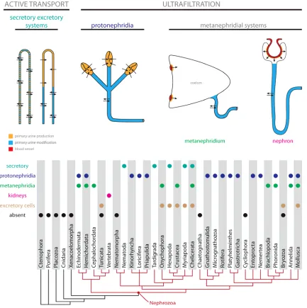
Figure 1. Excretory systems distribution across the animal tree. Schematic drawings and typology of basic types of
excretory organs with color coded sites of primary urine production and modification (see text for details). Distribution of different types of excretory organs in nephrozoan clades.
2. Diversity of cell types in excretory organs: a morphological perspective
Excretion in most of the Nephrozoa is a two-stage process (Ichimura and Sakai,
2017; Ruppert, 1994; Ruppert and Smith, 1988; Schmidt-Nielsen, 1997; Schmidt-Rhaesa,
2007). Initially the body fluid (e.g. blood, haemolymph or interstitial fluid) is roughly filtered
from large proteins and cells to produce the so-called primary urine (Ichimura and Sakai,
2017; Schmidt-Rhaesa, 2007). This initial product becomes later secondarily modified (e.g.
the ion concentration or water volume can be specifically changed), resulting in the finite
secondary urine, which is eventually expelled from the body (Schmidt-Nielsen, 1997;
performed, the excretory organs vary greatly regarding their morphology and physiology and,
as a consequence, in the diversity and spatial arrangement of the particular cell types, which
built them.
Traditionally, zoologists group excretory organs based on their physiological and
structural properties into categories, which reflect rather functional than evolutionary
similarities (Fig. 1; (Bartolomaeus and Ax, 1992; Ichimura and Sakai, 2017; Ruppert and
Smith, 1988; Schmidt-Rhaesa, 2007)). The most basic division relates to the mechanism of
primary urine production – in the secretory excretory organs the active intracellular transport
is used for that purpose, whereas in the ultrafiltration-based systems the primary urine is
filtered through semipermeable extracellular membrane – a filter composed of either
extracellular matrix (ECM) or slit diaphragm or both (Fig. 1; (Ichimura and Sakai, 2017;
Schmidt-Nielsen, 1997; Schmidt-Rhaesa, 2007)). The latter category includes
protonephridia, where ultrafiltration is driven by the ciliary action, and the metanephridial
system in which blood pressure is used to produce primary urine (Bartolomaeus and Ax,
1992; Ichimura and Sakai, 2017; Ruppert, 1994; Ruppert and Smith, 1988; Schmidt-Rhaesa,
2007). Among metanephridial systems it is possible to distinguish between vertebrate
kidneys, in which ultrafiltration and secondary urine modification occurs in the single
structural unit (i.e. nephron with glomerulus) and invertebrate metanephridial systems in
which ultrafiltration and secondary urine modification are spatially separated, the former
happening in the coelom lining and the latter in the metanephridium itself (Ichimura and
Sakai, 2017; Schmidt-Nielsen, 1997; Schmidt-Rhaesa, 2007).
Secretory excretory organs (Malpighian tubules and others)
Among the best-known examples of the secretory excretory organs are the
Malpighian tubules, the prevalent excretory organs in many Panarthropoda taxa. Although
Malpighian tubules are found in representatives of Eutardigrada, Chelicerata, Myriapoda and
Hexapoda (Fig. 1), it remains unknown if they are all homologous to each other or if they
rather evolved independently in particular lineages (Bitsch and Bitsch, 2004; Greven, 1982;
Paulus, 2000). Those organs are tubular invaginations of the gut (originating close to the
midgut-hindgut transition), which penetrate the haemocoel; distally they are blindly ended
and proximally they open to the gut lumen (Berridge and Oschman, 1969; Eichelberg and
Wessing, 1975; Nocelli et al., 2016; Schmidt-Nielsen, 1997; Schmidt-Rhaesa, 2007).
Malpighian tubules are surrounded by a basal lamina, which serves as a barrier for cells and
large proteins. Both their external and internal surface is greatly increased by respectively
basal infoldings and microvillar structures, which enhance cellular uptake and secretion
equipped with contractile muscle fibers and tracheae (e.g. (Bradley, 1983; Garayoa et al.,
1992; Li et al., 2015; Taylor, 1971b; Wall et al., 1975).
Often there are two basic cell types in Malpighian tubules – e.g. many insects
possess 1) primary cells (known as well as type I cells), which produce primary urine and are
characterized from numerous mitochondria-rich, external, basal infoldings and internal
microvilli and 2) less abundant secondary or stellate cells (known as well as type II cells) with
less developed microvilli, which are likely involved in the secondary urine modification (e.g.
(Berridge and Oschman, 1969; Dow and Davies, 2001; Kapoor, 1994; Li et al., 2015; Nocelli
et al., 2016; Pal and Kumar, 2012; Taylor, 1971a, b; Wall et al., 1975). In some animals
Malpighian tubules seem to include additional cell types – e.g. in tardigrades ”supportive”
cells are present next to primary and secondary cells (Møbjerg and Dahl, 1996; Węglarska,
1980).
It is evident that not only cell type diversity, but also their spatial distribution along the
organ is crucial for the evolution of Malpighian tubules. The two basic cell types – the primary
urine producing and the secondarily modifying ones – can be a) uniformly distributed along
the tubule as it is case in many insects (Berridge and Oschman, 1969; Dow and Davies,
2001; Kapoor, 1994; Nocelli et al., 2016; Pal and Kumar, 2012; Taylor, 1971a, b; Wall et al.,
1975), or b) restricted to the respectively proximal and distal portion of the tubule, resulting in
a clear division of the organ into distinct parts responsible for urine production and secondary
modification, as in e.g. tardigrades (Møbjerg and Dahl, 1996; Schill, 2019; Węglarska, 1980),
millipedes (Johnson and Riegel, 1977) or some insects (Arab and Caetano, 2002; Bradley,
1983; Green, 1979, 1980; Li et al., 2015; Nicholls, 1983; Nocelli et al., 2016). The subdivision
of Malpighian tubules into distinct regions with different cell types has been also shown in
spiders, however the function of the cells in each section is not clear (Hazelton et al., 2002;
Seitz, 1987).
Secretory excretory organs are also found among nematodes in which the two
different basic types can be recognized (reviewed in (Chitwood and Chitwood, 1950)). Some
of the free-living, mostly marine forms (once united into the group
Aphasmidia=Adenophorea) have a single large glandular cell used for excretion, the
so-called ventral gland, which opens to the exterior by the single pore. Members of the clade
Phasmidia (=Secernentea), which include most of the parasitic forms, as well as a model
species Caenorhabditis elegans, possess a specialized excretory organ, which consists of
several cells, each with different ultrastructure and function (e.g. (McLaren, 1974; Nelson et
al., 1983; Waddell, 1968). The exact number and types of cells might vary but generally the
organ always represents some modification of the so-called H-system (Chitwood and
Chitwood, 1950). There is an unpaired sinus cell which opens to the exterior by a
canals extend in the lateral hypodermal cords to the anterior and posterior portion of the
animal. Each canal is built by a single cell with intracellular lumen (sometimes canals are
projections of the sinus cell, e.g. (Nelson et al., 1983)), which can show spatial differentiation
into smooth and microvillar portions (McLaren, 1974; Waddell, 1968). Additional glandular,
sphincter or lip cells can be associated with the terminal duct (Chitwood and Chitwood, 1950;
McLaren, 1974; Nelson et al., 1983; Waddell, 1968). The molecular phylogeny of nematodes
shows that species with H-systems (or its modification) represents a monophyletic group
whereas species with single cell excretory gland are forming a basal grade (Holterman et al.,
2006; Smythe et al., 2019), hence the latter should be considered as plesiomorphic
nematode arrangement, whereas the former represent synapomorphy of Secernentea. Both
systems are nematode specific and represent derived excretory organs not easily
comparable with any structures found in remaining Nephrozoa.
Interestingly, the putative unicellular secretory excretory organs were also described
in the acoel Paratomella rubra (Ehlers, 1992), representing likely an evolutionary gain of
excretory organs independent from nephrozoan lineage (Andrikou et al., 2019). The
so-called dermonephridia are specialized epidermal cells, randomly distributed in the epidermis,
which lack cilia but possess modified long microvilli on their apical surface and intracellular
lacunar system of tubules and vacuoles, which communicate with the exterior. Taking into
account that dermonephridia are known only from a single acoel species they probably
represent a recent evolutionary novelty.
Protonephridia
Protonephridia use ultrafiltration through extracellular filters driven by ciliary action
(Bartolomaeus and Ax, 1992; Ichimura and Sakai, 2017; Ruppert, 1994; Ruppert and Smith,
1988; Schmidt-Nielsen, 1997; Schmidt-Rhaesa, 2007) (Figs. 1). Some of the simplest
protonephridia among entire animal kingdom are found in Gnathostomulida (Fig. 2;
(Lammert, 1985)), a group of microscopic marine worms closely related with rotifers. Each of
the gnathostomulid protonephridium consists of only three cells and can be used as an
example of the minimal and most basic cellular architecture required for this type of organs
(Fig. 2; (Lammert, 1985)). The most proximal (or terminal) cell has single cilium surrounded
by the cytoplasmic portion (so-called filtering area) traversed by irregular clefts closed with a
filtering membrane (i.e. slit diaphragm). The tubular filtering area tightly adjoins to the second
cell of the system – the canal cell – that harbors the intracellular canal into which the cilium of
the terminal cell protrudes. The canal cell is non-ciliated and has a complicated lacunar
system and microvilli facing the canal lumen, which both greatly increase the cellular surface.
through which a canal of the protonephridium communicates with the exterior. The terminal
cell provides the filter as well as the negative pressure necessary for the ultrafiltration and is
therefore the site of primary urine production. The canal cell, on the other hand, is
responsible for the secondary modification of the urine. Importantly, both components of the
organ show ultrastructural characteristics of epidermal cells, indicating that the entire organ
has an ectodermal origin (Lammert, 1985).
There are two ways in which this extremely simple system can be complicated in
other protonephridia-bearing animals. First of all, particular cells can be multiplied resulting in
e.g. many terminal cells opening to the single canal cell (e.g. in some gastrotrichs, Fig. 2,
(Kieneke et al., 2008; Kieneke and Hochberg, 2012; Teuchert, 1973) or some kinorhynchs,
Fig. 2, (Kristensen and Hay-Schmidt, 1989)), one terminal cell can open to the canal built of
many similar canal cells (e.g. in annelid Apodotrocha, Fig. 2; (Westheide, 1985)) or both
elements can be multiplied (e.g. in some Kinorhyncha and Loricifera; (Neuhaus, 1988;
Neuhaus and Kristensen, 2007) or in Nemertea (Bartolomaeus, 1985)). When terminal cells
are multiplied, each of them might form a separate filtering unit (Bartolomaeus, 1985;
Teuchert, 1973) or they can be merged together, with the filtering area formed by two or
more adjacent cells (Kieneke et al., 2008; Kieneke and Hochberg, 2012; Kristensen and
Hay-Schmidt, 1989; Neuhaus, 1988; Neuhaus and Kristensen, 2007).
Apart from cell multiplication, additional cell types might be present in the
protonephridial organs of some animals, increasing spatial diversification and functional
specification of the nephridium. For instance, in some platyhelminths the multicellular canal is
divided into the proximal ciliated zone to which the terminal cells open and a distal zone, built
exclusively by cells with extensive microvilli on the luminal surface (Rohde and Watson,
1993; Scimone et al., 2011; Vu et al., 2015). Additionally, it has been demonstrated, that in
planarians those two parts express different sets of solute carrier transporters and apparently
have different roles in the secondary urine modification (Vu et al., 2015). In microscopic
rotifers the canal is built of two or three distinct cell types (Ahlrichs, 1993a; Ahlrichs, 1993b;
Warner, 1969) and additionally in monogononts it opens to the muscular bladder, which
collects urine from paired protonephridial systems (Ahlrichs, 1993b; Warner, 1969). The
cellular complexity of the rotifer excretory system is further increased due to the fact that
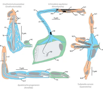
Figure 2. Morphological diversity of protonephridia. Schematic reconstruction of various protonephridial systems as inferred
from TEM studies, based on (Kristensen and Hay-Schmidt, 1989; Lammert, 1985; Teuchert, 1973; Westheide, 1985). In all drawings primary urine filtering cells (terminal cells) are in orange, primary urine modifying cells (canal cells) in blue, nephroporus cells in green, cell nuclei in dark grey and intracellular cavities in light gray. Abbreviations: cc canal cell, cl cilium, f protonephridial filter, la lacunar system, mv microvilli, nc nephroporus cell, sp sieve plate (cuticular opening for protonephridium), tc terminal cell, ve vesicle. Number after abbreviation indicate multiplication of particular cell type.
Protonephridia are present in many invertebrate taxa (sometimes only in larvae e.g.
in phoronids, some molluscs and annelids; see (Baeumler et al., 2011; Bartolomaeus, 1989;
Bartolomaeus and Quast, 2005; Goodrich, 1945; Hay-Schmidt, 1987; Koch et al., 2014;
Ruthensteiner et al., 2001; Temereva and Malakhov, 2006; Todt and Wanninger, 2010) and
even though they are often considered plesiomorphic (e.g. (Bartolomaeus and Ax, 1992)
they show remarkable diversity and evolutionary variation related with the fact that their
particular elements can be easily organized in various ways (Ichimura and Sakai, 2017). For
example, the filter might be built by a single cell with irregular openings, a single cell with
slits, two adjacent terminal cells with interdigitating processes or a terminal cell and a canal
diaphragm, with or without supporting microvilli, microvilli might be differentiated or uniform
etc.. The terminal cell might be monociliated (also called solenocyte), multiciliated with
independent cilia (then known as cyrtocyte) or multiciliated with all the cilia forming a single
structure, the so-called flame as in e.g. rotifers or some platyhelminthes (Riemann and
Ahlrichs, 2010; Rohde, 1991). The canal might be intracellular or multicellular, with or without
cilia, spatially diversified or uniform etc. However, despite all their diversity, protonephridia
are always straightforwardly comparable to each other (Bartolomaeus and Ax, 1992) and the
primary homology statements regarding their particular portions can be readily made. The
distribution of the protonephridia-bearing animals on the phylogenetic tree (Fig. 1), suggests
that those organs evolved either twice, once in Scalidophora and once in Spiralia, or are
homologous between those two groups. This makes protonephridia an excellent model for
studying the evolution of complexity and functionality of the excretory organs over long
evolutionary time, combining morphological, cellular and molecular levels.
Metanephridial systems
The typical metanephridia (Figs. 1, 3) are present in some annelids (e.g.
(Bartolomaeus and Quast, 2005; Goodrich, 1945), in brachiopods (Kuzmina and Malakhov,
2015; Lüter, 1995), adult phoronids (Bartolomaeus, 1989; Storch and Herrmann, 1978;
Temereva and Malakhov, 2006), cephalochordates (Ichimura and Sakai, 2017; Moller and
Ellis, 1974; Ruppert, 1994) and adult hemichordates (Balser and Ruppert, 1990; Dilly et al.,
1986; Mayer and Bartolomaeus, 2003). Furthermore, the onychophoran nephridia (Mayer,
2006), coxal glands of Chelicerata (Briggs and Moss, 1997; Koch et al., 2014), antennal
gland of Crustacea (Bartolomaeus et al., 2009; Khodabandeh et al., 2005), molluscan
heart-kidneys (Baeumler et al., 2011; Bartolomaeus, 1997) and the axial organ of echinoderms
(Balser and Ruppert, 1993; Ezhova et al., 2013, 2014; Welsch and Rehkamper, 1987;
Ziegler et al., 2009) can be also considered as metanephridial excretory systems, at least
from the functional point of view. The ultrafiltration occurs in invertebrate metanephridial
systems (Fig. 3) between blood vessels (or their functional equivalents, e.g. haemocoelic
sinus) and the coelomic lining where specialized cells – podocytes (Fig. 3) – are present
(Bartolomaeus and Ax, 1992; Ichimura and Sakai, 2017; Ruppert, 1994; Ruppert and Smith,
1988; Schmidt-Rhaesa, 2007). The primary urine is therefore synonymous with the coelomic
fluid in those animals (Bartolomaeus and Ax, 1992; Ruppert, 1994; Schmidt-Rhaesa, 2007).
The fluid leaves the coelom through the spatially independent structure, a metanephridium
(Fig. 3), which often consists of the proximal dilated portion (i.e. ciliated funnel) and the distal
region (i.e. nephroduct), which might be further subdivided into regions with differing
compared to protonephridia, many different cell types build metanephridial systems and their
exact number and qualitative composition varies from taxon to taxon, showing diverse
ultrastructural, developmental and functional characteristics. In some animals – e.g.
phoronids or some annelids - the metanephridia are ontogenetically predated by
protonephridia (Bartolomaeus, 1989; Bartolomaeus and Quast, 2005; Goodrich, 1945; Koch
et al., 2014; Temereva and Malakhov, 2006). In such instances the terminal cells of the
protonephridium degenerate during metamorphosis, and the adult metanephridium develops
from the larval protonephridial canal, whereas the podocytes develop de novo from the
myoepitheliocytes (Bartolomaeus, 1989; Bartolomaeus and Quast, 2005; Bartolomaeus et
al., 2009; Storch and Herrmann, 1978; Temereva and Malakhov, 2006). The term
nephromixium is sometimes used to refer to such definite organ of dual origin (Goodrich,
1945; Temereva and Malakhov, 2006). On the other hand in Panarthropoda taxa, the
podocyte-bearing cavity, called sacculus, does not seem to be formed by the coelom but
rather by dilatation of the blind end of the developing nephroduct (summarized and
discussed in (Koch et al., 2014)). Nephridia in Arthropoda are further distinguishable from
those found in other animals (including onychophorans) by lacking any ciliation (Mayer,
2006).
In the vertebrate kidney, the excretion is carried out in the structural unit called
nephron (Fig. 1; e.g. (Gérard, 1936; Ichimura and Sakai, 2017; Ruppert, 1994; Vize et al.,
1997)), which due to its importance in human physiology is very well studied on
morphological, physiological, developmental and molecular levels (e.g. (Bates, 2016;
Desgrange and Cereghini, 2015; Dressler, 2006; Lindström et al., 2018; Little et al., 2010;
McMahon, 2016; Quaggin and Kreidberg, 2008)). There are three types of nephrons found
among vertebrates: the closed glomerular (present in e.g. mammalian kidney), opened
glomerular (e.g. in salamanders) and aglomerular ones (found exclusively in some teleosts)
(Gérard, 1936; Schmidt-Nielsen, 1997). The first two are relatively similar: both have a
glomerular portion (also known as Bowman capsule), where filtering cells – podocytes –
tightly surround capillaries lined with the extremely thin-walled fenestrated endotheliocytes
(Bates, 2016; Ichimura and Sakai, 2017; Koriyama et al., 1992; Schmidt-Nielsen, 1997; Wolff
and Merker, 1966). The primary ultrafiltrate passes through the endothelium-ECM-podocytes
barrier and is subsequently accumulated inside the capsule, from where it is drained by the
proximal nephron tubule. Aside from the podocytes and endotheliocytes two additional cell
types of predominantly supportive function are found in the glomerulus – mesangial cells,
which support capillaries (but also contribute to filtration of some molecules directly from the
bloodstream) and parietal cells, which exclusively build the external wall of Bowman capsule
(Dressler, 2006; Ichimura and Sakai, 2017; Quaggin and Kreidberg, 2008; Vize et al., 1997).
to the urinary bladder. The tubule is differentiated into proximal, intermediate and distal
regions, which differs in function and consequently in cell types of which they are composed
(Bates, 2016; Desgrange and Cereghini, 2015; Lindström et al., 2018; Little et al., 2010;
Schmidt-Nielsen, 1997). The main difference between closed and opened glomerular
nephrons is that in the latter the additional ciliated canal (composed of yet another cell type)
connects its proximal tubule with the peritoneal cavity (Gérard, 1936). The aglomerular
nephron, on the other hand, is found only in some, mostly marine, teleosts (Gérard, 1936;
Ichimura and Sakai, 2017; Schmidt-Nielsen, 1997). It lacks glomerular capsule and in fact
utilize only active transport through the cells for primary urine production (Bulger, 1965;
Dobbs and Devries, 1975; Schmidt-Nielsen, 1997), which makes it actually an example of a
secretory excretory organ. It is, however, homologous to the glomerular nephron and, in
some species, it ontogenetically develops from the glomerular condition (Gérard, 1936).
Depending on the arrangement of nephrons in the organ it is possible to distinguish
between a mesonephros and a metanephros (Fig. 3; (Bates, 2016; Ichimura and Sakai,
2017; Vize et al., 1997)). In amniotes, they form an ontogenetic series with the metanephros
being a definite excretory organ and the mesonephros is found only in the embryonic or
larval stages (Bates, 2016; Vize et al., 1997). The earliest developmental stage of the
vertebrate kidney – pronephros – does not have separate nephrons: the glomerulus and
nephric duct are spatially separated, whereas the primary urine is accumulated inside
coelom (Fig. 3; e.g. (Møbjerg et al., 2000; Vize et al., 1997); from the functional perspective it
can be therefore categorized as the proper metanephridial system (Ichimura and Sakai,
Figure 3. Morphological diversity of metanephridial systems. Schematic drawings of metanephridial systems in
sites of primary urine modification in blue. Abbreviations: ac amebocyte, bc blood cell, bm basal membrane, bv blood vessel, ce coelom, cf ciliated funnel, co collar region, dm dorsal mesentery, gl glomerulus, hs heart sinus, in intestine, kd kidney, mf
myofibrils, mn metanephridium, ms musculature, nd nephric duct, np nephron, pb proboscis, pc podocyte, pd protocoel duct, pe pedicle, pp proboscis pore, sd slit diaphragm, tr trunk region, vm ventral mesentery.
The most distinctive cell type that seems to be shared by all metanephridial systems
(including kidneys) is a podocyte (Ichimura and Sakai, 2017; Ruppert, 1994). Cells of this
type are divided into the cell body and pedicles – long projections, which interdigitate with
each other (Fig. 3). On the junctions of the pedicles the proteins anchored in the podocyte
cell membrane might form the filtering membrane – a slit diaphragm (Fig. 3; e.g. (Gerke et
al., 2003; Ichimura and Sakai, 2017; Quaggin and Kreidberg, 2008; Storch and Herrmann,
1978; Tryggvason and Wartiovaara, 2001). Interestingly, a filtering region of terminal
protonephridial cells of planarians shares some ultrastructural and molecular similarities with
the filtering portion of the podocyte (Ichimura and Sakai, 2017; Vu et al., 2015). Taking into
account that metanephridia probably evolved independently at least few times in the animal
kingdom (Fig. 1; (Bartolomaeus, 1997; Bartolomaeus and Ax, 1992; Koch et al., 2014)), the
podocytes of various animal groups represent analogous cell types (Bartolomaeus and Ax,
1992), which likely evolved by independent modification of the same ancestral filtering
mechanism (e.g. from terminal cells of protonephridia) (Ruppert, 1994).
Excretory cells
In addition to multicellular excretory organs (and sometimes instead of them) some
animals possess specialized cells, which serve for excretory purposes (Haszprunar, 1996;
Ruppert, 1994). Instead of expelling toxic waste products from the animal body, these cells
accumulate the excreted substances inside their cytoplasm. Such accumulative excretory
cells have been reported in a wide range of animals (Fig. 1) and although they are generally
referred as nephrocytes, their ultrastructure and function differ from taxon to taxon.
The most studied excretory cells among nephrocytes are cells present in arthropods
and onychophorans (Coons et al., 1990; Crossley, 1972; El-Shoura, 1989; Hessler and
Elofsson, 1995; Seifert and Rosenberg, 1977; Shatrov, 1998; Vandenbulcke et al., 1998;
Weavers et al., 2009). These cells, located inside haemolymph-filled cavities, are surrounded
by ECM and resemble isolated podocytes with diaphragm-like structure. The waste products
and other toxic compounds are filtered through the ECM and diaphragm and accumulate
inside the nephrocytes (Vandenbulcke et al., 1998; Weavers et al., 2009). Insects have two
sets of nephrocytes with similar ultrastructural properties – garland cells around esophagus
and pericardial cells located on the heart walls (Crossley, 1972; Weavers et al., 2009). Cells
called rhogocytes (Haszprunar, 1996; Rivest, 1992; Ruthensteiner et al., 2001). It has been
proposed, based on molecular similarities (homology of the proteins forming diaphragm) and
intermediate morphological forms, that both arthropod nephrocytes and molluscan
rhogocytes are actually homologues of the filtering nephridial cells, which became spatially
separated from an ancestral excretory organ (Haszprunar, 1996; Hessler and Elofsson,
1995; Rivest, 1992; Ruppert, 1994; Weavers et al., 2009).
A very different cell type is the nephrocyte in tunicates. Tunicate nephrocytes
represent a fraction of blood cells, which accumulate nitrogenous waste products inside
voluminous vacuoles (Ballarin and Cima, 2005; Cima et al., 2014; George, 1939). A similar
type of excretory cells is also present in the coelomic fluid of Bryozoa (Schwaha et al., 2020).
Contrary to the excretory cells of insects and mollusks, nephrocytes in tunicates and
bryozoans lack both ECM and a slit diaphragm and they represent specialized haemo- and
coelomocytes, therefore they are evolutionarily unrelated to other excretory organs.
Excretory organs and adaptations to new environments
Excretory organs are not only responsible for the expulsion of metabolic waste
products but they are also the primary organs for osmoregulation and ion balance (Larsen et
al., 2014; Schmidt-Nielsen, 1997; Schmidt-Rhaesa, 2007). The maintenance of water
balance is especially challenging in environments such as freshwater and terrestrial habitats,
where water is excessive or sparse, respectively (Schmidt-Nielsen, 1997). Another problem
related to osmoregulation is faced by the organisms inhabiting the brackish and intertidal
realms, where salinity can rapidly and dramatically change in a short time span, which
requires ability to accommodate to different salinity regimes (Schmidt-Nielsen, 1997).
Moreover, differences in the environmental salinity and water availability result in adaptations
regarding the mechanisms of ammonia (one of the most toxic metabolites) excretion, such as
transforming it into less harmful nitrogenous end products (e.g. urea or uric acid) (Larsen et
al., 2014; Needham, 1935; Schmidt-Nielsen, 1997; Weihrauch and O'Donnell, 2017).
Therefore, modifications of excretory organs are believed to be crucial for colonizing new
environments (Schmidt-Rhaesa, 2007). In fact, studies on various nephrozoans have shown
that species inhabiting terrestrial, freshwater, brackish and intertidal environments exhibit
species-specific adaptations in the morphology (e.g. (Krishnamoorthi, 1963; Randsø et al.,
2019; Smith, 1984; von Nordheim and Schrader, 1994) and physiology (e.g. (Generlich and
Giere, 1996; Needham, 1935; Schmidt-Nielsen, 1997; Smith, 1970; Werntz, 1963) of their
excretory organs and these adaptations are not necessarily reflecting their evolutionary
3. Molecular identity of excretory organs: from development to function
In contrast to the large number of detailed morphological descriptions of excretory
organs in a variety of animals, molecular data are scarce. Most of the gene expression
studies have been conducted in vertebrates (mainly mammals, fish and frogs) (summarized
in (Desgrange and Cereghini, 2015) and a handful of invertebrates (mainly planarians and
flies) (Scimone et al., 2011; Vu et al., 2015; Weavers et al., 2009) and have revealed not only
common transcriptional programs governing the development of excretory organs, but also
analogous sub-localization of solute transporters and structural proteins within the
differentiated excretory compartments (Figure 4). Moreover, physiological studies in a large
array of animals have shown that a number of ammonium transporters and proton pumps
have conserved roles in excretory processes (Weihrauch and O'Donnell, 2017).
Nephron development
Kidneys, the excretory organs of vertebrates, emerge from the intermediate
mesoderm and develop through a sequential formation of up to three organs: pronephros,
mesonephros and metanephros (Figure 3) (Saxén and Saxén, 1987). Pronephros and
mesonephros are only transient structures in amniotes and function as the fetal excretory
organs, whilst metanephros is the definitive adult kidney. In fish and frogs, the adult kidney is
the mesonephros that replaces the embryonic pronephros. Both mesonephros and
metanephros are composed from a basic structural and functional unit, the nephron, which
shows a comparable regional organization (Wingert and Davidson, 2008). The formation of
kidneys is dictated by similar genetic interactions and morphogenetic events (Desgrange and
Cereghini, 2015) (Figure 4).
Nephron development in mammals starts with a mesenchyme-to-epithelial transition
(MET) of the anterior intermediate mesoderm (IM) and the specification of renal progenitor
cells. This process is induced by the interplay of an ectodermal BMP4 signaling (James and
Schultheiss, 2005) and the expression of odd skipped related gene (osr1) (James et al.,
2006) and lhx1 transcription factors (Tsang et al., 2000). Pax2 and pax8 genes are activated
shortly after and act redundantly in the nephric lineage specification (Bouchard et al., 2002).
The renal progenitor cells will form an epithelial tubule, the future nephric duct. As the tubule
extends posteriorly, a number of transcription factors, such as hox11, six1/2/4, eya, sall,
pax2, foxc and wt1 (Brophy et al., 2001; Kobayashi et al., 2008; Kreidberg et al., 1993; Kume
et al., 2000; Nishinakamura et al., 2001; Sajithlal et al., 2005; Wellik et al., 2002; Xu et al.,
2003) are expressed along its anterior-posterior axis and induce the expression of gdnf
branching of the ureteric bud (UB) at the posterior end of the nephric duct, which grows into
medially positioned metanephric mesenchyme, which in turn gives rise to a renal vesicle
(Costantini and Shakya, 2006). Other signaling pathways involved in UB formation and
branching include Wnt (Bridgewater et al., 2008; Carroll et al., 2005; Kispert et al., 1998),
sonic hedgehog (Shh) (Cain and Rosenblum, 2011), bone morphogenic protein (Bmp)
(Nishinakamura and Sakaguchi, 2014) and fibroblast growth factor (Fgf) (Bates, 2011).
Eventually, each renal vesicle forms a nephron through a series of morphogenetic
movements and patterning events. Mafb, wt1 and lmx1b drive podocyte specification(Miner
et al., 2002; Moriguchi et al., 2006) and Notch signaling together with pou3f3, hnf1b, irx1 and
irx2 are responsible for the proximal tubule fates (Cheng et al., 2007; Heliot et al., 2013;
Nakai et al., 2003). The specification of distal tubule is controlled from the molecular interplay
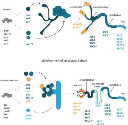
Figure 4. Development and structural correspondences of protonephridia and kidneys. Cartoon depiction of the
molecular programs governing the regeneration of planarian protonephridia and the development of vertebrate kidneys, based on Scimone et al 2011 and Vu et al 2015. The corresponding structural components of protonephridia (terminal cell, tubule, duct) and kidneys (podocyte, tubule, duct) and the expression domains of orthologous genes in relation to their components, are color coded. Abbreviations: bv blood vessel.
In the zebrafish Danio rerio, the renal progenitor field forms from the lateral-most IM
that express the transcription factors pax2a and pax8 (Pfeffer et al., 1998). Osr1 has also a
conserved expression since its endodermal expression during gastrulation promotes renal
lineages at the expense of blood/vascular ones (Mudumana et al., 2008). The renal
in the anterior-most domain express wt1a, wt1b, osr1, foxc1a and lhx1a (O'Brien et al., 2011;
Perner et al., 2007; Tomar et al., 2014) and will form the podocytes, whilst the remaining
cells will give rise to the proximal and distal tubule and express jagged, irx3b, evil and
pou3f3a/pou3f3b (Li et al., 2014; Ma and Jiang, 2007; Wingert et al., 2007). The
transcriptional interplay responsible for the formation of this boundary consists from pax2a,
which forms a negative feedback loop with wt1a (Majumdar et al., 2000) and hnf1b, a
suppressor of pax2a (Naylor et al., 2013). Tubule regionalization is also regulated by
Retinoic Acid (Ra) signaling pathway (Wingert et al., 2007).
In the frog Xenopus, the renal progenitors emerge after a MET of the caudo-lateral IM
resulting in the formation of a tubule. Once again, osr1 and osr2 are upstream of the
specification of the renal progenitor field (Tena et al., 2007). The first renal molecular
markers are lhx1 and pax8, followed by the expression of pax2, wt1 and hnf1β (Buisson et
al., 2015; Carroll and Vize, 1996, 1999; Carroll et al., 1999; Wild et al., 2000). Wt1 specifies
the future glomerulus whilst pax2 is restricted to the future tubular region. The subdivision of
the nephron into segments is governed by evi1 expression in the distal tubule and pronephric
duct (Van Campenhout et al., 2006) and irx1, irx2 and irx3 expression in the proximal and
intermediate tubule (Alarcon et al., 2008). The developing podocytes are specified from a
cross talk between wt1, foxc2, lmx1b and mafb genes (Haldin et al., 2008; White et al.,
2010). Signaling pathways with crucial roles in Xenopus nephrogenesis involve Wnt (Lavery
et al., 2008; Saulnier et al., 2002; Tetelin and Jones, 2010), Fgf (Urban et al., 2006), Bmp
(Bracken et al., 2008), Notch(McLaughlin et al., 2000; White et al., 2010) and Ra (Cartry et
al., 2006).
Protonephridial development
Although there are several morphological descriptions on protonephridial
development (Baeumler et al., 2011; Bartolomaeus, 1985; Hasse et al., 2010; Rohde et al.,
1988; Temereva and Malakhov, 2006; Wenning et al., 1993), molecular studies are
extremely limited. The most detailed work has been performed on the planarian Schmidtea
mediterranea (Scimone et al., 2011) that has shown a remarkable conservation of the
molecular programs between the regeneration of planarian protonephridia and the
development of vertebrate kidneys (Figure 4). Planarian protonephridia consists of four cells
types, the flame (terminal) cell, the ciliated tubule cell type (proximal tubule), the
tubule-associated cell type (distal tubule) and the duct. Regeneration and RNAi experiments on
amputated animals showed a conserved function of eya, six1/2, pou3, hunchback, sall and
osr genes in the regeneration and maintenance of protonephridia. Eya, six1/2, pou3, sall and
formation of the ciliated tubule cell type and the tubule-associated cell type. The ciliated
tubule cell type continues to express pou3, sall and osr, whilst the tubule-associated cells
express six1/2(Scimone et al., 2011).
The conserved role of pou3 is also reported in the nematode C. elegans, where an
orthologous gene, ceh-6 is required for the formation and function of its excretory cell
(Burglin and Ruvkun, 2001). Other conserved nephrogenesis-related transcription factors
has been shown to be pax3 in the developing nephridia of the leech Helobdella robusta
(Woodruff et al., 2007), pax2/5/8 in the nephridium of the cephalochordate Branchiostoma
floridae (Kozmik et al., 1999) and sall in the protonephridium precursors of the polychaete
Hydroides elegans (Arenas-Mena, 2013).
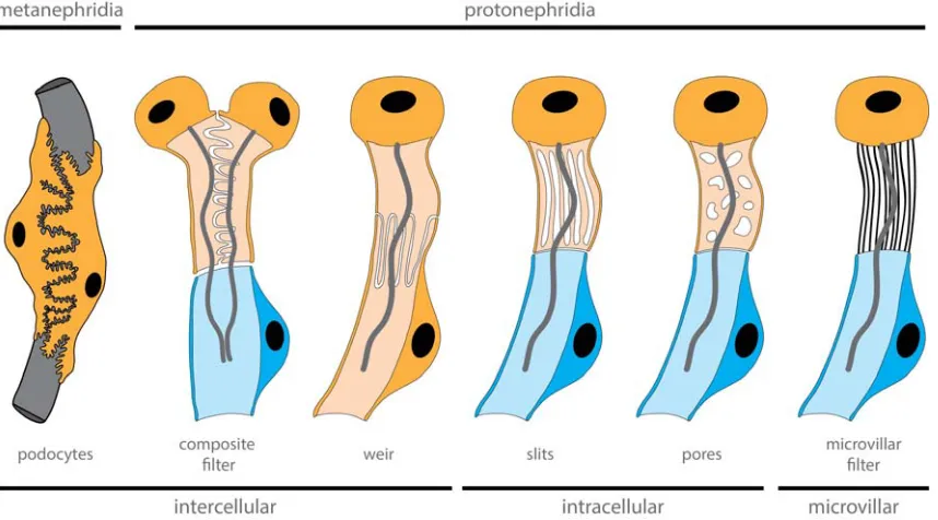
Figure 5. Diversity of filter-forming cells within nephridia. The filter apparatus can be formed by different cell features.
Intercalated cells form the filtration apparatus between cells, filtration can be performed by cellular pores and slits or microvilli
can build up the filtration apparatus. Cells forming extracellular filter are depicted in orange, while nephroduct cells in blue.
Development of insect Malpighian tubules
The Malpighian tubules of insects consist of two cell types; the primary cell (PC) and
the secondary (stellate) cell (SC). In Drosophila, these two cell types have a different
developmental origin; while the PC derive from an ectodermal primordium at the
hindgut/midgut junction and form the Malpighian tubule epithelium, the SC originate from the
posterior mesoderm and invades the tubule epithelium through MET (Denholm et al., 2003).
The molecular patterning of Malpighian tubule development appears quite different
of the hindgut marks the onset of PC development due to a Wnt signaling. Once specified,
these cluster of cells start to form bud-like branches under the control of Decapentaplegic
(BMP) signaling and Brinker (Hatton-Ellis et al., 2007). Later on, a regulatory interplay
between cut, barr, three rows, pebble, pimples, trachealess, ribbon, raw, crooked neck, faint
sausage, pant and schnurri, under a DPP/BMP signal, will result in the formation and
morphogenesis of tubules (Hatton-Ellis et al., 2007; Jack and Myette, 1999; Shim et al.,
2001). As the tubules elongate, a caudal mesodermal population that will become SC,
express the transcription factors tiptop and teashirt, undergo MET and intercalate in the
tubule (Denholm et al., 2003). The developmental process of Malpighian tubule development
is overall conserved, since studies in Tribolium have shown that the onset, morphogenesis
and molecular profile are similar. An important difference is the fact that in Drosophila only
differentiated PC are expressing cut, whilst in Tribolium cut is expressed in both PC and SC
(King and Denholm, 2014).
Molecular basis of functional compartmentalization
The terminal differentiated excretory organs are highly specialized and
compartmentalized into discrete segments, composed of distinct epithelial cell types that
carry out different functions. Each region is characterized by the expression of a set of
structural proteins and transporters, involved in excrete ultrafiltration and modification, such
as glucose and solute transport, and homeostasis. The spatial distribution of these proteins
on the different segments of excretory organs is, once more, remarkably conserved (Figure
4)(Kozmik et al., 1999; Vu et al., 2015; Weavers et al., 2009).
In vertebrates, the nephron is divided in five segments: the glomerulus, the proximal,
intermediate and distal tubule, and the collecting duct. Glomerulus has a central role in
ultrafiltration and is expressing a set of membrane-associated proteins, such as Nephrin
(NPHS1), KIRREL1 (NEPH1), CD2AP, ZO1, Nck and Stomatin/Podocin (summarised in
(Patari-Sampo et al., 2006)). The tubular segments are further subdivided in smaller regions;
each of them specialized in different aspects or excrete modification. The segmental
organization is highly conserved, with the solute carrier (SLC) protein family to be
sequentially expressed along the tubular segments of different types of nephrons, defining
their boundaries (Desgrange and Cereghini, 2015). SLCs are membrane transporters,
composed from 52 families, which can transport a number of different substrates (Hediger et
al., 2013). In both mammalian metanephros and Xenopus pronephros, subsets of cells of the
proximal tubule are specialized in reabsorbing: a) salts, expressing members from the
bicarbonate transporter SLC4, sodium- and chloride-dependent neurotransmitter transporter
organic cation/anion/zwitterion transporter SLC22 families; b) amino acids, expressing
members from the glutamate and neutral amino acid transporter SLC1, heavy subunits of the
heteromeric amino acid transporter SLC3 and cationic/glycoprotein-associated amino acid
transporter SLC7 families; and c) glucose, expressing members from the sodium-glucose
cotransporter SLC5 family (Landowski et al., 2008; Raciti et al., 2008). SLC9a3 expression is
also marking the proximal tubule of the pronephros of D. rerio, suggesting a similar function
of this segment in reabsorbing sodium (Wingert et al., 2007). The intermediate tubule of
nephron (Henle’s loop) is facilitating the concentration of excretes and does not share an
extensive molecular conservation among the different nephron types (Desgrange and
Cereghini, 2015). Cells of the distal tubule are specialized in the reabsorption and secretion
of ions, ammonium and water through the SLC9 and the electroneutral cation-chloride
cotransporter SLC12. Common transporters characterizing the distal tubule of mammalian
metanephros and Xenopus pronephros are members of SLC12, monocarboxylate
transporter SLC16, type III sodium-phosphate cotransporter SLC20 and ammonium
transporter SLC42/Rhesus families (Landowski et al., 2008; Raciti et al., 2008). Cells
comprising the distal tubule of Xenopus are additionally expressing SLC4a4 (Raciti et al.,
2008), whilst in D. rerio only members of the SLC12 family are seen (Wingert et al., 2007).
Finally, the collecting duct consists from highly specialized, electrically tight cell types with
dedicated roles in reabsorption and secretion of salts and water, and the concentration and
preparation of the urine. In mammals, the collecting duct expresses members of the
sodium/potassium exchanger SLC8, SLC16, SLC20, SLC22 and SLC42/Rhesus families
(Landowski et al., 2008), while in Xenopus the analogous segment, the collecting tubule,
expresses additionally SLC12a3 and doesn’t express SLC42/Rhesus (Raciti et al., 2008).
Even bigger differences we encounter in D. rerio, where the pronephric duct is not
expressing any of these transporters (Wingert et al., 2007). The fact that the distal tubule of
D. rerio and Xenopus pronephros expresses SLC12a3 and Xenopus do not express
SLC42/Rhesus in their collecting tubule but only in their distal tubule, suggests that the
function of distal tubule of D. rerio and Xenopus might be analogous to a mammalian
tubule/duct hybrid (Desgrange and Cereghini, 2015). Another family of transporters that
exhibit a nephron segment-specific expression is the one of aquaporins, which transports
mainly water, urea and glycerol (Gomes et al., 2009). In mammals, the proximal tubule is
expressing the water/nitrate/chloride transporter AQ1 (Nielsen et al., 1993), the
aquaglyceroporin AQ7 (Nejsum et al., 2000), the water/ammonia transporter AQ8 (Elkjaer et
al., 2001) and the super-aquaporin AQ11 (Morishita et al., 2005), while the collecting duct
expresses the water transporters AQ2 (Fushimi et al., 1993) and AQ4 (Terris et al., 1995),
the aquaglyceroporin AQ3 (Ecelbarger et al., 1995), the water/nitrate/chloride transporter
mammalian orthologs has been shown in the collecting duct. However, AQ1 expression was
restricted in glomerulus, in contrast to what is observed in mammals (Pandey et al., 2010).
The situation looks even more different in teleosts where the proximal tubules express one
AQ8, one AQ10-like and one AQ3 paralog (Engelund and Madsen, 2015; Santos et al.,
2004). Moreover, two copies of AQ1 have a renal expression but have acquired different
functions; AQ1a is expressed in the proximal tubule and AQ1b in the distal tubule (Engelund
and Madsen, 2015; Madsen et al., 2011). Finally, one AQ11 and one AQ12 ortholog are also
expressed in the teleost nephron but the exact expression domain has not yet been revealed
(Kim et al., 2014; Madsen et al., 2014).
In the planarian S. mediterranea protonephridia are divided in four major
compartments: the flame (terminal) cells, the proximal tubule, the distal tubule and the duct.
The flame cells are performing ultrafiltration and express Nephrin/Kirrel. In a similar fashion
with the metanephridial systems, the tubular compartments are further subdivided in smaller
specialized domains with different cell types being defined by the expression of a suite of
SLC transporters and having diverse roles in excrete modification (Vu et al., 2015). For
instance, the proximal tubule, primarily responsible for the recovery of filtered substances
and reabsorption of salts, is expressing members from the SLC1, SLC5, SLC4, SLC6,
SLC13 and SLC22 families. The distal tubule has a central role in homeostasis and
expresses members from the SLC4 and SLC12 families, which are also marking the duct.
The duct is additionally expressing SLC42/Rhesus, as seen in mammals but not in D. rerio
and Xenopus, and a number of other SLCs, which mark the proximal tubule of vertebrates,
such as SLC6, SLC7, SLC9 indicating a role of the planarian duct not only in the urine
concentration but also in reabsorption of salts and amino acids. Interestingly, aquaporins are
not expressed in any of the protonephridial compartments, suggesting divergent functions of
these transporters in planarians (Vu et al., 2015).
Malpighian tubules of some insects (e.g. the fly Drosophila melanogaster and the mosquito
Aedes aegypti) are also divided in distinct compartments with different physiological
functions: the initial, transitional, main and lower segments, and the ureter (Beyenbach et al.,
1993; Sozen et al., 1997). Each segment is populated by a different numeral and positional
combination of PC and SC cells, depending on the investigated species. Overall, the initial
and transitional segments are not participating in secretion but rather act as storage
segments and transport (Dow et al., 1994; Dube et al., 2000). The main segment has both
secretory and absorptive roles of salts and water and generates the primary urine. The PC
cells of the main segment express a basolateral sodium/potassium transporter (Torrie et al.,
2004), an inward-rectifier potassium transporter (Kir) (Evans et al., 2005), sodium/proton
exchangers of the NHA and NHE (SLC9) families (Pullikuth et al., 2006; Rheault et al., 2007)
absorptive role, express mainly a chloride transporter and an aquaporin (Kaufmann et al.,
2005; Kolosov and O'Donnell, 2020; O'Connor and Beyenbach, 2001; O'Donnell et al.,
1998). The expression of SLC4 exchanger has also been reported in the SC cells of some
species(Linser et al., 2012; Piermarini et al., 2010). Finally, the lower segment and rectum
are mostly dedicated in the reabsorption of salts and water and the concentration of urine
(O'Donnell and Maddrell, 1995).
Molecular similarities found at the building blocks of the ultrafiltration sites of proto-
and metanephridial systems are also seen in individual ultrafiltration cells. The
membrane-associated protein complex composed of Nephrin, Kirrel, Cd2ap, Zo1 and Stomatin/Podocin
is also forming the slit diaphragm of nephrocytes of the fly Drosophila melanogaster
(Weavers et al., 2009; Zhuang et al., 2009) that performs filtration of the haemolymph
(Weavers et al., 2009; Wigglesworth, 1943; Zhang et al., 2013). Nephrocytes are not only
involved in ultrafiltration but also in protein reabsorption, via receptors such as Cubilin and
Amnionless (AMN), similarly to mechanisms encountered in the renal proximal tubule cells
(Zhang et al., 2013). Moreover, Nephrin/Kirrel expression has been described in the
ultrafiltration apparatus of rhogocytes of the snail Biomphalaria glabrata (Kokkinopoulou et
al., 2014), through which proteins and ions are filtered (Kokkinopoulou et al., 2015).
Nitrogenous waste excretion
One of the main and most toxic products of excretion in animals is nitrogenous waste.
Nitrogenous waste products are the result of the amino acid catabolism (Campbell, 1991)
and exist in three forms: ammonia, urea, or uric acid. Most aquatic species excrete ammonia,
due to its high solubility in water, whilst semi-aquatic and terrestrial species usually convert
ammonia to less hazardous and less soluble forms, such as urea and uric acid. Ammonia
can occur either in its gaseous (NH3) or in its ionic form (NH4+); however, due to the high pK
of NH3, in physiological solutions the vast amount of ammonia exists as NH4+. The
excretory process of ammonia has been investigated in a large array of animals and shown
extensive conservation of the repertoire of the ammonia transporters and proton pumps,
independently of the presence of specialized excretory organs (Weihrauch and O'Donnell,
2017).
In kidneys, the epithelial cells of the proximal tubule secrete ammonia apically into the
luminal fluid. A significant fraction of this ammonia secretion occurs via Na+/NH4+ exchange
by the sodium/proton exchangers SLC9 (NHE) and some also takes place via diffusion
(Bourgeois et al., 2010; Knepper et al., 1989; Preisig and Alpern, 1990). Almost all secreted
ammonia is reabsorbed by cells of the thick ascending limb of Henle’s loop into the
(NKCC) (Good, 1994) and Na+/NH4+ exchange of the basolateral SLC9 exchangers
(Blanchard et al., 1998). Another cotransporter that plays an important role in NH4+
reabsorption is the basolateral bicarbonate transporter SLC4, which drives NH3 diffusion
across the basolateral membrane due to the bicarbonate transport into the cell and the
subsequent rise of the intracellular pH (Good et al., 1984; Lee et al., 2010). The accumulated
ammonia in the interstitium is forming a gradient that drives diffusion across the epithelium of
the cells of the collecting duct and secretion of the concentrated ammonia to the lumen. This
process is also supported by an proton gradient formed by the apical vacuolar H+-ATPase,
which creates an acidic environment facilitating NH4+ entrapment (Flessner et al., 1991; Star
et al., 1987) and a basolateral Na+/K+ ATPase (NKA) that actively transfers ammonia by
Na+/NH4+ exchange (Wall, 1996). Finally, the Rhesus ammonia transporters (RhBG and
RhCG), spatially restricted to the collecting duct, also participate to the secretion to the
lumen and preparation of the urine(Mak et al., 2006).
Species-specific excretory organs/sites have also recruited these transporters for
excreting ammonia. In Malpighian systems, ammonia is firstly secreted into the Malpighian
tubules and then actively absorbed by the hindgut and midgut, which express the ammonia
transporter Rhesus, sodium/proton exchangers (NHE) and a vacuolar H+-ATPase (Blaesse
et al., 2010; Weihrauch, 2006). Similarly, in the posterior rectum (anal papillae) of the aquatic
mosquito larvae Aedes aegypti, the ammonia transporters Rhesus and the Rhesus-related
AMTs, as well as a basal NKA and an apical vacuolar H+-ATPase are involved in ammonia
excretion (Chasiotis et al., 2016; Durant and Donini, 2018). In the excretory H-system of the
nematode C. elegans, ammonia enters the excretory cells via the basolateral NKA and
potassium transporters and then diffuses across the apical membrane through an
acid-trapping mechanism (Adlimoghaddam et al., 2015). Rhesus and the vacuolar H+-ATPase
are likely also involved in ammonia excretion in the plicate organ of mussels of the Mytilus
family (Thomsen et al., 2016). Other animal-specific ammonia excretory sites include the gills
of several crustacean species and the branchial appendages of the marine annelid Eurythoe
complanata, which all seem to express NKA, the vacuolar H+-ATPase, Rhesus, as well as
AMTs (in the case of the E. complanata)(Si et al., 2018; Thiel et al., 2017; Weihrauch et al.,
2017). Interestingly, NKA, NHE, Rhesus and the vacuolar H+-ATPase are also reported to
be involved in ammonia excretion even when this process is occurring through the
integument, as shown in the leech Nephelopsis obscura (Quijada-Rodriguez et al., 2015), the
planarian S. mediterranea (Weihrauch et al., 2012) and the nematode C. elegans
(Adlimoghaddam et al., 2016), or through digestive-associated tissues, as suggested in
members of the Xenacoelomorpha and Cnidaria (Andrikou et al., 2019).
The astonishing reported molecular similarities, not only at the developmental level
propose their common evolutionary origin (Haszprunar, 1996; Ruppert, 1994; Scimone et al.,
2011; Vu et al., 2015; Weavers et al., 2009). However, given the fact that correspondences
in molecular patterning cannot be the only criterion for supporting homology, especially in the
level of a complex organ system, such as a nephridium, these interpretations need to be
handled with caution.
4. The problem of homology
Although excretory organs are present across the Nephrozoa, their emergence and
evolutionary relationship remain unsolved. When mapping nephridia on the recent animal
phylogeny it is likely that the protonephridia are the ancestral form, from which at least the
metanephridia evolved (Bartolomaeus and Ax, 1992; Ruppert, 1994). The evolution of
metanephridia has likely happened multiple times independently, given that coeloms
emerged convergently at different animal lineages (Koch et al., 2014). The fact that
protonephridia can directly develop into metanephridia in some lineages indicates a close
evolutionary relationship between these two types of excretory organs (Bartolomaeus and
Ax, 1992; Ruppert, 1994). The homology of protonephridia and metanephridia is partly
supported by similar transcription factors that seem to be involved in patterning both
structures (Scimone et al., 2011). However, the taxon sampling is, so far, very narrow and
therefore needs to be extended to solidify this interpretation.
So far, the cell type perspective does not contribute much to the question on the
homology of excretory organs, mainly because it is dealing with a different level of homology
(Abouheif, 1997). Firstly, excretory organs are composed of different cells that perform
diverse functions. At what time in evolutionary history these cells originated and - if
homologous - have been assembled to a functional organ, remains unclear. Secondly, the
different cells that build a nephridium can perform similar functions with very different
structures, which could speak for their convergence. When comparing for example the
terminal ultrafiltration cell with other cell types that possess a “collar” or slit-like openings, it is
evident that these collar-like structures within nephridia are highly diverse and therefore
difficult to compare between species (Figure 5). Sometimes the filters are formed between
the cells, sometimes the openings are within the cells, and in other cases these slits are
formed by microvilli. All variations perform a similar task, namely the filtering of the primary
urine. Do these differences speak for a convergence of the cells or even of the whole organ?
On which level can these cellular structures be homologous? Can one use the presence of
microvilli to state cell type homology or can we only homologize the microvilli themselves? It
is clear that a cell is composed out of many different substructures that might have to be
et al., 2013). This raises the question of how many substructures are necessary to
characterize a “cell type” and whether these are sufficient to homologize these cell types
between species?
Additional problems are introduced when transcriptomic similarities are used for
characterizing a cell type. The transcriptomic differences between cell-types and cell-states
is a continuum, meaning that clear boundaries are established by the observer, and are
therefore artificial (Trapnell, 2015). It is furthermore in the nature of a cell that transcriptomic
noise (and in some cases technical noise), which can be to some extend stochastic,
obscures the potential signal that could be used to characterize the cell type/state (Ballouz et
al., 2019). But what kind of signal are we looking for? As mentioned already, subcellular
structures that are plesiomorphic for a clade (e.g. cilia, microvili for Metazoa) may provide a
signal, but cannot be used to homologize cell-types. Some authors propose the use of a
combination of transcription factors and effector genes to detect cell types within
transcriptomes (Arendt et al., 2016). Considering what we know about gene regulatory
networks and their flexibility and evolutionary exchangeability of key-regulators within these
networks, using “this gene combination” as definitions of cell type can lead to wrong
conclusions, especially when taking into account false positives and false negatives.
Moreover, the concentration of transcription factors on the protein level may impact the
output of a gene regulatory network and cannot yet be detected with the current single-cell
methods (Marx, 2019). Finally, given the fact that the consideration of sets of co-expression
of regulators and effectors without functional testing is very arbitrary, how do we then
discriminate between homoplasy and homology? (Shafer, 2019; Tschopp and Tabin, 2017).
In principle, we face similar problems in the homologization of cell types that we face with the
homologization of other biological levels. Noise, drift of underlying structures and
methodological problems may obscure the conclusions. The coming years of data
harvesting, comparative analyses and developments in these technologies will guide the way
for cell and organ comparisons between animals.
In summary, excretory systems with their structural and functional variation, diverse
cellular composition and variable embryology are an ideal showcase to test different
approaches currently used for unravelling the origin of organ systems. References
Abouheif, E., 1997. Developmental genetics and homology: a hierarchical approach. Trends Ecol Evol 12, 405-408.
Adlimoghaddam, A., Boeckstaens, M., Marini, A.M., Treberg, J.R., Brassinga, A.K., Weihrauch, D., 2015. Ammonia excretion in Caenorhabditis elegans: mechanism and evidence of ammonia transport of the Rhesus protein CeRhr-1. J Exp Biol 218, 675-683.
Adlimoghaddam, A., O'Donnell, M.J., Kormish, J., Banh, S., Treberg, J.R., Merz, D., Weihrauch, D., 2016. Ammonia excretion in Caenorhabditis elegans: Physiological and molecular characterization of the rhr-2 knock-out mutant. Comp Biochem Physiol A Mol Integr Physiol 195, 46-54.
Ahlrichs, W., 1993a. Ultrastructure of the Protonephridia of Seison annulatus (Rotifera). Zoomorphology 113, 245-251.
Ahlrichs, W.H., 1993b. On the protonephridial system of the brackish water rotifer Proales reinhardti (Rotifera, Monogononta). Microfauna Marina 8, 39-53.
Andrikou, C., Thiel, D., Ruiz-Santiesteban, J.A., Hejnol, A., 2019. Active mode of excretion across digestive tissues predates the origin of excretory organs. PLoS Biol 17, e3000408.
Arab, A., Caetano, F.H., 2002. Segmental specializations in the Malpighian tubules of the fire ant Solenopsis saevissima Forel 1904 (Myrmicinae): an electron microscopical study. Arthropod Struct Dev 30, 281-292.
Arenas-Mena, C., 2013. Brachyury, Tbx2/3 and sall expression during embryogenesis of the indirectly developing polychaete Hydroides elegans. Int J Dev Biol 57, 73-83.
Arendt, D., Musser, J.M., Baker, C.V.H., Bergman, A., Cepko, C., Erwin, D.H., Pavlicev, M., Schlosser, G., Widder, S., Laubichler, M.D., Wagner, G.P., 2016. The origin and evolution of cell types. Nat Rev Genet 17, 744-757.
Baeumler, N., Haszprunar, G., Ruthensteiner, B., 2011. Development of the excretory system in the polyplacophoran mollusc, Lepidochitona corrugata: the protonephridium. J Morphol 272, 972-986.
Ballarin, L., Cima, F., 2005. Cytochemical properties of Botryllus schlosseri haemocytes: indications for morpho-functional characterisation. European Journal of Histochemistry, 255-264.
Ballouz, S., Pena, M.T., Knight, F.M., Adams, L.B., Gillis, J.A., 2019. The transcriptional legacy of developmental stochasticity. bioRxiv, 2019.2012.2011.873265.
Balser, E.J., Ruppert, E.E., 1990. Structure, Ultrastructure, and Function of the Preoral Heart Kidney in Saccoglossus kowalevskii (Hemichordata, Enteropneusta) Including New Data on the Stomochord. Acta Zoologica 71, 235-249.
Balser, E.J., Ruppert, E.E., 1993. Ultrastructure of Axial Vascular and Celomic Organs in Comasterid Featherstars (Echinodermata, Crinoidea). Acta Zoologica 74, 87-101.
Bartolomaeus, T., 1985. Ultrastructure and development of the protonephridia of Lineus viridis (Nemertini). Microfauna Marina 2, 61-83. Bartolomaeus, T., 1989. Ultrastructure and relationship between protonephridia and metanephridia in Phoronis muelleri (Phoronida). Zoomorphology (Berlin) 109, 113-122.
Bartolomaeus, T., 1997. Ultrastructure of the renopericardial complex of the interstitial gastropod Philinoglossa helgolandica Hertling, 1932 (Mollusca: Opisthobranchia). Zoologischer Anzeiger 235, 165-176.
Bartolomaeus, T., Ax, P., 1992. Protonephridia and metanephridia - their relation within the Bilateria. Zeitschrift fuer Zoologische Systematik und Evolutionsforschung 30, 21-45.
Bartolomaeus, T., Quast, B., 2005. Structure and development of nephridia in Annelida and related taxa. Hydrobiologia 535-536, 139-165. Bartolomaeus, T., Quast, B., Koch, M., 2009. Nephridial development and body cavity formation in Artemia salina (Crustacea: Branchiopoda): no evidence for any transitory coelom. Zoomorphology (Berlin) 128, 247-262.
Bates, C.H., J.; Sims-Lucas, S., 2016. Embryonic development of the Kidney, in: al., A.e. (Ed.), Pediatric Nephrology. Springer-Verlag, Berlin Heidelberg, pp. 3-36.
Bates, C.M., 2011. Role of fibroblast growth factor receptor signaling in kidney development. Pediatr Nephrol 26, 1373-1379. Berridge, M.J., Oschman, J.L., 1969. A structural basis for fluid secretion by malpighian tubules. Tissue Cell 1, 247-272.
Beyenbach, K., Oviedo, A., Aneshansley, D., 1993. Malpighian tubules of Aedes aegypti: five tubules, one function. Journal of insect physiology 39, 639-648.
Bitsch, C., Bitsch, J., 2004. Phylogenetic relationships of basal hexapods among the mandibulate arthropods: a cladistic analysis based on comparative morphological characters. Zoologica Scripta 33, 511-550.
Blaesse, A.K., Broehan, G., Meyer, H., Merzendorfer, H., Weihrauch, D., 2010. Ammonia uptake in Manduca sexta midgut is mediated by an amiloride sensitive cation/proton exchanger: Transport studies and mRNA expression analysis of NHE7, 9, NHE8, and V-ATPase (subunit D). Comp Biochem Physiol A Mol Integr Physiol 157, 364-376.
Blanchard, A., Eladari, D., Leviel, F., Tsimaratos, M., Paillard, M., Podevin, R.A., 1998. NH4+ as a substrate for apical and basolateral Na(+)-H+ exchangers of thick ascending limbs of rat kidney: evidence from isolated membranes. J Physiol 506 ( Pt 3), 689-698.
Bouchard, M., Souabni, A., Mandler, M., Neubuser, A., Busslinger, M., 2002. Nephric lineage specification by Pax2 and Pax8. Genes Dev 16, 2958-2970.
Bourgeois, S., Meer, L.V., Wootla, B., Bloch-Faure, M., Chambrey, R., Shull, G.E., Gawenis, L.R., Houillier, P., 2010. NHE4 is critical for the renal handling of ammonia in rodents. J Clin Invest 120, 1895-1904.
Bracken, C.M., Mizeracka, K., McLaughlin, K.A., 2008. Patterning the embryonic kidney: BMP signaling mediates the differentiation of the pronephric tubules and duct in Xenopus laevis. Dev Dyn 237, 132-144.
Bradley, T.J., 1983. Functional design of microvilli in the Malpighian tubules of the insect Rhodnius prolixus. J Cell Sci 60, 117-135. Bridgewater, D., Cox, B., Cain, J., Lau, A., Athaide, V., Gill, P.S., Kuure, S., Sainio, K., Rosenblum, N.D., 2008. Canonical WNT/beta-catenin signaling is required for ureteric branching. Dev Biol 317, 83-94.
Briggs, R.T., Moss, B.L., 1997. Ultrastructure of the coxal gland of the horseshoe crab Limulus polyphemus: Evidence for ultrafiltration and osmoregulation. J Morphol 234, 233-252.
Brophy, P.D., Ostrom, L., Lang, K.M., Dressler, G.R., 2001. Regulation of ureteric bud outgrowth by Pax2-dependent activation of the glial derived neurotrophic factor gene. Development 128, 4747-4756.
Buisson, I., Le Bouffant, R., Futel, M., Riou, J.F., Umbhauer, M., 2015. Pax8 and Pax2 are specifically required at different steps of Xenopus pronephros development. Dev Biol 397, 175-190.
Bulger, R.E., 1965. The fine structure of the aglomerular nephron of the toadfish, Opsanus tau. Am J Anat 117, 171-191.
Burglin, T.R., Ruvkun, G., 2001. Regulation of ectodermal and excretory function by the C. elegans POU homeobox gene ceh-6. Development 128, 779-790.
Cain, J.E., Rosenblum, N.D., 2011. Control of mammalian kidney development by the Hedgehog signaling pathway. Pediatr Nephrol 26, 1365-1371.
Campbell, J., 1991. Excretory nitrogen metabolism. Environmental and metabolic animal physiology 1, 277-325.
Carroll, T.J., Park, J.S., Hayashi, S., Majumdar, A., McMahon, A.P., 2005. Wnt9b plays a central role in the regulation of mesenchymal to epithelial transitions underlying organogenesis of the mammalian urogenital system. Dev Cell 9, 283-292.
Carroll, T.J., Vize, P.D., 1996. Wilms' tumor suppressor gene is involved in the development of disparate kidney forms: evidence from expression in the Xenopus pronephros. Dev Dyn 206, 131-138.
Carroll, T.J., Vize, P.D., 1999. Synergism between Pax-8 and lim-1 in embryonic kidney development. Dev Biol 214, 46-59.
Carroll, T.J., Wallingford, J.B., Vize, P.D., 1999. Dynamic patterns of gene expression in the developing pronephros of Xenopus laevis. Dev Genet 24, 199-207.