a report of four cases
Donald R. Murphy,
DC, DACAN*§Charles W. Gay,
DC†* Clinical Director, Rhode Island Spine Center
Clinical Assistant Professor, Department of Community Health, Alpert Medical School of Brown University. Adjunct Associate Professor, Department of Research, New York Chiropractic College.
† Chiropractic Physician, Brooks Center for Rehabilitative Studies.
§ Corresponding author: Rhode Island Spine Center, 600 Pawtucket Avenue, Pawtucket RI 02860 USA. E-mail: [email protected]
© JCCA 2011
Purpose: To report and discuss four cases of ear pain which were treated successfully with manual therapy.
Methods: Report of four cases.
Results: Four patients with ear pain were referred for chiropractic consult. They were all treated with a combination of manual therapy and exercise with resolution of their ear symptoms.
Conclusions: The mechanism of idiopathic ear pain that may be amenable to manual therapy is not fully known. Further research is needed to investigate the etiology of this disorder and to determine whether manual therapy and exercise are viable options in some patients with idiopathic ear pain. In the meantime, it may be advantageous for otolaryngologists to seek input from physicians skilled in assessment and treatment of the musculoskeletal system in cases ear pain for which an otolarygologic etiology cannot be found.
(JCCA 2011; 55(1):40–46)
k e y w o r d s: earache; musculoskeletal manipulations; temporomandibular joint; pain
Objectif : Signaler et discuter de quatre cas d’otalgie qui ont été traités avec succès grâce à la thérapie manuelle.
Méthodes : Signalement de quatre cas.
Résultats : On a conseillé à quatre patients souffrant d’otalgie de consulter un chiropraticien. Ils ont tous été traités avec un ensemble de thérapies manuelles et d’exercices ayant conduit à la disparition des symptômes d’otalgie.
Conclusions : Le mécanisme d’otalgie idiopathique qui peut être traité par la thérapie manuelle n’est pas entièrement connu. Des recherches supplémentaires sont nécessaires afi n d’étudier l’étiologie de ce trouble et de déterminer si la thérapie manuelle et l’exercice sont des options viables chez des patients souffrant d’otalgies idiopathiques. Il peut également être bénéfi que pour les oto-rhino-laryngologistes de consulter des médecins compétents en matière d’évaluation et de traitement des systèmes musculosquelettique dans les cas où aucune étiologie oto-rhino-laryngologique ne peut être trouvée pour l’otalgie.
(JCCA 2011; 55(1):40–46)
m o t s c l é s : otalgie; manipulations
Introduction
Otalgia is said to be common although specifi c incidence and prevalence is not known. It has a number of potential causes. Otalgia is generally separated into two types. Pri-mary otalgia is that which arises from ear pathology, the most common of which is otitis media, but which also includes otitis externa (often referred to as “swimmer’s ear”) and Eustachian tube dysfunction.1 Less commonly,
primary otalgia may be attributed to primary neoplasms and benign tumors.2 However, up to 50% of cases are
classifi ed as secondary otalgia which involves referred pain from other areas, including chronic infection which spreads to other tissues such as the skull base, dental ab-normalities, sinus, pharyngeal or salivary gland infections, temporal arteritis, or cervical or temporomandibular joint dysfunction.1–3 It has also been reported to arise from
dis-orders of the cervical spine.4 It is not clear how frequently
ear pain involves musculoskeletal dysfunction that may be amenable to manual therapy, however it is the experience of the authors that a variety of problems in the musculo-skeletal system can cause or contribute to ear pain. Very little information about this can be found in the literature. The purpose of this paper is to report and discuss four cases of patients who complained of ear pain with a nor-mal otolaryngologic examination whose pain improved or resolved with a manual therapy/ exercise approach.
Case reports
The study protocol was reviewed by the Health Insurance Portability And Accountability Act (HIPAA) compliance offi cer of the facility at which the data were gathered and was deemed to be in compliance with HIPAA regulations. Informed consent was received from each patient.
Case 1
This was a 26-year-old woman who complained of bilat-eral ear pain which had developed insidiously one month previously. She had seen an otolaryngologist who did not fi nd any intra-aural pathology that would explain the symptoms and referred the patient for chiropractic con-sult. The pain was restricted to the intraural area bilateral-ly and was rated on a Numeric Rating Scale as 6/10. She denied tinnitus, hearing loss and loss of balance. She also denied hyperacusis, blurred vision, diplopia, dysarthria, dysphagia, vertigo or other bulbar symptoms. There were no particular exacerbating or remitting factors. Past
med-ical history was otherwise unremarkable and she was not taking any medications. She had no previous history of ear problems. She had no history of cervical trauma and had never seen a chiropractor before. Review of systems was unremarkable. She was married with no children. She did not smoke or drink alcohol and walked and used an el-liptical machine for exercise. Family history was remark-able for hypertension and heart disease in her father.
Blood pressure was 120/80 on the left. Oral temper-ature was 98.1 degrees Fahrenheit. Pulse was 80 per minute. Respirations were 16 per minute. Heel, toe and tandem walking were within normal limits. Romberg’s position was held with eyes closed without diffi culty. Examination of cranial nerves II through XII was within normal limits. Pupils were equal, round and reactive to light and accommodation. Funduscopic examination was unremarkable. Sensory examination to pin in the upper and lower extremities revealed no abnormalities. Motor strength was 5/5 bilaterally throughout. Muscle stretch refl exes were 2+ and symmetric throughout. Plantar re-sponses were downgoing bilaterally. Rapid alternating movements, heel to shin movements and fi nger to nose movements were carried out without dysmetria or tremor. There was no evidence of pronator drift. Joint play palpa-tion of the temporomandibular joint (TMJ)5 revealed mild
loss of joint play bilaterally. There was tenderness of the lateral pterygoid muscles bilaterally but mandibular gait5
was normal. Segmental palpation of the cervical zygapo-physeal joints6–9 revealed restriction of motion and pain
at approximately C1–2 bilaterally. This did not reproduce her ear pain. Myofascial trigger points were noted in the sternocleidomastoid (SCM) muscles bilaterally, palpa-tion of which reproduced the patient’s ear pain. No pain or perceived increased resistance to manual traction was noted upon palpation of the ears.
She was diagnosed with bilateral TMJ dysfunction, upper cervical joint dysfunction and SCM trigger points and was treated with manual mobilization of the TMJ,10
manipulation directed to the C1–2 segments using a non-thrusting muscle energy technique11 (the patient expressed
fear of “cracking” in the cervical spine) and ischemic compression and post-isometric relaxation to the SCM’s. She was also given TMJ exercises5 as well as a cervical
brace exercise.12 After fi ve treatments she reported
the following month, each of which was treated with reso-lution. She was then followed up two years later and re-mained pain-free with no further exacerbations.
Case 2
This was an 18-year-old woman who complained of left ear pain. This had begun 3 months previously when she awoke in the morning with it. She had seen an oto-laryngologist who did not fi nd any intra-aural pathology that explained the symptoms and referred the patient for chiropractic consult. The pain was well localized to the intraural area and was rated on a Numeric Rating Scale as 7–8/10. The pain was constant but worsened when she used her cellular phone. She described a “gushing” sound but no hearing loss. She denied hyperacusis, blurred vi-sion, diplopia, dysarthria, dysphagia, vertigo or other bul-bar symptoms. Past medical history was remarkable for a recent bout of mononucleosis from which she had fully recovered. She was not taking any medications. She had no previous history of ear problems. She had no history of cervical trauma and had never seen a chiropractor before. Review of systems was unremarkable. She was single with no children. She did not smoke or drink alcohol and did not exercise regularly. Family history was remarkable for hypertension and cancer in both parents.
Blood pressure was 120/64 on the left. Oral temperature was 97.8 degrees Fahrenheit. Pulse was 76 per minute. Respirations were 16 per minute. Heel, toe and tandem walking were within normal limits. Romberg’s position was held with eyes closed without diffi culty. Examination of cranial nerves II through XII was within normal limits. Pupils were equal, round and reactive to light and accom-modation. Funduscopic examination was unremarkable. Sensory examination to pin in the upper and lower ex-tremities revealed no abnormalities. Motor strength was 5/5 bilaterally throughout. Muscle stretch refl exes were 2+ and symmetric throughout. Plantar responses were downgoing bilaterally. Rapid alternating movements, heel to shin movements and fi nger to nose movements were carried out without dysmetria or tremor. There was no evidence of pronator drift.
Examination of the TMJ and its related muscles was unremarkable but there was pain and perceived increased resistance to manual traction of the left ear which repro-duced the patient’s ear pain. Segmental palpation of the cervical zygapophyseal joints6–9 revealed perceived
re-striction of motion and pain at approximately C1–2 and C2–3 on the left. This pain was at the point of palpation and did not reproduce the ear pain. She was diagnosed with idiopathic ear pain and upper cervical joint dysfunc-tion and was initially treated with manipuladysfunc-tion of the left ear. Upper cervical manipulation was deferred in order to monitor her response to treatment of the ear. She was taught self-mobilization maneuvers for the ear, which involved manually mimicking the practitioner-applied manipulative maneuver but with low-velocity oscillatory movements rather than high-velocity maneuver. After the initial treatment the patient verbally reported that her pain was “much better.” She was then treated twice more with manipulation of the ear and high-velocity, low-amplitude diversifi ed manipulation directed to the C1–2 and C2–3 segments on the left. She was advised to continue the self-mobilization maneuvers for the ear. After 3 treatments she was completely pain free. There was no pain or abnormal sounds in the ear. She was followed up 10 months later and remained symptom-free.
Case 3
not smoke or drink alcohol and did not exercise regularly. Family history was remarkable for cancer, heart disease, hypertension, and type 2 diabetes in her father and heart disease in her mother.
Blood pressure was 122/70 on the left. Oral temperature was 97.6 degrees Fahrenheit. Pulse was 84 per minute. Respirations were 12 per minute. Heel, toe and tandem walking were within normal limits. Romberg’s position was held with eyes closed without diffi culty. Examination of cranial nerves II through XII was within normal limits. Pupils were equal, round and reactive to light and accom-modation. Funduscopic examination was unremarkable. Sensory examination to pin in the upper and lower ex-tremities revealed no abnormalities. Motor strength was 5/5 bilaterally throughout. Muscle stretch refl exes were 2+ and symmetric throughout. Plantar responses were downgoing bilaterally. Rapid alternating movements, heel to shin movements and fi nger to nose movements were carried out without dysmetria or tremor. There was no evidence of pronator drift. Segmental palpation of the cervical zygapophyseal joints6–9 was unremarkable.
Examination of the TMJ and its related muscles was un-remarkable. There was perceived increased resistance to manual traction on palpation of the ears bilaterally and this reproduced the patient’s ear pain. Myofascial trigger points were noted in the SCM muscles bilaterally, which referred pain into the face but did not exactly reproduce the patient’s pain. She was diagnosed with idiopathic otalgia and SCM trigger points and treated with ma-nipulation of the ears along with ischemic compression and postisometric relaxation to the SCM muscles. She was also taught self-mobilization maneuvers for the ear. She was treated six times, after which she reported that she only had occasional mild ear pain, but the se-vere pain was gone. Pain rating was 0/10. There was no pain upon joint play palpation of the ears. The SCM mus-cles were non-tender to palpation. She was contacted by phone fi ve weeks later and reported that she remained pain-free.
Case 4
This was a 77-year-old man who complained of right sid-ed ear pain, neck pain and headache. This had developsid-ed insidiously 9 months previously. He saw his primary care doctor as well as an otolaryngologist, neither of whom
found evidence of intra-aural or other pathology. They both referred the patient for chiropractic consult. The pain was most severe deep within the right ear but he also re-ported pain in the right side of the cervical spine and the right parietal area. The pain was rated 5/10 in intensity. It was especially severe in the morning but there were no particular movements, positions or activities that ag-gravated the pain. He noted some decreased hearing acu-ity since the onset of the pain but denied blurred vision, diplopia, dysarthria, dysphagia, vertigo or other bulbar symptoms. He had a previous history of gout and cor-onary bypass surgery 17 years previously. Medications included atenolol, losartin, clopidogrel, lisinopril, rosuv-astatin and allopurinol. He had no previous history of ear problems. He had no history of cervical trauma and had never seen a chiropractor before. Review of systems was remarkable for occasional lightheadedness when he arose from a seated position quickly.
Blood pressure was 140/60 on the left. Oral temperature was 97.0 degrees Fahrenheit. Pulse was 48 per minute. Respirations were 24 per minute. Heel, toe and tandem walking were within normal limits. Romberg’s position was held with eyes closed without diffi culty. Examination of cranial nerves II through XII was within normal limits. Pupils were equal, round and reactive to light and accom-modation. Funduscopic examination was unremarkable. Sensory examination to pin in the upper and lower ex-tremities revealed no abnormalities. Motor strength was 5/5 bilaterally throughout. Muscle stretch refl exes were 2+ and symmetric throughout with the exception of the ankle jerks which were absent bilaterally. Plantar re-sponses were downgoing bilaterally. Rapid alternating movements, heel to shin movements and fi nger to nose movements were carried out without dysmetria or tremor. There was no evidence of pronator drift. Segmental pal-pation of the cervical zygapophyseal joints6–9 revealed
was treated with manipulation of the right ear, mobiliza-tion directed to the C2–3 joints bilaterally, manual mo-bilization of the right TMJ10 and postisometric relaxation
of the right lateral pterygoid muscle. He was also taught self-mobilization maneuvers for the right ear and exer-cises for the TMJ.5 He was treated fi ve times after which
he reported that his ear pain was resolved (rated 0/10). He still had some residual cervical and parietal area pain, but this was mild. Range of motion and joint play in the TMJ was nearly normal and there was no pain on palpation of the right ear. He had not yet been followed up by the time of this writing.
Discussion
The differential diagnosis in patients with ear pain in-cludes primary otalgia, which can arise from infectious processes, infl ammatory processes, direct trauma, per-foration of the tympanic membrane and Eustachian tube dysfunction and secondary otalgia, which can result from referred pain from neoplasm, cranial neuralgias, TMJ dysfunction, cervical joint pain, SCM triggers points, gastroesophageal refl ux or Eagle’s syndrome (symptom-atic elongation of the styloid process or calcifi cation of the stylohyoid ligaments).1 Therefore, a thorough workup
of the patient with ear pain, including a careful neuro-logic examination and assessment of the cervical spine and TMJ and its related muscles5 is essential. All patients
reported here were referred by otolaryngologists after having had primary otalgic causes ruled out. In cases of ear pain presenting to the non-surgical spine specialist it is advisable to seek otolaryngologic consult prior to pro-ceeding with manual therapy treatment.
Pain from the cervical spine may refer to the ear. Fein-stein et al13 found that when 6% saline solution was
in-jected into the intervertebral tissues at the C1 level, a referred pain pattern was created that included the ipsilat-eral ear. Some authors4 have reported that the C1
derma-tome includes the ear while others14 include the ear in the
C3 dermatome. The discrepancy may be refl ective of the general inaccuracy of dermatome maps when it comes to radicular pain.15 The superior aspect of the outer ear is
innervated by the trigeminal nerve.1 This area can still be
a source of referred pain from the cervical spine, how-ever, as nociceptive afferents from both the upper cervical spine and the trigeminal nerve synapse at a common area
in the cervical spinal cord known as the trigeminocervical nucleus.16 Simons et al17 describes the referred pain
pat-tern of trigger points in the SCM as including the ear. Finally, the TMJ is reported to commonly cause referred pain into the ear.3,18,19
Little has been previously published regarding manual treatment and otalgia. Cowin and Bryner20 reported a
pa-tient with hearing loss, tinnitus, otalgia, vertigo, unsteadi-ness and disorientation who was treated over a period of seven years with “fi xed stylus, compression-wave adjust-ments” to the cervical spine with reported positive results. Kaye21 reported a patient with left-sided otalgia along
with headache, neck pain and upper extremity pain who was treated with diversifi ed manipulation to the lower cervical and upper thoracic spine, home stretching and trigger point injections followed by strength training ex-ercise with resolution of all symptoms.
The method of manipulation of the ear reported here has not previously been described. Channell22 described
two osteopathic techniques. The fi rst, called the Galbreath Technique, is an attempt at lymphatic drainage by apply-ing inferior and medial pressure across the mandible. The second is the Muncie Technique, which attempts to cor-rect Eustachian tube dysfunction by applying a pumping action with the index fi nger in the vicinity of the palantine tonsil. Channell reports a case of a patient with vertigo who was successfully treated with a modifi cation of the Muncie technique,22 however, no studies on either
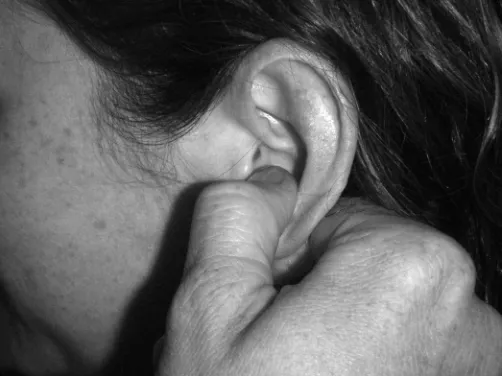
tech-nique are presented. The techtech-nique used in the cases re-ported here was one in which the thumb is placed just inside the intertragic notch, with the proximal interphal-geal joint of the index fi nger contacting just inside the lobule (fi gure 1). A gentle lateral movement is applied and the patient is asked whether this produces pain. The practitioner also attempts to assess the degree of resist-ance to the movement (the reliability and validity of this assessment is unknown). If manipulation is deemed indi-cated, a high-velocity, low-amplitude thrust is performed in a straight lateral direction. An audible release typically occurs. The patient can then be taught self-mobilization in the same direction, applying low-velocity, low-amplitude oscillatory maneuvers.
pressure with the atmosphere, resulting in distortion of the mucosa of the middle ear and tympanic membrane.1
This can cause otalgia with or without otitis media. As several muscles are involved in opening and closing the Eustachian tube (salpingopharyngeus, levator veli pal-atini, tensor veli palpal-atini, and tensor tympani), it is pos-sible that disruption of the tone of these muscles can lead to Eustachian tube dysfunction and that manipulation of the ear restores normal tone. Alternately, as the middle ear ossicles are synovial joints,23 it is possible that these
joints can become painful, as do other synovial joints, and that ear manipulation affects these joints in a similar way that spinal manipulation affects zygapophyseal joints.24,25
However it is unknown whether external manipulation of the ear has any effect on these muscles or joints.
Mobilization or manipulation was applied to the upper cervical spine in three of these four cases. The decision to apply this treatment was based on, fi rst, the presence of painful joint dysfunction7 at the involved levels and,
second, previous literature that suggested the possible role of referred pain from the cervical spine in the causa-tion of in some cases of secondary otalgia. However, joint palpation in these cases did not exactly reproduce the ear pain in any case, thus there is no way to determine wheth-er the cwheth-ervical fi ndings wwheth-ere directly involved in any in-dividual case. Also because the treatment of these cases was multi-modal, there is no way to determine the extent to which any individual treatment modality may or may
not have contributed to the perceived benefi cial outcome. In addition, there is no way to determine on the basis of a case report whether the perceived benefi cial outcome oc-curred as a result of the management strategy applied or whether it occurred by natural history. Further research is needed to confi rm or deny the theoretical model by which the treatment may have benefi cial, as well as to determine whether the fi ndings reported here are generalizable.
The treatment in these cases was done at a primary spine care center at which chiropractors and physiotherapists provide all examination and treatment and for which 80% of its patients are referred by medical doctors. A number of these patients are those with ear and face pain who are referred by otolaryngologists. This referral relationship arose as a result of communication by the clinical director of the spine center with local otolaryngologists through Grand Rounds presentations and personal communication regarding the role of the cervical spine and other aspects of the neuromusculoskeletal system may play in symptoms referable to the ear, mouth and face. It is the experience of the authors that otolaryngologists see a considerable number of such patients for which an otolaryngological etiology cannot be identifi ed. Despite this, it is uncom-mon for chiropractors to be consulted in these cases. In the 2010 edition of the “Practice Analysis of Chiroprac-tic” which is published by the National Board of Chiro-practic Examiners and which documents the frequency with which US chiropractors evaluate and treat various health conditions, “otalgia” and “ear pain” were not even listed. “Cranial nerve disorders” were seen “rarely” and “signifi cant ear pathology” and “eye, ear, nose or throat tumor” were seen “virtually never.” “TMJ syndrome,” on the other hand, was seen “sometimes.” In our opinion, a chiropractor or chiropractor-physiotherapist team may be a useful resource for these practitioners for those patients who report pain in the ear and face for which a primary otalgic explanation cannot be found. It may be useful for chiropractors to communicate with otolaryngologists in order to provide a resource for these patients.
Conclusion
to have at their disposal clinicians who are skilled at the examination and manual treatment of cervical spine and the musculoskeletal system in general, who can provide input regarding the possible involvement of these tissues.
References
1 Shah RK, Blevins NH. Otalgia. Otolaryngol Clin North Am. 2003; 36(6):1137–51.
2 Thaller SR, De Silva A. Otalgia with a normal ear. Am Fam Physician. 1987; 36(4):129–36.
3 Kuttila S, Kuttila M, Le Bell Y, Alanen P, Suonpaa J. Characteristics of subjects with secondary otalgia. J Orofac Pain. 2004; 18(3):226–34.
4 Lamer TJ. Ear pain due to cervical spine arthritis: treatment with cervical facet injection. Headache. 1991; 31(10):682–3.
5 Skaggs CD. Diagnosis and treatment of
temporomandibular disorders. In: Murphy DR, ed. Conservative Management of Cervical Spine Syndromes. New York: McGraw-Hill 2000; 579–92.
6 Jull G, Bogduk N, Marsland A. The accuracy of manual diagnosis for cervical zygapophysial joint pain syndromes. Med J Australia. 1988; 148:233–6.
7 Jull G, Zito G, Trott P, Potter H, Shirley D. Inter-examiner reliability to detect painful upper cervical joint dysfunction. Aust Physiother. 1997; 43:125–9.
8 Jull G, Amiri M, Bullock-Saxton J, Darnell R, Lander C. Cervical musculoskeletal impairment in frequent intermittent headache. Part 1: Subjects with single headaches. Cephalalgia. 2007; 27(7):793–802.
9 Uthaikhup S, Sterling M, Jull G. Cervical musculoskeletal impairment is common in elders with headache. Man Ther. 2009; 14(6):636–41.
10 Medlicott MS, Harris SR. A systemic review of the effectiveness of exercise, manual therapy, electrotherapy, relaxation training, and biofeedback in the management of temporomandibular disorder. Phys Ther. 2006; 86(7):955– 73.
11 Chaitow L, Liebenson C. Muscle Energy Techniques. 2nd ed. Edinburgh: Churchille Livingstone; 2001.
12 Murphy DR. Sensorimotor training and cervical stabilization. In: Murphy DR, ed. Conservative Management of Cervical Spine Syndromes. New York: McGraw-Hill 2000; 607–40.
13 Feinstein B, Langton JNK, Jameson RM, Schiller F.
Experiments on pain referred from deep somatic tissues. J Bone Joint Surg. 1954; 36A(5):981–97.
14 Poletti CE. C2 and C3 pain dermatomes in man. Cephalalgia. 1991; 11(3):155–9.
15 Murphy DR, Hurwitz EL, Gerrard JK, Clary R. Pain patterns and descriptions in patients with radicular pain: Does the pain necessarily follow a specifi c dermatome. Research Agenda Conference; 2007; Phoenix, AZ; 2007. 16 Bendtsen L. Central sensitization in tension-type
headache-possible pathophysiological mechanisms. Cephalagia. 2000; 20(5):486–508.
17 Simons DG, Travell JG, Simons LS. Myofascial Pain and Dysfunction: The Trigger Point Manual. Volume 1. Baltimore: Williams and Wilkens; 1999.
18 Kuttila S, Kuttila M, Le Bell Y, Alanen P, Jouko S. Aural symptoms and signs of temporomandibular disorder in association with treatment need and visits to a physician. Laryngoscope. 1999; 109(10):1669–73.
19 Curl D. Orofacial pain. In: Murphy DR, ed. Conservative Management of Cervical Spine Syndromes. New York: McGraw-Hill 2000; 237–54.
20 Cowin RC, Bryner P. Hearing loss, otalgia and neck pain: A case report on long-term chiropractic care that helped to improve quality of life. Chiropr J Aust. 2002; 22(4):119– 30.
21 Kaye MJ. Evaluation and treatment of a patient with upper quarter myofasical pain syndrome. J Sports Chiropr Rehabil. 2001; 15(1):26–3.
22 Channell MK. Modifi ed Muncie technique: osteopathic manipulation for eustachian tube dysfunction and illustrative report of case. J Am Osteopath Assoc. 2008; 108(5):260–3.
23 Whyte JR, Gonzalez L, Cisneros AI, Yus C, Torres A, Sarrat R. Fetal development of the human tympanic ossicular chain articulations. Cells Tissues Organs. 2002; 171(4):241–9.
24 Cramer GD, Tuck NR, Knudsen JT, Fonda SD, Schliesser JS, Fournier JT, et al. Effects of side-posture positioning and side posture adjusting on the lumbar zygapophyseal joints as evaluated by magnetic resonance imaging. J Manipulative Physiol Ther. 2000; 23(6):380–94. 25 Ianuzzi A, Khalsa PS. High loading rate during spinal